HAL Id: hal-03015954
https://hal.archives-ouvertes.fr/hal-03015954
Preprint submitted on 20 Nov 2020
HAL is a multi-disciplinary open access
archive for the deposit and dissemination of sci-entific research documents, whether they are pub-lished or not. The documents may come from teaching and research institutions in France or
L’archive ouverte pluridisciplinaire HAL, est destinée au dépôt et à la diffusion de documents scientifiques de niveau recherche, publiés ou non, émanant des établissements d’enseignement et de recherche français ou étrangers, des laboratoires
host infection
Laurent Camborde, Andrei Kiselev, Pel Michiel, Aurelie Leru, A. Jauneau,
Pouzet Cécile, Bernard Dumas, Elodie Gaulin
To cite this version:
Laurent Camborde, Andrei Kiselev, Pel Michiel, Aurelie Leru, A. Jauneau, et al.. A DEAD 1 BOX RNA helicase from Medicago truncatula is hijacked by an RNA-binding effector from the root pathogen Aphanomyces euteiches to facilitate host infection. 2020. �hal-03015954�
A DEAD BOX RNA helicase from Medicago truncatula is hijacked by an RNA-binding 1
effector from the root pathogen Aphanomyces euteiches to facilitate host infection 2
3
L. Camborde1, A. Kiselev1, M.J.C. Pel1, *, A. Leru2, A. Jauneau2, C. Pouzet2, B. Dumas1, 4 E. Gaulin1 5 6 7 Affiliations 8 1
Laboratoire de Recherche en Sciences Végétales (LRSV), Université de Toulouse, CNRS,
9
UPS, France
10
2Plateforme d’Imagerie FRAIB-TRI, Université de Toulouse, CNRS, France 11
*Present address: Bacteriology Group, National Reference Centre (NRC), Dutch National
12
Plant Protection Organization (NPPO-NL), P.O. Box. 9102, 6700 HC Wageningen, the
13
Netherlands
14 15
Corresponding author: Elodie Gaulin
16
LRSV, UMR CNRS 5546 Université de Toulouse,
17 24, Chemin de Borde-Rouge 18 31320 Auzeville, France 19 Mail : gaulin@lrsv.ups-tlse.fr 20 21 22
Keywords: effectors, RNA-helicase, plant development, ribosome biogenesis pathway,
23
oomycete, Medicago, nucleolar stress
24 25
Abstract 26
Microbial effectors from plant pathogens are molecules that target host components to
27
facilitate colonization. While eukaryotic pathogens are virtually able to produce hundreds of
28
effectors, the molecular mechanisms allowing effectors to promote infection are still largely
29
unexplored. Here we show that the effector AeSSP1256 from the soilborne oomycete
30
pathogen Aphanomyces euteiches is able to interact with plant RNA. Heterologous expression
31
of AeSSP1256 delays Medicago truncatula host roots development and facilitate pathogen
32
colonization. Transcriptomic analyses of AeSSP1256-expressing roots show a downregulation
33
of genes implicated in ribosome biogenesis pathway. A yeast-two hybrid approach reveals
34
that AeSSP1256 associates with a nucleolar L7 ribosomal protein and a M. truncatula RNA
35
helicase (MtRH10) orthologous to the Arabidopsis RNA helicase RH10. Association of
36
AeSSP1256 with MtRH10 impaired the capacity of MtRH10 to bind nucleic acids.
37
Promoter:GUS composite plants revealed that MtRH10 is expressed preferentially in the
38
meristematic root cells. Missense MtRH10 plants displayed shorter roots with developmental
39
delay and are more susceptible to A. euteiches infection. These results show that the effector
40
AeSSP1256 facilitates pathogen infection by causing stress on plant ribosome biogenesis and
41
by hijacking a host RNA helicase involved in root development and resistance to root
42
pathogens.
Introduction 44
Plant pathogens divert host cellular physiology to promote their own proliferation by
45
producing effector proteins that interact with molecular targets (Gaulin et al., 2018).
46
Numerous studies indicate large variation in the effector repertoire of plant pathogens
47
suggesting that a large number of molecular mechanisms are targeted.
48
Oomycetes constitute a large phylum that includes important eukaryotic pathogens,
49
and many of which are destructive plant or animal pathogens (Kamoun et al., 2015; van West
50
and Beakes, 2014). They share common morphological characteristics with true fungi as
51
filamentous growth, osmotrophic feeding or the presence of a cell wall, but they evolved
52
independently (Judelson, 2017). Oomycetes are included in the Stramenopile lineage and have
53
diatoms and brown algae as closest cousins. These filamentous microorganisms have the
54
capacity to adapt to different environment as illustrated by their capacity to develop resistance
55
to anti-oomycete chemicals or quickly overcome plant resistance (Rodenburg et al., 2020).
56
Comprehensive identification of oomycete proteins that act as effectors is challenging. Up to
57
now, computational predictions of effector proteins have provide a fast approach to identify
58
putative candidate effectors in oomycetes (Haas et al., 2009; Tabima and Grünwald, 2019).
59
Based on their predictive subcellular localization within the host cells they are classified as
60
extracellular (apoplasmic) or intracellular (cytoplasmic) effectors. As example, RxLR and
61
Crinklers (CRNs) constitute the two largest family of oomycetes intracellular effectors that
62
contain hundreds of members per family (McGowan and Fitzpatrick, 2017). While oomycete
63
effector proteins have probably different mechanisms of action, what they have in common
64
might be the ability to facilitate pathogen development. Nonetheless, computational
65
predictions do not give any clues regarding the putative role of theses effectors since
66
numerous effectors are devoid of any functional domains. Therefore, biochemical and
67
molecular studies are used to discover and confirm the functional activity of these proteins.
68
To promote infection oomycete intracellular effectors interfere with many host routes which
69
include for example signaling such as MAPKinase cascades (King et al., 2014),
70
phytohormone-mediated immunity (Boevink et al. 2016; Liu et al. 2014), trafficking vesicles
71
secretion (Du et al., 2015) or autophagosome formation (Dagdas et al., 2016). Growing
72
evidences point to plant nucleus as an important compartment within these interactions thanks
73
to the large portfolio of putative nucleus-targeted effectors predicted in oomycete genomes.
74
The study of subcellular localization of fifty-two Phytophthora infestans RxLR effectors
75
upregulated during the early stage of host infection show that nucleocytoplasmic distribution
is the most common pattern, with 25% effectors that display a strong nuclear association
77
(Wang et al. 2019). The CRN family was firstly reported as a class of nuclear effector from P.
78
infestans (Schornack et al., 2010), around 50% of predicted NLS-containing CRN effectors
79
from P. capsici showed nuclear localization (Stam et al., 2013) and numerous CRNs effectors
80
from P. sojae such as PsCRN108, PsCRN63 or PsCRN115 harbor a nuclear localization
81
(Song et al., 2015; Zhang et al., 2015). In agreement with this, different mechanisms of action
82
at the nuclear level have been reported for oomycete effectors such as the alteration of genes
83
transcription (Wirthmueller et al., 2018; Song et al., 2015; He et al., 2019), the mislocalisation
84
of transcription factor (Mclellan et al., 2013),the suppression of RNA silencing by inhibition
85
of siRNA accumulation (Qiao et al., 2015; Xiong et al., 2014)or the induction of plant
DNA-86
damage (Camborde et al. 2019; Ramirez-Garcés et al. 2016). However specific function has
87
been assigned to very few effectors.
88
We previously use comparative genomics and predictive approaches on the
89
Aphanomyces genus to identify putative effectors and characterized a large family of small
90
secreted proteins (SSPs) (Gaulin et al., 2018). SSPs harbor a predicted N secretion signal, are
91
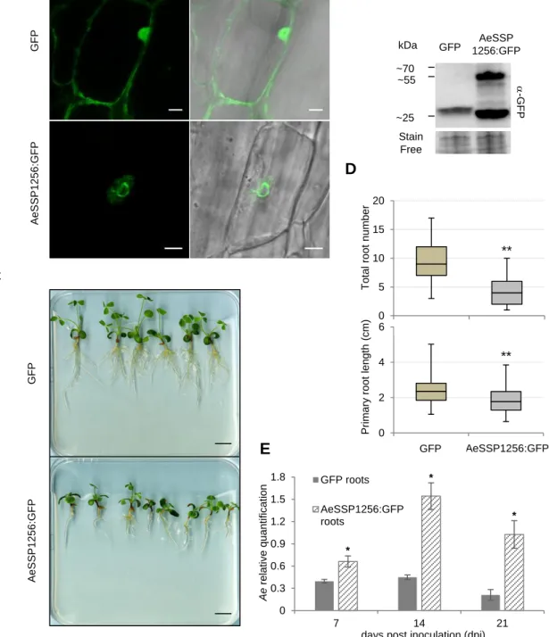
less than 300 residues in size and devoid of any functional annotation. More than 290 SSPs
92
are predicted in the legume pathogen A. euteiches (AeSSP) while 138 members with no
93
obvious similarity to AeSSP members are reported in the crustacean parasite A. astaci (Gaulin
94
et al., 2018). This specific SSP repertoire suggests its role in adaption of Aphanomyces
95
species to divergent hosts. We have previously identified one AeSSP (AeSSP1256) based on
96
a screen aiming to identify SSP able to promote infection of Nicotiana benthamiana plants by
97
the leaf pathogen Phytophthora capsici. AeSSP1256 harbors a nuclear localization signal
98
indicating its putative translocation to host nucleus. However, the function of this protein
99
remained to be identified.
100
Here we report on the functional analysis of AeSSP1256 and the characterization of its
101
plant molecular target. We show that AeSSP1256 binds RNA in planta, induces
102
developmental defects when expressed in M. truncatula roots and promotes A. euteiches
103
infection. This phenotype is correlated with a downregulation of a set of ribosomal protein
104
genes. A yeast two-hybrid approach identified a host RNA helicase (MtRH10) and a L7
105
ribosomal protein as interactors of AeSSP1256. By FRET-FLIM analyses we reveal that
106
AeSSP1256 co-opts MtRH10 to abolish its nucleic acid binding capacity. We provide a
107
mechanistic explanation of this observation by demonstrating the implication of MtRH10 in
observed that silenced-MtRH10 roots are highly susceptible to A. euteiches infection like
110
AeSSP1256-expressing roots, showing that MtRH10 as AeSSP1256 activities modify the
111
outcome of the infection. We now present results supporting effector-mediated manipulation
112
of a nuclear RNA helicase as a virulence mechanism during plant-eukaryotic pathogens
113
interactions.
114 115
Results 116
117
AeSSP1256 contains RGG/RG domains and binds RNA in planta 118
AeSSP1256 is a member of a large family of A. euteiches effectors devoid of any predicted
119
functional domain, except the presence of a signal peptide at the N-terminus (Gaulin et al.,
120
2018). As showed in Figure 1A, AeSSP1256 protein is enriched in glycine residues (30% of
121
the amino acid sequence). Analysis using the Eukaryotic Linear Motif database (Gouw et al.,
122
2018) revealed 3 GGRGG motifs (positions 81-85; 95-99 and 99-103). These motifs are
123
variant arginine methylation site from arginine-glycine(-glycine) (RGG/RG) domains,
124
presents in many ribonucleoproteins and involved in RNA binding (Thandapani et al., 2013;
125
Bourgeois et al., 2020). We then noticed the presence of two di-RGG domains (RGG(X 0-126
5)RGG) (position 75-85 and 97-103) and one di-RG domains (RG(X0-5)RG) (position 123-127
126) corresponding to RGG or RG motifs that are spaced less than 5 residues (Chong et al.,
128
2018). According to RGG/RG definition, those repeats occur in low-complexity region of the
129
protein (position 60-180) (Chong et al., 2018) and are associated with di-glycine motifs and
130
GR or GGR sequences (Figure 1A), which are also common in RGG/RG-containing proteins
131
(Chong et al., 2018). Considering that RGG/RG domains are conserved from yeast to humans
132
(Rajyaguru and Parker, 2012) and represent the second most common RNA binding domain
133
in the human genome (Ozdilek et al., 2017), we therefore investigated the RNA binding
134
ability of AeSSP1256. To test this, we performed a FRET-FLIM assay on N. benthamiana
135
agroinfiltrated leaves with AeSSP1256:GFP fusion protein in presence or absence of Sytox
136
Orange to check its capacity to bind nucleic acids (Camborde et al. 2017). Briefly
137
AeSSP1256:GFP construct is transiently express in N. benthamiana leaves where it
138
accumulates in the nucleus (Gaulin et al., 2018). Samples are collected 24h after treatment
139
and nucleic acids labeled with the Sytox Orange dye. In presence of Sytox, if the GFP fusion
140
protein is in close proximity (<10nm) with nucleic acids, the GFP lifetime of the GFP tagged
141
protein will significantly decrease, due to energy transfer between the donor (GFP) and the
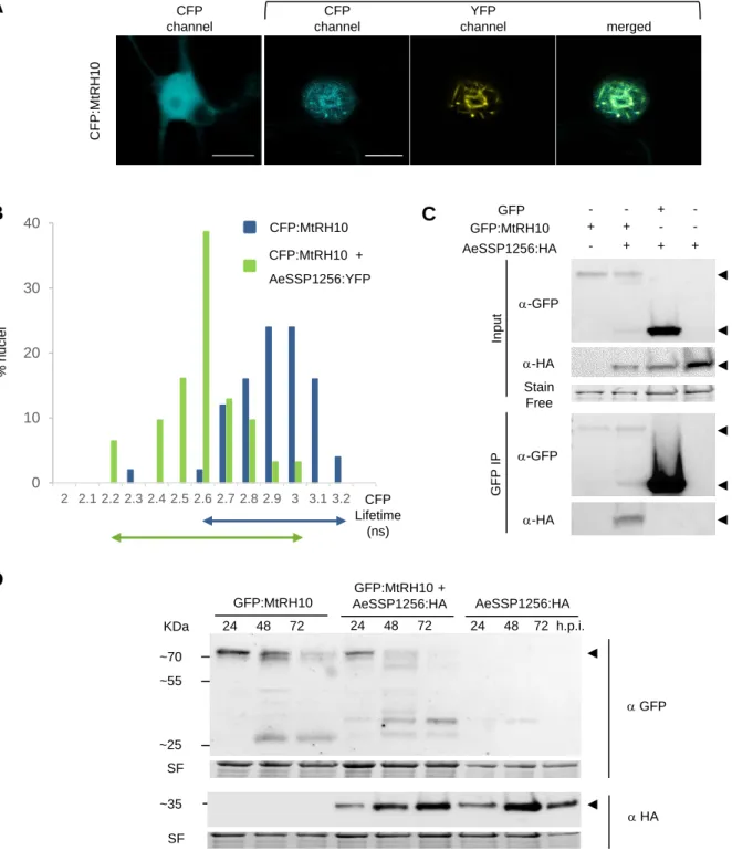
142
acceptor (Sytox). To distinguish RNA interactions from DNA interactions, an RNase
143
treatment can be performed. In the case of a specific RNA-protein interaction, no FRET
144
acceptor will be available due to RNA degradation and the lifetime of the GFP tagged protein
145
will then return at basal values. It appeared that GFP lifetime of AeSSP1256:GFP decreased
146
significantly in presence of Sytox Orange as reported in table 1 and in Figure 1B, decreasing
nucleic acids. After an RNase treatment, no significant difference on GFP lifetime was
149
observed in absence (2.01 ns +/- 0.02) or in presence (1.96 ns +/- 0.02) of Sytox Orange,
150
meaning that the FRET was not due to DNA interaction but was specific to RNA (table 1 and
151
Figure 1B). These results indicate that AeSSP1256 is able to bind nuclear RNA in plant cells. 152
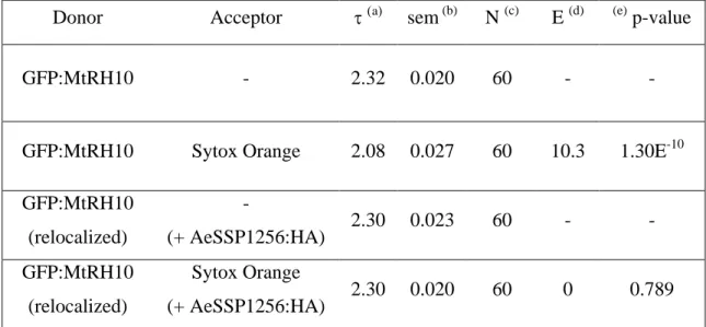
153
Table 1: FRET-FLIM measurements for AeSSP1256:GFP with or without Sytox Orange
154
Donor Acceptor a sem (b) N (c) E (d) (e)p-value
AeSSP1256:GFP - 2.06 0.020 78 - -
AeSSP1256:GFP Sytox 1.84 0.026 77 11 1.34E-09
AeSSP1256:GFP -
(+ RNase) 2.01 0.026 50 - -
AeSSP1256:GFP Sytox
(+ RNase) 1.96 0.027 50 2.6 0.17
mean life-time in nanoseconds (ns). (b) s.e.m.: standard error of the mean. (c) N: total number of measured
155
nuclei. (d) E: FRET efficiency in %: E=1-(DA/D). (e) p-value (Student’s t test) of the difference between the
156
donor lifetimes in the presence or absence of acceptor.
157 158
AeSSP1256 impairs M. truncatula root development and susceptibility to A. euteiches 159
160
To check whether expression of AeSSP1256 may have an effect on the host plant, we
161
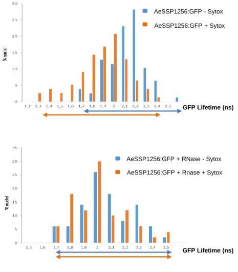
transformed M. truncatula (Mt) roots, with a native version of GFP tagged AeSSP1256. As
162
previously observed (Gaulin et al., 2018), confocal analyses confirmed the nuclear
163
localization of the protein in root cells, with accumulation around the nucleolus as a
164
perinucleolar ring (Figure 2A) despite the presence of a signal peptide (Gaulin et al., 2018).
165
Anti-GFP western blot analysis on total proteins extracted from transformed roots confirmed
166
the presence of GFP-tagged AeSSP1256 (46.7 kDa expected sizes) (Figure 2B). We noticed
167
the presence of a second band around 28 kDa, which is probably free GFP due to the cleavage
168
of the tagged protein. AeSSP1256:GFP transformed plants showed delayed development
169
(Figure 2C), with total number of roots and primary root length per plant being significantly
170
lower than values obtained with GFP control plants (Figure 2D). As previously observed in
171
N. benthamiana, when a KDEL-endoplasmic reticulum (ER) retention signal is added to the
172
native AeSSP1256 construct (Gaulin et al., 2018), AeSSP1256:KDEL:GFP proteins mainly
accumulates in the ER (Supplemental Figure 1A-C) and roots showed no significant
174
differences in development as compared to GFP control roots (Supplemental Figure 1D and
175
E). In contrast a construct devoid of a native signal peptide (SP) shows that the proteins 176
accumulated in root cell nuclei (Supplemental Figure 1B), leading to abnormal root
177
development, with symptoms similar to those observed in presence of the AeSSP1256:GFP
178
construct, including shorter primary root and lower number of roots (Supplemental Figure
179
1D and E). Altogether these data show that within the host, AeSSP1256 triggers roots 180
developmental defects thanks to its nuclear localization.
181
To investigate whether AeSSP1256 modifies the outcome of the infection,
AeSSP1256-182
transformed roots were inoculated with A. euteiches zoospores. RT-qPCR analyses at 7, 14
183
and 21 days post inoculation were performed to follow pathogen development. At each time
184
of the kinetic, A. euteiches is more abundant in M. truncatula roots expressing the effector
185
than in GFP control roots (respectively 1.5, 3 and 5 times more) (Figure 1E). This indicates
186
that roots are more susceptible to A. euteiches in presence of AeSSP1256. Transversal
187
sections of A17-transformed roots followed by Wheat-Germ-Agglutinin (WGA) staining to
188
detect the presence of A. euteiches, showed that the pathogen is still restricted to the root
189
cortex either in the presence or absence of AeSSP1256 (Supplemental Figure 2). This
190
phenotype is similar to the one observed in the natural A17 M. truncatula tolerant line
191
infected by A. euteiches (Djébali et al., 2009). This data suggests that defence mechanisms
192
like protection of the central cylinder (Djébali et al., 2009) are still active in
AeSSP1256-193
expressing roots.
194 195
AeSSP1256 affects the expression of genes related to ribosome biogenesis 196
To understand how AeSSP1256 affects M. truncatula roots development and facilitates A.
197
euteiches infection, we performed expression analyses by RNASeq using
AeSSP1256-198
expressing roots and GFP controls roots. 4391 genes were differentially express (DE) between
199
the two conditions (p adjusted-value <10-5) (Supplemental Table 1a). Enrichment analysis of
200
‘Biological process’ GO-terms showed the presence of ‘ribosome biogenesis’ and
201
‘organonitrogen compound biosynthetic, cellular amide metabolic’ processes terms among the
202
most enriched in AeSSP1256 roots as compared to GFP-expressing roots (Supplemental
203
Table 1b). We noticed that over 90% of DE-genes from ‘ribosome biogenesis’ and 204
expression of the effector within the roots affects ribosome biogenesis pathway
206
(Supplemental Table 1a). To evaluate whether expression of AeSSP1256 mimics infection
207
of M. truncatula by A. euteiches infection through downregulation of genes related to
208
ribosome biogenesis, we analyzed RNASeq data previously generated on the susceptible
209
F83005.5 M. truncatula line nine daysafter root infection (Gaulin et al., 2018). As shown on
210
the Venn diagram depicting the M. truncatula downregulated genes in the different conditions
211
(Figure 3A, Supplemental Table1c), among the 270 common downregulated genes between
212
AeSSP1256-expressing roots and susceptible F83-infected lines, 58 genes (>20%) are
213
categorized in the ‘ribosome biogenesis’ and ‘translation’ GO term (Figure 3B). We next
214
selected seventeen M. truncatula genes to confirm the effect via qRT-PCR. First, we selected
215
ten A. thaliana genes related to plant developmental control (i,e mutants with shorter roots
216
phenotype) (Supplemental Table 1d) by Blast searches (>80% identity) in A17 line r5.0
217
genome portal (Pecrix et al., 2018). In addition, seven nucleolar genes coding for ribosomal
218
and ribonucleotides proteins and related to the ‘ribosome biogenesis’ in M. truncatula were
219
selected for expression analysis based on KEGG pathway map
(https://www.genome.jp/kegg-220
bin/show_pathway?ko03008) (Supplemental Table 1d). As shown on Figure 3C, all of the
221
selected genes from M. truncatula are downregulated in presence of AeSSP1256, supporting
222
the RNAseq data. Altogether these expression data show that the effector by itself mimics
223
some effects induced by pathogen infection of the susceptible F83 line. At this stage of the
224
study, results point to a perturbation of the ribosome biogenesis pathway of the host plant by
225
the AeSSP1256 effector.
226 227
AeSSP1256 targets a DEAD-box RNA helicase and a L7 ribosomal protein 228
To decipher how AeSSP1256 can affect ribosome biogenesis pathway of the host plant and
229
knowing that numerous RNA-binding proteins interact with protein partners, we searched for
230
AeSSP1256 host protein targets. For this, a Yeast two hybrid (Y2H) library composed of
231
cDNA from M. truncatula roots infected with A. euteiches was screened with the mature form
232
of the effector. Eight M. truncatula coding genes were identified as potential protein targets
233
(Supplemental Table 2a), all these genes but one (a lecithin retinol acyltransferase gene)
234
correspond to putative nuclear proteins in accordance with the observed subcellular
235
localization of AeSSP1256.
To confirm the Y2H results, we first expressed AeSSP1256 and candidates in N. benthamiana
237
cells to observe their subcellular localization and performed FRET-FLIM experiments to
238
validate protein-protein interactions. Only two candidates showed co-localization with
239
AeSSP1256, a L7 ribosomal protein (RPL7, MtrunA17_Chr4g0002321) and a predicted RNA
240
helicase (RH) (MtrunA17_Chr5g0429221). CFP-tagged version of RPL7 displays a nucleolar
241
localization, with partial co-localization areas in presence of AeSSP1256 (Supplemental
242
Figure 3A, Table 2b). FRET-FLIM measurements confirmed the interaction of RPL7:CFP 243
protein with AeSSP1256:YFP effector (Supplemental Figure 3B, Table 2b), with a mean
244
CFP lifetime of 2.83 ns +/- 0.03 in absence of the SSP protein, leading to 2.46 ns +/- 0.03 in
245
presence of AeSSP1256:YFP (Supplemental Table 2b).
246
The second candidate is a predicted DEAD-box ATP-dependent RNA helicase
247
(MtrunA17_Chr5g0429221), related to the human DDX47 RNA helicase and the RRP3 RH
248
in yeast. Blast analysis revealed that the closest plant orthologs were AtRH10 in Arabidopsis
249
thaliana and OsRH10 in Oryza sativa. Consequently the M. truncatula protein target of
250
AeSSP1256 was named MtRH10. The conserved domains of DEAD-box RNA helicase are
251
depicted in the alignment of MtRH10 with DDX47, RRP3, AtRH10, OsRH10 proteins
252
(Supplemental Figure 4A) (Schütz et al., 2010; Gilman et al., 2017). MtRH10 CFP-tagged 253
fusion protein harbors nucleocytoplasmic localization when transiently express in N.
254
benthamiana cells (Figure 4A), in accordance with the presence of both putative nuclear
255
export signals (NESs) (position 7-37; 87-103; 261-271) and nuclear localization signal (NLS)
256
sequences (position 384-416). When MtRH10 is co-expressed with YFP-tagged version of
257
AeSSP1256, the fluorescence is mainly detected as a ring around the nucleolus, indicating a
258
partial relocalisation of MtRH10 to the AeSSP1256 sites (Figure 4A). FRET-FLIM
259
measurements on these nuclei confirm the interaction between AeSSP1256 and the Medicago
260
RNA helicase (Figure 4B), with a mean CFP lifetime of 2.86 ns +/- 0.02 in absence of the
261
effector protein, to 2.53 ns +/- 0.03 in presence of AeSSP1256:YFP (Table 2).
262
Table 2: FRET-FLIM measurements of CFP:MtRH10 in presence or absence of
263
AeSSP1256:YFP
264
Donor Acceptor a sem (b) N (c) E (d) (e) p-value
CFP:MtRH10 AeSSP1256:YFP 2.53 0.031 31 11.1 2.56E-12
mean life-time in nanoseconds (ns). (b) s.e.m.: standard error of the mean. (c) N: total number of measured
265
nuclei. (d) E: FRET efficiency in % : E=1-(DA/D). (e) p-value (Student’s t test) of the difference between the
266
donor lifetimes in the presence or absence of acceptor.
267 268
To confirm this result, co-immunoprecipitation assays were carried out. A GFP:MtRH10
269
construct was co-transformed with AeSSP1256:HA construct in N. benthamiana leaves. As
270
expected, the localization of GFP:MtRH10 protein in absence of AeSSP1256 was
271
nucleocytoplasmic while it located around the nucleolus in the presence of the effector
272
(Supplemental Figure 4B). Immunoblotting experiments using total proteins extracted from
273
infiltrated leaves (24hpi) showed that AeSSP1256:HA proteins were co-immunoprecipitated
274
with GFP:MtRH10, but not with the GFP alone (Figure 4C). These data indicate that
275
AeSSP1256 associates with MtRH10 in the nucleus. To go further we checked the stability of
276
the two proteins when expressed alone or in combination in N. benthamiana cells during 72
277
hours. While GFP:MtRH10 was still detected at 72h after agroinfiltration, it started to be
278
degraded 48hpi (Figure 4D). Expression of the effector alone is stable along the time. In
279
contrast, when the two proteins are co-expressed, GFP:MtRH10 is almost entirely processed
280
at 48h, and no more detectable at 72h (Figure 4D), suggesting that the effector enhance
281
instability of its host target. Taken together, these results strongly suggest an interaction
282
between AeSSP1256 and two type of components, a ribosomal protein and a nuclear RNA
283
helicase from M. truncatula.
284 285
AeSSP1256 alters the RNA binding activity of MtRH10 286
DEAD-box RNA helicases are RNA binding proteins involved in various RNA-related
287
processes including pre-rRNA maturation, translation, splicing, and ribosome assembly
288
(Jarmoskaite and Russell, 2011). These processes are dependent to the RNA binding ability of
289
the proteins. Therefore we checked whether MtRH10 is able to bind nucleic acids in planta
290
using FRET-FLIM assays as described previously. As reported in Table 3 and in Figure 5A,
291
GFP lifetime of GFP:MtRH10 decreased in presence of the acceptor, from 2.32 ns +/- 0.02 to
2.08 ns +/- 0.03 due to FRET between GFP and Sytox, confirming as expected that MtRH10
293
protein is bounded to nucleic acids.
294 295
Table 3: FRET-FLIM measurements for GFP:MtRH10 with or without Sytox Orange, in
296
presence or in absence of AeSSP1256:HA
297
Donor Acceptor a sem (b) N (c) E (d) (e) p-value
GFP:MtRH10 - 2.32 0.020 60 - -
GFP:MtRH10 Sytox Orange 2.08 0.027 60 10.3 1.30E-10
GFP:MtRH10 (relocalized) - (+ AeSSP1256:HA) 2.30 0.023 60 - - GFP:MtRH10 (relocalized) Sytox Orange (+ AeSSP1256:HA) 2.30 0.020 60 0 0.789
mean life-time in nanoseconds (ns). (b) s.e.m.: standard error of the mean. (c) N: total number of measured
298
nuclei. (d) E: FRET efficiency in % : E=1-(DA/D). (e) p-value (Student’s t test) of the difference between the
299
donor lifetimes in the presence or absence of acceptor.
300
To evaluate the role of AeSSP1256 on the function of MtRH10 we reasoned that the effector
301
may perturb its binding capacity since it is required for the activity of numerous RH protein
302
family (Jankowsky, 2011). We then co-expressed the GFP:MtRH10 construct with
303
AeSSP1256:HA in N. benthamiana leaves and performed FRET-FLIM assays. Measurements
304
made in nuclei where both proteins are detected due to the re-localization of MtRH10
305
indicated that GFP lifetime of GFP:MtRH10 remained unchanged with or without Sytox (2.3
306
ns in both conditions) showing that MtRH10 was not able to bind nucleic acids in the
307
presence of the effector (Table 3 and Figure 5B). These data reveal that AeSSP1256 hijacks
308
MtRH10 binding to RNA, probably by interacting with MtRH10.
309 310
MtRH10 is expressed in meristematic root cells and its deregulation in M. truncatula 311
impacts root architecture and susceptibility to A. euteiches infection 312
To characterize the function of MtRH10, we firstly consider the expression of the gene by
313
mining public transcriptomic databases including Legoo
(https://lipm-314
browsers.toulouse.inra.fr/k/legoo/), Phytozome (https://phytozome.jgi.doe.gov/pz/portal.html)
315
and MedicagoEFP browser on Bar Toronto
(http://bar.utoronto.ca/efpmedicago/cgi-316
bin/efpWeb.cgi). No variability was detected among the conditions tested in the databases and
317
we do not detect modification of MtRH10 expression upon A. euteiches inoculation in our
318
RNAseq data. To go further in the expression of the MtRH10 gene, transgenic roots
319
expressing an MtRH10 promoter-driven GUS (-glucuronidase) chimeric gene were
320
generated. GUS activity was mainly detectable in meristematic cells, at the root tip or in
321
lateral emerging roots (Figure 6A) suggesting a role in meristematic cell division. We
322
complete MtRH10 analyses by overexpressing a GFP-tagged version in Medicago roots. The
323
observation by confocal analyses of the subcellular localization of MtRH10 confirms its
324
nucleocytoplasmic localisation as previously observed in N. benthamiana cells (Figure 6B).
325
We also noticed the presence of brighter dots in the nucleolus corresponding probably to the
326
fibrillar center. No developmental defects were detected in roots overexpressing MtRH10
327
(Figure 6C-D). To assess the effect of MtRH10 on root physiology and resistance to A.
328
euteiches, a pK7GWiWG2:RNAi MtRH10 vector was design to specifically silence the gene
329
in Medicago roots. RNA helicase gene expression was evaluated by qPCR 21 days after
330
transformation. Analyses confirmed a reduced expression (from 3 to 5 times) compared to
331
roots transformed with a GFP control vector (Supplemental Figure 5). Missense MtRH10
332
plants display a reduced number of roots coupled with shorter primary roots (Figure 6C-D)
333
and a delay in development which starts with a shorter root apical meristem (RAM) (Figure
334
6E-F). This reduction in not due to smaller RAM cortical cell size (Figure 6F) suggesting a 335
decrease in cell number. Longitudinal sections of roots expressing either RNAi MtRH10 or
336
AeSSP1256 performed in elongation/differentiation zone (EDZ) revealed comparative defects
337
in cortical cell shape or cell size (Supplemental Figure 6A). Cell area in missense MtRH10
338
or in AeSSP1256 roots is approximately reduced 2 times compared to GFP control roots
339
(Supplemental Figure 6B) but proportionally the perimeter of those cells is longer than GFP
340
cells, indicating a difference in cell shape (Supplemental Figure 6C). We noticed that most
341
of EDZ cells in GFP roots present a rectangular shape, which seem impaired in missense
342
MtRH10 and AeSSP1256 expressing roots. Thus we measured the perimeter-bounding
343
rectangle (PBR) which calculates the smallest rectangle possible to draw with a given cell. A
344
perimeter/PBR ratio of 1 indicates that the cell is rectangular. As presented in Supplemental
345
Figure 6D, the perimeter/PBR ratio in GFP roots is close to 1 and significantly different than 346
those observed in RNAi MtRH10 and AeSSP1256 roots. This analysis reveals that the
347
reduction of MtRH10 expression or the expression of the effector AeSSP1256 in Medicago
348
roots, impair the cortical cell shape. The similar phenotypic changes observed on
MtRH10-349
silenced roots and AeSSP1256-expressing roots, suggests that the effector may affect
350
MtRH10 activity in cell division regions of the roots.
351
Having shown that MtRH10 is implicated in M. truncatula roots development, we test
352
whether this biological function is related to pathogen colonisation. We therefore investigate
353
by qPCR the presence of A. euteiches in silenced and overexpressed MtRH10 roots infected
354
by the pathogen. As shown on Figure 7, overexpression of MtRH10 reduce the amount of
355
mycelium in roots after 7, 14 and 21 dpi (1.8, 3.3 and 1.6 times less, respectively). We note by
356
western-blot analyses a slight decrease in MtRH10 amount upon the time probably due to the
357
accumulation of the AeSSP1256 effector (Supplemental Figure 7). As expected in roots
358
where MtRH10 is silenced to 2 to 3 times as compared to GFP control roots, qPCR analyses
359
revealed approximately 5 to 10 times more of the pathogen at 7, 14 and 21 dpi (Figure 7).
360
Taken together these infection assays show that MtRH10 is involved in conferring basal
361
resistance to A. euteiches at the root level.
362 363
Discussion 364
365
Protein effectors from filamentous plant pathogens such as fungi and oomycetes facilitate host
366
colonization by targeting host components. However the molecular mechanisms that enhance
367
plant susceptibility to the pathogen are still poorly understood. Here we report that the A.
368
euteiches AeSSP1256 RNA-binding effector facilitate host infection by downregulating
369
expression of plant ribosome-related genes and by hijacking from its nucleic target MtRH10,
370
a Medicago nuclear RNA-helicase (RH). Thus the current study unravels a new strategy in
371
which pathogenic oomycete triggers plant nucleolar stress to promote infection.
372
AeSSP1256 is an effector from the oomycete root pathogen A. euteiches previously shown to
373
enhance oomycete infection (Gaulin et al., 2018). Despite the absence of any functional
374
domain, in silico RGG/RG RNA-binding motif prediction (see for review (Thandapani et al.,
375
2013)) prompt us to show by FRET/FLIM analysis that the secreted AeSSP1256 effector is an
376
RNA-binding protein (RBP). RNAs play essential role in cell physiology and it is not
377
surprising that filamentous plant pathogens may rely on RNA-dependent process to control
378
host infection (for review see (Göhre et al., 2013; Pedersen et al., 2012)). Moreover RBPs are
379
key players in the regulation of the post-transcriptional processing and transport of RNA
380
molecules (Yang et al., 2018). However to our knowledge only three examples of RBPs
381
acting as virulence factor of plant pathogens are known. This includes the glycine-rich protein
382
MoGrp1 from the rice pathogen Magnaporthe oryzae (Gao et al., 2019), the UmRrm75 of
383
Ustilago maydis (Rodríguez-Kessler et al., 2012) and the secreted ribonuclease effector
384
CSEP0064/BEC1054 of the fungal pathogen Blumeria graminis which probably interferes
385
with degradation of host ribosomal RNA (Pennington et al., 2019). This situation is probably
386
due to the absence of conventional RNA-binding domain which render this type of RBP
387
undetectable by prediction algorithms. The future studies that will aim to unravel the atlas of
388
RNA-binding effector in phytopathogens should not only rely on computational analysis but
389
will have to use functional approaches such as crystallization of the protein to validate
390
function as performed with CSEP0064/BEC1054 effector (Pennington et al., 2019) screening
391
method like the RNA interactome capture (RIC) assay develops in mammals (Castello et al.,
392
2012) or FRET/FLIM assays to detect protein / nucleic acid interactions (Camborde et al.,
393
2017).
We observed that when expressed inside roots of the partially resistant Jemalong A17 M.
395
truncatula line, AeSSP1256 triggers developmental defects such as shorter primary roots and
396
delay in root development. Defects in roots development and retarded growth are typical
397
characteristics of auxin-related and ribosomal proteins mutants reported in Arabidopsis
398
(Ohbayashi et al., 2017; Wieckowski and Schiefelbein, 2012). In addition, those composite
399
Medicago promote infection of A. euteiches. This modification in the output of the infection is
400
highly relevant since we previously observed that M. truncatula quantitative resistance to A.
401
euteiches is correlated to the development of secondary roots (Rey et al., 2016). This activity
402
is dependent on the nucleolar rim localization of AeSSP1256, closed to the nucleolus.
403
The nucleolus is a membrane-free subnuclear compartment essential for the highly complex
404
process of ribosome biogenesis organized in three domains including the fibrillar center that
405
contain rDNA, which are not yet engaged in transcription (reviewed in (Shaw and Brown,
406
2012). Ribosome biogenesis is linked to cell growth and required coordinated production of
407
processed ribosomal RNA (rRNA), ribosomal biogenesis factors and ribosomal proteins (RP).
408
In the nucleolus, ribosome biogenesis starts with the transcription of pre-rRNAs from rRNA
409
genes, followed by their processing and assembly with RPs into two ribosome subunits (ie
410
small and large subunit). In animals, perturbation of any steps of ribosome biogenesis in the
411
nucleolus can cause a nucleolar stress or ribosomal stress which stimulates specific signaling
412
pathway leading for example to arrest of cell growth (Pfister, 2019). The nucleolar rim
413
localization of AeSSP1256 within the host cells suggested that this effector could interfere
414
with ribosome biogenesis pathway to facilitate infection. This speculation was further
415
strengthened by RNAseq experiments which showed that, within A17-roots, AeSSP1256
416
downregulated numerous genes implicated in ribosome biogenesis pathway, notably
417
ribosomal protein genes. This effect was also detected in susceptible F83 M. truncatula lines
418
infected by A. euteiches indicating that AeSSP1256, mimics some A.euteiches effects during
419
roots invasion.
420
An Y2H approach led to the identification of putative AeSSP1256 plant targets and all but
421
one correspond to predicted nuclear M. truncatula proteins. By a combination of multiple
422
experiments as FRET-FLIM to detect protein/protein interactions, a L7 ribosomal protein
423
(MtrunA17_Chr4g0002321) and a DExD/H box RNA helicase ATP-dependent
424
(MtrunA17_Chr5g0429221) were confirmed as AeSSP1256-interacting proteins. The
425
DExD/H (where x can be any amino acid) box protein family include the largest family of
as ‘RNA chaperone’ promoting the formation of optimal RNA structures by unwinding
428
locally the RNA (for review see (Fuller-Pace, 2006)). These proteins are of major interest due
429
to their participation to all the aspects of RNA processes such as RNA export and translation,
430
splicing but the most common function of these proteins is in ribosome biogenesis including
431
assembly (Jarmoskaite and Russell, 2011). Specific function of RH is probably due to the
432
presence of a variable C-terminal ‘DEAD’ domain in contrast to the well conserved
N-433
terminal ‘helicase core’ domain (for review see (Fuller-Pace, 2006)). This structural
434
organization was detected in the MtRH10. This M. truncatula protein corresponds to the
435
ortholog of the nucleolar human DDX47 (Sekiguchi et al., 2006), the nuclear yeast RRP3
436
(O’Day, 1996) and the nucleolar Arabidopsis AtRH10 RNA-helicases, all involved in 437
ribosome biogenesis (Liu and Imai 2018; Matsumura et al. 2016), and the nucleolar rice
438
OsRH10 (TOGR1) involved in rRNA homeostasis (Wang et al. 2016).
439
Like its human ortholog DDX47 (Sekiguchi et al., 2006), MtRH10 possesses a bipartite
440
nuclear transport domain which can function as a nuclear localization signal (NLS) and two
441
nuclear export signal (NES), and thereby it probably shuttles between the cytoplasm and the
442
nucleus as reported for many others RNA helicases involved in rRNA biogenesis and splicing
443
function (Sekiguchi et al. 2006; Wang et al. 2009). Fluorescence analysis showed a
444
relocalization of the nucleocytoplasmic MtRH10 in the nucleoli periphery, when it is
445
transiently co-express with AeSSP1256 in N. benthamiana cells. The change in MtRH10
446
distribution suggests that the interaction between the two proteins caused a mislocation of
447
MtRH10 that can probably affect its activity. We thereby check the nucleic acid binding
448
capacity of MtRH10 by FRET-FLIM approach. The decrease in the lifetime of GFP revealed
449
the ability of MtRH10 to bind nucleic acids. Knowing that both proteins display the same
450
properties, we further provided evidence that the presence of AeSSP1256 effector inhibits the
451
nucleic binding capacity of MtRH10. This mechanism was also reported for the RNA-binding
452
HopU1 effector from the plant bacterial pathogen Pseudomonas syringae which associates to
453
the glycin-rich RNA binding 7 protein (GRP7) of Arabidopsis to abolish GRP7 binding to
454
immune gene transcripts (ie FLS2 receptor, (Nicaise et al., 2013)). Here we cannot exclude
455
that AeSSP1256 also blocks the putative helicase activity of MtRH10, but we favored an
456
inhibitory mechanism of AeSSP1256 on MtRH10 activity as complex and at least in part due
457
to both protein-protein interaction and nucleic acid interaction with the two proteins.
458
Interestingly, we also noticed that co-expression of both proteins led to decrease in MtRH10
459
probably due to degradation of the protein. While this observation warrants further analyses,
this effect is reminiscent of other effector activities which destabilize their targets (for review
461
see (Langin et al., 2020)).
462
Plant genomes encode a large variety of DExD/H RH family in comparison to other
463
organisms and numerous studies have shown that several are associated through their activity
464
with plant development, hormone signaling or responses to abiotic stresses (for review see
465
(Liu and Imai 2018)). Very few studies reported that DExD/H RH could also be involved in
466
biotic stresses, like responses to pathogens. One example is the DExD/H RH OsBIRH1 from
467
rice that enhanced disease resistance against Alternaria brassicicola and Pseudomonas
468
syringae through activation of defense-related genes (Li et al. 2008). A recent study on
469
oomycete reports the binding of the Phytophthora sojae RxLR PSR1 effector to a putative
470
nuclear DExD/H RH. Although the affinity for nucleic acids was not evaluated for the RH,
471
association of both partners promote pathogen infection by suppressing small RNA biogenesis
472
of the plant (Qiao et al., 2015). Here we showed that MtRH10 knockdown tolerant A17 lines
473
supported higher-level accumulation of A. euteiches in contrast to overexpressed MtRH10
474
lines, indicating the importance of MtRH10 for M. truncatula roots defense against soil-borne
475
pathogens.
476
This works reveals that MtRH10 expression is restricted at the root apical meristematic zone
477
(RAM) where cells divide (ie, primary and lateral roots). Missense MtRH10 roots harbor
478
defects in the primary root growth and reduced number of roots. Longitudinal sections in
479
elongation zone (EDZ) of these composite roots show a significant reduction in the size and
480
shape modification of cortical cells indicating that MtRH10 is required for normal cell
481
division. Defect in primary roots elongation is also detected in silenced AtRH10 and OsRH10
482
mutant (Matsumura et al. 2016; Wang et al. 2016). Thus MtRH10 plays a role on Medicago
483
root development as its orthologs OsRH10 and AtRH10. At the cellular level we also
484
observed in AeSSP1256-expressing roots, reduction in cell size in elongation zone, with
485
defects in cell shape and in adhesion between cells of the cortex, maybe due to a modification
486
of the middle lamella (Zamil and Geitmann, 2017). Thus AeSSP1256 triggers similar or
487
enhanced effect on host roots development as the one detected in defective MtRH10
488
composite plants, supporting the concept that the activity of the effector on MtRH10
489
consequently leads to developmental roots defects. Several reports have indicated that
490
Arabidopsis knockout of genes involved in rRNA biogenesis or in ribosome assembly cause
491
abnormal plant development including restriction and retardation in roots growth (Ohtani et
common mechanism that regulate growth in response to insults of the ribosome biogenesis
494
pathway, known as nucleolar stress response (for review see (Ohbayashi and Sugiyama,
495
2018)). How plant cells sense perturbed ribosome biogenesis and nucleolar problems is still
496
an open question (Sáez-Vásquez and Delseny, 2019), but the ANAC082 transcription factor
497
from Arabidopsis can be a ribosomal stress response mediator (Ohbayashi et al., 2017). In
498
addition the recent report on the activity of the nucleolar OsRH10 (TOGR1, MtRH10
499
ortholog) implicated in plant primary metabolism through is activity on rRNA biogenesis,
500
suggests that metabolites may play a role in this process. Finally our current study indicates
501
that nuclear RNA-binding effector like AeSSP1256, by interacting with MtRH10, can act as a
502
stimulus of the ribosomal stress response.
503
This work established a connection between the ribosome biogenesis pathway, a nuclear
504
DExD/H RH, root development and resistance against oomycetes. Our data document that the
505
RNA binding AeSSP1256 oomycete effector downregulated expression of ribosome-related
506
genes of the host plant. The effector hijacked MtRH10, a nuclear DExD/H RH involved in
507
root development, to promote host infection. This work not only provides insights into
plant-508
root oomycete interactions but also reveals the requirement of fine-tuning of plant ribosome
509
biogenesis pathways for infection success.
510 511
Material and Methods 512
513
Plant material, microbial strains, and growth conditions 514
M. truncatula A17 seeds were in vitro-cultured and transformed as previously described
515
(Boisson-Dernier et al., 2001; Djébali et al., 2009). A. euteiches (ATCC 201684) zoospore
516
inoculum were prepared as in (Badreddine et al., 2008). For root infections, each plant was
517
inoculated with a total of 10µl of zoospores suspension at 105 cells.ml-1. Plates were placed in
518
growth chambers with a 16h/8h light/dark and 22/20°C temperature regime. N. benthamiana
519
plants were grown from seeds in growth chambers at 70% of humidity with a 16h/8h
520
light/dark and 24/20°C temperature regime. E.coli strains (DH5α, DB3.5), A. tumefaciens
521
(GV3101::pMP90) and A. rhizogenes (ARQUA-1) strains were grown on LB medium with
522
the appropriate antibiotics.
523 524
Construction of plasmid vectors and Agrobacterium-mediated transformation 525
GFP control plasmid (pK7WGF2), +SPAeSSP1256:GFP and +SPAeSSP1256:YFP (named
526
AeSSP1256:GFP and AeSSP1256:YFP in this study for convenience) and minus or plus
527
signal peptide AeSSP1256:GFP:KDEL constructs were described in (Gaulin et al., 2018).
528
Primers used in this study are listed in Supplemental Table 3. M. truncatula candidates
529
sorted by Y2H assay (MtrunA17_Chr7g0275931, MtrunA17_Chr2g0330141,
530
MtrunA17_Chr5g0407561, MtrunA17_Chr5g0429221, MtrunA17_Chr1g0154251,
531
MtrunA17_Chr3g0107021, MtrunA17_Chr7g0221561, MtrunA17_Chr4g0002321) were
532
amplified by Pfx Accuprime polymerase (Thermo Fisher; 12344024) and introduced in
533
pENTR/ D-TOPO vector by means of TOPO cloning (Thermo Fisher; K240020) and then
534
transferred to pK7WGF2, pK7FWG2 (http://gateway.psb.ugent.be/),
pAM-PAT-535
35s::GTW:CFP or pAM-PAT-35s::CFP:GTW binary vectors.
536
Using pENTR/ D-TOPO:AeSSP1256, described in (Gaulin et al., 2018), AeSSP1256 was
537
transferred by LR recombination in pAM-PAT-35s::GTW:3HA for co-immunoprecipitation
538
and western blot experiments to create a AeSSP1256:HA construct and in pUBC-RFP-DEST
539
(Grefen et al., 2010) to obtain a AeSSP1256:RFP construct for FRET-FLIM analysis. For
540
RNAi of MtRH10 (MtrunA17_Chr5g0429221), a 328 nucleotides sequence in the 3’UTR was
541
amplified by PCR (see Supplemental Table 3), introduced in pENTR/D-TOPO vector and
542
LR cloned in pK7GWiWG2(II)-RedRoot binary vector (http://gateway.psb.ugent.be/) to
red fluorescent marker DsRED under the constitutive Arabidopsis Ubiquitin10 promoter
545
(http://gateway.psb.ugent.be/), to facilitate screening of transformed roots. For MtRH10
546
promoter expression analyses, a 1441nt region downstream of the start codon of MtRH10
547
gene was amplified by PCR (see Supplemental Table 3), fused to β-glucuronidase gene
548
(using pICH75111 vector (Engler et al., 2014)) and inserted into pCambia2200:DsRED
549
derivative plasmid (Fliegmann et al., 2013) by Golden Gate cloning to generate
550
PromoterMtRH10:GUS vector.
551
Generation of M. truncatula composite plants was performed as described by
(Boisson-552
Dernier et al., 2001) using ARQUA-1 A. rhizogenes strain. For leaf infiltration, GV3101 A.
553
tumefaciens transformed strains were syringe-infiltrated as described by (Gaulin et al., 2002).
554 555
Cross-section sample preparation for confocal microscopy 556
M. truncatula A17 plants expressing GFP or AeSSP1256:GFP constructs were inoculated
557
with A. euteiches zoospores 21 days after transformation as indicated previously. Roots were
558
harvested 21 days post inoculation, embedded in 5% low-melting point agarose and cutted
559
using a vibratome (VT1000S; Leica, Rueil-Malmaison, France) as described in (Djébali et al.,
560
2009). Cross-sections were stained using Wheat Germ Agglutin (WGA)-Alexa Fluor 555
561
conjugate (Thermo Fischer; W32464), diluted at 50 μg/ml in PBS for 30min to label A.
562 euteiches. 563 564 RNA-Seq experiments 565
Roots of composite M. truncatula A17 plants expressing GFP or AeSSP1256:GFP constructs
566
were harvested one week later after first root emergence. Before harvest, roots were checked
567
for GFP-fluorescence by live macroimaging (Axiozoom, Carl Zeiss Microscopy, Marly le
568
Roi, France) and GFP-positive roots were excised from plants by scalpel and immediately
569
frozen in liquid nitrogen. Four biological replicates per condition were performed (GFP vs
570
AeSSP1256-expressing roots), for each biological replicate 20-40 transformed plants were
571
used. Total RNA was extracted using E.Z.N.A.® total RNA kit (Omega bio-tek) and then
572
purified using Monarch® RNA Cleanup Kit (NEB). cDNA library was produced using
573
MultiScribe™ Reverse Transcriptase kit using mix of random and poly-T primers under
574
standard conditions for RT-PCR program. Libraries preparation was processed in GeT-PlaGe
575
genomic platform (https://get.genotoul.fr/en/; Toulouse, France) and sequenced using
576
Illumina HiSeq3000 sequencer. The raw data was trimmed with trmigalore (version 0.6.5)
577
(https://github.com/FelixKrueger/TrimGalore) with cutadapt and FastQC options, and mapped
to M. truncatula cv. Jemalong A17 reference genome V. 5.0 (Pecrix et al., 2018) using Hisat2
579
(version 2.1.0) (Kim et al., 2019). Samtools (version 1.9) algorithms fixmate and markdup (Li
580
et al. 2009) were used to clean alignments from duplicated sequences. Reads were counted by
581
HTseq (version 0.9.1) (Anders et al., 2015) using reference GFF file. The count files were
582
normalized and different expression were quantified using DESeq2 algorithm (Love et al.,
583
2014), false-positive hits were filtered using HTS filter (Rau et al., 2013). GO enrichment
584
were done using ErmineJ (Lee et al., 2005) and topGO (Alexa and Rahnenfuhrer 2020)
585
software. RNASeq experiments on F83005.5 (F83) susceptible plants infected by A. euteiches
586
and collected nine days after infection are described in (Gaulin et al., 2018).
587 588
RNA extraction and qRT-PCR 589
RNA was extracted using the E.Z.N.A® Plant RNA kit (Omega Bio-tek). For reverse
590
transcription, 1µg of total RNA were used and reactions were performed with the
High-591
Capacity cDNA Reverse Transcription Kit from Applied Biosystems and cDNAs obtained
592
were diluted 10 fold. qPCR reactions were performed as described in (Ramirez-Garcés et al.,
593
2016) and conducted on a QuantStudio 6 (Applied Biosystems) device using the following
594
conditions: 10min at 95°C, followed by 40 cycles of 15s at 95°C and 1min at 60°C. All
595
reactions were conducted in triplicates.
596
To evaluate A. euteiches’s infection level, expression of Ae α-tubulin coding gene
597
(Ae_22AL7226, (Gaulin et al., 2008)) was analyzed and histone 3-like gene and EF1 gene
598
of M. truncatula (Rey et al., 2013) were used to normalize plant abundance during infection.
599
For Aphanomyces infection in plant over-expressing GFP, AeSSP1256:GFP or GFP:MtRH10,
600
cDNAs from five biological samples were analyzed, given that a sample was a pool of 3 to 5
601
plants, for each time point, on three independent experiments, representing 45 to 75
602
transformed plants per construct. M. truncatula roots were harvested 7, 14 and 21 dpi. For
603
missense MtRH10 experiments, downregulation of MtRH10 gene was first verified using
604
cDNAs from five biological samples, given that a sample was a pool of 5 plants, harvested 21
605
days post transformation. For A. euteiches inoculation, three biological samples were
606
analyzed, given that a sample was a pool of 3 plants, for each time point, on two independent
607
experiments, representing around 50 transformed missense MtRH10 plants. Relative
608
expression of Ae α-tubulin or MtRH10 helicase genes were calculated using the 2-∆∆Ct method
609
(Livak and Schmittgen, 2001). For qPCR validation of RNAseq experiment, cDNAs from five
610
biological replicates (pool of three plants) of AeSSP1256-expressing roots were extracted 21
613
Yeast Two Hybrid assays 614
An ULTImate Y2H™ was carried out by Hybrigenics‐services
(https://www.hybrigenics-615
services.com) using the native form of AeSSP1256 (20-208 aa) as bait against a library
616
prepared from M. truncatula roots infected by A. euteiches. The library was prepared by
617
Hybrigenics‐services using a mixture of RNA isolated from uninfected M. truncatula
618
F83005.5 (+/- 12%), M. truncatula infected with A. euteiches ATCC201684 harvested one
619
day post infection (+/- 46%) and M. truncatula infected with A. euteiches harvested six days
620
post infection (+/- 42%). This library is now available to others customers on Hybrigenics‐
621
services. For each interaction identified during the screen performed by Hybrigenics (65
622
millions interaction tested), a ‘Predicted Biological Score (PBS)’ was given which indicates
623
the reliability of the identified interaction. The PBS ranges from A (very high confidence of
624
the interaction) to F (experimentally proven technical artifacts). In this study we kept eight
625
candidates with a PBS value from ‘A and C’ for validation.
626 627
Analysis of amino acid sequence of MtRH10 628
Conserved motifs and domains of DEAD-box RNA helicase were found using ScanProsite
629
tool on ExPASy web site (https://prosite.expasy.org/scanprosite/). MtRH10 putative NLS
630
motif was predicted by cNLS Mapper with a cut-off score of 4.0 (Kosugi et al., 2009), and
631
the putative NES motifs were predicted by NES Finder 0.2
632
(http://research.nki.nl/fornerodlab/NES-Finder.htm) and the NetNES 1.1 Server (la Cour et
633 al., 2004). 634 635 Immunoblot analysis 636
N. benthamiana leaves, infected M. truncatula roots or roots of M. truncatula composite
637
plants were ground in GTEN buffer (10% glycerol, 25 mM Tris pH 7.5, 1 mM EDTA, 150
638
mM NaCl) with 0.2% NP-40, 10mM DTT and protease inhibitor cocktail 1X (Merck;
639
11697498001). Supernatants were separated by SDS-PAGE and blotted to nitrocellulose
640
membranes. For GFP and GFP variant fusion proteins detection, anti-GFP from mouse IgG1κ
641
(clones 7.1 and 13.1) (Merck; 11814460001) were used when monoclonal Anti-HA antibodies
642
produced in mouse (Merck; H9658) were chosen to detect HA recombinant proteins. After
643
incubation with anti-mouse secondary antibodies coupled to horseradish peroxidase (BioRad;
644
170-6516), blots were revealed using ECL Clarity kit (BioRad; 170-5060).
645 646
647 648
Co-immunoprecipitation assay 649
Co-immunoprecipitation was performed on N. benthamiana infiltrated leaves expressing GFP,
650
GFP:MtRH10 or AeSSP1256:HA tagged proteins. Total proteins were extracted with GTEN
651
buffer and quantified by Bradford assay. 50µg of total proteins were incubated 3H at 4°C with
652
30µl of GFP-Trap Agarose beads (Chromotek; gta-20) under gentle agitation for GFP-tagged
653
protein purification. After four washing steps with GTEN buffer containing 0,05% Tween-20,
654
beads were boiled in SDS loading buffer.
655 656
Confocal microscopy 657
Scanning was performed on a Leica TCS SP8 confocal microscope. For GFP and GFP variant
658
recombinant proteins, excitation wavelengths were 488 nm (GFP) whereas 543nm were used
659
for RFP variant proteins. Images were acquired with a 40x water immersion lens or a 20x
660
water immersion lens and correspond to Z projections of scanned tissues. All confocal images
661
were analyzed and processed using the Image J software.
662 663
Cytological observations of transformed roots 664
Roots of composite plants expressing GFP, AeSSP1256:GFP, GFP:MtRH10 or RNAi
665
MtRH10 were fixed, polymerized and cutted as described in (Ramirez-Garcés et al., 2016).
666
NDPview2 software was used to observe longitudinal root sections of GFP or missense
667
MtRH10 plants and to measure RAM size. Image J software was used for all others
668
measurements. Average RAM cells size were estimated by measuring all the cells from a
669
same layer from the quiescent center to the RAM boundary. Mean values were then calculated
670
from more than 200 cells. In the elongation zone (EDZ) of GFP, AeSSP1256:GFP or
671
missense MtRH10 roots, cell area and cell perimeter were measured in rectangular selection
672
of approximately 300x600µm (two selections per root). To obtain a normalized cell perimeter,
673
each cell perimeter is proportionally recalculated for a of 500µm² area standard cell. To
674
estimate cell shape differences, considering that cortical cells in EDZ of GFP control roots are
675
mostly rectangular, we measured the perimeter bounding rectangle (PBR), which represent
676
the smallest rectangle enclosing the cell. Then we calculated the ratio perimeter / PBR.
677
Rectangular cells have a perimeter / PBR ratio close to 1. Three roots per construct from three
678
independent experiments were used.
FRET / FLIM measurements 681
For protein-protein interactions, N. benthamiana agroinfiltrated leaves were analysed as
682
described in (Tasset et al., 2010). For protein-nucleic acid interactions, samples were treated
683
as described in (Camborde et al., 2017; Escouboué et al., 2019). Briefly, 24 h agroinfiltrated
684
leaf discs were fixed with a 4% (w/v) paraformaldehyde solution. After a permeabilization
685
step of 10 min at 37°C using 200 µg/ml of proteinase K (Thermo Fisher; 25530049), nucleic
686
acid staining was performed by vaccum-infiltrating a 5 µM of Sytox Orange (Thermo Fisher;
687
S11368) solution. For RNase treatment, foliar discs were incubated 15 min at room
688
temperature with 0.5 mg/ml of RNAse A (Merck; R6513) before nucleic acid staining. Then
689
fluorescence lifetime measurements were performed in time domain using a streak camera as
690
described in (Camborde et al., 2017). For each nucleus, fluorescence lifetime of the donor
691
(GFP recombinant protein) was experimentally measured in the presence and absence of the
692
acceptor (Sytox Orange). FRET efficiency (E) was calculated by comparing the lifetime of
693
the donor in the presence (DA) or absence (D) of the acceptor: E=1-(DA) / (D). Statistical 694
comparisons between control (donor) and assay (donor + acceptor) lifetime values were
695
performed by Student t-test. For each experiment, nine leaf discs collected from three
696
agroinfiltrated leaves were used.
697 698
Accession Numbers 699
Transcriptomic data are available at the National Center for Biotechnology Information
700
(NCBI), on Gene Expression Omnibus (GEO) under accession number [GEO:GSE109500]
701
for RNAseq corresponding to M. truncatula roots (F83005.5 line) infected by A. euteiches
702
(9dpi) and Sequence Read Archive (SRA) under accession number PRJNA631662 for
703
RNASeq samples corresponding to M. truncatula roots (A17) expressing either a GFP
704
construct or a native AeSSP1256:GFP construct. SRA data will be release upon acceptation
705
of the manuscript.
706 707