Computational Prediction of Coiled-coil Interaction
Structure Specificity
by
Karl N. Gutwin
Submitted to the Department of Biology
in partial fulfillment of the requirements for the degree of Doctor of Philosophy at the
ARCHIVES
MASSACHUSETTS INSTITUTE OF TECHNOLOGYJUN 01 2009
LIBRARIES
MASSACHUSETTS INSTITUTE OF TECHNOLOGYMay 2009
© Karl N. Gutwin, MMIX. All rights reserved.
The author hereby grants to MIT permission to reproduce and to distribute publicly paper and electronic copies of this thesis document in whole or in part in any medium now
known or hereafter created.
// 1-- --Author... Department of Biology May 22, 2009
//
Certified by ... ... Amy E. Keating Associate Professor Thesis Supervisor Accepted by ... ... Tania Baker Program Director, Committee on Graduate StudentsComputational Prediction of Coiled-coil Interaction
Structure Specificity
by
Karl N. Gutwin
Submitted to the Department of Biology on May 22, 2009, in partial fulfillment of the
requirements for the degree of Doctor of Philosophy
Abstract
The alpha-helical coiled coil is a protein sequence and structural motif that consists of two or more helices in a parallel or antiparallel orientation supercoiling around a central axis. Coiled coils have been observed in a wide range of protein families, and many studies have focused on their sequence and structural diversity over the past half-century. In particular, the observation that coiled coils can be involved in determining protein-protein interactions and protein-protein architectures has prompted the developments of methods to predict the structure of a coiled-coil complex from sequence information alone. In this thesis, I discuss the development of a structurally annotated database of coiled-coil sequence useful for training statistics-based methods of coiled-coil structure prediction. This database was used to retrain and stringently cross-validate the Multicoil method of predicting coiled-coil oligomerization state. In addition, I describe recent work using implicit and explicit structure models to predict dimeric coiled-coil orientation and alignment. Improvements to existing models, insight into coiled-coil structure determinants, and the future of coiled-coil prediction are also discussed.
Thesis Supervisor: Amy E. Keating Title: Associate Professor
Acknowledgements
This thesis would not have been possible without the help of so many people. First and foremost, I would like to thank my parents, Paul and Sharon, who have encouraged me all the way through over two decades of my education. They have given me the best opportunities possible, and have kept me going when things got tough. I wouldn't have been able to come this far without their tireless support of me and my dreams. I would also like to thank my sisters, Rebecca and Anna, who have given me many good times and many great stories. Finally, I must thank my grandparents, especially Otto and Leona, who have always been an inspiration to me as well as great friends.
My advisor, Amy Keating, has been instrumental to my career here at MIT. She has been an advocate, mentor and friend, and I have always appreciated her persistent encouragement to grow as a scientist. I have learned far more than I ever expected as a member of her lab, and I am grateful to the discussions and insights that she has invested in my work. In addition, each and every member of the Keating lab has been a great friend and colleague, and I have thoroughly enjoyed working with you all. Each of you has been been helpful and kind, and I have benefited from our scientific and personal discussions. In particular, I would like to thank James Apgar for our fruitful collaboration, without whom I would still be searching for successful models. I would also like to thank Dr. William Cutter and the MIT Chamber Chorus for being a welcome distraction from the scientific rigors of MIT.
I would also like to thank my thesis committee members, Tania Baker, Chris Burge, and Jonathan King, for their support and their suggestions. I have particularly appreciated their encouragement and their advice on scientific and career matters.
Finally, I cannot help but thank my wife, Rebecca, for everything. She has been my strongest ally and my best friend. Throughout the trials and triumphs of our life together, she has never failed to encourage and strengthen me. Her spirit of dedication has inspired me to always work harder, and her enjoyment of the simple pleasures of life has enriched my soul. I never would have made it had it not been for her help, with matters as mundane as the dishes and as profound as the questions of life. As we embark on our next adventure, I thank God that we will be working together as partners and friends.
Contents
Introduction
1.1 Protein structure-function relationships and the study of conserved
structural motifs ... 17
1.2 The history of coiled-coil structure ... ... 20
1.3 Computational prediction of coiled coil structures ... 23
1.4 Statistical models for coiled-coil structure prediction ... 25
1.5 Coiled-coil sequence databases... 27
1.6 Structural models for coiled-coil prediction ... 29
1.6.1 Simple implicit structure models ... .... 30
1.6.2 Statistical contact potential-based implicit structure models...33
1.6.3 Explicit structure models ... ... 34
1.7 Summary of thesis work ... 37
1.8 R eferences ... ... 38
2 Discriminating coiled coil dimers vs. trimers using an annotated sequence database and Multicoil2 2.1 A bstract ... 45 2.2 Introduction... ... 46 2.3 M ethod ... ... 49 2.3.1 Database construction ... 49 2.3.2 D atabase form at ... 51 2.3.3 Database analysis ... 53
2.3.4 Multicoil in brief... ... ... 53
2.3.5 Multicoil rewrite ... 55
2.3.6 Multicoil2 training database ... ... 55
2.3.7 Estimating prior class probabilities... ... 56
2.3.8 Assessing performance using cross-validation ... 57
2.4 R esults ... .... 59
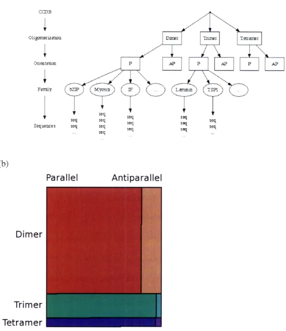
2.4.1 A database of structurally-annotated sequences ... 59
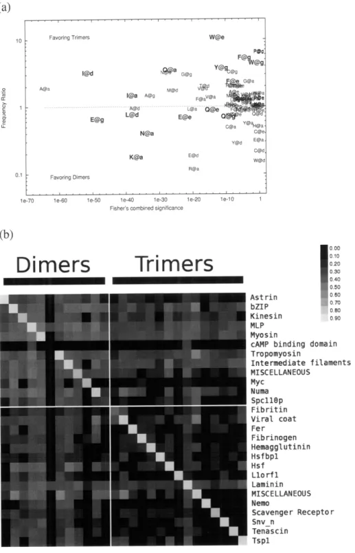
2.4.2 Features of the dimer and trimer sequences...61
2.4.3 Retraining Multicoil to Multicoil2 ... ... 63
2.4.4 V alidation ... ... 65
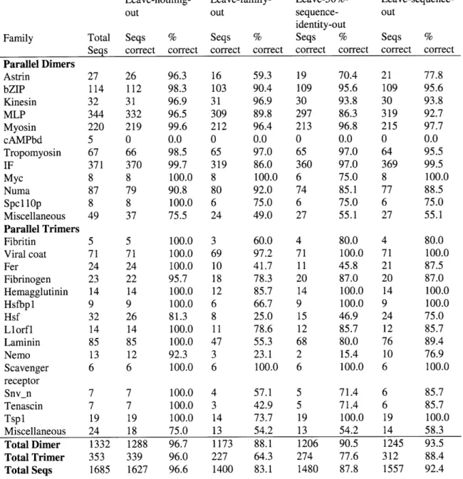
2.4.4.1 Leave-family-out testing ... 66
2.4.4.2 Leave-percent-identity-out testing ... 68
2.4.4.3 Leave-sequence-out testing... .... 70
2.4.5 Improvement over Multicoil (1997) ... 71
2 .5 D iscussion ... 73
2.6 R eferences ... 77
3 Predicting helix orientation for coiled-coil dimers 3.1 A b stract ... ... 8 1 3.2 Introduction ... 83 3.3 M ethods... ... 87 3.3.1 Coiled-coil database ... ... 87 3.3.2 Crick parameterization... 88 3.3.3 Generation of backbones...90 3.3.4 Evaluation of structures ... ... 91
3.3.5 Energy functions - ESMs ... ... 92
3.3.6 Energy functions - ISMs ... ... 94
3.4 R esults ... . ... 95
3.4.1 Performance of explicit structure models ... 99
3.4.2 Performance of implicit structure models ... 103
3.4.3 Analysis... ... 105
3.4.4 C onfidence ... 116
3.5 D iscussion ... 117
3.6 Acknowledgements ... 122
3.7 References ... ... 123
4 Structure-based approaches to the prediction of coiled-coil alignment 4.1 Introduction... 127 4.2 Methods...130 4.2.1 Fram ew ork ... 130 4.2.2 Test sets... ... 132 4.2.3 Scoring models...133 4.2.4 Performance metrics ... ... 136 4.2.5 Homodimer preference ... ... 137 4.2.6 Model optimization... ... 137 4 .3 R esults ... . 138 4.3.1 Performance of ISMs ... .. ... 140 4.3.2 Homotypic bias ... 143 4.3.3 Performance of ESMs...146 4.3.4 Model optimization... ... 149 4.4 D iscussion ... 15 1 4.5 R eferences... 156
5 Conclusions and Future Directions 5.1 Prediction of coiled coil structure ... 159
5.2 Coiled-coil databases and statistics-based prediction ... 160
5.3 Current prediction of coiled-coil structural features ... 162
5.4 The future of coiled-coil prediction methods... 164
5.4.1 Improvements to existing methods ... 164
5.4.2 Folding-based models ... ... 165
5.5 Applications of coiled-coil structure prediction ... 166
5.5 R eferences ... 169
Appendix A Residue frequencies from NPS database ... 171
Appendix B Supplementary material for Chapter 3: Predicting helix orientation for coiled-coil dim ers... 179
List of Figures
1-1 Illustration of coiled-coil structure... .... 22
1-2 Growth of known genomic and coiled-coil sequence databases ... 28
2-1 Overview of the NPS coiled-coil database ... .... 52
2-2 Flow chart of validation m ethod ... 58
2-3 Characteristics of the NPS coiled-coil database ... 62
2-4 Distributions of raw scores resulting from cross-validation testing ... 66
2-5 Leave-family-out cross-validated raw score plots per family...69
2-6 Effect of leave-N%-identity-out threshold on prediction performance ... 71
3-1 Crick parameterization of parallel and antiparallel coiled coils ... 84
3-2 Parallel vs. antiparallel discrimination performance of different methods.... 100
3-3 Overview of prediction performance and component analysis...107
3-4 Energy component contributions to performance... 110
3-5 Distribution of Ca-Ca distances for core residues in parallel and antiparallel coiled coils ... 14
3-6 Performance as a function of increasing the gap requirement...117
4-1 Alignment prediction framework...131
4-2 Prediction perform ance of ISM s... 141
4-3 Component analysis of selected ISMs ... ... 142
4-4 H om odim er bias analysis ... ... 144
4-5 Perform ance of ESM s ... 147
4-6 Component analysis of selected ESMs ... 148
4-7 Performance of modified, unoptimized ESMs...149
B-i Native coiled-coil variation described using Crick parameterization...186
B-2 Histogram of the parallel Crick parameters generated by fitting parallel test-set structures to the best possible Crick backbone...187
B-3 Histogram of the antiparallel Crick parameters generated by fitting antiparallel test-set structures to the best possible Crick backbone...189
B-4 Antiparallel OIA and DB correlation for all structures in the test set... 192
List of Tables
1-1 Key coiled-coil interactions ... 31
2-1 Estimates of the coiled-coil dimer and trimer content of various genomes...64
2-2 Multicoil2 prediction performance for all families under different testing protocols... ... 67
3-1 Test set of coiled-coil dimers of known orientation ... 97
3-2 Summary of pair terms used in ISM models ... 103
4-1 Summary of alignment test sets ... 132
4-2 Pair term s used in ESM s... ... 134
B-1 List of PQS structures in the test set ... 180
B-2 Chi angle recovery of repacked structures ... 184
Chapter 1
Introduction
1.1 Protein structure-function relationships and the study of conserved structural motifs
Significant effort in current biomedical research is devoted to understanding how proteins function and malfunction. Proteins are known to have highly specific yet diverse structures, and in each instance, structure and function are closely intertwined. Many significant advancements in molecular biology have been made through studying protein structure, making methods of experimental structural characterization and computational structure prediction important for modem biology[l].
Studies across the large number of solved protein structures have revealed that proteins often contain conserved structural domains[2]. These domains, despite being present in diverse proteins, often share evolutionary history, significant sequence identity, and basic functions. Understanding the structures and functions of commonly recurring
conserved domains is one strategy adopted by protein scientists to broaden our understanding of structure-function relationships. Here, I illustrate this by briefly reviewing a few of the most common and most studied domains along with progress on predicting their occurrences and annotating their functions.
One large class of protein domains is involved in mediating protein-peptide associations. This class includes, among many others, the SH2, SH3 and PDZ domains. SH2 and SH3 domains have been primarily identified in signal transduction processes, in which SH2 domains preferentially bind phosphotyrosine-containing peptides, while SH3 domains bind proline-rich peptides and are often associated with protein kinases[3]. PDZ domains have been found to form scaffolding interactions, also primarily in signal transduction[4]. Because these domains mediate many critical protein-protein interaction networks, much work has been done to characterize the structure and specificity of these domains[5,6]. However, while identifying domains is relatively straightforward using sequence comparison methods, identifying putative interaction peptides and measuring or predicting the interaction specificity of these domains is an area of active research[7,8,9,10].
Another important class of domains are the zinc fingers. Each of these short (20-30 residue) domains coordinates a zinc ion through combinations of cysteine and histidine residues. Although found primarily as specific DNA binding domains in a wide array of transcription factors and other nucleic acid binding proteins[11], zinc fingers have also been identified as protein-lipid and protein-protein interaction domains[12]. Zinc fingers can be recognized by their sequence similarity and are extremely common, being identified in approximately 2% of all human proteins[ 11]. In their function as DNA
recognition domains, chains of zinc fingers have shown combinatorial specificity for DNA sequence[13]. This combinatorial aspect has important implications for evolutionary mechanisms of DNA-binding specificity and has been exploited in protein engineering [14]. As with the protein-peptide interaction domains, the prediction of DNA-binding specificity is important for understanding transcriptional regulation networks, and has been addressed using machine learning approaches[15].
A highly conserved catalytic structure is that of the protein kinase catalytic domains, which preferentially phosphorylate key serine, threonine or tyrosine residues in their specific substrates. These kinase domains generally share similar structures, which are conserved from yeast to humans[16]. However, despite this domain conservation, kinases are known to have diverse substrate specificity[17]. This specificity plays an important role in determining signal transduction pathways that are critical to all biological processes. Therefore, many diverse approaches to predicting the specificity of kinases and substrates have been developed[17,18,19].
The conserved structural domain that forms the subject of this thesis is the alpha-helical coiled coil. Many proteins, a few of which are highlighted below, have been identified to contain coiled coils, and a recent survey of 22 proteomes using a coiled-coil detection method has estimated that between 2% and 8% of all proteins in any given proteome contain coiled-coil motifs[20]. Unlike other protein domains, however, the function of coiled coils is not always easily inferred from sequence similarity. Many diverse coiled coils share very low levels of sequence similarity, and these have a wide range of functions. Coiled-coil-containing fibrous proteins are involved in cellular architecture, shorter coiled coils act as critical structural and mechanical elements of
globular proteins, and other coiled coils mediate protein-protein interactions to regulate key cellular functions[21]. Because each coiled coil has a function that is influenced strongly by its structure, understanding the functions of novel coiled coils would be significantly enhanced through methods of predicting coiled-coil structure.
In this chapter, I describe some of the history and structure of the coiled coil, how prediction of coiled-coil structure can play an important part in understanding protein function, and how coiled-coil modeling is related to modeling of other types of protein folds. Compared to predicting the structures of proteins generally, there are both significant advantages as well as particular challenges involved in the prediction of coiled-coil structure. Statistical approaches, previously demonstrated for secondary structure prediction, have been successfully used in the prediction of coiled-coil structure. However, coiled-coil databases used for training such methods, particularly those annotated with structural information, have lagged behind the growth of sequence and structure databases. In addition, structural approaches such as fold recognition have been widely used in the prediction of protein structure, and the application of such techniques to the coiled-coil geometry is discussed. Finally, this chapter summarizes the recent advancements in coiled-coil structure prediction as presented in this thesis.
1.2 The history of coiled-coil structure
The coiled coil was first proposed in 1952 by Francis Crick as a solution to the structure of certain fibrous proteins such as keratin[22]. Earlier models and experiments had previously established the alpha helix as a key protein structural element[23];
however, X-ray fiber diffraction data of keratin did not exactly fit the alpha-helix model. Crick's proposal was that the alpha helix, with only minor distortion, could be twisted into a supercoil and pack against other alpha-helices having the same supercoil. This packing was suggested to occur via a "knobs-into-holes" mechanism, where a side chain protruding from one helix (the knob) packs into the space between four side chains from the opposing helix (the hole)[24]. Figure 1-1 illustrates this basic structure. Later analyses of alpha-helix interactions have shown that knobs-into-holes packing is the most common of several classes of possible helix packing modes, including ridges-into-grooves and knobs-onto-knobs[25,26].
Crick and others also noted that a seven-residue periodicity overlaid on the helices involved in a coiled-coil interaction would result in a consistent set of residues present in the core and peripheral positions[24,27]. This "heptad" periodicity, denoted by the letters (a-b-c-d-e-f-g), placed hydrophobic residues primarily at the a and d positions, creating a hydrophobic core that stabilized the interaction. This was first confirmed through sequence analysis of the coiled-coil region of tropomyosin which showed a seven-residue periodicity of hydrophobic residues[28,29].
Crick recognized that this simple combination of knobs-into-holes interaction and heptad repeat could be achieved through the interaction of two, three or more helices together in parallel or antiparallel relative orientations[24]. But it was not until 1963 that tropomyosin was demonstrated to form a dimer[30], and the parallel orientation of myosin was not confirmed until 1967[31]. These and further studies on fibrous proteins led to the idea that the coiled coil was most often long, parallel and dimeric[32]. However, it soon became clear that significant coiled-coil structural diversity could be
a)
c)
d)
cz'
e)
f)
g)
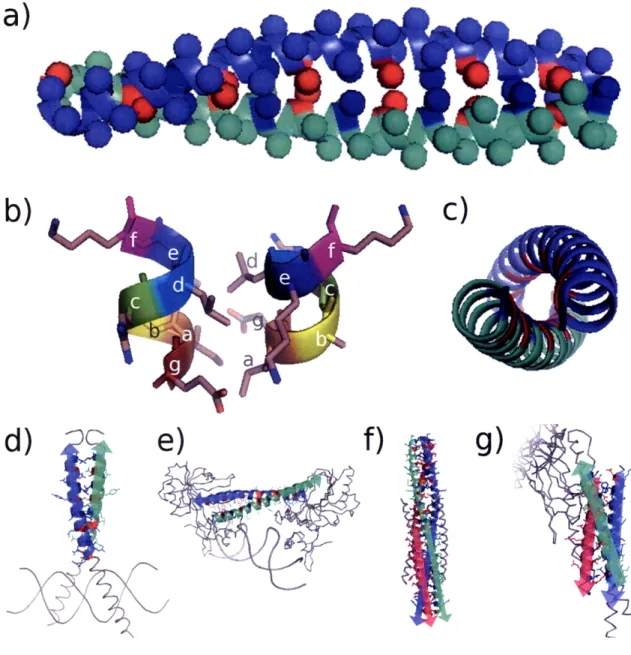
Figure 1-1. Illustration of coiled-coil structure. (a) Side view of a parallel dimeric coiled coil. Spheres denote location of C3 atoms. Red and blue spheres represent a and d position residues, respectively. (b) Structure of the heptad repeat in a parallel dimeric configuration. Note hydrophobic interactions of a-a' and d-d' residues, as well as Glu-Lys g-e' charge pair interaction. (c) Top view of parallel dimer, showing consistency of hydrophobic interaction (red and blue segments) along the supercoil. (d-g) Examples of various coiled-coil structures. Colored cartoon region is coiled-coil assigned by SOCKET[33]. Arrows denote N--C sequence polarity. (d) Parallel dimer (e) Antiparallel dimer (f) Parallel trimer (g) Antiparallel trimer)
found in a wide range of protein families. The crystal structure of influenza hemagglutinin was published in 1981, showing a three-stranded coiled coil as the core of this globular protein[34]. ROP, a protein involved in plasmid replication in E. coli, was shown to consist entirely of a dimer of antiparallel-associated helices, creating a four-stranded coiled coil[35]. In addition, crystal structures of E. coli seryl-tRNA synthetase clearly showed a five-heptad-long antiparallel dimeric coiled coil, possibly involved in binding tRNA[36]. Crystallographic studies of the leucine-zipper-containing protein GCN4 demonstrated that it contained a two-stranded, parallel coiled coil[37], settling a standing debate over the structure of the leucine zipper motif[38].
1.3 Computational prediction of coiled-coil structures
Since the original discovery of the coiled coil, increasing numbers of coiled-coil domains have been found to be relevant for a wide array of protein structures and interactions [39]. For example, the gp41 protein from the HIV virus contains a coiled-coil trimer at its core that is crucial for viral-membrane fusion[40]. The SNAREs are a class of yeast and mammalian membrane fusion proteins that associate as hetero-tetrameric coiled-coil complexes comprised of three distinct proteins[41]. Motor proteins such as myosin and kinesin use coiled coils as rigid rods to transmit force between load and substrate[21]. The alphavirus capsid consists of capsid proteins that are known to dimerize through a coiled-coil domain[42]. This dimerization is crucial for proper capsid assembly, as mutations to the coiled-coil domain that promote trimerization disrupt proper assembly[43]. In all of these examples, the structure of the coiled coil is critical to
the overall activity of the protein. However, over the past several decades, the growth of sequence databases has significantly outpaced that of structure databases[44]. There remain many coiled-coil-containing proteins with unsolved structures and important functions. For example, the yeast spindle pole body is hypothesized to contain many coiled coils[45], some of which have already been implicated in determining the architecture of the large complex[46]. Therefore, computational methods that could predict the structure of a coiled coil from its sequence alone would significantly enhance our understanding of many aspects of biology.
The prediction of coiled-coil structure is a subproblem of the general protein structure prediction problem. Many approaches have been devised to predict protein structure generally. The first methods focused solely on predicting secondary structure through simple statistical models[47,48]. Later, the concept of "fold recognition" was introduced as an inverse protein folding problem; that is, the problem of finding a sequence compatible with an observed fold[49,50]. Such approaches were the first reasonably successful general protein structure prediction methods, but they can only make accurate predictions for sequences that adopt a previously crystallized fold. Despite this constraint, the consistent growth in protein structure databases, as well as developments in structure evaluation potentials, have contributed to the continuing success of fold recognition[51].
For structures without appropriate templates, modern advancements in structure sampling algorithms, scoring functions and computational power have enabled the development of ab initio or "free modeling" prediction methods that do not require a complete template model[52,53]. These approaches have shown great promise on certain
prediction problems, such as high-resolution structure prediction[54,55,56], improving homology models[57], and flexible-backbone protein-protein docking[58]. However, such methods are still relatively unreliable, computationally expensive and not yet
available for genome-scale applications[51].
Given the many advancements in general protein structure prediction, it would be natural to conclude that such approaches may be directly useful for predicting the structure of the coiled coil. While this is a possibility, it is more likely that methods specifically designed to utilize features of the coiled coil, such as the regular supercoil and the heptad repeat, will reduce the need for crystal-based templates or extensive structural sampling, and may be more accurate in their predictions. However, there are unique challenges to predicting coiled-coil structure, such as fewer topological restraints when predicting coiled-coil interactions, and many candidate template structures with very similar energetics, as discussed in Section 1.6. These challenges have hindered the development of methods that are able to completely predict coiled-coil structure. However, much work has been done to address various subproblems of this goal, which is summarized below.
1.4 Statistical models for coiled-coil structure prediction
One of the key challenges in coiled-coil structure prediction is identifying regions of sequence that have the propensity to form coiled coils. This is similar to approaches that assign secondary structure propensity to un-annotated sequence. Chou and Fasman first developed such a method, which was trained using statistical information derived
from crystallographic observations[47]. This was later improved through the GOR method, which considers pairwise residue information[48]. While these initial methods were not highly accurate, modern approaches can achieve up to 70-80% prediction accuracy through sequence-profile information and neural network approaches[59,60].
The first practical method of coiled-coil propensity prediction was suggested by Parry, who proposed an algorithm based on the geometrical average of heptad-position-specific residue frequencies determined from a small collection of diverse coiled-coil sequences[61]. This approach was extended by Lupas et al., who suggested the use of a maximum-over-window function to smooth scores and calibrated the results to a large database of protein sequences in order to estimate the probability that a certain sequence corresponds to a coiled-coil structure[62]. The Lupas approach, as originally captured in the program COILS, is highly effective in many situations, but it is vulnerable to false positives. The Paircoil algorithm extends the heptad-position-specific approach to include pairwise heptad positions, similar to how the GOR method considers the effect of neighboring residues on secondary structure propensity[48]. Paircoil was shown to be more specific and yields fewer false positives[63]; however, the corresponding drastic increase in parameter space leaves open the question of whether or not the available sequence information is sufficient for training[64].
The success of the Paircoil method inspired several extensions of its approach to different problems in coiled-coil structure prediction. The Learncoil method[65], which applies Paircoil in an iterative training process to improve detection of under-represented families, was successfully used to predict coiled coils found in viral membrane fusion proteins [66] and histidine kinases[67]. In addition, the recognition that known
coiled-coil dimers and trimers exhibit significantly different residue preferences[68] prompted development of the Multicoil method[69], which uses pairwise residue correlations from a large dimer and relatively small trimer database to predict the propensity of sequences to form one of those two structures. Finally, recognition of the significant growth in protein sequence databases prompted updating the original Paircoil training set, resulting in Paircoil2[70] which showed improved performance.
One aspect of all previously mentioned approaches is that they rely on a window-based method to smooth scores for more accurate predictions. The Marcoil method does not use such a window; instead, it is based on a hidden Markov model that uses residue state transition probabilities to model the coiled-coil region[71]. Eliminating the window makes it possible to predict much shorter coiled coils. A recent review of the above methods suggests that while performance has improved significantly from the original, simple approaches, it is still not possible to recognize all structurally confirmed coiled coils with high confidence[64].
1.5 Coiled-coil sequence databases
All existing coiled-coil detection methods require a database of known coiled-coil sequence for training purposes, and all except Marcoil require accurate heptad annotations. The size and composition of the training databases plays an important role in the ability of any given method to detect both known and previously unknown coiled-coil sequence. As seen in Figure 1-2, the amount of sequence annotated with coiled-coil structure initially grew to match the pace of growth in all known genomic sequence;
1E+08 1 E+07 1E + 0 6 - " 1E+05 1E+04 + 1E+03 1E+02 1E+01 1E+00 Year --- Genbank Sequences + Coiled-coil residues cO
&
@
ob ob oFigure 1-2. Growth of known genomic and coiled-coil sequence databases. Genbank sequence size from [73]. Coiled coil database sizes from [61,62,63,69,70,74] and Chapter 2.
however, after a certain amount of time, that growth slowed significantly. This is likely due to the exponential growth in sequence information overwhelming the predominantly manual approaches used for curating such databases. The vast majority of sequences in the original databases were collected from long fibrous coiled coils such as myosin, tropomyosin and keratin[62]. Recently, the development of the SOCKET algorithm[33], which detects the characteristic knobs-into-holes packing of coiled coils in PDB structures, has opened the possibility of including many previously poorly annotated short coiled coils in sequence databases[72]. However, the relatively small size of the protein structure database has minimized the impact of such tools on general coiled-coil sequence databases. Currently, the most common way to obtain new coiled-coil sequence
is to use homology searches from existing annotated sequence and to individually discover and characterize new families using experimental data[70].
Predicting additional features of the coiled coil, such as dimerization versus trimerization preference, requires databases that are annotated beyond the heptad repeat. Early coiled-coil sequence databases consisted of sequences with uniform (parallel stranded) structure. Subsequently, Woolfson and Alber studied a small database of two-and three-strtwo-anded coiled coils (-2000 residues each) two-and identified patterns of residue preferences that could distinguish between these two sets[68]. These successes prompted the development of the Multicoil method and database[69], which contained 6,319 three-stranded coiled-coil residues along with the 58,191-residue two-three-stranded database created for Paircoil[63]. However, as discussed in Chapter 2, this three-stranded database does not capture enough diversity to accurately predict some coiled coils. The primary aim of the work described in Chapter 2 was to increase these database sizes and characterize performance of the Multicoil method under more rigorous validation standards than were possible when the method was first developed.
1.6 Structural models for coiled-coil prediction
The previously described prediction methods, with the exception of Multicoil, only predict the propensity of a sequence to form generic coiled-coil structure. However, as discussed above, coiled coils have been found to adopt a wide variety of structures[75], including variations in number of helices, helix orientation, alignment and partnering preference (Figure 1-1). Because protein sequence encodes structure, given the
proper model, it should be possible to predict structure from sequence. However, in some cases, closely related sequences have been observed to form different structures, and the energetic threshold between these structures can be low. For example, studies of point mutations in the GCN4 leucine zipper (bZIP) domain showed variation not only in interaction stability but in helix number, helix orientation and partnering preference[76,77]. A solvent-exposed mutation in a GCN4 variant was shown to specify the formation of parallel or antiparallel tetramers[78]. Studies of a model parallel dimeric coiled coil demonstrated the formation of antiparallel dimers simply by moving the position of one core asparagine residue[79], and further investigation showed the overall contribution of the resulting core polar interaction was roughly equivalent to that of a single interhelical electrostatic interaction, approximately 2.1 kcal/mol[80]. Therefore, in order to accurately predict what type of structure a sequence will form, accurate structural models that consider the entire complex are likely necessary.
1.6.1 Simple implicit structure models
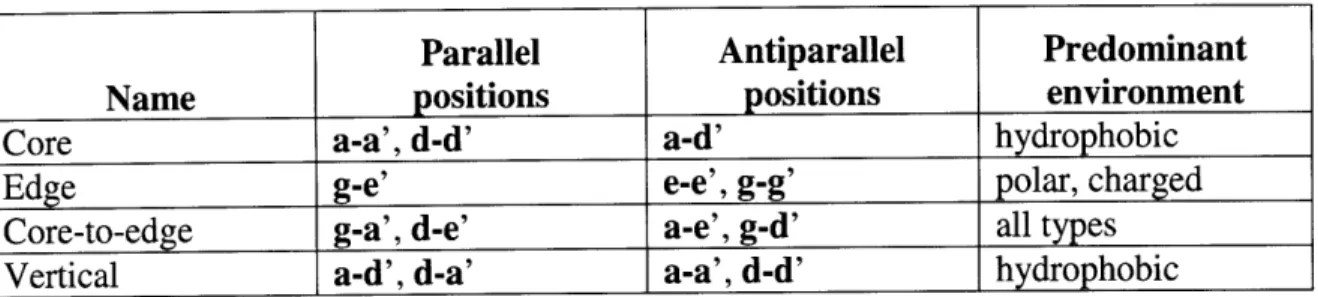
One of the earliest innovations in the prediction of coiled-coil structure was the recognition that a key set of interactions between specific positions of the heptad (summarized in Table 1-1) are most important in determining structural preference[81]. This led to the development of a simple "charge-patterning" model where charge-charge
interactions between g and e' residues were scored according to their
complementarity[82]. A similar charge-patterning model was successful at predicting the hetero-trimerization of laminins[83]. This rational approach of distilling complex
Parallel Antiparallel Predominant
Name positions positions environment
Core a-a', d-d' a-d' hydrophobic
Edge g-e' e-e', g-g' polar, charged
Core-to-edge g-a', d-e' a-e', g-d' all types
Vertical a-d', d-a' a-a', d-d' hydrophobic
Table 1-1. Key coiled-coil interactions. Interacting positions (such as g-e') denote an interaction between a g position residue on one helix and an e position residue on the other (prime) helix.
interactions into simple integer scores was later extended to include simple core patterning terms, and was successful at predicting the association preference of a set of bZIP proteins[84]. A similar rational model which considered charge and core patterning along with a helix-propensity term was recently shown to correlate well with a set of melting temperatures of bZIPs[85]. However, these types of models are extremely low-resolution and tend to assign the same interaction weights to a group of related residues, even when experiments have suggested that this is not strictly appropriate.
In order to refine such simplistic association models, the technique of double-mutant thermodynamic cycle analysis was used. Experimental coupling energies between residues commonly found at both e-g'[86,87] and a-a'[88,89] heptad pair positions (Table 1-1) were measured in a model system derived from the avian bZIP coiled coil VBP. A small set of antiparallel coupling energies have also been derived using synthetic peptides[90,91]. The advantage of these coupling energies is that the thermodynamic cycle allows for isolation and quantification of the interaction between a specific pair of residues[92]. On a test of predicting a set of known bZIP associations, coupling energies were shown to consistently perform better than a set of simple charge-patterning weights[93]. While this approach appears to provide useful data, its major disadvantage is
the large number of experiments necessary to create and characterize the mutant proteins. This has limited the number of residue pairs for which data is available.
Due to the significant amount of coiled-coil sequence available, machine learning approaches are an attractive alternative to extensive experimental characterization. Singh and Kim used a support vector machine (SVM) approach to determine residue pair weights for core, edge and core-to-edge interactions (Table 1-1, [74]). The major advantage of the SVM framework is that it allows for learning from heterogeneous data derived from both sequence and experimental observations. The resulting weights were shown to give good performance in predicting dimeric coiled-coil alignment, as well as partner prediction among both the keratin[74] and bZIP families[93]. However, the resulting weights are not always physically interpretable, as they are optimized only to provide the greatest separation between "interacting" and "non-interacting" datasets.
Despite their diversity of construction, each of the previously described methods shares a common trait: pairwise residue interactions used in evaluating models are predefined according to knowledge about the structural relationships in canonical coiled coils. These interactions are assumed to be independent at the pairwise level, and their contributions to the stability of the complex are assumed to be additive. Therefore, stability can be calculated simply by summing the scores assigned to all relevant interactions. This class of models effectively has structure information "baked in" in an implicit form, hence the name "implicit structure model" (ISM).
1.6.2 Statistical contact potential-based implicit structure models
As mentioned previously, fold recognition techniques have been widely used in the prediction of protein structures. The basic process of fold recognition involves collecting a set of structural templates, then "threading" a sequence to be predicted onto those templates using a sequence/structure alignment protocol[94]. A key distinction among fold recognition methods is the scoring function used to evaluate candidate structural solutions. One common class of scoring functions is the statistical residue-based contact potentials. Potentials such as these are constructed from the frequencies of pairwise residue "contacts" (residue-residue distances below a given cutoff) in a large database of protein structures, that are normalized to generate additive scores[95]. Other, more detailed functions have been developed and tested, and have shown improved performance at the expense of computational complexity[96].
The growing number of protein-protein interactions observed in structural data has enabled the development of multimeric threading approaches that predict both protein tertiary and quaternary structure. The MULTIPROSPECTOR method considers as templates protein pairs that are observed to interact; candidate sequences are threaded first onto individual components of the complex and then onto the complex itself. At the complex stage, models are scored using a statistical contact potential specifically derived from observed protein-protein interfaces. This method, along with others, has been shown to recapitulate pieces of the protein interaction networks observed through experimental
This work on structural prediction of protein-protein interactions using fold-recognition methods was used as a starting point for predicting coiled-coil association, as discussed in Chapters 3 and 4. We developed a statistical contact potential, known as RISP, that is similar to those discussed above but is derived exclusively from heterotypic protein-protein interfaces. While we first tested an approach where interhelical contacts were defined using distance constraints derived from 3D models, as is common in fold recognition, we found that the implicit structure modeling framework used in conjunction with residue contact potentials was not only less computationally intensive but also more accurate. Implicit structure models in the form of position-specific scoring matrices derived from statistical potentials have been used previously to predict SH2-phosphopeptide associations[8]. Unfortunately, the major drawback to such approaches is the effort needed to develop new models for each possible structural variant. This process is not always straightforward, as discussed in Chapter 4, where we observed that different structures and even different sequence families are predicted best with different pairwise heptad interactions. In addition, the representation of residue interactions as a single value approximates the "true" potential, which can include multi-body effects such as rotamer selection, packing strain and desolvation[99].
1.6.3 Explicit structure models
In recognition of the limitations of the implicit-structure approach, an alternative class of structure modeling considers the full 3D representation of the interaction at the atomic level. This detailed model allows for consideration of multi-body effects, as well
as having much finer granularity of interatomic distances. Such models were originally developed for detailed modeling and refinement of protein structures[100,101], and modern high-resolution scoring functions form the basis of the ab initio structure prediction methods described above[ 102]. These potentials are applicable to the coiled-coil structure as long as a suitable structural modeling framework is used.
One challenge with traditional fold recognition approaches is that coiled coils have significant diversity in their backbone structures, not only in gross architectural parameters but also in fine details such as superhelical pitch, radius and interhelical displacement[103]. Instead of the fragment library approach used by methods such as ROSETTA[54], the mathematical description of the coiled coil originally proposed by Crick prompted the development of parameterized models of coiled-coil backbones. These form the basis of most current coiled-coil explicit structure models. The first example of this work was in the prediction of the coiled-coil structure of GCN4 to within 1.75 A, several months before the crystal structure was published[104,105]. Harbury and coworkers used structure-energy minimization techniques to predict the rotamer preferences of GCN4[106], as well as to design a coiled coil with a slight right-handed superhelical twist[107]. Keating et al. later extended this technique to predict the association preferences of core-position mutants in designed parallel heterodimers[ 108].
In contrast to the above approaches, which use parameterized constraints during structure minimization to refine each modeled structure, Grigoryan and Keating modeled a large number of native bZIP sequences on a fixed coiled-coil backbone[109]. This approach, after correcting for concerns with the unfolded reference state and poorly
modeled core-position interactions, was shown to be superior to implicit-structure models derived from coupling energies[109].
In Chapters 3 and 4 of this thesis, we have investigated the combination of modeling backbone flexibility using parameterized backbones along with evaluating the resulting structures with a variety of previously published energy functions. Our results indicate that careful modeling can elucidate some of the determinants of structural specificity; however, issues with structural sampling as well as reference state models can diminish performance significantly.
While explicit structure models show some predictive ability, these models suffer from several important limitations. First, the computational time necessary to build and evaluate such detailed models makes this approach prohibitive for genome-scale prediction. Computational time constraints also limit the amount of structural sampling necessary to achieve an optimal model; several studies have demonstrated the detrimental effect of even small inaccuracies in an evaluated structure[110,111]. However, cluster expansion methods[ll112,113], which can be used to derive a set of sequence-based weights from explicitly computed energies, effectively build a bridge between the explicit and implicit structure models, making it possible to efficiently use explicit structure models in computationally intensive tasks. For example, this method has recently proven highly useful for designing novel heterospecific inhibitors of bZIP interactions[ 114].
1.7 Summary of thesis work
Progress has been made in the field of coiled-coil structure prediction, yet several major issues remain. This thesis describes our recent work in updating statistical coiled-coil prediction methods using new sequences, introducing more stringent tests of method performance and exploring the behavior of a range of both implicit and explicit structure models for two important coiled-coil structure prediction problems. Dramatic growth of protein sequence and structure databases prompted us to develop updated coiled-coil sequence databases. These databases, as described in Chapter 2, are organized by structure and useful for training and validating prediction methods. The significantly larger amount of available sequence expands the types of tests that can be used for method validation. In addition, two major aspects of coiled-coil structure - helix orientation and helix alignment - have yet to be comprehensively treated. Chapters 3 and 4 address these two problems, respectively, and show that useful prediction performance can be achieved using both simple implicit structure models as well as detailed explicit structure models. Finally, Chapter 5 discusses the progress made in coiled-coil structure prediction and proposes future approaches towards the ultimate goal of a unified, accurate coiled-coil structure prediction framework.
1.8 References
1. Cohen C (2007) Seeing and Knowing in Structural Biology. J Biol Chem 282: 32529-32538.
2. Kohn W, Mant C, Hodges R (1997) alpha -Helical Protein Assembly Motifs. J Biol Chem 272: 2583-2586.
3. Engen JR, Wales TE, Hochrein JM, Meyn MA, Banu Ozkan S, et al. (2008) Structure and dynamic regulation of Src-family kinases. Cellular and molecular life sciences : CMLS 65: 3058-3073.
4. Ranganathan R, Ross E (1997) PDZ domain proteins: Scaffolds for signaling complexes. Current Biology 7: R770-R773.
5. Wiedemann U, Boisguerin P, Leben R, Leitner D, Krause G, et al. (2004) Quantification of PDZ domain specificity, prediction of ligand affinity and rational design of super-binding peptides. Journal of molecular biology 343: 703-718.
6. Tonikian R, Zhang Y, Sazinsky S, Currell B, Yeh J-H, et al. (2008) A Specificity Map for the PDZ Domain Family. PLoS Biology 6: e239.
7. Li L, Wu C, Huang H, Zhang K, Gan J, et al. (2008) Prediction of phosphotyrosine signaling networks using a scoring matrix-assisted ligand identification approach. Nucleic acids research 36: 3263-3273.
8. Sanchez IE, Beltrao P, Stricher F, Schymkowitz J, Ferkinghoff-Borg J, et al. (2008) Genome-wide prediction of SH2 domain targets using structural information and the FoldX algorithm. PLoS computational biology 4.
9. Hou T, Xu Z, Zhang W, McLaughlin W, Case D, et al. (2008) Characterization of domain-peptide interaction interface: a generic structure-based model to decipher the binding specificity of SH3 domains. Mol Cell Proteomics: M800450-MCP800200.
10. Fernandez-Ballester G, Beltrao P, Gonzalez JM, Song Y-H, Wilmanns M, et al. (2009) Structure Based Prediction of the S. cerevisiae SH3-Ligand Interactions. Journal of molecular biology.
11. Matthews JM, Sunde M (2002) Zinc fingers--folds for many occasions. IUBMB life 54: 351-355.
12. Brayer KJ, Segal DJ (2008) Keep your fingers off my DNA: protein-protein interactions mediated by C2H2 zinc finger domains. Cell biochemistry and biophysics 50: 111-131.
13. Choo Y (1997) Physical basis of a protein-DNA recognition code. Current Opinion in Structural Biology 7: 117-125.
14. Henikoff S, Greene EA, Pietrokovski S, Bork P, Attwood TK, et al. (1997) Gene families: the taxonomy of protein paralogs and chimeras. Science 278: 609-614. 15. Persikov AV, Osada R, Singh M (2008) Predicting DNA recognition by Cys2His2
zinc finger proteins. Bioinformatics (Oxford, England).
16. Hanks SK, Quinn AM, Hunter T (1988) The protein kinase family: conserved features and deduced phylogeny of the catalytic domains. Science 241: 42-52.
17. Kobe B, Kampmann T, Forwood JK, Listwan P, Brinkworth RI (2005) Substrate specificity of protein kinases and computational prediction of substrates. Biochim Biophys Acta 1754: 200-209.
18. Wong YH, Lee TY, Liang HK, Huang CM, Wang TY, et al. (2007) KinasePhos 2.0: a web server for identifying protein kinase-specific phosphorylation sites based on sequences and coupling patterns. Nucleic acids research 35.
19. Miller ML, Blom N (2009) Kinase-specific prediction of protein phosphorylation sites. Methods in molecular biology (Clifton, NJ) 527.
20. Rose A, Schraegle SJ, Stahlberg EA, Meier I (2005) Coiled-coil protein composition of 22 proteomes--differences and common themes in subcellular infrastructure and traffic control. BMC evolutionary biology 5.
21. Rose A, Meier I (2004) Scaffolds, levers, rods and springs: diverse cellular functions of long coiled-coil proteins. Cellular and molecular life sciences : CMLS 61:
1996-2009.
22. Crick FH (1952) Is alpha-keratin a coiled coil? Nature 170: 882-883.
23. Pauling L, Corey R, Branson HR (1951) The Structure of Proteins. Proceedings of the National Academy of Sciences of the United States of America 37: 205-211. 24. Crick FHC (1953) The packing of alpha-helices: simple coiled-coils. Acta
Crystallographica 6: 689-697.
25. Chothia C, Levitt M, Richardson D (1977) Structure of proteins: packing of alpha-helices and pleated sheets. Proc Natl Acad Sci U S A 74: 4130-4134.
26. Walther D, Eisenhaber F, Argos P (1996) Principles of helix-helix packing in proteins: the helical lattice superposition model. J Mol Biol 255: 536-553.
27. Pauling L, Corey R (1953) Compound Helical Configurations of Polypeptide Chains: Structure of Proteins of the [alpha]-Keratin Type. Nature 171: 59-61.
28. Parry DA (1975) Analysis of the primary sequence of alpha-tropomyosin from rabbit skeletal muscle. Journal of molecular biology 98: 519-535.
29. Hodges RS, Sodek J, Smillie LB, Jurasek L (1973) Tropomyosin: Amino Acid Sequence and Coiled-Coil Structure. Cold Spring Harbor Symposia on Quantitative Biology 37: 299-310.
30. Cohen C, Holmes KC (1963) X-ray diffraction evidence for alpha-helical coiled-coils in native muscle. Journal of molecular biology 6: 423-432.
31. Slayter H, Lowey S (1967) Substructure of the Myosin Molecule as Visualized by Electron Microscopy. Proceedings of the National Academy of Sciences of the United States of America 58: 1611-1618.
32. Caspar DL, Cohen C, Longley W (1969) Tropomyosin: crystal structure, polymorphism and molecular interactions. Journal of molecular biology 41:
87-107.
33. Walshaw J, Woolfson DN (2001) Socket: a program for identifying and analysing coiled-coil motifs within protein structures. J Mol Biol 307: 1427-1450.
34. Wilson IA, Skehel JJ, Wiley DC (1981) Structure of the haemagglutinin membrane glycoprotein of influenza virus at 3 [angst] resolution. Nature 289: 366-373. 35. Banner D (1987) Structure of the ColE1 Rop protein at 1.7 [angs] resolution. Journal
36. Cusack S, Berthet-Colominas C, Hirtlein M, Nassar N, Leberman R (1990) A second class of synthetase structure revealed by X-ray analysis of Escherichia coli seryl-tRNA synthetase at 2.5 A. Nature 347: 249-255.
37. O'Shea EK, Klemm JD, Kim PS, Alber T (1991) X-ray structure of the GCN4 leucine zipper, a two-stranded, parallel coiled coil. Science 254: 539-544.
38. Landschulz W, Johnson P, McKnight S (1988) The Leucine Zipper: A Hypothetical Structure Common to a New Class of DNA Binding Proteins. Science 240: 1759-1764.
39. Grigoryan G, Keating A (2008) Structural specificity in coiled-coil interactions. Current Opinion in Structural Biology 18: 477-483.
40. Chan D, Fass D, Berger J, Kim P (1997) Core Structure of gp41 from the HIV Envelope Glycoprotein. Cell 89: 263-273.
41. Sutton B, Fasshauer D, Jahn R, Brunger A (1998) Crystal structure of a SNARE complex involved in synaptic exocytosis at 2.4 A resolution. Nature 395: 347-353.
42. Perera R, Owen K, Tellinghuisen T, Gorbalenya A, Kuhn R (2001) Alphavirus Nucleocapsid Protein Contains a Putative Coiled Coil (alphal-Helix Important for Core Assembly. J Virol 75: 1-10.
43. Perera R, Navaratnarajah C, Kuhn RJ (2003) A heterologous coiled coil can substitute for helix I of the Sindbis virus capsid protein. Journal of virology 77: 8345-8353. 44. Westbrook J, Feng Z, Chen L, Yang H, Berman HM (2003) The Protein Data Bank
and structural genomics. Nucleic Acids Res 31: 489-491.
45. Zizlsperger N, Malashkevich VN, Pillay S, Keating AE (2008) Analysis of coiled-coil interactions between core proteins of the spindle pole body. Biochemistry 47:
11858-11868.
46. Kilmartin JV, Dyos SL, Kershaw D, Finch JT (1993) A spacer protein in the Saccharomyces cerevisiae spindle poly body whose transcript is cell cycle-regulated. The Journal of cell biology 123: 1175-1184.
47. Chou PY, Fasman GD (1974) Prediction of protein conformation. Biochemistry 13:
222-245.
48. Garnier J, Osguthorpe D, Robson B (1978) Analysis of the accuracy and implications of simple methods for predicting the secondary structure of globular proteins. Journal of Molecular Biology 120: 97-120.
49. Ponder J, Richards F (1987) Tertiary templates for proteins *lUse of packing criteria in the enumeration of allowed sequences for different structural classes. Journal of Molecular Biology 193: 775-791.
50. Bowie JU, Lithy R, Eisenberg D (1991) A method to identify protein sequences that fold into a known three-dimensional structure. Science 253: 164-170.
51. Zhang Y (2008) Progress and challenges in protein structure prediction. Current Opinion in Structural Biology 18: 342-348.
52. Das R, Baker D (2008) Macromolecular modeling with rosetta. Annual review of biochemistry 77: 363-382.
53. Wu S, Skolnick J, Zhang Y (2007) Ab initio modeling of small proteins by iterative TASSER simulations. BMC Biology 5: 17.
54. Bradley P, Misura K, Baker D (2005) Toward High-Resolution de Novo Structure Prediction for Small Proteins. Science 309: 1868-1871.
55. Das R, Qian B, Raman S, Vernon R, Thompson J, et al. (2007) Structure prediction for CASP7 targets using extensive all-atom refinement with Rosetta@home. Proteins 69: 118-128.
56. Zhang Y (2007) Template-based modeling and free modeling by I-TASSER in CASP7. Proteins: Structure, Function, and Bioinformatics 69: 108-117.
57. Misura KM, Chivian D, Rohl CA, Kim DE, Baker D (2006) Physically realistic homology models built with ROSETTA can be more accurate than their templates. Proc Natl Acad Sci U S A 103: 5361-5366.
58. Wang C, Bradley P, Baker D (2007) Protein-Protein Docking with Backbone Flexibility. Journal of Molecular Biology 373: 503-519.
59. Rost B, Sander C (1993) Prediction of Protein Secondary Structure at Better than 70% Accuracy. Journal of Molecular Biology 232: 584-599.
60. Jones DT (1999) Protein secondary structure prediction based on position-specific scoring matrices. J Mol Biol 292: 195-202.
61. Parry DA (1982) Coiled-coils in alpha-helix-containing proteins: analysis of the residue types within the heptad repeat and the use of these data in the prediction of coiled-coils in other proteins. Bioscience reports 2: 1017-1024.
62. Lupas A, Van Dyke M, Stock J (1991) Predicting coiled coils from protein sequences. Science (New York, NY) 252: 1162-1164.
63. Berger B, Wilson DB, Wolf E, Tonchev T, Milla M, et al. (1995) Predicting coiled coils by use of pairwise residue correlations. Proceedings of the National Academy of Sciences of the United States of America 92: 8259-8263.
64. Gruber M, S6ding J, Lupas AN (2006) Comparative analysis of coiled-coil prediction methods. J Struct Biol 155: 140-145.
65. Berger B, Singh M (1997) An iterative method for improved protein structural motif recognition. Journal of computational biology : a journal of computational
molecular cell biology 4: 261-273.
66. Singh M, Berger B, Kim PS (1999) LeamCoil-VMF: computational evidence for coiled-coil-like motifs in many viral membrane-fusion proteins. Journal of molecular biology 290: 1031-1041.
67. Singh M, Berger B, Kim P, Berger J, Cochran A (1998) Computational learning reveals coiled coil-like motifs in histidine kinase linker domains. Proceedings of the National Academy of Sciences of the United States of America 95: 2738-2743.
68. Woolfson DN, Alber T (1995) Predicting oligomerization states of coiled coils. Protein science : a publication of the Protein Society 4: 1596-1607.
69. Wolf E, Kim PS, Berger B (1997) MultiCoil: a program for predicting two- and three-stranded coiled coils. Protein science : a publication of the Protein Society 6: 1179-1189.
70. McDonnell AV, Jiang T, Keating AE, Berger B (2006) Paircoil2: improved prediction of coiled coils from sequence. Bioinformatics 22: 356-358.
71. Delorenzi M, Speed T (2002) An HMM model for coiled-coil domains and a comparison with PSSM-based predictions. Bioinformatics 18: 617-625.
72. Testa OD, Moutevelis E, Woolfson DN (2008) CC+: a relational database of coiled-coil structures. Nucleic acids research.
73. Benson DA, Karsch-Mizrachi I, Lipman DJ, Ostell J, Sayers EW (2009) GenBank. Nucleic acids research 37.
74. Singh M, Kim P. Towards predicting coiled-coil protein interactions; 2001. ACM. pp. 279-286.
75. Moutevelis E, Woolfson D (2009) A Periodic Table of Coiled-Coil Protein Structures. Journal of Molecular Biology 385: 726-732.
76. Harbury PB, Zhang T, Kim PS, Alber T (1993) A switch between two-, three-, and four-stranded coiled coils in GCN4 leucine zipper mutants. Science 262: 1401-1407.
77. Zeng X, Herndon A, Hu J (1997) Buried Asparagines Determine the Dimerization Specificities of Leucine Zipper Mutants. Proceedings of the National Academy of Sciences of the United States of America 94: 3673-3678.
78. Yadav M, Leman L, Price D, Brooks C, Stout D, et al. (2006) Coiled Coils at the Edge of Configurational Heterogeneity. Structural Analyses of Parallel and Antiparallel Homotetrameric Coiled Coils Reveal Configurational Sensitivity to a Single Solvent-Exposed Amino Acid Substitution. Biochemistry 45: 4463-4473. 79. Oakley MG, Kim PS (1998) A buried polar interaction can direct the relative
orientation of helices in a coiled coil. Biochemistry 37: 12603-12610.
80. McClain DL, Binfet JP, Oakley MG (2001) Evaluation of the energetic contribution of interhelical Coulombic interactions for coiled coil helix orientation specificity. J Mol Biol 313: 371-383.
81. Cohen C, Parry D (1990) alpha-Helical coiled coils and bundles: How to design an alpha-helical protein. Proteins: Structure, Function, and Genetics 7: 1-15.
82. Vinson CR, Hai T, Boyd SM (1993) Dimerization specificity of the leucine zipper-containing bZIP motif on DNA binding: prediction and rational design. Genes & development 7: 1047-1058.
83. Beck K, Dixon TW, Engel J, Parry DA (1993) Ionic Interactions in the Coiled-coil Domain of Laminin Determine the Specificity of Chain Assembly. Journal of Molecular Biology 231: 311-323.
84. Vinson C, Myakishev M, Acharya A, Mir AA, Moll JR, et al. (2002) Classification of human B-ZIP proteins based on dimerization properties. Mol Cell Biol 22: 6321-6335.
85. Mason JM, Schmitz MA, Miiller KM, Arndt KM (2006) Semirational design of Jun-Fos coiled coils with increased affinity: Universal implications for leucine zipper prediction and design. Proc Natl Acad Sci U S A 103: 8989-8994.
86. Krylov D, Mikhailenko I, Vinson C (1994) A thermodynamic scale for leucine zipper stability and dimerization specificity: e and g interhelical interactions. The EMBO journal 13: 2849-2861.
87. Krylov D, Barchi J, Vinson C (1998) Inter-helical interactions in the leucine zipper coiled coil dimer: pH and salt dependence of coupling energy between charged amino acids. J Mol Biol 279: 959-972.
88. Acharya A, Ruvinov SB, Gal J, Moll JR, Vinson C (2002) A heterodimerizing leucine zipper coiled coil system for examining the specificity of a position interactions: amino acids I, V, L, N, A, and K. Biochemistry 41: 14122-14131.
89. Acharya A, Rishi V, Vinson C (2006) Stability of 100 homo and heterotypic coiled-coil a-a' pairs for ten amino acids (A, L, I, V, N, K, S, T, E, and R). Biochemistry 45: 11324-11332.
90. Hadley E, Gellman S (2006) An Antiparallel [alpha]-Helical Coiled-Coil Model System for Rapid Assessment of Side-Chain Recognition at the Hydrophobic Interface. Journal of the American Chemical Society 128: 16444-16445.
91. Hadley E, Testa O, Woolfson D, Gellman S (2008) Preferred side-chain constellations at antiparallel coiled-coil interfaces. Proceedings of the National Academy of Sciences: 0709068105.
92. Horovitz A (1996) Double-mutant cycles: a powerful tool for analyzing protein structure and function. Folding and Design 1: R121-R126.
93. Fong JH, Keating AE, Singh M (2004) Predicting specificity in bZIP coiled-coil protein interactions. Genome Biol 5.
94. Jones DT, Taylor WR, Thornton JM (1992) A new approach to protein fold recognition. Nature 358: 86-89.
95. Vajda S, Sippl M, Novotny J (1997) Empirical potentials and functions for protein folding and binding. Curr Opin Struct Biol 7: 222-228.
96. Lazaridis T (2000) Effective energy functions for protein structure prediction. Current Opinion in Structural Biology 10: 139-145.
97. Lu L, Lu H, Skolnick J (2002) MULTIPROSPECTOR: an algorithm for the prediction of protein-protein interactions by multimeric threading. Proteins 49:
350-364.
98. Aloy P, Russell RB (2002) Interrogating protein interaction networks through structural biology. Proc Natl Acad Sci U S A 99: 5896-5901.
99. Boas FE, Harbury PB (2007) Potential energy functions for protein design. Curr Opin Struct Biol.
100. Brooks B, Bruccoleri R, Olafson B, States D, Swaminathan S, et al. (1983) CHARMM: A program for macromolecular energy, minimization, and dynamics calculations. Journal of Computational Chemistry 4: 187-217.
101. Cornell W, Cieplak P, Bayly C, Gould I, Merz K, et al. (1995) A Second Generation Force Field for the Simulation of Proteins, Nucleic Acids, and Organic Molecules. Journal of the American Chemical Society 117: 5179-5197.
102. Simons KT, Ruczinski I, Kooperberg C, Fox BA, Bystroff C, et al. (1999) Improved recognition of native-like protein structures using a combination of sequence-dependent and sequence-insequence-dependent features of proteins. Proteins 34: 82-95. 103. Crick FHC (1953) The Fourier Transform of a Coiled-Coil. Acta Crystallography 6:
685-689.
104. Nilges M, Briinger AT (1991) Automated modeling of coiled coils: application to the GCN4 dimerization region. Protein Eng 4: 649-659.
105. Nilges M, Briinger AT (1993) Successful prediction of the coiled coil geometry of the GCN4 leucine zipper domain by simulated annealing: comparison to the X-ray structure. Proteins 15: 133-146.
106. Harbury PB, Tidor B, Kim PS (1995) Repacking protein cores with backbone freedom: structure prediction for coiled coils. Proc Natl Acad Sci U S A 92:
107. Harbury P, Plecs J, Tidor B, Alber T, Kim P (1998) High-Resolution Protein Design with Backbone Freedom. Science 282: 1462-1467.
108. Keating AE, Malashkevich VN, Tidor B, Kim PS (2001) Side-chain repacking calculations for predicting structures and stabilities of heterodimeric coiled coils. Proc Natl Acad Sci U S A 98: 14825-14830.
109. Grigoryan G, Keating AE (2006) Structure-based prediction of bZIP partnering specificity. J Mol Biol 355: 1125-1142.
110. Grigoryan G, Ochoa A, Keating AE (2007) Computing van der Waals energies in the context of the rotamer approximation. Proteins 68: 863-878.
111. Kono H, Saven JG (2001) Statistical theory for protein combinatorial libraries. Packing interactions, backbone flexibility, and the sequence variability of a main-chain structure. J Mol Biol 306: 607-628.
112. Grigoryan G, Zhou F, Lustig SR, Ceder G, Morgan D, et al. (2006) Ultra-fast evaluation of protein energies directly from sequence. PLoS Comput Biol 2. 113. Apgar JR, Hahn S, Grigoryan G, Keating AE (2009) Cluster expansion models for
flexible-backbone protein energetics. Journal of computational chemistry.
114. Grigoryan G, Reinke A, Keating A (2009) Design of protein-interaction specificity gives selective bZIP-binding peptides. Nature 458: 859-864.
Chapter 2
Discriminating coiled-coil dimers vs. trimers using an
annotated sequence database and Multicoil2
Author Contributions
This work was done in collaboration with Andrew V. McDonnell, Bonnie Berger and Amy E. Keating. A.V.M. reimplemented the Multicoil2 algorithm in Java and contributed cross-validation code. A.V.M. and B.B. contributed Paircoil2 training databases.
2.1 Abstract
The alpha-helical coiled coil can adopt a variety of topologies, among the most common of which are parallel and antiparallel dimers and trimers. In order to facilitate computational approaches to predicting the structural specificity of coiled coils, we
![Figure 1-2. Growth of known genomic and coiled-coil sequence databases. Genbank sequence size from [73]](https://thumb-eu.123doks.com/thumbv2/123doknet/14439370.516629/28.918.159.779.144.504/figure-growth-genomic-coiled-sequence-databases-genbank-sequence.webp)