This was confirmed after contrast injec-tion, with massive passage of micro-bub-bles through a PFO.
Despite measures designed to reduce especially the right heart pressure, the shunt continued to be clinically significant, therefore we decided to close the PFO, under fluoroscopy and TEE guidance using an Amplatzer PFO occluder via a percuta-neous approach [3]. At the end of the pro-cedure the shunt was virtually absent on both fluoroscopy and TEE (Fig. 1). This was further corroborated by a marked im-provement in arterial oxygenation during the subsequent days. On day 16, the patient underwent successful cardiac transplanta-tion with a favourable clinical course.
Due to a combination of VAD-induced left atrial and ventricular unloading a RLS is generally observed in the presence of an inter-atrial communication [2]. Conserva-tive measures to reduce the degree of RLS by lowering right-sided heart pressures, such as reducing PEEP, reduction of the pressure support ventilation and careful volume management, are often insufficient. In such cases the closure of the intra-atrial communication is necessary, by either a surgical or percutaneous approach [3]. Be-cause PFOs are present in up to 30% of the general population, peri-operative screen-ing with a TEE should be considered prior to implantation of a VAD [4]. Furthermore, the TEE should be preferably performed immediately after the VAD is activated, because almost 20% of PFOs can be
detect-ed only in the presence of this artificial, de-vice-generated, right-left atrial gradient [5]. In conclusion, we suggest that when a VAD (left, right or bi-ventricular) has to be implanted, patients should be screened for an inter-atrial communication by TEE [4, 5]. If this is present, surgical closure should be considered during the VAD-im-plantation [2]. However, if a clinically apparent shunt is documented after VAD implantation, percutaneous closure can be performed safely with a high success rate [3], and should probably be considered first-line treatment.
References
1. Schiessler A, Warnecke H, Friedel N, Hennig E, Hetzer R (1990) Clinical use of the Berlin Biventricular Assist Device as a bridge to transplantation. ASAIO Trans 36:M706–708
2. Baldwin RT, Duncan JM, Frazier OH, Wilansky S (1991) Patent foramen ovale: a cause of hypoxemia in patients on left ventricular support. Ann Thorac Surg 52:865–867
3. Windecker S, Wahl A, Chatterjee T, Garachemani A, Eberli FR, Seiler C, Meier B (2000) Percutaneous closure of patent foramen ovale in patients with paradoxical embolism: long-term risk of recurrent thromboembolic events. Circulation 101:893–898
Intensive Care Med (2005) 31:602–603
DOI 10.1007/s00134-004-2466-3
C O R R E S P O N D E N C E
Robert F. Bonvini
Vitali Verin
René Lerch
Isabelle Gerard
Jorge Sierra
James C. Spratt
Percutaneous closure
of patent foramen ovale
in a patient presenting arterial
hypoxaemia and supported
with bi-ventricular assist device
Accepted: 7 September 2004Published online: 14 January 2005 © Springer-Verlag 2005
Sir: Due to a shortage of donors, ventricu-lar assist devices (VADs)—left, right or bi-ventricular—have been developed as a bridge to cardiac transplantation for pa-tients with severe congestive heart failure (CHF) [1]. The Berlin Heart VAD is an external implantable device utilising an extracorporeal pump (or “ventricle”) with a compressed air system, which aspirates blood from the atria or ventricles and pumps it through the aorta and/or the pul-monary trunk [1]. In the presence of an inter-atrial communication, i.e. patent fora-men ovale (PFO) or atrial septum defect (ASD), a significant right-to-left shunt (RLS) may develop, leading to systemic arterial desaturation [2]. We report the case of a patient with a Berlin Heart bi-ventricu-lar assist device (BiVAD), implanted as a bridge to cardiac transplantation for de-compensated CHF, complicated by a se-vere refractory arterial hypoxemia caused by a RLS via a PFO.
A 46-year-old male presented in cardio-genic shock with radiological evidence of pulmonary oedema. The coronary angiogra-phy revealed severe three-vessel coronary artery disease (CAD) with 10% left ventric-ular ejection fraction at echocardiography. Immediate revascularisation was not con-sidered feasible and because of the rapid clinical deterioration of the patient a Bi-VAD, Berlin Heart Excor, was inserted as a bridge to possible cardiac transplantation.
Due to the ameliorated haemodynam-ics, a more aggressive vasodilatatory thera-py was instituted, however, on the follow-ing day unexplained recurrent arterial desaturation was observed, with arterial oxygen saturations as low as 75%. A trans-oesophageal echocardiography (TEE) was performed to investigate a suspected RLS, through an inter-atrial communication.
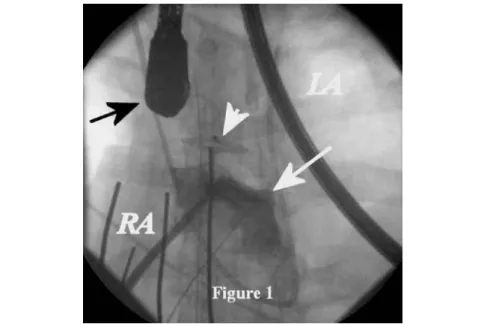
Fig. 1 Fluoroscopy image showing contrast medium (c.m. white arrow) injection in
the right atrium (RA) without passage of c.m. through the Amplatzer PFO occluder (arrowhead). LA left atrium, black arrow TEE transductor
603
J. Sierra
Cardiovascular Surgical Department, University Hospital of Geneva, Rue Micheli-du-Crest 24,
1211 Geneva 14 Geneva, Switzerland 4. Nguyen DQ, Das GS, Grubbs BC,
Bolman RM 3rd, Park SJ (1999) Trans-catheter closure of patent foramen ovale for hypoxemia during left ventricular assist device support. J Heart Lung Transplant 18:1021–1023
5. Liao KK, Miller L, Toher C, Ormaza S, Herrington CS, Bittner HB, Park SJ (2003) Timing of transesophageal echo-cardiography in diagnosing patent fora-men ovale in patients supported with left ventricular assist device. Ann Thorac Surg 75:1624–1626
R. F. Bonvini (
✉
) · V. Verin · R. Lerch · J. C. SprattCardiology Department, University Hospital of Geneva, Rue Micheli-du-Crest 24,
1211 Geneva 14 Geneva, Switzerland e-mail: [email protected] Tel.: +41-22-3727200
Fax: +41-22-3727229 I. Gerard
Intensive Care Unit,
University Hospital of Geneva, Rue Micheli-du-Crest 24,