Original article
The value of FDG-PET in patients with painful total knee
arthroplasty
Katrin D. M. Stumpe1, Jose Romero3, 4, Oliver Ziegler3, 5, Ehab M. Kamel1, 6, Gustav K. von Schulthess1, Klaus Strobel1, Juerg Hodler2
1Department of Medical Radiology, Division of Nuclear Medicine, University Hospital, Zurich, Switzerland 2Department of Radiology, Orthopaedic University Hospital Balgrist, Forchstrasse 340, 8008 Zurich, Switzerland 3Orthopaedic Surgery, Orthopaedic University Hospital Balgrist, Zurich, Switzerland
4EndoClinic Zurich, Center for Joint Diseases at Hirslanden Clinic, Zurich, Switzerland 5Ortho Zentrum Rosenheim, Rosenheim, Germany
6Division of Nuclear Medicine, Centre Hospitalier Universitaire Vaudois (CHUV), Lausanne, Switzerland Received: 9 January 2006 / Accepted: 16 March 2006 / Published online: 24 May 2006
© Springer-Verlag 2006
Abstract. Purpose: The purpose of this study was to evaluate18F-fluorodeoxyglucose (FDG) uptake in patients with painful total knee arthroplasty and to relate FDG uptake to the location of soft tissue pain.
Methods: Twenty-eight patients with painful total knee arthroplasty had a clinical examination, standard radio-graphs, CT measurement of rotation of the femoral component and FDG-PET (18 PET/CT, 10 PET). The diagnosis of infection was based on microbiological examinations of surgical specimens (n=12) or clinical follow-up for at least 6 months (n=16), 99mTc-labelled monoclonal antibody scintigraphy and joint aspiration. Results: Twenty-seven of 28 patients presented with diffuse synovial FDG uptake. Additional focal extrasyno-vial FDG uptake was observed in 19 knees. Twenty-four of the 28 patients had a diagnosis of internal femoral malrotation. The remaining four patients showed no rotation (0°) and 3°, 4° and 7° of external rotation, respectively. Three patients presented with the additional diagnosis of an infected total knee replacement. Pain was described as diffuse (n=10) or focal (n=18). In two knees a relationship between pain location and FDG uptake was observed. Of ten patients with a severe internal femoral component rotation (>6°), seven had focal uptake, four in the femoral periosteum and three in the tibial periosteum. The difference between knees with severe malrotation and the remaining knees was not significant (p=1.000, Fisher’s Exact Test).
Conclusion: Diffuse synovial and focal extrasynovial FDG-PET uptake is commonly found in patients with malrotation of the femoral component and is not related to
pain location. The information provided by FDG-PET does not contribute to the diagnosis and management of individual patients with persistent pain after total knee replacement.
Keywords: FDG-PET– Knee prostheses – Malrotation – Infection– Synovitis
Eur J Nucl Med Mol Imaging (2006) 33:1218–1225 DOI 10.1007/s00259-006-0127-1
Introduction
Persistent knee pain may represent a frustrating problem after total knee replacement. Several different types of complication have been associated with such pain, including malrotation of the femoral and/or tibial compo-nent [1–3], joint instability [2, 4–7], muscle imbalance [7–9], arthrofibrosis [10], component loosening [11, 12] and fractures [13]. Periprosthetic osteolysis due to the formation of granuloma is not as common as in total hip replacements. This difference has been attributed to the size of shedded polyethylene particles, which are larger in knee than in hip arthroplasty [14]. Infection is relatively rare after total knee replacement (1.7% in patients with osteoarthritis and 4.4% in rheumatoid arthritis [15]). More than 50% of revisions are performed within 2 years after total knee replacement. They are typically performed due to axial or rotational malalignment, instability and fixation failure [16–18].
Complications of total knee replacements are most commonly assessed with conventional radiographs and bone scintigraphy. Less commonly, computed tomography (CT) is performed in suspected rotational malalignment of the femoral or tibial component [1]. Ultrasonography may
Juerg Hodler ()) Department of Radiology,
Orthopaedic University Hospital Balgrist, Forchstrasse 340,
8008 Zurich, Switzerland e-mail: [email protected]
be useful in suspected soft tissue infection and arthrofi-brosis [10]. In suspected infection, combined111In-labelled leucocyte scintigraphy/99mTc-sulphur colloid marrow scin-tigraphy or leucocyte scinscin-tigraphy with monoclonal antibodies may be performed [19]. Cultures of joint fluid have been shown to be positive in 80–90% of patients with infected total knee replacements [20–22].
18F-fluorodeoxyglucose positron emission tomography
(FDG-PET) uses a completely different physical principle of imaging and demonstrates increased glucose uptake related to increased glucose metabolism, which is asso-ciated with a multitude of conditions in addition to infection. FDG-PET results in suspected implant-asso-ciated infections in trauma surgery and in chronic muscu-loskeletal infections without prior surgery have proved encouraging [23,24]. It was less successfully employed in infected total joint replacements of the hip and knee [25–27]. FDG-PET may be positive in synovial prolifer-ation in osteoarthritis and other synovial abnormalities [28]. It has also been shown to sensitively demonstrate recent muscle activity such as that occurring during running and level walking [29] and may also demonstrate abnormal muscle activity after joint replacement. There-fore, FDG-PET has the potential to demonstrate many of the possible abnormalities presenting with pain after total knee replacements.
The purpose of this investigation was to assess FDG uptake in patients with painful total knee replacement. In addition, we evaluated whether the location of FDG uptake is related to specific structures which may be responsible for pain associated with total knee replacement.
Materials and methods
The study was approved by the institutional review board responsible for the institution leading this investigation. Informed consent was obtained from all patients. Thirty-seven consecutive patients with total knee replacements were recruited for this investigation in a consecutive fashion if they had suffered from pain for more than 6 weeks. Three patients had bilateral total knee replacements, with each having a symptomatic and an asymptomatic side.
Study exclusion criteria were: surgery dating back less than 6 months (n=7) [30–32], arthrofibrosis (n=2), rejection of further evaluation (n=0), periprosthetic fractures (n=0) and rheumatoid arthritis (n=0).
Of the 28 patients included in the study, 15 were women (mean age 67 years, range 50–86 years) and 13 men (59 years, 50–79 years). The mean time interval between the last surgical intervention at the site of the involved knee replacement and study inclusion was 28 months (range 6–108 months). Fifteen of the 28 patients had a cemented and 13 an uncemented prosthesis. Twenty-four patients were examined after primary implantation and four patients after revision surgery of the knee. Fourteen patients had an LCS prosthesis (Complete Mobile-Bearing Knee System, DePuy Orthopaedics, Inc., a Johnson & Johnson company, Warsaw, IN, USA), five had a Natural Knee prosthesis (Zimmer, Warsaw, IN, USA), three had an SAL (Self-Aligning Total Knee Prosthesis, Centerpulse, Winterthur, Switzerland), two had an Innex (Zimmer, Winterthur, Switzerland),
two had a Wallaby (Centerpulse/Groupe Guepar, Winterthur, Switzerland), one had a p.f.c. SigmaTM RP Knee System (DePuy Orthopaedics, Inc., a Johnson & Johnson company, Warsaw, IN, USA) and one had a CKS prosthesis (STRATEC Medical, Oberdorf, Switzerland).
The following examinations were performed in all 28 patients: clinical examination by a knee surgeon, standard anteroposterior and lateral radiographs, functional (varus and valgus stress) radiographs in flexion and extension, CT measurement of rotation of the femoral component and FDG-PET. All patients had 99mTc-labelled mono-clonal antibody scintigraphy (BW 250/183) and fluoroscopically guided knee aspiration to exclude infection. In 99mTc-labelled monoclonal antibody scintigraphy (BW 250/183), radionuclide uptake was judged as positive for infection if the intensity of accumulation around the bone–prosthesis interface exceeded phys-iological bone marrow uptake or if the intensity of radionuclide uptake increased from 4 to 24 h.
Clinical aspects
Clinical assessment was performed by or under the supervision of a single orthopaedic staff surgeon specialised in knee surgery. For the purpose of this investigation, any pain was described as either localised or diffuse. The following anatomical locations were described for localised pain: patellar ligament, patella, suprapatellar recess, medial and lateral parapatellar recess, tibial tuberosity, pes anserinus, medial and lateral joint space, Hoffa’s fat pad, popliteal fossa, hamstrings, adductors and dorsal, anterior and peroneal compartments of the lower leg. The knee was tested for varus and valgus instability.
CT and PET imaging
For determination of femoral component rotation, 5-mm slices were obtained through the distal femur on a Siemens Plus 4 CT scanner (Siemens Medical Solutions, Erlangen, Germany). Table increments were 6 mm, and a high-resolution kernel was employed. The transepicondylar axis and the dorsal surface of the femoral compo-nent were used for calculation of femoral compocompo-nent rotation. The femoral component should be parallel to this axis or slightly externally rotated [1,33] (Figs.1d,2d,3d,4e). Femoral malrotation was considered to be severe in >6° of internal and >8° of external rotation [3].
Ten patients had FDG-PET examinations, and the remaining 18, PET/CT studies. The PET studies were obtained on a GE Advance scanner (GE Medical Systems, Waukesha, WI, USA). The scanner acquires 35 2D sections of 4.25 mm thickness per increment with an axial field of view of 14.6 cm. Images were obtained at three or four bed positions, covering the leg from the middle third of the femur to the middle third of the tibia. After fasting for at least 4 h, 300– 400 MBq of FDG was injected intravenously 40 min before scanning. Attenuation correction was performed using the built-in rotating 68Ge sources. A multiplicative iterative reconstruction algorithm for improvement of image quality and reduction of computation time was employed [34]. Uncorrected images were acquired in addition to the corrected images. Coronal and sagittal reformations were obtained. PET/CT was performed with a
DISCOVERY LS integrated device (GE Medical Systems, Milwaukee, WI, USA). CT data were acquired with the following parameters: tube rotation time, 0.5 s per revolution; 140 kV; 80 mA; 22.5 mm per rotation; pitch of 6; acquisition time of 22.5 s and a scan length of 867 mm. Subsequently, PET emission data were acquired in the two-dimensional mode. Emission counts were collected over 4 min for each table position. Adjacent fields of view shared one overlapping slice. Matched CT and PET images were reconstructed with a field of view of 500 mm and a 4.25-mm slice thickness (Figs.1c,2c,3c,4c).
PET image evaluation
Two board-certified nuclear physicians evaluated the PET examina-tions in consensus. Both had at least 8 years of PET experience and at least 3 years of CT experience. They were blinded with regard to the results of other imaging studies. eNTEGRA software (GE Medical Systems, Milwaukee, WI, USA) was used for this evaluation.
The anatomical distribution of the FDG uptake was described according to the clinical examinations. FDG accumulation was described as either diffuse synovial or focal extrasynovial. The FDG uptake was interpreted regardless of intensity according to the results of several studies [25,35]. In order to exclude artefacts associated with misregistration between attenuation-corrected and uncorrected scans caused by the implant, only the uncorrected scans were used to make a diagnosis.
Standard of reference
The final diagnosis was based on a combination of patient history, clinical examination and standard radiographs (n=28) and CT (femoral component rotation) (n=28). The presence or absence of infection was evaluated by fluoroscopically guided joint aspiration (n=28), 99mTc-antigranulocyte antibody scintigraphy (n=28) and either microbiological evaluation of surgical specimens (n=12) or clinical follow-up for at least 6 months (n=16). Microbiological findings were available in all three patients with infection.
Results
Twenty-four patients showed instability clinically (varus instability in 16, valgus instability in two and combined instability in six). In four patients the diagnoses of severe varus deformity (n=1), loosening of the tibial component (n=1) and non-specific pain (n=2) were made. Three patients presented with the additional diagnosis of an infected total knee replacement. Staphylococcus aureus was identified in two patients and Staphylococcus epidermidis in one patient. In all 12 patients undergoing surgery, results of analysis of histological specimens of capsular tissue and bone were available. Eleven of these 12 patients presented with chronic synovial inflammation. Three patients presented with capsular hypertrophy. Four of the 12 operated patients had granulation tissue with giant cells and macrophages at the prosthesis–bone interface.
Ten patients complained about diffuse pain. The remaining 18 patients had localised pain at the pes
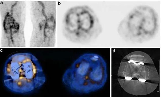
Fig. 1. Maximum intensity projection (MIP) PET (a), axial PET (b), axial PET/CT (c) and CT measurement of component rotation (d) in a 79-year-old man with knee replacement on the right. Pain at pes anserinus. There is strongly increased FDG accumulation in the
synovial membrane of the right knee, including the suprapatellar recess. FDG accumulation is also seen in the contralateral left joint with painful osteoarthritis. Rotational CT demonstrates 10° internal rotation of the femoral component
anserinus (n=6), the medial and lateral joint space (n=6), the lateral joint space alone (n=2), the medial joint space alone (n=1), the patella (n=1), the tibial tuberosity (n=1) and the pes anserinus in combination with the patella (n=1). Twenty-seven of the 28 patients presented with diffusely increased FDG uptake in the entire synovial membrane (Fig. 1). Extrasynovial focal FDG uptake was observed in 19 patients: in the tibial periosteum (n=9), femoral periosteum (n=5), anterior tibial muscle (n=2) (Fig.2) and in one case each at the fibular head periosteum, the tibial tuberosity (Fig.3) and the femur/tibia combined. Extrasynovial focal FDG uptake was demonstrated in both cemented and non-cemented prosthesis (in 10 of 15 patients with cemented prostheses and in 9 of 13 patients with non-cemented prostheses).
One of three patients with infection had diffuse synovial FDG uptake and focal periprosthetic FDG uptake adjacent to the proximal femoral prosthetic component. Another patient with infection presented with diffuse synovial FDG uptake in the suprapatellar recess. The third patient with infection had diffuse FDG accumulation in the soft tissues surrounding the painful total knee replacement (Fig. 4). The last-mentioned patient presented with patellofemoral focal FDG uptake on the attenuation-corrected images. On the non-attenuation-corrected images the focal uptake was absent, indicating an artefact related to the prosthetic device.
99mTc-labelled monoclonal antibody scintigraphy was
negative for infection in 26 of 28 cases. In one patient with infection, 99mTc-labelled monoclonal antibody scintigra-phy was false negative for infection. Joint aspiration was false negative in two of three cases with infection.
The three FDG-PET scans of the three patients with bilateral total knee replacements were all negative on the asymptomatic side (not further evaluated in this study).
In two knees a direct relationship between pain location and FDG uptake was observed. This included the single patient with pain at the tibial tuberosity (Fig.3) and one of the seven patients with pain at the pes anserinus.
Twenty-four of 28 femoral components were incorrectly internally rotated based on CT measurements. The mean rotation in these 24 patients was 6.2° (range 2–12°). The remaining four patients had no rotation (0°) and 3°, 4° and 7° of external rotation, respectively. Of the 24 internally rotated femoral components, ten were severely malrotated (>6°) (Fig.1).
Except for one patient with an infected total knee replacement (internal femoral component rotation: 11°), none of the ten patients with diffuse pain had severe malrotation (range of internal rotation in this group: 2–5°) (Fig. 2). Six of the ten patients with an internal femoral component rotation of >6° had localised pain at the pes anserinus (including the patient with additional painful patella) (Table1).
Of ten patients with a severe internal femoral compo-nent rotation (>6°), seven had focal uptake, four in the femoral periosteum and three in the tibial periosteum; the incidence of focal uptake in these patients was higher than in the remaining patients. The difference between knees with severe malrotation and the remaining knees was not significant (p=1.000, Fisher’s Exact Test). Of the remain-ing 18 patients, 12 had focal FDG uptake [tibial perios-teum, n=7; tibial tuberosity, n=1; tibial and femoral periosteum combined, n=1; fibula (biceps insertion), n=1; anterior tibial muscle, n=2].
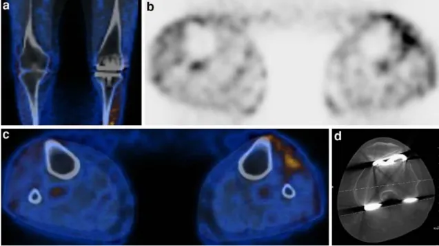
Fig. 2. Coronal PET-CT (a), axial PET (b), axial PET/CT (c) and CT measurement of component rotation (d) in a 66-year-old woman with diffuse pain. Increased FDG accumulation is observed in the
left anterior tibial muscle. Rotational CT demonstrates 3° internal rotation of the femoral component
Discussion
Persistent pain after total knee arthroplasty is bothersome to both patient and surgeon, and is difficult to treat. It is clinically relevant to be able to better recognise the pathogenesis and to more precisely diagnose the reason for this problem. Besides malalignment and instability, which were the main problem in our patient population, patellar problems (commonly associated with malalign-ment), infection, aseptic loosening, polyethylene wear, arthrofibrosis and periprosthetic fractures have been recognised as relevant abnormalities.
Infection is a severe but fortunately relatively rare occurrence in total knee arthroplasty. The reported prevalence is between 1% and 4% [15]. 111In-labelled white blood cells in combination with 99mTc-sulphur colloid marrow imaging is currently the method of choice in the assessment of infection in total joint replacement, with a sensitivity, specificity and accuracy of 100%, 97% and 98%, respectively [11,19,27,36–39].99mTc-labelled antigranulocyte antibody scintigraphy is emerging as a new radionuclide technique to detect infected prosthetic devices without the time-consuming process of labelling the cells outside the body [19]. FDG-PET appears to be promising in the diagnosis of musculoskeletal infection [23, 35, 40]. This method has also shown good results in implants used in a trauma population [23].
FDG uptake is based on increased glucose uptake by inflammatory cells like phagocytes (neutrophils, eosino-phils and mononuclear phagocytes). These cells start
metabolising large quantities of glucose by way of the hexose monophosphate shunt, and their rates of oxygen uptake increase greatly when activated (so-called respira-tory burst), this increase being especially pronounced in infection. Data on FDG-PET in patients with infected prosthetic devices are controversial [25,27,35]. Zhuang et al. [35] found PET to be more accurate in detecting infections in patients with hip replacements than in patients with knee replacements. There were ten false positive findings in 36 patients with total knee replacements using periprosthetic FDG uptake as a criterion. The sensitivity, specificity and accuracy of PET for detecting infection in patients with total knee replacements were 91%, 72% and 78%. The authors assumed that in addition to postsurgical changes, other factors must have contributed to the false positive results. Our results confirmed this. In our study, one patient with infection and 16 patients with femoral malrotation showed periprosthetic focal bone uptake. In a recent study including 40 and 19 patients with failed total hip and knee replacements, respectively, Love et al. [27] showed that a periprosthetic FDG uptake pattern was neither sensitive nor specific for infection. Periprosthetic uptake was found both in infection and in aseptic loosening. One possible explanation for periprosthetic uptake in the absence of infection might be aberrant but otherwise normal bone marrow [27]. Bone marrow distri-bution may be altered by an orthopaedic device. Van Acker et al. [15] used focal FDG uptake at the bone–prosthesis interface as the criterion for infection. FDG-PET had a sensitivity of 100% and a specificity of 73% for the
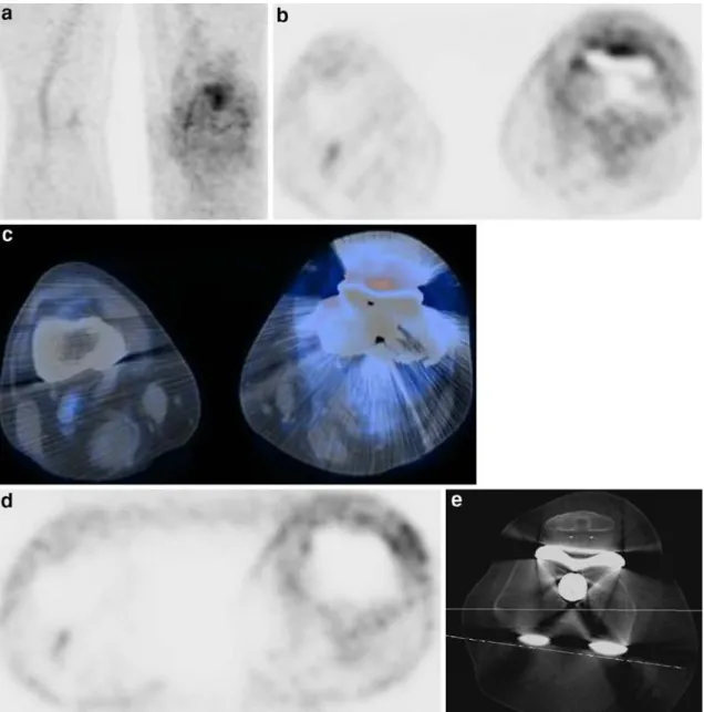
Fig. 3. Maximum intensity projection (MIP) PET (a), axial PET (b), axial PET/CT (c) and CT measurement of component rotation (d) in a 50-year-old woman with pain at the left tibial tuberosity. Strongly increased FDG accumulation is present in the suprapatellar recess
and tibial tuberosity periosteum, including adjacent soft tissue. Rotational CT demonstrates 4° internal rotation of the femoral component
diagnosis of infection but offered no added benefit in comparison to white blood cell scintigraphy in combina-tion with bone scintigraphy.
Apparently, the level of FDG uptake does not improve the performance of PET in diagnosing infection in patients with total knee replacement. FDG uptake has been shown to be even stronger in patients with aseptic prosthetic loosening [25, 27, 35]. Several studies [25–27] indicate that FDG-PET may not be able to differentiate aseptic from septic prosthetic loosening because of the remarkably similar histopathological morphology.
FDG-PET also demonstrates synovial proliferation in osteoarthritis and in patients with total knee replacements [28,40]. Manthey et al. [40] examined 28 patients with 14 Table 1. Effect of severely internally rotated femoral component
(>6°) Internal rotation of femoral component No. Focal FDG uptake Pain at pes anserinus Diffuse pain >6° 10 7 (70%) 6 (60%) 0 <6° 18 12 (67%) 1 (6%) 10 (56%) Total 28 19 (68%) 7 (25%) 10 (36%)
Fig. 4. Maximum intensity projection (MIP) PET (a), axial attenuation-corrected PET (b), axial PET/CT (c), axial non-attenu-ation-corrected PET (d) and CT measurement of component rotation (e) in a 55-year old man with infection of the left knee replacement. Pain was present in the medial and lateral left joint space. Diffusely increased FDG uptake is observed in the soft tissues surrounding the
knee replacement, and there is focally increased FDG uptake in the patellofemoral joint space on the attenuation-corrected PET and PET/CT images. On the axial non-attenuation-corrected PET image, focal patellofemoral uptake was absent; this represented an artefact related to the prosthetic device
hip and 14 knee prostheses with FDG-PET in order to differentiate different sources of pain in orthopaedic prostheses. Nine of the 14 patients with a painful knee prosthesis had synovitis diagnosed with PET. Synovitis was found more frequently in patients with total knee replacement than in patients with total hip replacement (nine versus four prostheses). In another investigation with 21 consecutive patients with painful total knee replace-ments, diffusely increased FDG uptake in the synovial membrane was found [15]. The authors reported that comparison of FDG-PET with bone scintigraphy facilitated the detection of focal FDG uptake at the bone–prosthesis interface, which is easily overlooked owing to intensified adjacent synovial uptake. In our study, 27 of 28 patients had increased synovial FDG uptake (Fig. 1). In most patients with knee arthroplasty, FDG uptake seems to be related to chronic synovitis, as analysis of histological specimens in 11 of the 12 patients who underwent surgery revealed chronic synovial inflammation; this is consistent with previously published data [15]. The clinical relevance of this finding, however, is not obvious. FDG accumula-tions in joints are frequently found without symptoms [28]. Such accumulations increase with age. They are most likely a result of subclinical synovial proliferation. In addition, four of 12 patients in our study showed granu-lomatous tissue with giant cells and macrophages at the knee prosthesis–bone interface. A foreign body reaction due to polyethylene and metal wear with shedding of particles was most likely to be responsible for this reaction. Malalignment of knee replacement may lead to soft tissue imbalance resulting in abnormal muscle activity. FDG-PET demonstrates recent muscle activity [29]. In this regard, the results of our investigation are disappointing, with increased muscle activity present in few patients and without correlation between PET activity and pain location. On the other hand, the standardised protocol employed for this investigation, with several hours of resting before imaging, may have obscured any intermittent findings relating to muscle activity.
Our study had limitations. The prevalence of infection was low, which was related to the prospective nature of the study. Nevertheless, our results may explain periprosthetic activity, which is often present in total knee replacement and may cause false positive findings. There is no simple relationship between clinical symptoms, FDG uptake and malrotation of the femoral component, which had a high prevalence in our population. However, patients with severe malrotation appear to have localised pain, com-monly located at the pes anserinus, and focal FDG uptake not directly related to the location of pain. Based on this combination, malrotated prostheses may not primarily be caused by changed muscle metabolism (e.g. because of strain due to changes in mechanical axes or instability). Pain may rather be related to synovitis, which would explain the common diffuse FDG uptake.
In conclusion, diffuse synovial and focal extrasynovial FDG-PET uptake is commonly found in patients with malrotation of the femoral component and is not related to pain location. The information provided by FDG-PET does
not contribute to the diagnosis and management of individual patients with persistent pain after total knee replacement.
References
1. Berger RA, Crossett LS, Jacobs JJ, Rubash HE. Malrotation causing patellofemoral complications after total knee arthro-plasty. Clin Orthop Relat Res 1998;356:144–153
2. Berger RA, Rubash HE. Rotational instability and malrotation after total knee arthroplasty. Orthop Clin North Am 2001;32:639–647, ix
3. Hofmann S, Romero J, Roth-Schiffl E, Albrecht T. Rotational malalignment of the components may cause chronic pain or early failure in total knee arthroplasty. Orthopade 2003;32: 469–476
4. Clarke HD, Scuderi GR. Flexion instability in primary total knee replacement. J Knee Surg 2003;16:123–128
5. Argenson JN, Aubaniac JM. Total knee arthroplasty in femorotibial instability. Orthopade 2000;29 Suppl 1:S45–47 6. Nabeyama R, Matsuda S, Miura H, Kawano T, Nagamine R,
Mawatari T, et al. Changes in anteroposterior stability following total knee arthroplasty. J Orthop Sci 2003;8:526–531 7. Malo M, Vince KG. The unstable patella after total knee arthroplasty: etiology, prevention, and management. J Am Acad Orthop Surg 2003;11:364–371
8. Parker DA, Dunbar MJ, Rorabeck CH. Extensor mechanism failure associated with total knee arthroplasty: prevention and management. J Am Acad Orthop Surg 2003;11:238–247 9. Sharkey PF, Hozack WJ, Rothman RH, Shastri S, Jacoby SM.
Insall Award paper. Why are total knee arthroplasties failing today? Clin Orthop Relat Res 2002;404:7–13
10. Boldt JG, Munzinger UK, Zanetti M, Hodler J. Arthrofibrosis associated with total knee arthroplasty: gray-scale and power Doppler sonographic findings. AJR Am J Roentgenol 2004;182:337–340
11. Hendrix RW. Radiographic evaluation of prosthetic joints. Compr Ther 1988;14:36–48
12. Ducheyne P, Kagan A, 2nd, Lacey JA. Failure of total knee arthroplasty due to loosening and deformation of the tibial component. J Bone Joint Surg Am 1978;60:384–391
13. Hirsh DM, Bhalla S, Roffman M. Supracondylar fracture of the femur following total knee replacement. Report of four cases. J Bone Joint Surg Am 1981;63:162–163
14. Bauer TW, Schils J. The pathology of total joint arthroplasty.II. Mechanisms of implant failure. Skeletal Radiol 1999;28: 483–497
15. Van Acker F, Nuyts J, Maes A, Vanquickenborne B, Stuyck J, Bellemans J, et al. FDG-PET,99mTc-HMPAO white blood cell SPET and bone scintigraphy in the evaluation of painful total knee arthroplasties. Eur J Nucl Med 2001;28:1496–1504 16. Briard JL, Hungerford DS. Patellofemoral instability in total
knee arthroplasty. J Arthroplast 1989;4 Suppl:S87–97 17. Merkow RL, Soudry M, Insall JN. Patellar dislocation
following total knee replacement. J Bone Joint Surg Am 1985;67:1321–1327
18. Romero J, Duronio JF, Sohrabi A, Alexander N, MacWilliams BA, Jones LC, et al. Varus and valgus flexion laxity of total knee alignment methods in loaded cadaveric knees. Clin Orthop Relat Res 2002;394:243–253
19. Sciuk J, Puskas C, Greitemann B, Schober O. White blood cell scintigraphy with monoclonal antibodies in the study of the infected endoprosthesis. Eur J Nucl Med 1992;19:497–502
20. Freeman MA, Sudlow RA, Casewell MW, Radcliff SS. The management of infected total knee replacements. J Bone Joint Surg Br 1985;67:764–768
21. Goksan SB, Freeman MA. One-stage reimplantation for infected total knee arthroplasty. J Bone Joint Surg Br 1992;74:78–82
22. Simmons TD, Stern SH. Diagnosis and management of the infected total knee arthroplasty. Am J Knee Surg 1996;9: 99–106
23. Schiesser M, Stumpe KD, Trentz O, Kossmann T, Von Schulthess GK. Detection of metallic implant-associated infections with FDG PET in patients with trauma: correlation with microbiologic results. Radiology 2003;226:391–398 24. de Winter F, van de Wiele C, Vogelaers D, de Smet K, Verdonk
R, Dierckx RA. Fluorine-18 fluorodeoxyglucose-position emis-sion tomography: a highly accurate imaging modality for the diagnosis of chronic musculoskeletal infections. J Bone Joint Surg Am 2001;83-A:651–660
25. Stumpe KD, Notzli HP, Zanetti M, Kamel EM, Hany TF, Gorres GW, et al. FDG PET for differentiation of infection and aseptic loosening in total hip replacements: comparison with conventional radiography and three-phase bone scintigraphy. Radiology 2004;231:333–341
26. Love C, Tomas MB, Marwin SE, Pugliese PV, Palestro CJ. Role of nuclear medicine in diagnosis of the infected joint replacement. Radiographics 2001;21:1229–1238
27. Love C, Marwin SE, Tomas MB, Krauss ES, Tronco GG, Bhargava KK, et al. Diagnosing infection in the failed joint replacement: a comparison of coincidence detection18F-FDG
and111In-labeled leucocyte/99mTc-sulfur colloid marrow imag-ing. J Nucl Med 2004;45:1864–1871
28. von Schulthess GK, Meier N, Stumpe KD. Joint accumulations of FDG in whole body PET scans. Nuklearmedizin 2001;40:193–197
29. Oi N, Iwaya T, Itoh M, Yamaguchi K, Tobimatsu Y, Fujimoto T. FDGPET imaging of lower extremity muscular activity during level walking. J Orthop Sci 2003;8:55–61
30. Meyer M, Gast T, Raja S, Hubner K. Increased F-18 FDG accumulation in an acute fracture. Clin Nucl Med 1994;19: 13–14
31. Zhuang H, Sam JW, Chacko TK, Duarte PS, Hickeson M, Feng Q, et al. Rapid normalization of osseous FDG uptake following traumatic or surgical fractures. Eur J Nucl Med Mol Imaging 2003;30:1096–1103
32. Kaim AH, Gross T, von Schulthess GK. Imaging of chronic posttraumatic osteomyelitis. Eur Radiol 2002;12:1193–1202 33. Akagi M, Yamashita E, Nakagawa T, Asano T, Nakamura T.
Relationship between frontal knee alignment and reference axes in the distal femur. Clin Orthop Relat Res 2001;388:147–156 34. Schmidlin P. Improved iterative image reconstruction using
variable projection binning and abbreviated convolution. Eur J Nucl Med 1994;21:930–936
35. Zhuang H, Duarte PS, Pourdehnad M, Maes A, Van Acker F, Shnier D, et al. The promising role of 18F-FDG PET in detecting infected lower limb prosthesis implants. J Nucl Med 2001;42:44–48
36. Ryd L, Gustafson T, Lindstrand A. 99mTc-diphosphonate
scintigraphy in successful knee arthroplasty and its relation to micromotion. Clin Orthop Relat Res 1993;287:61–67 37. Rosenthall L, Lepanto L, Raymond F. Radiophosphate uptake
in asymptomatic knee arthroplasty. J Nucl Med 1987;28: 1546–1549
38. Becker W, Pasurka B, Borner W. Significance of leukocyte scintigraphy of the infected total endoprosthesis. Rofo 1989;150:284–289
39. Palestro CJ, Kim CK, Swyer AJ, Capozzi JD, Solomon RW, Goldsmith SJ. Total-hip arthroplasty: periprosthetic indium-111-labeled leukocyte activity and complementary technetium-99m-sulfur colloid imaging in suspected infection. J Nucl Med 1990;31:1950–1955
40. Manthey N, Reinhard P, Moog F, Knesewitsch P, Hahn K, Tatsch K. The use of [18F]fluorodeoxyglucose positron emis-sion tomography to differentiate between synovitis, loosening and infection of hip and knee prostheses. Nucl Med Commun 2002;23:645–653