Silke Grabherr Christine Cooper Susi Ulrich-Bochsler Tanya Uldin Steffen Ross Lars Oesterhelweg Stephan Bolliger Andreas Christe Pierre Schnyder Patrice Mangin Michael J. Thali Received: 10 March 2008 Revised: 12 June 2008 Accepted: 3 July 2008
Published online: 3 September 2008 # European Society of Radiology 2008
Estimation of sex and age of
“virtual
skeletons
”–a feasibility study
Abstract This article presents a fea-sibility study with the objective of investigating the potential of multi-detector computed tomography (MDCT) to estimate the bone age and sex of deceased persons. To obtain virtual skeletons, the bodies of 22 deceased persons with known age at death were scanned by MDCT using a special protocol that consisted of high-resolution imaging of the skull, shoulder girdle (including the upper half of the humeri), the symphysis
pubis and the upper halves of the femora. Bone and soft-tissue recon-structions were performed in two and three dimensions. The resulting data were investigated by three anthropol-ogists with different professional ex-perience. Sex was determined by investigating three-dimensional mod-els of the skull and pelvis. As a basic orientation for the age estimation, the complex method according to Nemeskéri and co-workers was applied. The final estimation was effected using additional parameters like the state of dentition, degenera-tion of the spine, etc., which where chosen individually by the three ob-servers according to their experience. The results of the study show that the estimation of sex and age is possible by the use of MDCT. Virtual skeletons present an ideal collection for anthro-pological studies, because they are obtained in a non-invasive way and can be investigated ad infinitum. Keywords Skeletal age . Age estimation . Sex estimation . Virtual skeleton . Virtopsy . Digital osteology
Introduction
In recent years, modern cross-sectional imaging techniques have revolutionised forensic medicine. Magnetic resonance (MR) imaging and especially multi-detector computed tomography (MDCT) are becoming more and more widely
used for post-mortem examinations [1]. Computed tomog-raphy (CT) systems have already been installed in institutes of forensic medicine all over the world, and in the future this trend will continue [2]. The advantage of having access to CT and MR can also influence other specialists working in collaboration with institutes of S. Grabherr (*) . P. Mangin
Institute of Forensic Medicine, University Hospital of Lausanne, Rue du Bugnon 21, 1005 Lausanne, Switzerland e-mail: [email protected] Tel.: +41-21-3147067 Fax: +41-21-3147090 S. Grabherr . S. Ross . L. Oesterhelweg . S. Bolliger . A. Christe . M. J. Thali Institute of Forensic Medicine, University of Bern,
Buehlstrasse 20, 3012 Bern, Switzerland S. Grabherr . P. Schnyder Service of Diagnostic and Interventional Radiology, University Hospital of Lausanne, Rue du Bugnon 46,
1011 Lausanne, Switzerland
C. Cooper . S. Ulrich-Bochsler Institute for the History of Medicine, Historical Anthropology, University of Bern, Fabrikstrasse 29d, 3012 Bern, Switzerland T. Uldin Service of Osteo-Archaeology, Klusstrasse 43, 4147 Aesch, Switzerland A. Christe
Institute of Diagnostic Radiology, University of Bern,
Inselspital,
forensic medicine. In this context, the Institute of Forensic Medicine of the University of Lausanne is working on creating an“anthropological database” that should contain data from a large number of cadavers which have been examined by CT. These data will consist of “virtual skeletons” with well-documented age, sex, illnesses, origin, etc. that can be used to perform anthropological studies in order to optimise anthropological methods or to develop new techniques.
This article deals with a feasibility study that demon-strates how CT can be used for anthropological purposes. In the context of the Virtopsy project (www.virtopsy.com), the potential of MDCT to perform anthropological estimation of skeletal sex and age was evaluated. The aim of this first study was to show if different parts of the skeleton can be visualised by MDCT in a quality that allows examinations as they are performed on real skeletons. Therefore, MDCT data of 22 cases were collected at the Institute of Forensic Medicine in Bern. A special CT protocol that should allow detailed high-resolution imaging with reconstruction of high quality three-dimensional (3D) models was developed. The obtained “virtual skeletons” were examined by three anthropologists with different professional experience.
All the disposable methods for age estimation lead to an appraisal of the biological age but not to an exact calculation of the chronological age of an individual. Examples of well-established methods for age estimation on adult skeletons are aspartic acid racemisation [3] and tooth cementum annulation [4], both techniques that are not adaptable for our question. For a first appraisal of the age, we decided to use the complex method of Nemeskéri et al. [5]. Despite the criticism concerning this method [6, 7], it seemed to us to be the most interesting choice for our question because it consists of a unique multi-factor concept, including four different skeletal age parameters, which are combined and put in relation to each other. This aspect was important, as the goal of this study was to investigate different parts of the skeleton and not just one parameter, as has already been reported [8].
The complex method according to Nemeskéri et al. [5] includes the following parameters: (1) the endocranial obliteration of the sutures; (2) the structure of the spongiosa of the proximal humerus; (3) the proximal femur; (4) the texture of the symphyseal surface of the pubic bone. Optimally, the deviation should be ±2.5 years (confidence ca. 80–85%). After assigning a state to each parameter as depicted in [5], the estimated age range can be withdrawn from tables published by Ferembach et al. [9] and Sjøvold [10]. In German speaking regions, the method according to Nemeskéri and co-workers is widely applied by anthro-pologists, especially those who are working with historical skeletal materials. To test the work on virtual bones, the involved anthropologists preferred to apply this method because they are most familiar with it. Other techniques, like the method described by Suchey and co-workers [11–
13] and Iscan et al. [14–15], would rule out age estimations in many cases. Suchey and Katz [16] pointed out that the assessment of the symphysis, which is used for this kind of age estimation, is nearly impossible in individuals of more than 40 years old, whereas the complex method allows differentiation up to an age of over 50 years. These factors contributed to our decision to use the Nemeskéri method in this study despite the criticism concerning this method.
Acsádi and Nemeskéri [17] emphasised that an over-simplified mechanistic employment of the method should be avoided by all means and that the final age diagnosis should be established in a most circumspect manner. In our study, the age estimated by the complex method was used as a starting point for further estimations. These depended mostly on the experience and preferences of the three observers and did not follow rules of specific methods. Characteristics of interest here were the obliteration of ectocranial sutures, degenerative joint disease, degree of epiphyseal fusion, presence of osteophytes, the ossification of the rib and laryngeal cartilage, degeneration of the spine, the state of the dentition and the texture of the clavicle. In the most general of terms, the absence of degenerative changes, osteophytes and advanced ossification of epiph-yses, sutures and cartilages is indicative of a younger age, their presence however points towards a more mature age. Such age changes are highly individual, though, and depend on different factors like lifestyle and genetic disposition. The assessment of these skeletal changes and the weight attached to them depends on the observer’s judgement. Thus, the final age estimation is a process of weighing and balancing, therefore including many factors that cannot be assessed by an exact method. With regard to these aspects, an aggregated interpretation (final estima-tion) was performed by each observer which revealed the estimated age of each individual.
Materials and methods
Subjects
The study was performed on 22 blinded cases of the Institute of Forensic Medicine in Bern. The cases were chosen by the following criteria: (1) known and confirmed age at death, (2) intact skeletal system. The study group was composed of seven female and 15 male corpses, representing ages between 17 and 92 years at the time of death. Cause of death was either intoxication (drugs and/or medicine) or sudden death due to cardiac insufficiency.
MDCT
MDCT was performed on a Siemens Somatom Sensation 6 unit, using a special CT protocol including high-resolution imaging of regions relevant for the complex analysis: skull,
shoulder girdle including the upper half of the humeri, the symphysis pubis and the upper halves of the femora (see Fig.1).
CT parameters were 0.63-mm slice width, 6×0.5-mm detector-collimation and a reconstruction increment of 0.5-mm (tube voltage: 130 kV; effective mAs: 90; radiation dose: 12.24 mGy). Reconstructions were performed using a kernel of B30 (soft-tissue reconstruction) and B90 (bone reconstruction).
Estimation of sex
Estimation of sex was performed by three anthropologists (observers 1–3) with 5 to more than 30 years of professional experience by examination of the skull and the pelvis independently from each other. Therefore, 3D models reconstructed in a B30 kernel or B90 kernel from the CT data of regions 1 and 3 (see Fig. 1) were examined (see Figs.2a,3a). The determination was based on the secondary sex characters of the skull and the pelvis [17–21]. The parameters used for the estimations are listed in Table1.
Estimation of age
Estimation of age was performed by the same three anthropologists. The observers investigated the anonymous virtual skeletons independently from each other.
A first appraisal was performed using the complex method [5]. Therefore, the endocranial sutures of the skull were examined on a 3D model (see Figs.2b,3b) calculated from the data of a reconstruction in a B90 kernel. After virtually cutting the facial skeleton and the cranial base, the sutures could be examined. To evaluate the trabecular structure of the humeri and the femora, reconstructions in 2D (see Figs.2c–d,3c–d) were performed. Therefore, coronal section images with a thickness of 2.5 cm where reconstructed from the data or the images were viewed in a maximum-intensity projection (MIP). This allows producing images that are similar to conventional X-ray images, which are often used for age estimation by anthropologists. The pubic bones were examined on a 3D model calculated from the data of reconstructions in a B90 kernel. By the use of the“virtual scalpel”, the symphysis can be cut in the midline and the symphyseal surface of each pubic bone can be shown on the 3D model (see Figs.2e,3e).
Results
Estimation of sex
By examining the virtual skull and pelvis the sex of all individuals was determined correctly by all anthropologists.
Estimation of age
The results of the age estimation are shown in Tables 2 (detailed) and 3 (summary). We have separately investi-Fig. 1 Scheme showing the skeletal regions of interest of the
scanning protocol. The first region (yellow box) includes the whole skull. The second region (pink box) contains the shoulder girdle with the sterno-clavicular articulations and the proximal half of both humeri. The third region (red box) includes the pubic angle with the two pubic bones and the symphysis. The forth region (green boxes) includes the proximal halves of the two femora
gated the results using only the complex method and the results of the final estimation including more parameters. To ease the interpretations, we grouped the results according to their correctness by criteria we defined beforehand. Group 1 (green numbering in Table2) includes all cases in which the age of the victim is situated inside of the estimated age range. Group 2 (blue numbering in Table 2) contains cases in which the calendar age of the
individual differs 1–3 years from the estimated age range. Group 3 (violet numbering in Table2) includes cases with a difference of 3–5 years between the age at death and the estimated age range. In group 4 (red numbering in Table2), cases are listed which show a difference of more than 5 years between the estimated age range and the calendar age. Estimations with results situated in group 1 and 2 were interpreted as“correct estimations”. Regarding the fact that Fig. 2 a–e Example of
recon-structions in 2D and 3D of case no. 15, a 17-year-old female. a For sex determination, 3D models of the skull and the pelvis were used. b–e The four parameters according to the complex method were exam-ined. The endocranial sutures of the skull were graded by inves-tigating the 3D model of the skull, calculated from the data of a reconstruction in a kernel of 90 (b). For grading the trabecu-lar structure of the humerus (c) and the femur (d), coronar cross-sectional images were calculated with a thickness of 2.5 cm. The symphyseal surface of the pubic bone was investi-gated on a 3D model (e), calculated from the data of a bone tissue reconstruction in a 90 kernel. In this case, all observers postulated female sex and grade I for cranial sutures, humerus, femur and symphysis. Observer 1 did not apply the complex method due to an estimated age of under 20 years
the skeletal age is compared with the calendar age, the interval of confidence of 3 years seems to be advisable, as there is often a difference between the two ages.
Results using only the complex method
By applying only the complex method, most of the misinterpretations were observed (Fig. 4). Observer 1 presented only one estimation (4.8%) in group 1. In two
cases (9.5%), the results were situated in group 2. Four cases (19%) were situated in group 3. Fourteen cases (66.7%) were interpreted wrongly and were placed in group 4. In one case, the observer did not apply the complex method due to an estimated age of under 20 years. If cases in groups 1 and 2 are interpreted as “correct estimations”, observer 1 reached only three correct results (14.3%) with the use of the complex method.
Observer 2 provided the best results of all observers. He presented five results (22.7%) in group 1. Three cases Fig. 3 a–e Example of
recon-structions in 2D and 3D of case no. 12, a 49-year-old male. The sex determination by investigat-ing the 3D reconstructions of the skull and the pelvis (a) was not difficult for the three observers, who all appraised male sex. Difficulties were observed with grading the four parameters of the complex method. The endo-cranial sutures (b) were assigned to state I, state IV and state V by the three anthropologists. The trabecular structure of the hu-merus (c) was classified as state III, III–IV and IV by the three observers. The trabecular struc-ture of the femur (d) was assigned to state IV, IV–V and V. The symphyseal surface of the pubic bone (e) was found to show a texture of grade IV by all three observers
(13.6%) were situated in group 2. Two results (9.1%) were observed in group 3. Like observer 1, this second observer showed the highest number of cases with results in group 4 (12 cases, 54.5%). Correct estimations (group 1 and 2) were obtained by observer 2 in eight cases (36.3%).
Observer 3 presented four results (18.2%) with an age inside the estimated range (group 1). One result (4.5%) was placed in group 2 and two (9.1%) in group 3. Fifteen cases (68.2%) were wrongly estimated (group 4). Observer 3 presented five cases (22.7%) with correct estimations (group 1 and 2) using the complex method.
Results of the final estimations
The results of the final estimations are shown in Fig.5. By adding more parameters to the results of the complex method, the age estimations of all three observers improved
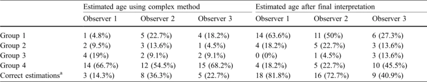
Table 2 Detailed demonstration of the results of age estimation using the complex method and the final estimation of age of the three observers
The real data of the cases are given in the first three columns. The different estimations were divided into four groups according to their correctness by criteria defined beforehand. Group 1 (green): age at death inside the estimated range; group 2 (blue): estimated range differs from the age at death by 1–3 years; group 3 (violet): difference of 4–5 years between the age at death and the estimated age range; group 4 (red): estimated age range differs more than 5 years from age at death. Observer 1 did not apply the complex method to case number 15 due to an estimated age of under 20 years Table 1 The secondary sex characters of the skull and the pelvis on
which the determination of the sex of the individuals were based
Skull Pelvis
Morphology of glabella and superciliary arch
Subpublic angle
Morphology of the forehead Morphology of the symphysis Morphology of the orbits Morphology of the ischium Morphology of the supraorbital
margin
Obturator for a men Morphology of the facial skeleton Diameter of the acetabulum Morphology of the mandible
Mandibular angle
Morphology of the mastoid processes
Morphology of the occipital region
significantly and wrong interpretations were conspicuously reduced (Fig.4).
Observer 1 reached the highest number of results in group 1 with 14 cases (63.6%). Four results (18.2%) were placed in group 2. None of the cases showed a result according to group 3 and four results (18.2%) could be found in group 4. Regarding his final interpretation, observer 1 reached 18 (81.8%) correct estimations (groups 1 and 2). By adding more parameters and his personal experience to the results of the complex method, the observer was able improve his estimations from three correct cases (14.3%) to 18 (81.8%).
Observer 2 presented 11 results in group 1 (50%). Five estimations (22.7%) showed results according to group 2. One case (4.5%) was placed in group 3 and in five cases (22.7%) the results were placed in group 4. In conclusion, observer 2 reached 16 (72.7%) correct estimations (group 1 and 2). Adding other parameters to the complex method improved the results of observer 2 from eight (36.3%) to 16 (72.7%) correct estimations.
Observer 3 presented six cases (27.3%) of group 1. Three cases (13.6%) were estimated with results in group 2 and group 3. With ten (45.5%) cases in group 4, observer 3 showed the highest number of wrong interpretations. Nine Table 3 Results of age estimation by the three observers according to the complex method and the final interpretation giving the number of cases with results in the different groups explained in Table2
Estimated age using complex method Estimated age after final interpretation
Observer 1 Observer 2 Observer 3 Observer 1 Observer 2 Observer 3
Group 1 1 (4.8%) 5 (22.7%) 4 (18.2%) 14 (63.6%) 11 (50%) 6 (27.3%) Group 2 2 (9.5%) 3 (13.6%) 1 (4.5%) 4 (18.2%) 5 (22.7%) 3 (13.6%) Group 3 4 (19%) 2 (9.1%) 2 (9.1%) 0 (0%) 1 (4.5%) 3 (13.6%) Group 4 14 (66.7%) 12 (54.5%) 15 (68.2%) 4 (18.2%) 5 (22.7%) 10 (45.5%) Correct estimationsa 3 (14.3%) 8 (36.3%) 5 (22.7%) 18 (81.8%) 16 (72.7%) 9 (40.9%) a
Cases with results situated in groups 1 and 2 were interpreted as correct. The percentages are shown in parentheses. Because observer 1 did not apply the complex method to one case, the total number of cases for this observer using the complex method is reduced and therefore the percentage differs in correlation to the other two observers
Fig. 4 Demonstration of the results of the final age estimations in a chart. The results using the complex method only are not shown. The results were grouped according to their correctness by criteria we defined beforehand, as shown in the form of colour coding in Table 2. Cases with age at death inside the estimated age range prevail for observers 1 and 2 (14 and 11 cases), while observer 3 shows the most wrong estimations (ten cases). Cases with age at
death inside and 3 years over or under the estimated range were considered as correct estimations and are presented in the last group of bars. Using this consideration, observer 1 shows 18 correct estimations, observer 2 reached 15 correct results and observer 3, nine. These values correspond respectively to 81.8% 72.7% and 40.9% of correct estimations
cases (40.9%) could be assessed as correct estimations. Therefore, the observer increased the number of correct estimations from five (22.7%) by using the complex method to nine (40.9%) by adding other parameters.
Discussion
The results of our first feasibility study show that MDCT is well able to perform estimations of the sex and age at death of deceased persons. The sex was determined correctly by all three investigators using 3D models of the skull and pelvis. The age estimation was more difficult for the investigators, but presented the same problems already described in the literature. Additionally, at least at the beginning of the study, some difficulties arose from a lack of experience in evaluating digital images.
Most misinterpretations occurred when applying the complex method, whereas the routine of observation lowered the error rate only a little. Possible reasons for this apparent high rate of misestimation have already been described in the literature. As Rösing et al. [7] affirmed, the range of ±2.5 years (including all four parameters) is too narrow. The skeleton collection on which the method was developed has also been criticised in the literature [6]. Only 105 individuals (61 male and 44 female), who died due to accidents, with ages between 23 and 93 years and an over-representation of older ages served as random sample. Individuals under 20 years old cannot be aged using the complex method. Other points of criticism were the absence of an assumption range and a tolerance range [22]. However, the complex method is used regularly for age estimations, especially in German-speaking regions.
In further analysing our results, an overestimation of young ages and an underestimation of old ages can be observed. This phenomenon has also been described in the literature, and it was called the “attraction of the middle” [23]. It was speculated that this effect might be caused by a biological effect in the sense of a“regression to the middle” [7]. Other authors, like Aykroyd et al. [24], declare that this error is a consequence of the statistical procedure.
By adding further parameters to the estimation according to the complex method and therefore by considering as many parameters as possible, the number of misestimations was decreased by all three observers (see Fig. 4). This amelioration is significant and constitutes in lowering the number of wrong estimations to about half. Correct results (groups 1 and 2) were increased from ~14% to ~82% (observer 1), from ~36% to ~73% (observer 2) and from ~23% to~41% (observer 3).
To perform exact investigations of the“virtual” bone, the use of a well-reasoned CT protocol is of greatest importance. If the bone is examined with a resolution that is normally used for post-mortem whole-body CT (slice thickness of 1.25cm), the diagnosis of various lesions is no problem. But to investigate smallest details of the skeleton, like cranial sutures, trabecular bone structure etc., such a resolution will inevitably lead to errors and misinterpretations. A high-resolution CT protocol is there-fore essential; furthermore, radiation risk is no problem. For our CT protocol, a slice width of 0.63mm was chosen, which in clinical radiology is only used to investigate the inner ear. With such thin CT slices, even small structures like the texture of the symphyseal surface of the pubic bone can be investigated. A second important parameter is the reconstruction increment. A reconstruction increment of Fig. 5 Chart showing the
diminution of wrong estimations (more than 5 years’ difference between estimated age range and age at death). Using the complex method observer 1 (blue bar) featured 14 wrong estimations (66.7%). Including other criteria, the final result showed only four wrong esti-mations (18.2%). Observer 2 (red bar) reached 12 wrong estimations (54.5%) by using the complex method. The final estimation showed a reduction to five wrong estimations (22.7%). Observer 3 (yellow bar) was able to reduce the wrong results from 15 (68.2%) to ten (45.5%) by adding other parameters to the estimation of the complex method
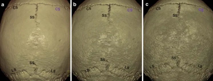
0.5mm, as used in our study, indicates that slices used for calculating 3D models are overlapping. This leads to much more precise reconstructions than those with an increment of 1.0mm or more. Finally, we want to point out that the chosen reconstruction parameter (kernel) is also important. If a 3D model is calculated from a dataset reconstructed in a soft tissue reconstruction (kernel of ~30), the computer will smooth the surface of the model. This leads so handsome images but also to artefacts that can be of importance if small structures have to be investigated. Figure6gives an example of this effect on the appearance of the cranial sutures on 3D models calculated from differently recon-structed data. While in a bone-reconstruction model the suture appears open, it seems to be already closed in a soft-tissue reconstruction. The same effect happens if other software with smoothing effect of the surface is used; therefore, such programs have to be avoided. In conclu-sion, all parameters that are important for the age estimation have to be investigated on models calculated from data with bone reconstructions, high resolution and low increment. These parameters are not essential for the determination of the sex, where importance is given to the shape of the pelvis and the skull.
A disadvantage of high-resolution CT slices is the generation and subsequent extraction of a large quantity of data. Therefore, we decided to examine only those skeletal regions that are important for the age estimation instead of the entire skeleton. Due to the results, we believe that the estimations could be improved if also one dataset showing the whole skeleton would be added. The difference
between the results using the complex method and those using further characteristics indicates that the investigation of more skeletal elements can be useful as well. A problem we observed in our study was the appearance of artefacts due to gas bubbles in the bone marrow recognisable in the 2D reconstructions of the humerus and femur of some decomposed bodies. These bubbles, visible as black dots with no X-ray absorption, complicated the interpretation of the structure of the spongiosa and can lead to misinterpre-tation of the state.
From a medico-legal point of view the work on“virtual skeletons” can bring many advantages. If a decomposed body is found and anthropological age estimation has to be performed, time-consuming maceration of the bones can be avoided. This fact is also of high importance from an ethical point of view because the bones are investigated in a non-invasive way. The unidentified body can be examined using the described protocol. This CT procedure takes only 10–20 min, depending on the cooling time of the machine and the routine of the CT personnel. The data can be stored in a database or simply on a CD or DVD. The fact that the data can be duplicated without an end or put on an internet server leads to an interactive data exchange. Experts like anthropologists can start their age estimation even before or while the autopsy of the body is performed. They do not have to wait until the bones are removed and macerated, and even experts in different places can give their opinions at the same time. This fact is especially important for the possibility of performing age estimations of skeletons which were found in common graves or if archaeologists
Fig. 6 a–c Demonstration of the importance to choose correct scanning parameters. A 3D model of the same skull, scanned with the same resolution (slice-thickness of 0.63mm and reconstruction increment of 0.5mm), but calculated from different reconstructed data. For the 3D model of the skull in a, a soft-tissue reconstruction was used (kernel: 30). The model in b was reconstructed with a kernel between soft-tissue and bone reconstruction (kernel: 50) and the model in c was reconstructed in a hard-bone reconstruction (kernel: 90). All 3D models show the cranial sutures (CS coronar
suture, LS lambdoid suture, SS sagittal suture) but with small differences. Using the soft-tissue reconstruction, which is most often used for creating 3D models, the program adds an automatical surface smoothing. Therefore, the model is the most handsome and unpitted one, but investigating the sutures the smoothing effect can lead to errors. The right coronar suture (violet lettering) seems to be closed in the soft-tissue reconstruction (a). In a bone-reconstruction (c), it is apparent that the suture is not completely closed
are investigating gravesites. By examining the bones and sending them to different experts via protected internet platforms, age and sex estimations of the bones can be performed without moving the other experts to the site. Also in cases of mass disasters, such approaches present an important saving of time. The application of MDCT for identification of disaster victims has already been dis-cussed in the literature [25]. The possibility to perform bone age estimation with the resulting data is a further argument to demonstrate the usefulness of an MDCT system on the site of disasters.
The virtual skeletons can be handled virtually without destroying the original sample. They can be cut and remerged, zoomed and reproduced and images can be stored simply as different files. Furthermore, estimation of sex and age of a body by using MSCT presents another step in the direction of the goal of the Virtopsy research group: to develop a minimally invasive autopsy.
The greatest advantages of an“anthropological database” as it is planned at the Institute of Forensic Medicine in Lausanne is that it leads to a large number of dated skeletons that can be stored and investigated ad infinitum. We think that such collections are important, especially with regard to the requirement of evaluating the existing age-estimation methods. A similar database exists in Bern in the context of the Virtopsy project. Because this database was created for medico-legal purposes and not for anthropological ones, the CT parameters were not adapted to the needs of anthro-pological studies. The CT resolution of these virtual skeletons, in particular, is not high enough. Until now, performing anthropological studies is difficult because collections of skeletons with known calendar age are rare, especially if collectives including a huge number of individuals are needed. The present feasibility study was an important first step on the way to our optimised database. The biggest limitation of the virtual approach is, of course, the access to an MDCT. In spite of the big impact of forensic radiology all over the world and the implementa-tion of MDCT in forensic institutes, most of the instituimplementa-tion do not have access to CT and MRI. To realise forensic
radiological working groups, a close collaboration between forensic pathologists and radiologists is of the highest importance. Also, the problem of the huge number of resulting data has to be considered, if anthropological studies should be performed. Thus, digital databases with high storage capacity are important.
Due to the low number of cases reported in our study, statistical analysis would not be significant. Therefore, a large evaluative study including at least 100 male and 100 female individuals will be realised in the near future, in order to perform correct statistics that can also be used as comparative studies for further approaches with other age-estimation methods. Actually, there is a leak of large anthropological studies, especially such using the complex method of age estimation. Therefore, a comparison of the virtual approach with studies carried out on real bone material was not possible. To perform such comparative studies, it would be necessary to remove and macerate the bones of the bodies, which is beyond the scope in forensic cases, seen from an ethical and juridical point of view. Performing CT scans of already existing bone collections, and comparing the sex and age estimation performed on the real bones with those performed on virtual bones would be an alternative. The problem, however, is the difficulty in getting access to the rare collections of dated bones. However, such a study is important for further develop-ment of the virtual anthropological approach.
Conclusion
In conclusion, this article demonstrates the potential of MDCT for estimating the sex and age of an individual in a non-invasive way. By creating a virtual skeleton with 2D and 3D reconstructions, the bones can be investigated without sampling and maceration. Virtual skeletons can be stored easily, handled without destroying the sample and therefore investigated ad infinitum. These abilities mean that virtual skeletons should be collected in order facilitate future anthropological studies.
References
1. Dirnhofer R, Jackowski C, Vock P et al (2006) VIRTOPSY: minimally inva-sive, imaging-guided virtual autopsy. Radiographics 26:1305–1333
2. Grabherr S, Stephan BA, Buck U et al (2007) Virtopsy–radiology in forensic medicine. Imaging Decis MRI 11:2–9 3. Ritz-Timme S, Rochholz G, Schutz
HW et al (2000) Quality assurance in age estimation based on aspartic acid racemisation. Int J Legal Med 114:83– 86
4. Wittwer-Backofen U, Gampe J, Vaupel JW (2004) Tooth cementum annulation for age estimation: results from a large known-age validation study. Am J Phys Anthropol 123:119–129
5. Nemeskéri J, Harsányi L, Acsádi G (1960) Methoden zur Diagnose des Lebensalters von Skelettfunden. Anthrop Anz 24:70–95 6. Kemkes-Grottenthaler A (1993)
Kritischer Vergleich osteomorphognos-tischer Verfahren zur Lebensalterbes-timmung Erwachsener. Inaug-Diss (thesis), Mainz
7. Rösing FW, Graw M, Marré B et al (2007) Recommendations for the fo-rensic diagnosis of sex and age from skeletons. Homo 58(1):75–89 8. Verhoff MA, Ramsthaler F, Krähahn J
et al (2008) Digital forensic osteology– possibilities in cooperation with the Virtopsy project. Forensic Sci Int 174:152–156
9. Ferembach D, Schwidetzky I, Stloukal M (1979) Empfehlungen für die Alters-und Geschlechtsdiagnose am Skelett. Homo 30:289–321
10. Sjøvold T (1975) Tables of the com-bined method for determination of age at death given by Nemeskéri, Harsányi, Acsádi. Anthrop Közl 19:9–22 11. Suchey JM, Wiseley DV, Katz D
(1986) Evaluation of the Todd and McKern-Stewart methods for aging the male os pubis. In: Reichs KJ (ed) Forensic osteology. Advances in the identification of human remains, 1st edn. Charles C Thomas, Springfield 12. Suchey JM, Katz D (1997)
Applica-tions of pubic age determination in a forensic setting. In: Reichs KJ (ed) Forensic osteology. Advances in the identification of human remains, 2nd edn. Charles C Thomas, Springfield 13. Brooks S, Suchey JM (1990) Skeletal
age determination based on the os pubis: a comparison of the Acsádi-Nemeskéri and Suchey-Brooks meth-ods. Hum Evol 5:227–238
14. Iscan MY, Loth SR, Wright RK (1984) Age estimation from the rib by phase analysis: white males. J Forensic Sci 29:1094–1104
15. Iscan MY, Loth SR, Wright RK (1985) Age estimation from the rib by phase analysis: white females. J Forensic Sci 30:853–863
16. Suchey JM, Katz D (1986) Skeletal age standards derived from an extensive multiracial sample of modern Ameri-cans. Proc 55th Annu Meeting Am Assoc Phys Anthrop, Albuquerque 17. Acsádi G, Nemeskéri J (1970) History
of human life span and mortality. Akadémiai Kiadó, Budapest 18. Byers SN (2005) Introduction to
fo-rensic anthropology, 2nd edn. Pearson, Boston New York
19. Graw M, Czarnetzki A, Haffner HT (1999) The form of the supraorbital margin as a criterion in identification of sex from the skull: investigations based on modern human skulls. Am J Phys Anthropol 108:91–96
20. Krogman WM,İşcan MY (1986) The human skeleton in forensic medicine, 2nd edn. Charles C Thomas, Springfield
21. Sjøvold T (1988) Geschlechtsdiagnose am skelett. In: Knußmann R (ed) Anthropologie. Handbuch der vergle-ichenden Biologie des Menschen 1, part 1. Gustav Fischer, Stuttgart New York
22. Rösing FW (2001) Forensische Altersdiagnose: Statistik, Arbeitsregeln und Darstellung. In: Oehmichen M, Geserick G (ed) Osteologische Identi-fikation und Altersschätzung. Schmidt-Römhildm, Lübeck
23. Masset C (1989) Age estimation on the basis of cranial sutures. In: Thomas CC, Iscan MY (eds) Age markers in the human skeleton. Charles C Thomas, Springfield
24. Aykroyd RG, Lucy D, Pollard AM et al (1997) Technical note: regression anal-ysis in adult age estimation. Am J Phys Anthropol 104:259–265
25. Sidler M, Jackowski C, Dirnhofer R et al (2007) Use of multislice computed tomography in disaster victim identifi-cation–advantages and limitations. Forensic Sci Int 169:118–128