158
The inspiratory partial pressure of oxygen (PIO2) at sea level is approximately 160 mm Hg. At altitudes of 2000 and 4500 m above sea level, the PIO2is about 120 and 85 mm Hg, respectively. Concomitantly, in the alve-olar air, PO2 is reduced to 75 and 55 mm Hg. In the healthy individual, the resulting hypoxemia causes a vari-ety of respiratory, circulatory, and electrocardiographic (ECG) changes,1including an increase in sympathetic tone, with pulmonary hypertension, hyperventilation, and an enhanced diuresis. The ECG changes have mainly been attributed to the synergistic effects of catecholamine secretion and vagal withdrawal rather than to direct hypoxic effects.2In patients with marginal cardiocircula-tory reserve or coronary artery disease (CAD), these
effects may cause cardiac decompensation3 and silent ischemia during normal daily activities, even at a modest altitude of 2000 m.4Thus it is current clinical practice to advise patients with CAD not to exceed moderate alti-tudes of about 2000 to 2500 m.5-7Similar conditions are encountered in most airplanes during flight, raising some concerns about the risk of air travel for patients with CAD.8,9
In experimental animals, acute hypoxemia10 and chronic hypoxemia have been shown to increase base-line11 and maximal drug-induced12 and exercise-induced11 myocardial blood flow (MBF). In contrast, only a few studies have examined the influence of acute hypoxic hypoxia on coronary circulation in human beings. A close relation between cardiac effort, myocar-dial oxygen consumption, and coronary flow has been repeatedly reported.13-15Four decades ago, Gregg16was the first to point out that most of the oxygen is normally removed from the coronary blood so that the oxygen con-tent of coronary sinus (venous) blood cannot be reduced by very much. Therefore increased oxygen delivery must be achieved primarily by increased MBF. In fact, MBF is regulated to maintain constant myocardial tissue oxygen tension.17 However, Kaijser et al18 found an enhanced oxygen extraction at rest during acute exposure to an alti-tude of 2300 m and no change in MBF, which increased
Philipp A. Kaufmann, MD,aChristian Schirlo, MD,cVojtech Pavlicek, MD,c Thomas Berthold,aCyrill Burger, PhD,bGustav K. von Schulthess, MD, PhD,b Erwin A. Koller, MD,cand Alfred Buck, MDa
Background. Although only poor data exist on changes in myocardial blood flow (MBF)
under acute hypoxia, patients with known coronary artery disease are advised not to exceed a moderate altitude exposure of about 2000 m above sea level.
Methods and Results. We measured MBF with positron emission tomography using
O-15–labeled water in 8 healthy human volunteers (aged 26 ± 3 years [mean ± SD]) at baseline (450 m above sea level, Zurich, Switzerland) and during acute hypoxic hypoxemia induced by inhala-tion of 2 hypoxic gas mixtures corresponding to altitudes of 2000 and 4500 m. MBF remained unchanged at 2000 m (increase of 10%, not significant) but increased significantly at 4500 m (62%, P < .001), exceeding the relative increase in rate pressure product.
Conclusions. Our results may explain why exposure to an altitude of 2000 m (corresponding
to the cabin pressure in most airplanes during flight) is clinically well tolerated, even by patients with reduced coronary flow reserve, such as those with coronary artery disease. However, at an altitude of 4500 m, MBF increases significantly, supporting the recommendation that patients with impaired flow reserve avoid exposure to higher altitudes. (J Nucl Cardiol 2001;8:158-64.)
Key Words: Myocardial blood flow • high altitudes • positron emission tomography
From the Departments of Nuclear Cardiologyaand Nuclear Medicine,b University Hospital and the Institute of Physiology,cUniversity of Zurich, Zurich, Switzerland.
P.A.K. was funded by a grant from the Swiss National Science Foundation (SCORE B, grant no. 3232-055002.98).
Received for publication July 28, 2000; final revision accepted Oct 10, 2000.
Reprint requests: Philipp A. Kaufmann, MD, Cardiovascular Center, Nuclear Cardiology, University Hospital, Raemistr 100, CH-8091 Zurich, Switzerland; [email protected].
Copyright © 2001 by the American Society of Nuclear Cardiology. 1071-3581/2001/$35.00 + 0 43/1/112537
during physical exercise. Thus it remains unclear whether a reduced coronary vasodilator capacity (such as that in patients with CAD) represents a risk factor during acute exposure to hypoxia.
The purpose of this study was to assess the influence of different stages of acute hypoxic hypoxia on MBF in healthy human volunteers.
METHODS
The study protocol was approved by the local ethics com-mittee. Each volunteer gave informed written consent after the investigative nature of the study and its risks and merits had been carefully explained.
Study Population
We studied 8 healthy volunteers (1 woman and 7 men, aged 26 ± 3 years [mean ± SD], range 23-32 years) who were nonacclimated (ie, they had no sojourn at or above 2700 m 4 weeks before the experiment and were all living at altitude of Zurich, Switzerland). None of the subjects had a history of car-diovascular disease or smoking. Further inclusion criteria were normal heart rate, blood pressure, and electrocardiography and low probability of CAD development.19
Study Protocol and Radiopharmaceuticals
The simulation of altitude hypoxia (hypoxic hypoxia) was achieved by inhalation of moistened hypoxic gas mixtures (through a mouthpiece), consisting of approximately 17.5% and 12.5% oxygen in nitrogen, which correspond to altitudes of 2000 and 4500 m, respectively. The 2 periods of hypoxic breathing were interposed by normoxic resting ventilation for 15 minutes. Heart rate and peripheral arterial oxygen satura-tion (SaO2) were recorded with a finger pulse oximeter (Nellcor N-200E; Nellcor Inc, Hayward, Calif). Arterial blood pressure was measured by the oscillometric Riva-Rocci method with an upper arm cuff (Bosotron 2; Bosch & Sohn, Jungingen, Germany). End-tidal partial pressure of carbon dioxide (PEtCO2) and respiratory frequency were determined with a capnograph (Dräger Capnodig; Dräger, Lübeck, Germany).
At baseline 1 (450 m, Zurich), all volunteers breathed through a mouthpiece with occluded nasal airways to avoid con-founding errors due to irritation by this installation. After a car-diorespiratory steady state was achieved, as assessed by a steady PEtCO2, SaO2, and heart rate, positron emission tomography (PET) measurements were performed after 15 minutes at base-line 1, 2000 m, and 4500 m and at the end of the experiment at baseline 2 (450 m, Zurich). By use of a bolus technique, 800 MBq of O-15–labeled water ([15O]H
2O) was injected into a
peripheral vein of each volunteer at all of the above-mentioned conditions, while acquisition of the serial transaxial tomo-graphic images of the heart was started. The 20 minutes between each [15O]H
2O injection allowed ample time for [15O] decay to
occur (halftime = 2 minutes). After the second baseline
mea-surement, 350 MBq of [13N]ammonia was injected for
visual-ization of the myocardium to draw the regions of interest (ROIs) for quantitative analysis. Patient positioning for each image set was aided by marking the patient’s chest with a felt-tip pen and aligning the marks with the reference laser beam of the tomo-graph.
PET Protocol
All images were acquired on a GE positron emission tomo-graph (GE Medical Systems, Milwaukee, Wis). This device records 35 image planes simultaneously. The axial field of view is 14.5 cm. A 30-minute blank scan was recorded as part of the daily routine procedures. Correct positioning of the volunteer’s heart within the axial field of view of the tomograph was ascer-tained on a 4-minute rectilinear transmission scan. This was fol-lowed by a 20-minute transmission scan for photon attenuation correction. Beginning with the intravenous administration of [15O]H
2O, twelve 10-second frames, four 30-second frames, and
one 60-second frame were recorded. Twelve-lead electrocardio-grams were monitored continuously throughout the study. Heart rate and blood pressure were recorded at 1- and 5-minute inter-vals, respectively.
Data Analysis
Two representative midventricular slices were chosen from the acquired sets of 35 transaxial images for quantita-tive analysis. The images acquired after the ammonia injec-tion were used to define a septal, anterior, and lateral ROI of the myocardial territory.20 Furthermore, a spheric ROI was
placed in the blood pool of the left ventricle. All ROIs were then pasted to the dynamically acquired image sets. Myocardial and blood pool time-activity curves were gener-ated from the dynamic frames and corrected for radioisotope decay.
Estimates of MBF
MBF was estimated by model fitting of the blood pool and myocardial activity curves. The PET-measured time-activity curves were fitted with a 1-compartment model.21
MBF values for each ROI were calculated and averaged in each patient to create a mean MBF value for the entire myocardium. Because resting MBF is determined by cardiac workload,22we also considered baseline MBF normalized for
the rate pressure product (RPP),23an index of myocardial
oxy-gen consumption, by dividing MBF by RPP × 10–4at each
study condition for each patient.
Statistical Analysis
Values are given as mean ± SD, unless stated otherwise. Comparisons of hemodynamic and MBF values between the different study conditions were performed with analysis of vari-ance statistics for repeated measures. If the P value was below .05, the Scheffé test was applied.
RESULTS
All subjects tolerated the simulated ascent to high altitude subjectively well and without major objective impairment.
Hemodynamic and ECG Findings
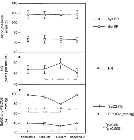
Heart rate was similar at baseline 1 (68 ± 7 beats/min) and 2000 m (68 ± 9 beats/min), increased at 4500 m (80 ± 10 beats/min, P < .05), and decreased at baseline 2 (64 ± 8 beats/min). No significant changes were found for systolic and diastolic arterial blood pressure, which averaged 116 ± 8 mm Hg and 66 ± 8 mm Hg, respectively, at all altitudes (Figure 1). No significant ST-segment depression and no arrhythmia were found in any subject.
Respiratory Reactions
PEtCO2 was 39 ± 3 mm Hg at baseline 1 and
remained unchanged at 2000 m (38 ± 4 mm Hg); it
decreased significantly at 4500 m (34 ± 4 mm Hg, P < .05 vs baseline 1). SaO2 decreased from 97% ± 1% at baseline 1 to 94% ± 2% at 2000 m (P < .05) and 83% ± 4% at 4500 m (P < .0001). The respiratory frequency was about 14 ± 3 breaths/min and remained constant. Myocardial Blood Flow
Estimates of mean MBF were 0.97 ± 0.21 mL/min/g at baseline 1 (Zurich, 450 m), 1.07 ± 0.30 mL/min/g at 2000 m (not significant [NS]), 1.56 ± 0.36 mL/min/g at 4500 m (P < .0001 vs baseline 1 and vs 2000 m), and 1.07 ± 0.20 mL/min/g at baseline 2 (Table 1). Thus there was no significant change in MBF at 2000 m (increase of 10%, NS). In contrast, MBF increased significantly by 62% at 4500 m (P < .001) and decreased at baseline 2 (Figure 2). After normalization for the RPP, MBF aver-aged 1.34 ± 0.44 mL/min/g at baseline 1, 1.34 ± 0.39 mL/min/g at 2000 m, 1.74 ± 0.48 mL/min/g at 4500 m (P < .01 vs baseline 1 and 2000 m), and 1.43 ± 0.49
Figure 1. Hemodynamics and SaO2/PEtCO2. Although no significant changes were found for systolic and diastolic arte-rial blood pressure, heart rate increased significantly at 4500 m and was normalized after return to baseline. PEtCO2was similar at baseline and at 2000 m but decreased significantly at 4500 m. SaO2decreased slightly at 2000 and 4500 m and was normalized after return to baseline. sys, Systolic; dia, diastolic; BP, blood pressure; HR, heart rate.
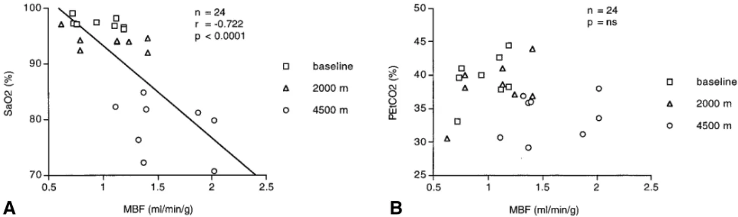
mL/min/g at baseline 2 (NS vs baseline 1). Thus, even after normalization of MBF for workload (ie, RPP), there was a significant increase in MBF at 4500 m (36%, P < .05) in excess of the increase in myocardial oxygen (Figure 2). In fact, the percentage increase in MBF at 4500 m was greater than the percentage increase in RPP (62% vs 18%, P < .005). MBF was inversely correlated with SaO2(r = –0.722, P < .0001), an extrapolated index of arteriolar PO2, but not with PEtCO2(Figure 3).
DISCUSSION
In this study the changes in MBF during high-alti-tude exposure were assessed noninvasively by PET. In agreement with the findings of Grubbstrom et al24 and
Kaijser et al,18our results show that there are no signifi-cant changes in MBF in healthy human beings at a mod-erate altitude of 2000 m. This may explain the fact that even patients with CAD and impaired left ventricular function tolerate these conditions well,25 because no sizeable flow reserve seems to be required to meet the myocardial oxygen demand during the slight decrease in PO2at 2000 m. However, at high altitudes such as 4500 m, an increase in MBF of 62% is observed despite the concomitant decrease in PEtCO2. A hypoxia-induced increase in heart rate due to sympathetic activation and vagal withdrawal could increase myocardial oxygen con-sumption because of an increase in cardiac work26and thus could also account for increased MBF.27There was still a significant increase in MBF (36%, P < .05),
how-Figure 3. MBF was inversely correlated with SaO2(an index of arteriolar PO2) (A) but not with PEtCO2(B), supporting the theory that diminished oxygen tension may be the main stimulus for coronary dilation and that carbon dioxide plays a minor role at this altitude.
Figure 2. Influence of altitude on MBF. MBF did not change significantly at 2000 m but increased by 62% at 4500 m (P < .001) and decreased at baseline 2. After normalization for RPP, there was still a significant increase in blood flow (baseline MBF normalized [MBFnorm] 36%, P < .05), indicating that the correlation between MBF and RPP does not hold at these conditions. This suggests that an important part of the MBF increase is due to direct hypoxia-induced vascular dilation.
ever, even after we accounted for the hypoxia-induced increase in workload by normalizing for RPP. This sug-gests that an important part of the MBF increase is due to direct, hypoxia-induced dilation. In fact, MBF was inversely correlated with SaO2(an extrapolated index of PaO2) but not with PEtCO2 (Figure 3). This is in agree-ment with findings in experiagree-mental animals showing that diminished oxygen tension in the myocardium may be the main stimulus for coronary dilation28and that carbon dioxide only plays a minor role at this altitude.27The lack of significance in the MBF increase at 2000 m, with a significant but slight decrease in SaO2, does not necessar-ily imply an increase in the oxygen extraction ratio. According to our findings, the decreasing arterial oxygen supply seems to be met by an autoregulatory increase of coronary flow, but the correlation between MBF and RPP no longer holds true under these conditions. In fact, this study, using a noninvasive technique, is the first full report describing an increase in MBF in human beings during acute hypoxic hypoxia, providing evidence for the assumed mechanism of coronary autoregulation. The exact nature of the coronary autoregulatory mechanism remains unclear because MBF is influenced by many fac-tors.29For example, chemoreceptor stimulation with neu-rogenic vasodilation mediated through vagal efferent fibers may occur during acute hypoxia.30
The oxygen deprivation at high altitudes can be toler-ated only because of the increase in ventilation, which defends the alveolar PO2against the reduced PIO2. In agree-ment with previous findings31 the respiratory frequency remained unchanged during our experiment, indicating that the hyperventilation was due to an increase in tidal volume. Initially, the hyperventilation results in a low arterial PCO2, causing respiratory alkalosis. This increases the oxygen affinity of hemoglobin and accelerates the oxygen loading
of the pulmonary capillary blood. Stamler et al32recently found that deoxygenation induces an allosteric transition in S-nitrosohemoglobin, causing the release of a nitric oxide group, which relaxes the vessels to bring local blood flow in line with oxygen requirements. This mechanism could contribute to the MBF regulation. Furthermore, adenosine may play an important role in this context because, in vitro, its production is linked to myocardial PO233,34and the major stimulus for adenosine formation seems to be an imbalance between oxygen delivery and oxygen demand.35,36 Cardiac adenosine production has been reported in myocytes37and in endothelial cells,33,37,38and coronary as well as myocardial adenosine receptors have been isolated in vitro.39This endothelium-mediated mech-anism is potentially susceptible to harm by risk factors such as hypertension40and hypercholesterolemia,41which have been shown to cause endothelial dysfunction, even in the absence of angiographically detectable CAD. Future stud-ies on the influence of hypoxia in these patients and those with CAD should provide more insight.
Study Limitations
Realignment to the transmission image to correct for subject motion, as reported by Hoh et al,42was not per-formed. However, during MBF measurements at rest, lit-tle if any subject movement occurs43 because subject motion is most often due to side effects of potent vasodilators such as adenosine or dipyridamole, neither of which was used in this study. Furthermore, the sub-jects were fixed with thoracic elastic bands after they were placed in the PET scanner.
Irritation caused by breathing through the mouth-piece might have influenced the subjects’ respiration, leading to an altered pattern of breathing and possible Table 1. Myocardial blood flow (milliliters per minute per gram of tissue)
Volunteer Baseline1 2000 m
No. Baseline 1 normalized 2000 m normalized 4500 m
1 1.19 1.30 1.41 1.53 2.02 2 1.10 1.33 1.13 1.14 1.32 3 1.12 2.22 1.24 1.70 1.87 4 0.94 1.17 1.13 1.34 1.39 5 1.19 1.71 1.41 2.01 2.02 6 0.72 0.88 0.62 1.00 1.11 7 0.73 1.13 0.79 1.14 1.37 8 0.76 0.94 0.79 0.86 1.37 Mean ± SD 0.97 ± 0.21 1.34 ± 0.44 1.07 ± 0.30 1.34 ± 0.39 1.56 ± 0.36
sympathetic activation due to discomfort. To minimize this effect, all measurements, including both baseline measurements, were performed in the same way (the mouthpiece and the noseclip in place). A habituation effect over the study time that could affect the repro-ducibility of repeated measures can most likely be excluded because no differences were found between baseline 1 and baseline 2 with regard to MBF, PEtCO2, and SaO2.
The age range of the study population was lower than that of the typical CAD age group. However, no difference in the physiologic response to hypoxia over a wide age range from 8 to 83 years has been reported.44
No additional measurements were performed dur-ing hyperemia to assess flow reserve (hyperemic/base-line flow) because of ethical reasons (restricted amount of radioactivity). Thus no firm conclusion on the influ-ence of hyperemia on the myocardial flow reserve can be drawn. However, there is no evidence to suggest that hypoxia would affect the ceiling of maximal hyperemic MBF usually achieved by vasodilators such as adeno-sine or dipyridamole. This leads to the conclusion that, in hypoxia, flow reserve might be reduced because basal MBF is increased with no change in maximal MBF.
Conclusions
Acute exposure to an altitude of 2000 m (corre-sponding to the cabin pressure in most airplanes during flight) induces no changes in MBF at rest. This might explain why these conditions are clinically well toler-ated, even by patients with reduced coronary flow reserve, such as those with CAD. However, at an
alti-tude of 4500 m, MBF increases significantly at rest, suggesting that patients with reduced flow reserve as seen in CAD can generally travel safely to moderate altitudes but should avoid exposure to higher altitudes. Although the exact mechanism of the coronary autoreg-ulation remains unclear, adenosine is likely to play an important role.
References
1. Laciga P, Koller EA. Respiratory, circulatory, and ECG changes during acute exposure to high altitude. J Appl Physiol 1976;41:159-67. 2. Koller EA, Drechsel S, Hess T, Macherel P, Boutellier U. Effects of
atropine and propranolol on the respiratory, circulatory, and ECG responses to high altitude in man. Eur J Appl Physiol 1988;57: 163-72.
3. Lomazzi F, Gurtner HP. Staying at high altitudes and plane travel for heart patients [in German]. Schweiz Med Wochenschr 1981;111:618-24. 4. Volker K, Hoppe B, Krestin M, Rost R. Cross-country and downhill skiing in patients with myocardial infarct. Can silent ischemia be pre-vented by drug therapy? [in German]. Fortschr Med 1990;108:273-5. 5. Gemelli A. Some cardiological problems in high mountains. Correlation
with humans in flight [in German]. Minerva Med 1980;71:1985-9. 6. Roach RC, Houston CS, Honigman B, et al. How well do older persons
tolerate moderate altitude? West J Med 1995;162:32-6.
7. Alexander JK. Coronary problems associated with altitude and air travel. Cardiol Clin 1995;13:271-8.
8. Beighton PH, Richards PR. Cardiovascular disease in air travellers. Br Heart J 1968;30:367-72.
9. Skjenna OW, Evans JF, Moore MS, Thibeault C, Tucker AG. Helping patients travel by air. Can Med Assoc J 1991;144:287-93.
10. Manohar M, Parks CM, Busch MA, et al. Regional myocardial blood flow and coronary vascular reserve in unanesthetized young calves exposed to a simulated altitude of 3500 m for 8-10 weeks. Circ Res 1982;50:714-26.
11. Paridon SM, Bricker JT, Dreyer WJ, et al. The effects of hypoxemia on myocardial blood flow during exercise. Pediatr Res 1989;25:280-4. 12. Reller MD, Morton MJ, Giraud GD, Wu DE, Thornburg KL. Maximal
myocardial blood flow is enhanced by chronic hypoxemia in late ges-tation fetal sheep. Am J Physiol 1992;263:H1327-9.
13. Korner PI. Circulatory adaptations in hypoxia. Physiol Rev 1959;39: 687-730.
14. Katz LM, Feinberg H. The relation of cardiac effort to myocardial oxy-gen consumption and coronary flow. Circ Res 1958;6:656-69. 15. Schrader J. Mechanisms of coronary circulation regulation [in
German]. Z Kardiol 1984;2:41-6.
16. Gregg DW. Regulation of the collateral and coronary circulation of the heart. In: McMichael J, editor. Circulation. Oxford (UK): Blackwell Scientific Publications; 1958.
17. Grover RF, Lufschanowski R, Alexander JK. Alterations in the coro-nary circulation of man following ascent to 3,100 m altitude. J Appl Physiol 1976;41:832-8.
18. Kaijser L, Grubbstrom J, Berglund B. Coronary circulation in acute hypoxia. Clin Physiol 1990;10:259-63.
19. Diamond G, Forrester J. Analysis of probability as an aid in the clinical diagnosis of coronary artery disease. N Engl J Med 1979;300:1350-8. 20. Hutchins GD, Schwaiger M, Rosenspire KC, Krivokapich J, Schelbert
H, Kuhl DE. Noninvasive quantification of regional blood flow in the human heart using N-13 ammonia and dynamic positron emission tomographic imaging. J Am Coll Cardiol 1990;15:1032-42.
21. Iida H, Kanno I, Takahashi A, et al. Measurement of absolute
myocar-4500 m Baseline 2
normalized Baseline 2 normalized
2.00 1.31 1.59 1.32 1.12 1.43 2.12 1.21 1.94 1.31 0.90 1.12 2.60 1.31 2.32 1.74 0.98 1.12 1.23 0.85 1.09 1.61 0.85 0.86 1.74 ± 0.48 1.07 ± 0.20 1.43 ± 0.49
dial blood flow with H215O and dynamic positron-emission tomography.
Strategy for quantification in relation to the partial-volume effect [pub-lished correction appears in Circulation 1988;78:1078]. Circulation 1988;78:104-15.
22. Camici P, Marraccini P, Marzilli M, et al. Coronary hemodynamics and myocardial metabolism during and after pacing stress in normal humans. Am J Physiol 1989;257:E309-17.
23. Uren NG, Melin JA, De BB, Wijns W, Baudhuin T, Camici PG. Relation between myocardial blood flow and the severity of coronary artery stenosis. N Engl J Med 1994;330:1782-8.
24. Grubbstrom J, Berglund B, Kaijser L. Myocardial blood flow and lac-tate metabolism at rest and during exercise with reduced arterial oxy-gen content. Acta Physiol Scand 1991;142:467-74.
25. Erdmann J, Sun KT, Masar P, Niederhauser H. Effects of exposure to altitude on men with coronary artery disease and impaired left ventric-ular function. Am J Cardiol 1998;81:266-70.
26. Czernin J, Muller P, Chan S, et al. Influence of age and hemodynamics on myocardial blood flow and flow reserve. Circulation 1993;88:62-9. 27. Rushmer RF. The coronary system. In: Rushmer RF, editor. Cardiovascular dynamics. 2nd ed. Philadelphia: Saunders; 1961. p. 218-9.
28. Wolpers HG, Hoeft A, Korb H, Lichtlen PR, Hellige G. Heterogeneity of myocardial blood flow under normal conditions and its dependence on arterial PO2. Am J Physiol 1990;258(2 Pt 2):H549-55.
29. Marcus ML. The coronary circulation in health and disease. New York: McGraw-Hill; 1983. p. 65-92.
30. Heistad DD, Abboud FM. Circulatory adjustments to hypoxia. Circulation 1980;61:463-70.
31. Koller EA, Lesniewska B, Bührer A, Bub A, Kohl J. The effects of acute altitude exposure in Swiss highlanders and lowlanders. Eur J Appl Physiol 1993;66:146-54.
32. Stamler JS, Jia L, Eu JP, et al. Blood flow regulation by S-nitrosohe-moglobin in the physiological oxygen gradient. Science 1997; 276:2034-7.
33. Deussen A, Moser G, Schrader J. Contribution of coronary endothelial cells to cardiac adenosine production. Pflugers Arch 1986;406:608-14. 34. Smolenski RT, Schrader J, de Groot GH, Deussen A. Oxygen partial pressure and free intracellular adenosine of isolated cardiomyocytes. Am J Physiol 1991;260:C708-14.
35. Bardenheuer H, Schrader J. Supply-to-demand ratio for oxygen determines formation of adenosine by the heart. Am J Physiol 1986;250:H173-80. 36. Schrader J, Bardenheuer H, Orntyd J. Relationship between myocardial
metabolism and coronary blood flow regulation. Cardiologia 1985; 30:915-8.
37. Deussen A, Lloyd HG, Schrader J. Contribution of S-adenosylhomo-cysteine to cardiac adenosine formation. J Mol Cell Cardiol 1989;21: 773-82.
38. Kroll K, Schrader J, Piper HM, Henrich M. Release of adenosine and cyclic AMP from coronary endothelium in isolated guinea pig hearts: relation to coronary flow. Circ Res 1987;60:659-65.
39. Schrader J, Kroll K, Henrich M, Piper HM. Coronary and myocardial adenosine receptors. Biomed Biochim Acta 1987;46:S421-6. 40. Frielingsdorf J, Seiler C, Kaufmann P, Vassalli G, Suter T, Hess OM.
Normalization of abnormal coronary vasomotion by calcium antago-nists in patients with hypertension. Circulation 1996;93:1380-7. 41. Kaufmann PA, Frielingsdorf J, Mandinov L, Seiler C, Hug R, Hess OM.
Reversal of abnormal coronary vasomotion by calcium antagonists in patients with hypercholesterolemia. Circulation 1998;97:1348-54. 42. Hoh CK, Dahlbom M, Harris G, et al. Automated iterative
three-dimen-sional registration of positron emission tomography images. J Nucl Med 1993;34:2009-18.
43. Nitzsche EU, Choi Y, Czernin J, Hoh CK, Huang S, Schelbert HR. Noninvasive quantification of myocardial blood flow in humans: a direct comparison of the [13N]ammonia and the [15O]water tech-niques. Circulation 1996;93:2000-6.
44. Veglio M, Maule S, Cametti G, Cogo A, Lussiana L, Madrigale G, et al. The effects of exposure to moderate altitude on cardiovascular auto-nomic function in normal subjects. Clin Auton Res 1999;9:123-7.
RECEIVE TABLES OF CONTENTS BY E-MAIL
To receive the tables of contents by e-mail, sign up through our Web site at
http://www.mosby.com/nuclcard
Choose E-mail Notification.
Simply type your e-mail address in the box and click the Subscribe button. Alternatively, you may send an e-mail message to [email protected]. Leave the subject line blank, and type the following as the body of your message:
subscribe nuclcard_toc
You will receive an e-mail to confirm that you have been added to the mailing list. Note that TOC e-mails will be sent out when a new issue is posted to the Web site.