O R I G I N A L A R T I C L E
Expression of Atrophy mRNA Relates to Tendon Tear Size
in Supraspinatus Muscle
Silvia Schmutz PhD, Thomas Fuchs,
Felix Regenfelder MD, Patrick Steinmann MD, M. Zumstein MD, Bruno Fuchs MD, PhD
Received: 12 November 2007 / Accepted: 24 September 2008 / Published online: 22 October 2008 Ó The Association of Bone and Joint Surgeons 2008
Abstract Skeletal muscle atrophy and fatty infiltration develop after tendon tearing. The extent of atrophy serves as one prognostic factor for the outcome of surgical repair of rotator cuff tendon tears. We asked whether mRNA of genes involved in regulation of degradative processes leading to muscle atrophy, ie, FOXOs, MSTN, calpains, cathepsins, and transcripts of the ubiquitin-proteasome pathway, are overexpressed in the supraspinatus muscle in patients with and without rotator cuff tears. We evaluated biopsy specimens collected during surgery of 53 consecu-tive patients with different sizes of rotator cuff tendon tears and six without tears. The levels of corresponding gene transcripts in total RNA extracts were assessed by semi-quantitative reverse transcriptase-polymerase chain reaction (RT-PCR) analysis. Supraspinatus muscle atrophy was assessed by MRI. The area of muscle tissue (or atro-phy), decreased (increased) with increasing tendon tear size. The transcripts of CAPN1, UBE2B, and UBE3A were upregulated more than twofold in massive rotator cuff tears as opposed to smaller tears or patients without tears. These atrophy gene products may be involved in cellular pro-cesses that impair functional recovery of affected muscles after surgical rotator cuff repair. However, the damaging
effects of gene products in their respective proteolytic processes on muscle structures and proteins remains to be investigated.
Introduction
Atrophy and fatty infiltration of skeletal muscle are con-sequences of rotator cuff tendon tears in animal models and in humans [4,14,26]. Muscle atrophy depends on the size of tear and time elapsed since the tear [13, 26, 34, 42]. Even after structurally successful surgical tendon repair, skeletal muscles of the rotator cuff only partially recover from atrophy [13,33]. Thus, the original force-generating capacity of the muscle is not restored and muscle function remains restricted. If atrophy could be prevented or reversed, the clinical outcome after rotator cuff tendon repair could be improved.
Skeletal muscle atrophy results from reduced protein synthesis and/or increased protein degradation [16, 23]. Various signaling proteins, such as transcription factors, reportedly regulate skeletal muscle atrophy. In the absence of growth or survival signals, the transcription factor FOXO localizes to the nucleus (Fig.1) and activates genes involved in cell death and cell cycle inhibition [35]. Specifically, FOXO induces atrogin-1 and MURF1 and thus leads to muscle atrophy [22, 31]. This signaling orchestrates three known proteolytic systems working together in muscle atro-phy, ie, the calcium-dependent calpain system, the lysosomal protease system (cathepsins), and the ubiquitin-proteasome system [21]. The ubiquitous calpains (CAPN1 and CAPN2) are involved in the disassembly of sarcomeric proteins in atrophy models [20,28,41]. Therefore, myofibrillar proteins become available for ubiquitination, a prerequisite for deg-radation by proteasomes that are not able to degrade intact One or more of the authors (CG, BF) have received funding from the
Swiss National Science Foundation (SNF #320000-113424). Each author certifies that his or her institution has approved the human protocol for this investigation, that all investigations were conducted in conformity with ethical principles of research, and that informed consent for participation in the study was obtained. S. Schmutz, T. Fuchs, F. Regenfelder, P. Steinmann, M. Zumstein, B. Fuchs (&)
Department of Orthopedics, University Hospital Balgrist, University of Zurich, Forchstrasse 340, 8008 Zurich, Switzerland e-mail: bruno.fuchs@balgrist.ch
myofibrils [1]. Different from the ubiquitous calpains, calpain 3 is not involved in the initial proteolytic events [9], and may play a role in sarcomere maintenance in mature muscle cells [9]. The conjugation of ubiquitin to proteins in the ubiquitin-proteasome system is mediated by a series of sequential reactions conducted by ubiquitin-activating enzymes (E1), ubiquitin-conjugating enzymes (E2), and ubiquitin-protein ligases (E3). Proteins ubiquitinated by polyubiquitin chains are recognized and degraded by the proteasome [21]. If the protein substrate is monoubiquitinated or diubiquitinated, it is degraded by the lysosomal pathway [21]. Cathepsins L, B, D, and H are the major lysosomal proteases [3] and have dif-ferent substrate specificities [24, 27, 32]. They hydrolyze various myofibrillar proteins and are involved in skeletal muscle atrophy [3].
Although several studies explore acute skeletal muscle atrophy (eg, disuse, spaceflight, immobilization, or dener-vation [19, 29, 36, 37], the molecular mechanisms of chronic atrophy after tendon tears are less well understood [11]. We presume an understanding of the cascade(s) of cellular processes that lead to muscle atrophy with tendon damage could lead to focused pharmacologic treatments (eg, proteolysis inhibitors) reversing the atrophy.
We therefore (1) compared the expression levels of the selected gene transcripts of patients with defined rotator cuff defects compared with the respective transcript levels in the supraspinatus muscle of patients with intact rotator cuffs and (2) determined whether the expression varied by cuff size and age.
Materials and Methods
To determine the expression status of genes involved in protein degradation, we enrolled 59 consecutive patients undergoing shoulder surgery: 53 patients had various sizes of tendon tears (Table1); six patients had an intact rotator cuff and underwent surgery for other reasons (impingement syndrome [n = 2], shoulder instability with Bankart lesion [n = 1], acromioclavicular arthropathy [n = 2], and gle-nohumeral degenerative disease [n = 1]). To examine the influence of defect size, we defined three groups of the 53 patients with rotator cuff tendon tears: tears as much as1.3 of the rotator cuff tendons in sagittal diameter (n = 27), as much as2.3(n = 13), and full tears (n = 13). Thus, we had four groups: three with varying size tears and one without Fig. 1 The three known proteolytic systems with the corresponding
proteins are shown. The black boxes indicate increases (p \ 0.05) and gray boxes decreases in the transcript level with increasing size of the rotator cuff tear (Kruskal-Wallis test over all groups). The dotted boxes indicate a trend for an upregulation (p \ 0.1). FOXO1A = forkhead box protein O1A; FOXO3A = forkhead box protein O3A; CAP-NS1 = calpain small subunit 1; CAPN1 = calpain-1 catalytic subunit;
CAPN2 = calpain-2 catalytic subunit; CAPN3 = calpain-3; Ub = ubiquitin; E1 = ubiquitin-activating enzyme; E2 = ubiquitin-conju-gating enzyme; UBE2J1 = ubiquitin-conjuubiquitin-conju-gating enzyme E2 J1; UBE2B = conjugating enzyme E2 B; E3 = ubiquitin-protein ligase; UBE3A = ubiquitin-ubiquitin-protein ligase E3A; MURF1 = muscle-specific RING finger protein 1; UBE4B = ubiquitin conjuga-tion factor E4 B; CTSB = cathepsin B; CTSL = cathepsin L.
tears. The median patient age was 57.3 years (range, 27– 87 years). There was no difference in the distribution of men and women among the groups without rotator cuff tendon tears and with different sizes of tendon tears in sagittal diameter (Table1). Patients with tendon tears were on average 20 years older (p \ 0.001) than patients with intact tendons. The body mass index (median, 25.7 kg/m2) was not related to the size of the rotator cuff tendon tear. Our power analysis indicated, to detect a threshold of differential expression of 2.5, presuming this increase would influence protein expression and ultimately clini-cally relevant proteolysis, six patients per group were required. The study was approved by the institutional investigational review board. All patients provided signed informed consent to participate in this study.
We (SS, PS, FR) determined the Constant-Murley score [7] and Goutallier stages on standardized MRI (parasagittal T1-weighted turbo spin echo) preoperatively [10, 17]. Goutallier Stage 0 corresponds to a completely normal muscle, without any fatty streak; Stage 1, the muscle contains some fatty streaks; Stage 2, fatty infiltration is important, but there is still more muscle than fat present; Stage 3, there is as much fat as muscle; and Stage 4, there is more fat than muscle present [17, 18]. For the 59 patients, the Goutallier stage increased with increasing size of the rotator cuff tendon tear (p = 0.002), and the Constant-Murley score also was related to tendon tear size (p = 0.019), with maximal values observed in Group 1 (Table1). To measure the extent of atrophy, we (FR, MZ) determined the muscle cross-sectional area of the supra-spinatus in the most lateral MR image where the scapular spine is in contact with the rest of the scapula [34]. This muscle area was standardized to the area of the supraspi-natus fossa, which is considered representative for body constitution [42]. Areas were measured on the MR console using the manufacturer’s standard software. We found the muscle cross-sectional area standardized to the area of the supraspinatus fossa decreased with increasing tear size (p \ 0.001), indicating ongoing muscle atrophy (Table1). Biopsy specimens of the supraspinatus muscle of approximately 20 mg were collected during surgery (before repair) and sampled immediately thereafter, which was
described previously [11]. To avoid the influence on gene expression of muscular inhomogeneity, eg, from different fiber type composition in different parts of the muscle [2,25], biopsy specimens always were taken from the same region of the muscle, ie, at the anatomic location which corresponds on MRI to the most lateral part where the scapular spine is in contact with the rest of the scapula to produce a Y-shaped appearance as described previously [11,42].
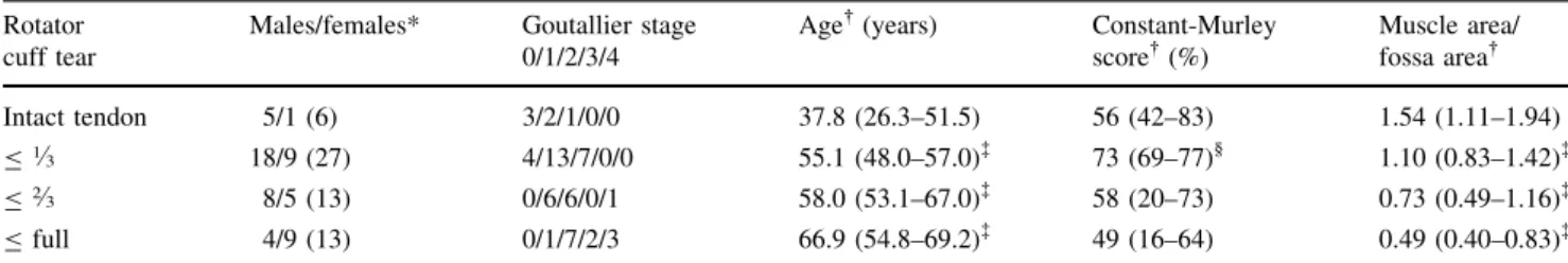
Total RNA was isolated from 25-lm cryosections of the biopsy specimens [11, 40]. Specifically, the integrity of the RNA was analyzed by agarose gel electrophoresis, and RNA concentrations were estimated with the RiboGreen1 RNA quantification kit (Invitrogen, Carlsbad, CA). The expression levels of transcripts encoding proteins involved in muscle atrophy were analyzed by semiquantitative RT-PCR in triplicate and normalized to glyceraldehyde-3-phosphate dehydrogenase (GAPDH) transcript levels for all samples as described previously [8,11]. Briefly, cDNA was synthesized from individual RNA extracts in triplicate using the StrataScript1 First Strand cDNA Synthesis kit (Stratagene, La Jolla, CA) with oligo-dT primers and 750 ng total RNA template in a total volume of 50 lL. The primer pairs for PCR amplification of selected gene tran-scripts were designed with the LightCycler1Probe Design Software 2.0 (Roche, Rotkreuz, Switzerland) (Table 2). As quality control for quantification of PCR products in the linear range of amplification, PCR reactions were per-formed with 4 lL and 8 lL of the RT reaction containing cDNA template equivalent to 5 ng and 10 ng total RNA, respectively, extracted from individual biopsy specimens. The following conditions revealed amplification within the linear range for all selected gene transcripts, indicated by ratios of 2 ± 0.3 between the detected amounts of PCR product of a distinct gene transcript generated in parallel reactions with twofold different amounts of template. After initial denaturation at 94°C for 3 minutes, cDNA repre-senting GAPDH or individual atrophy-related transcripts were amplified by 25 or 35 PCR cycles (denaturation at 94°C for 40 seconds, primer pair-dependent annealing at a specific temperature [Table2] for 40 seconds, and elon-gation at 72°C for 20 seconds), respectively. PCR was completed by a final elongation step at 72°C for 7 minutes. Table 1. Patient characteristics
Rotator cuff tear
Males/females* Goutallier stage 0/1/2/3/4
Age (years) Constant-Murley score (%) Muscle area/ fossa area Intact tendon 5/1 (6) 3/2/1/0/0 37.8 (26.3–51.5) 56 (42–83) 1.54 (1.11–1.94) B1.3 18/9 (27) 4/13/7/0/0 55.1 (48.0–57.0)à 73 (69–77)§ 1.10 (0.83–1.42)à B2.3 8/5 (13) 0/6/6/0/1 58.0 (53.1–67.0)à 58 (20–73) 0.73 (0.49–1.16)à B full 4/9 (13) 0/1/7/2/3 66.9 (54.8–69.2)à 49 (16–64) 0.49 (0.40–0.83)à * Values expressed as number of males/females, with the total number of patients in parentheses; values expressed as median, with 95% confidence interval in parentheses;àdifferent from patients without rupture (p \ 0.05);§tends to be different from patients without rupture (p \ 0.1).
PCR products were analyzed by agarose gel electrophoresis and quantified with Quantity One1 1D Analysis software provided by the VersaDocTM Imaging System (Bio-Rad Laboratories, Hercules, CA) [11].
Data are presented as median and 95% confidence interval. We used a chi square test to determine whether the categories of nominal or ordinal scaled data (gender, Goutallier stage, dominance) were distributed equally among the four groups. We used a correlation analysis to determine whether any of the transcripts varied with age.
For the transcripts, the gene expression level of each group was normalized to the median of the control group. We determined the differences in transcript levels of the respective genes of interest with respect to the size of the rotator cuff tendon tear using the Kruskal-Wallis test. We determined differences of gene expression between any two respective groups with differing tear sizes (Table3) using the Mann-Whitney U test; we did not use post hoc tests for multiple comparisons. We used SPSS1 for Windows111.5.0 (SPSS Inc, Chicago, IL).
Table 2. Genes and primer sequences of the analyzed transcripts
Gene name GenBank ID Forward primer (50–30) Annealing temperature (°C) Reverse primer (50–30) FOXO1A NM_002015 CTTGTATGTTAATTGCATCTTCATTGGCTTGGTA 65 GCTGTGCTTAGAGGAACTTGGGT FOXO3A NM_001455 TCATGATGACACAGTCGGACCC 64 TGGTGGTGGAGCAAGTTCTGATT MSTN NM_005259 TGGTAGTAGACCGCTGTGGGTG 64 CTTGTATGATTTGTTTGGATGGTTAAATGCC CAPNS1 NM_001749 TGAGGCCAACGAGAGTGAGGA 64 CTTGCCTGTGGTGTCGCTAT CAPN1 NM_005186 GCAAGTGCTCTCAGAAGAGGAGATTGACG 67 CCATTGCCATCACGATCCATGAGGTT CAPN2 NM_001748 CCCTGTCAACTCCACCAAGTCATCGT 68 AAGTACTGAGAAACAGAGCCAAGAGATAAGGTCG CAPN3 NM_000070 GCCTCCCAGCGAGTACGTCA 67 GCTCCTTGTTGCTGTTTGCTCTGTCC UBE2J1 NM_016021 AACAGCCTTCCCTCCGTT 64 AGCAGGTGGCTAGCTGACA UBE2B NM_003337 ATGAGCAGAGAACTGATGCGACTTGT 67 CCCAGCCTATAAGCATTTATCCAATCAATGT UBE3A NM_000462 CTATGTCTGTGCCTCCCTTCTTTATTGG 64 AAGGCTCAACCTCAAGCAGTAATAAAC Atrogin-1 NM_058229 TGCAGCCAAGAAGAGAAAGAAGGACA 66 TCCAACAGCCGGACCACGTAG MURF1 NM_032588 GGGCTTTGAGAACATGGACTTCTTTACTTTGGATT 68 GGTGTCCTTCTTCCTTCCCTTCTGTGG UBE4B NM_006048 ATGAAAGTCTGGAGTCTCTGAAGCGAA 66 GGGCGAGGTAAGAGCGGGACA CTSB BC001908 ACAGGCCATGTGAGCCACCG 68 CGCTTTCCATTCCTGCGTCTCTGTCTTG CTSL NM_145918 ATGAATCCTACACTCATCCTTGCTGCCTTT 68 CCACACTGCTCTCCTCCATCCTTCTT GAPDH NM_002046 TGAACGGGAAGCTCACTGGCATGG 67 TGGGTGTCGCTGTTGAAGTCAGAGGAGA
MSTN = myostatin; GAPDH = glyceraldehyde-3-phosphate dehydrogenase; FOXO1A = forkhead box protein O1A; FOXO3A = forkhead box protein O3A; CAPNS1 = calpain small subunit 1; CAPN1 = calpain-1 catalytic subunit; CAPN2 = calpain-2 catalytic subunit; CAPN3 = calpain-3; UBE2J1 = conjugating enzyme E2 J1; UBE2B = conjugating enzyme E2 B; UBE3A = ubiquitin-protein ligase E3A; MURF1 = muscle-specific RING finger ubiquitin-protein 1; UBE4B = ubiquitin conjugation factor E4 B; CTSB = cathepsin B; CTSL = cathepsin L.
Results
Several genes involved in protein degradation were upregulated at the mRNA level in the muscles of massive rotator cuff tears (Table3). However, we identified no specific degradation pathway where upregulation was substantially greater than another.
Of the transcription factors analyzed in this analysis, FOXO1A was upregulated in the muscles of massive tears compared with smaller tears and controls, whereas FOXO3A was downregulated if the tear became larger than 1.3 (Table3). In the calpain group, CAPN1 was upregulated in massive tears. In the ubiquitin-proteasome pathway group, UBE2B and UBE3A were upregulated in the muscles of massive tears compared with smaller tears and controls. In the lysosomal group, CTSB was upregulated in massive tears compared with smaller tears. Of all genes analyzed, CAPN1, UBE2B, and UBE3A were upregulated greater than twofold in the muscles of massive tears compared with smaller tears and controls.
Gene expression of each transcript did not correlate (r2\ 0.1) with age (Fig.2; Table4). Thus, at most 10% variation of the expression levels could be explained by age of the patients.
Discussion
Rotator cuff tears lead to atrophy, fatty infiltration of skeletal muscles, pain, and loss of shoulder function. Therefore, massive rotator cuff tears with considerable atrophy and high muscular fat content of the corresponding muscles have a poor outcome after surgery. We presume identification of the molecular mechanisms of chronic atrophy could lead to pharmacologic treatments (eg, blocking agents to proteolysis). To recognize indicator molecules for increased muscle structure degradation causing atrophy in supraspinatus muscle of patients with rotator cuff tears, we investigated expression at the tran-script level of selected genes, which encode proteins representative of four groups that orchestrate proteolysis. These include transcription factors and proteins represent-ing the calpain, lysosomal, and ubiquitin-proteasome proteolytic pathways.
Owing to limitations of such a study in patients, the findings need critical discussion. Gene expression can be influenced by the timing of responses to injury and we did not know the time that had elapsed between the rotator cuff tendon tear and surgery. By analyzing patients’ histories, we estimated the time between tendon tearing until sur-gery, which ranged from months to years. Therefore, the Table 3. Results of the RT-PCR analysis
Protein category Gene name p Value Intact tendon B1.
3 B2
.
3 B Full
Transcription factor/signaling FOXO1A* 0.002 1.0 (0.4–1.7) 1.1 (0.7–1.2) 0.8 (0.5–1.4) 1.6 (1.3–2.2) FOXO3A* 0.008 1.0 (0.3–1.3) 1.0 (0.8–1.1) 0.5 (0.2–0.6) 0.6 (0.2–1.0) MSTN 0.147 1.0 (0.3–2.1) 1.2 (1.1–1.6) 1.7 (0.9–2.1) 1.9 (0.4–3.1) Ca2+-dependent calpains CAPNS1 0.051 1.0 (0.5–1.3) 0.9 (0.8–1.0) 1.0 (0.7–1.1) 1.2 (1.0–2.1)
CAPN1 0.071 1.0 (0.5–6.3) 2.2 (1.7–2.9) 2.0 (1.5–4.0) 2.7 (1.9–4.7) CAPN2 0.118 1.0 (0.8–4.2) 1.4 (1.1–1.9) 1.2 (0.5–1.6) 1.7 (1.1–2.1) CAPN3* 0.047 1.0 (0.3–1.4) 1.2 (1.0–1.3) 0.8 (0.5–0.9) 1.2 (0.9–1.3) Ubiquitin- proteasome pathway UBE2J1 0.685 1.0 (0.7–1.3) 1.0 (0.6–1.9) 1.2 (1.1–1.3) ND
UBE2B* 0.006 1.0 (0.4–1.4) 1.2 (1.1–1.9) 1.2 (1.0–1.3) 2.1 (1.2–2.7) UBE3A* 0.007 1.0 (0.3–7.1) 0.7 (0.5–1.1) 1.9 (0.5–3.0) 3.5 (0.8–6.5) Atrogin-1 0.195 1.0 (0.7–1.6) 0.8 (0.8–1.0) 1.0 (0.8–1.5) 1.1 (0.8–1.6) MURF1 0.491 1.0 (0.3–1.1) 0.9 (0.7–1.1) 0.7 (0.6–1.0) 0.8 (0.4–1.0) UBE4B 0.564 1.0 (0.4–1.2) 0.9 (0.8–1.0) 0.9 (0.5–1.1) 1.1 (0.7–1.4) Lysosomal enzymes CTSB* 0.015 1.0 (0.3–3.6) 0.9 (0.6–1.5) 0.8 (0.5–1.4) 1.5 (1.0–3.4) CTSL 0.245 1.0 (0.7–1.4) 0.9 (0.7–1.4) 0.7 (0.5–0.9) 0.9 (0.4–2.5) Values expressed as median ratio of mRNA levels of patients with different extent of rotator cuff tendon tears to patients with intact tendon, with 95% confidence intervals in parentheses; *transcripts that are altered compared with intact tendon (p \ 0.05; Kruskal-Wallis test over all groups); p values refer to entire cohort of patients including all four subgroups; different from the group with the next smaller size tendon tear (p \ 0.05; Mann-Whitney U test comparing two groups); ND = not detected; MSTN = myostatin; FOXO1A = forkhead box protein O1A; FOXO3A = forkhead box protein O3A; CAPNS1 = calpain small subunit 1; CAPN1 = calpain-1 catalytic subunit; CAPN2 = calpain-2 catalytic subunit; CAPN3 = calpain-3; UBE2J1 = ubiquitin-conjugating enzyme E2 J1; UBE2B = ubiquitin-conjugating enzyme E2 B; UBE3A = ubiquitin-protein ligase E3A; MURF1 = muscle-specific RING finger protein 1; UBE4B = ubiquitin conjugation factor E4 B; CTSB = cathepsin B; CTSL = cathepsin L.
muscle biopsies performed in our study represent only the chronic phase of atrophy and must be distinguished from the acute phase. Another potential limitation of our study was the age difference in the group with intact rotator cuff versus the other groups with varying sizes of rotator cuff tendon tears because age may influence gene expression of proteins involved in skeletal muscle atrophy [6, 15, 39]. We analyzed expression values using the Kruskal-Wallis test including all four groups together. Additionally, we compared the expression values of each group with any extent of tendon tear with the group with intact tendons and the massive tear group with the next smaller extent of tendon tear. The latter analysis revealed the main differ-ences in expression occurred in massive tears compared
with the second largest tear size group. For these two groups, there was no difference in age. Given less than 10% of the variation in expression levels could be explained by age, we presume age is not a causative factor. The significantly upregulated or downregulated gene tran-scripts we observed indicate altered proteolysis in supraspinatus muscle of patients with a rotator cuff tear. However, the findings must be confirmed at the protein level, and the biological role of these proteins in interaction with cellular mechanisms that contribute to muscle atrophy remain to be investigated because expression at the mRNA level may not correlate with protein expression or biolog-ical relevance.
To understand and interpret our findings, the temporal expression patterns of genes must be considered. The expression level of mRNA encoding proteins involved in skeletal muscle atrophy varies with time after a tendon tear. For example, atrogin-1 and MURF1, which represent key regulators of muscle atrophy, appear only transiently ele-vated during rapid muscle mass loss in the acute phase of skeletal muscle atrophy [5,30]. Therefore, these two genes showed no changes in expression levels because our analysis included only chronic tears and mRNA levels normalized again after the acute phase. The fact that MURF1 was not upregulated also may reflect the fact that age may not influence our findings as it is upregulated in aged skeletal muscles [6,15,39]. Also, the expression of FOXO1A is upregulated in acute tears and persists at an elevated level [30]. These findings may be consistent with our increased expression of FOXO1A. The greatest dif-ferential expression was identified in UBE2B and UBE3A, which act in concert in the ubiquitination of soluble muscle proteins and in CAPN1 [38]. We found them upregulated particularly in massive rotator cuff tears. This may indicate it is the UBE genes in the ubiquitin-proteasome pathway and not the atrogin-1 and MURF1 genes which play an important role in mediating the molecular processes of atrophy in massive rotator cuff tears. One key player of the lysosomal pathway, CTSB, also is upregulated. This indi-cates all gene families involved in protein degradation are represented in the atrophy process in patients with massive
Age compared with Atrogin-1
R2 = 0.001 0,0 0,5 1,0 1,5 2,0 2,5 3,0 3,5 4,0 20 40 60 80 Age [years] Atrogin-1 /GAPDH A
Age compared with FOXO1A
R2 = 0.095 0,0 0,5 1,0 1,5 2,0 20 40 60 80 Age [years] FOXO1A /GAPDH B
Fig. 2A–B Scatterplots of (A) atrogin-1 and (B) FOXO1A mRNA levels indicate the expression of these genes is not age related.
Table 4. Coefficient of determination of age and gene expression
Gene name R2 FOXO1A 0.095 FOXO3A 0.022 MSTN 0.002 CAPNS1 0.049 CAPN1 0.027 CAPN2 0.012 CAPN3 0.002 UBE2J1 0.021 UBE2B 0.084 UBE3A 0.064 Atrogin-1 0.001 MURF1 0.002 UBE4B 0.010 CTSB 0.083 CTSL 0.029
R2= coefficient of determination; MSTN = myostatin; FOXO1A = forkhead box protein O1A; FOXO3A = forkhead box protein O3A; CAPNS1 = calpain small subunit 1; CAPN1 = calpain-1 catalytic subunit; CAPN2 = calpain-2 catalytic subunit; CAPN3 = calpain-3; UBE2J1 = conjugating enzyme E2 J1; UBE2B = ubiquitin-conjugating enzyme E2 B; UBE3A = ubiquitin-protein ligase E3A; MURF1 = muscle-specific RING finger protein 1; UBE4B = ubiquitin conjugation factor E4 B; CTSB = cathepsin B; CTSL = cathepsin L.
rotator cuff tears, suggesting the cellular processes involved in this atrophy are complex and these pathways are orchestrated among each other (Fig.1). However, in view of this complexity, our analysis represents a pre-liminary study providing the rationale for in vitro studies that require more detailed analysis at the protein and functional levels to better characterize the role of these genes in the atrophy process of rotator cuff tears.
Our data suggest mRNA levels of atrophy-related genes were increased preferentially in biopsy specimens from our patients with large or massive rotator cuff tendon tears. Massive rotator cuff tears were associated with increased muscular atrophy and fatty infiltration [13]. Therefore, the repair of massive tears often is followed by retears of the tendons and accompanied by additional muscular degen-eration [13]. Consequently, the outcome after surgical repair of massive tears is often less satisfactory than out-come in patients with smaller tears [12]. Considering the fact that protein degradation genes are overexpressed in the muscles of massive rotator cuff tears, our results parallel the clinical observations of increased atrophy and muscular degeneration in large tears compared with small tears. Therefore, it may be that the activity of atrophy genes in muscles of patients with massive rotator cuff tendon tears may contribute to the unsatisfying results after surgical repair.
We found the transcript levels of distinct genes involved in skeletal muscle atrophy increased in patients with mas-sive rupture of the rotator cuff tendons compared with patients with smaller tears or without tears. Increased activity of these genes may contribute to impaired func-tional recovery of atrophic muscles after surgical rotator cuff repair. Taken together, the observations suggest upregulation of the transcripts encoding FOXO1A, cal-pains, UBE2B, UBE3A, and CTSB, which reflect molecular events in the rotator cuff muscle after tendon tears and concomitant muscle atrophy.
Acknowledgments We thank Walter Born, PhD, for helpful advice on the manuscript and Christian Gerber, MD, for helping to recruit the patients.
References
1. Bartoli M, Richard I. Calpains in muscle wasting. Int J Biochem Cell Biol. 2005;37:2115–2133.
2. Barton ER, Gimbel JA, Williams GR, Soslowsky LJ. Rat supraspinatus muscle atrophy after tendon detachment. J Orthop Res. 2005;23:259–265.
3. Bechet D, Tassa A, Taillandier D, Combaret L, Attaix D. Lysosomal proteolysis in skeletal muscle. Int J Biochem Cell Biol. 2005;37:2098–2114.
4. Bjorkenheim JM. Structure and function of the rabbit’s supra-spinatus muscle after resection of its tendon. Acta Orthop Scand. 1989;60:461–463.
5. Bodine SC, Latres E, Baumhueter S, Lai VK, Nunez L, Clarke BA, Poueymirou WT, Panaro FJ, Na E, Dharmarajan K, Pan ZQ, Valenzuela DM, DeChiara TM, Stitt TN, Yancopoulos GD, Glass DJ. Identification of ubiquitin ligases required for skeletal muscle atrophy. Science. 2001;294:1704–1708.
6. Clavel S, Coldefy AS, Kurkdjian E, Salles J, Margaritis I, Derijard B. Atrophy-related ubiquitin ligases, atrogin-1 and MuRF1 are up-regulated in aged rat tibialis anterior muscle. Mech Ageing Dev. 2006;127:794–801.
7. Constant CR, Murley AH. A clinical method of functional assessment of the shoulder. Clin Orthop Relat Res. 1987;214:160–164.
8. Dheda K, Huggett JF, Bustin SA, Johnson MA, Rook G, Zumla A. Validation of housekeeping genes for normalizing RNA expression in real-time PCR. Biotechniques. 2004;37:112–114, 116, 118–119.
9. Duguez S, Bartoli M, Richard I. Calpain 3: a key regulator of the sarcomere? FEBS J. 2006;273:3427–3436.
10. Fuchs B, Weishaupt D, Zanetti M, Hodler J, Gerber C. Fatty degeneration of the muscles of the rotator cuff: assessment by computed tomography versus magnetic resonance imaging. J Shoulder Elbow Surg. 1999;8:599–605.
11. Fuchs B, Zumstein M, Regenfelder F, Steinmann P, Fuchs T, Husmann K, Hellermann J, Jost B, Hodler J, Born W, Gerber C. Upregulation of alpha-skeletal muscle actin and myosin heavy polypeptide gene products in degenerating rotator cuff muscles. J Orthop Res. 2008;26:1007–1011.
12. Gazielly DF, Gleyze P, Montagnon C. Functional and anatomical results after rotator cuff repair. Clin Orthop Relat Res. 1994; 304:43–53.
13. Gerber C, Fuchs B, Hodler J. The results of repair of massive tears of the rotator cuff. J Bone Joint Surg Am. 2000;82:505–515. 14. Gerber C, Meyer DC, Schneeberger AG, Hoppeler H, von Rechenberg B. Effect of tendon release and delayed repair on the structure of the muscles of the rotator cuff: an experimental study in sheep. J Bone Joint Surg Am. 2004;86:1973–1982.
15. Giresi PG, Stevenson EJ, Theilhaber J, Koncarevic A, Parkington J, Fielding RA, Kandarian SC. Identification of a molecular signature of sarcopenia. Physiol Genomics. 2005;21:253–263. 16. Goldspink DF, Morton AJ, Loughna P, Goldspink G. The effect
of hypokinesia and hypodynamia on protein turnover and the growth of four skeletal muscles of the rat. Pflugers Arch. 1986;407:333–340.
17. Goutallier D, Postel JM, Bernageau J, Lavau L, Voisin MC. Fatty muscle degeneration in cuff ruptures: pre- and postoperative evaluation by CT scan. Clin Orthop Relat Res. 1994;304:78–83. 18. Goutallier D, Postel JM, Lavau L, Bernageau J. [Impact of fatty degeneration of the suparspinatus and infraspinatus muscles on the prognosis of surgical repair of the rotator cuff][in French]. Rev Chir Orthop Reparatrice Appar Mot. 1999;85:668–676. 19. Graebe A, Schuck EL, Lensing P, Putcha L, Derendorf H.
Physiological, pharmacokinetic, and pharmacodynamic changes in space. J Clin Pharmacol. 2004;44:837–853.
20. Huang J, Forsberg NE. Role of calpain in skeletal-muscle protein degradation. Proc Natl Acad Sci USA. 1998;95:12100–12105. 21. Jackman RW, Kandarian SC. The molecular basis of skeletal
muscle atrophy. Am J Physiol Cell Physiol. 2004;287:C834– C843.
22. Kandarian SC, Jackman RW. Intracellular signaling during skeletal muscle atrophy. Muscle Nerve. 2006;33:155–165. 23. Loughna P, Goldspink G, Goldspink DF. Effect of inactivity and
passive stretch on protein turnover in phasic and postural rat muscles. J Appl Physiol. 1986;61:173–179.
24. Matsukura U, Okitani A, Nishimuro T, Kato H. Mode of degra-dation of myofibrillar proteins by an endogenous protease, cathepsin L. Biochim Biophys Acta. 1981;662:41–47.
25. Meyer DC, Pirkl C, Pfirrmann CW, Zanetti M, Gerber C. Asymmetric atrophy of the supraspinatus muscle following ten-don tear. J Orthop Res. 2005;23:254–258.
26. Nakagaki K, Ozaki J, Tomita Y, Tamai S. Fatty degeneration in the supraspinatus muscle after rotator cuff tear. J Shoulder Elbow Surg. 1996;5:194–200.
27. Noda T, Isogai K, Hayashi H, Katunuma N. Susceptibilities of various myofibrillar proteins to cathepsin B and morphological alteration of isolated myofibrils by this enzyme. J Biochem (Tokyo). 1981;90:371–379.
28. Purintrapiban J, Wang MC, Forsberg NE. Degradation of sar-comeric and cytoskeletal proteins in cultured skeletal muscle cells. Comp Biochem Physiol B Biochem Mol Biol. 2003;136: 393–401.
29. Rittweger J, Frost HM, Schiessl H, Ohshima H, Alkner B, Tesch P, Felsenberg D. Muscle atrophy and bone loss after 90 days’ bed rest and the effects of flywheel resistive exercise and pamidro-nate: results from the LTBR study. Bone. 2005;36:1019–1029. 30. Sacheck JM, Hyatt JP, Raffaello A, Jagoe RT, Roy RR,
Edgerton VR, Lecker SH, Goldberg AL. Rapid disuse and denervation atrophy involve transcriptional changes similar to those of muscle wasting during systemic diseases. FASEB J. 2007;21:140–155.
31. Sandri M, Sandri C, Gilbert A, Skurk C, Calabria E, Picard A, Walsh K, Schiaffino S, Lecker SH, Goldberg AL. Foxo tran-scription factors induce the atrophy-related ubiquitin ligase atrogin-1 and cause skeletal muscle atrophy. Cell. 2004;117:399– 412.
32. Schwartz W, Bird JW. Degradation of myofibrillar proteins by cathepsins B and D. Biochem J. 1977;167:811–820.
33. Thomazeau H, Boukobza E, Morcet N, Chaperon J, Langlais F. Prediction of rotator cuff repair results by magnetic resonance imaging. Clin Orthop Relat Res. 1997;344:275–283.
34. Thomazeau H, Rolland Y, Lucas C, Duval JM, Langlais F. Atrophy of the supraspinatus belly: assessment by MRI in 55 patients with rotator cuff pathology. Acta Orthop Scand. 1996;67:264–268.
35. Tran H, Brunet A, Griffith EC, Greenberg ME. The many forks in FOXO’s road. Sci STKE. 2003;2003:RE5.
36. Tyml K, Mathieu-Costello O. Structural and functional changes in the microvasculature of disused skeletal muscle. Front Biosci. 2001;6:D45–D52.
37. Tyml K, Mathieu-Costello O, Cheng L, Noble EG. Differential microvascular response to disuse in rat hindlimb skeletal muscles. J Appl Physiol. 1999;87:1496–1505.
38. Ventadour S, Attaix D. Mechanisms of skeletal muscle atrophy. Curr Opin Rheumatol. 2006;18:631–635.
39. Welle S, Brooks AI, Delehanty JM, Needler N, Thornton CA. Gene expression profile of aging in human muscle. Physiol Genomics. 2003;14:149–159.
40. Wittwer M, Fluck M, Hoppeler H, Muller S, Desplanches D, Billeter R. Prolonged unloading of rat soleus muscle causes distinct adaptations of the gene profile. FASEB J. 2002;16:884–886. 41. Xiao YY, Wang MC, Purintrapiban J, Forsberg NE. Roles of
mu-calpain in cultured L8 muscle cells: application of a skeletal muscle-specific gene expression system. Comp Biochem Physiol C Toxicol Pharmacol. 2003;134:439–450.
42. Zanetti M, Gerber C, Hodler J. Quantitative assessment of the muscles of the rotator cuff with magnetic resonance imaging. Invest Radiol. 1998;33:163–170.