Denis R. Morel Jean-Louis Frossard Banu Cikirikcioglu Maxime Tapponnier Catherine M. Pastor
Time course of lung injury
in rat acute pancreatitis
Received: 22 February 2006 Accepted: 6 June 2006 Published online: 15 July 2006 © Springer-Verlag 2006
This work was supported by the Fonds National de la Recherche Scientifique (no. 3200-100868 to C.M.P. and no.
3200-100764 to J.-L.F.)
Electronic supplementary material The electronic reference of this article is http://dx.doi.org/10.1007/s00134-006-0264-9. The online full-text version of this article includes electronic supplementary material. This material is available to authorised users and can be accessed by means of the ESM button beneath the abstract or in the structured full-text article. To cite or link to this article you can use the above reference. C. M. Pastor (u)
Hôpitaux Universitaires de Genève, Laboratoire de Physiopathologie Hépatique et Imagerie Moléculaire,
Rue Micheli-du-Crest 24, 1205 Geneva, Switzerland e-mail: Catherine.Pastor@hcuge.ch Tel.: +41-22-3729353 Fax: +41-22-3729366 D. R. Morel · B. Cikirikcioglu · M. Tapponnier
Hôpitaux Universitaires de Genève, Division d’Investigations
Anesthésiologiques,
Rue Micheli-du-Crest 24, 1205 Geneva, Switzerland
J.-L. Frossard
Hôpitaux Universitaires de Genève, Division de Gastroentérologie et Hépatologie,
Rue Micheli-du-Crest 24, 1205 Geneva, Switzerland
Abstract Objective: Lung injury is
a severe complication of acute pancre-atitis that increases the mortality rate of the disease. The pathophysiology of acute pancreatitis has been studied in several experimental models, but the kinetics of pulmonary compli-cations in relation to the pancreatic disease is not completely understood. We then studied the severity of acute pancreatitis-associated lung injury over 18 h in rats that had taurocholic acid injection in the pancreatic duct and determined whether blood col-lected from rats with pancreatitis is toxic enough to induce injury in normal lungs. Design and setting: Prospective, randomized, and con-trolled animal study in an animal research laboratory in a university hospital. Interventions: We isolated lungs from rats with acute pancre-atitis 2, 6, and 18 h after taurocholic acid injection in the biliopancreatic duct and perfused them with blood collected from the same rats. Addi-tionally, blood collected from rats with acute pancreatitis (time-points: 2 and 6 h) was perfused in normal lungs. Measurements and results: Tauro-cholic acid injection induced a severe pancreatic injury that started as early as 2 h after the injection and persisted without recovery over the 18-h study period. In contrast, the pulmonary injury was transient, appearing at the 6-h time point with recovery by the end of the study. Pulmonary injury
was moderate and evidenced mostly during lung reperfusion. Interestingly, blood collected at the 2-h time point in pancreatic rats induced pulmonary injury in normal lungs while blood collected at the 6-h time-point was not toxic. Conclusions: While pancreatic injury persists over the full experimental period, pulmonary injury is transient in our experimental model. The recovery of lung injury by 18 h might be explained by a decrease in the overall toxicity of pancreatic blood over time.
Keywords Rats · Lung injury ·
Introduction
During acute pancreatitis the incidence of pulmonary complications varies from 15% to 55%, and their severity ranges from mild hypoxemia without clinical or radio-logical abnormalities to severe acute respiratory distress syndrome [1, 2, 3]. The early phase of the pancreatic disease is associated with a systemic inflammation while the second is characterized by the appearance of local complications such as fat necrosis, pseudocyst formation, and pancreatic abscesses. Two peaks of pulmonary com-plications during the early phase of acute pancreatitis have been observed by Berry et al. [4]. The first one is observed on hospital admission, and radiological abnormalities have been found in 15% of patients while by day 5 new radiological abnormalities are reported in additional 71% patients of the study. However, the kinetics of pulmonary complications in relation to the pancreatic disease is not well established.
Moreover, the mechanisms of propagation of the dis-ease from pancreas to lungs is largely unknown. Media-tors released by the inflamed pancreas, the peripancreatic tissues, or ascites have been administered in healthy ro-dents to reproduce the lung injury observed in severe acute pancreatitis. For example, trypsin [5, 6, 7] and elastase [5] can reproduce lung injury in healthy rodents. Moreover, ascites collected from rats with acute pancreatitis and in-jected to normal rats also reproduce the injury [8, 9]. An-other way to investigate the propagation of the pancreatic disease to lungs might be to perfuse normal lungs with blood collected from rats with acute pancreatitis. By
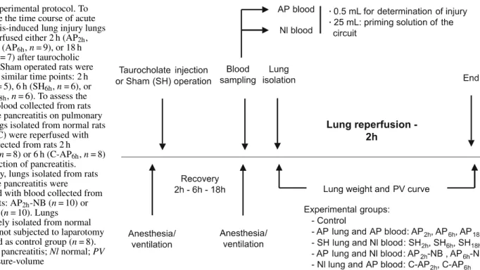
col-Fig. 1 Experimental protocol. To investigate the time course of acute pancreatitis-induced lung injury lungs were reperfused either 2 h (AP2h,
n = 7), 6 h (AP6h, n = 9), or 18 h
(AP18h, n = 7) after taurocholic
injection. Sham operated rats were studied at similar time points: 2 h (SH2h, n = 5), 6 h (SH6h, n = 6), or
18 h (SH18h, n = 6). To assess the
effect of blood collected from rats with acute pancreatitis on pulmonary injury lungs isolated from normal rats (control, C) were reperfused with blood collected from rats 2 h (C-AP2h, n = 8) or 6 h (C-AP6h, n = 8)
after induction of pancreatitis. Conversely, lungs isolated from rats with acute pancreatitis were reperfused with blood collected from normal rats: AP2h-NB (n = 10) or
AP6h-NB (n = 10). Lungs
immediately isolated from normal rats were not subjected to laparotomy and served as control group (n = 8). AP Acute pancreatitis; Nl normal; PV lung pressure-volume
lecting blood from pancreatic rats at various time points we might be able to determine which phase of the pancreatic disease is the more susceptible to induce injury in healthy lungs.
We then studied the severity of acute pancreatitis-associated lung injury over 18 h in rats that had tauro-cholic acid injection in the pancreatic duct and determined whether blood collected from rats with pancreatitis is toxic enough to induce injury in normal lungs.
Methods
Induction of acute pancreatitis
Sprague-Dawley rats (350–400 g) were anesthetized with isoflurane, intubated, and mechanically ventilated (FIO2 0.4) with a rodent ventilator (tidal volume 7 ml/kg; pos-itive end-expiratory pressure, PEEP, 2.5 cm H2O; respira-tory rate 70–80/bpm). After laparotomy the pancreatic duct was cannulated with a PE-10 tubing. A clip was placed on the duct close to the liver and taurocholic acid (4%, 0.5 ml sodium salts; Sigma) was infused at a constant rate. The clip was then withdrawn and the tubing was left in place, the lower extremity being secured into the duodenum to al-low a normal bile secretion. Before abdomen closure rats had saline solution (5 ml) into the peritoneum. Rats re-ceived buprenorphine and were looked after until lung iso-lation (2, 6, or 18 h later; Fig. 1). Sham rats were similarly anesthetized and ventilated and after laparotomy received intraperitoneal fluid and buprenorphine injection. All
pro-cedures were performed according to the guidelines stated in the National Institutes of Health “Guide for the Care and Use of Laboratory Animals.” The experimental proto-col was reviewed and approved by the Ethics Committee for Animal Research at the University Medical Center and by the Veterinary Office in Geneva, Switzerland.
Lung isolation
One hour before lung reperfusion rats were anesthetized, tracheotomized, ventilated, and monitored as previously described. In addition, the femoral artery was cannulated for blood sampling, continuous blood pressure monitoring, and fluid replacement. Rats were anticoagulated and 25 ml blood was gently withdrawn via the arterial cannula and replaced by an equal volume of 6% hydroxyethyl starch to maintain a mean systemic blood pressure above 50 mm Hg to minimize lung ischemia (normovolemic hemodilution). This blood volume was reconcentrated (hematocrit close to 30%) and used as a priming volume into the circuit. After a median sternotomy a polyethylene catheter was placed into the main pulmonary artery via the right ventricular outflow track. Catheters were also placed into the left ven-tricle for collection of effluent blood, and in the left atrium for continuous left atrial pressure (Pla) recording. Follow-ing pulmonary artery cannulation lungs were flushed with 30 ml cold (10°C) hydroxyethyl starch solution (perfusion pressure 30 cm H2O) to minimize warm ischemia until ex vivo reperfusion.
In vitro lung reperfusion
In a humidified Plexiglas chamber (37°C) the heart-lung block was suspended to an isometric force displacement transducer that continuously measures lung weight. Lungs were ventilated with room air mixed with 5% CO2 at a respiratory rate of 30/min, a tidal volume of 7 ml/kg, and a PEEP of 2.7 cm H2O. Sighs were applied every 5 min (21 ml/kg). The circuit was primed with blood volume collected from the same or from a different rat (see protocol in Fig. 1). Lungs were then perfused via the pulmonary artery cannula from an arterial reservoir placed at a fixed height to induce a mean pulmonary arterial pressure (Ppa) close to 17.5 mm Hg. Pulmonary blood flow (PBF) was continuously measured by a transit-time flowmeter. Effluent blood was drained through the left ventricle cannula whose distal extremity was placed at a height sufficient to obtain a Plaof 7.5± 0.5 mm Hg.
Assessment of lung function
Airway pressure (Paw), tidal volume, and airflow re-sistance were measured. Continuous breath by breath
dynamic lung compliance (Cdyn) and expiratory airway resistance (Raw) were assessed by the following formulas: Cdyn= tidal volume/(peak Paw– PEEP) and Raw= (peak Paw– PEEP)/maximal expiratory air flow. Inspiratory and expiratory quasistatic pressure-volume curves were performed before lung reperfusion and at the end of the 2-h reperfusion period by inflating and deflating lungs at a constant rate (0.3 ml/s) from PEEP level to a volume of 7 ml or 25 cm H2O (whichever occurred first) using an automated infusion pump. The volumes obtained at an inspiratory and expiratory Paw of 10 cm H2O were recorded (V10insp and V10exp, respectively). Pulmonary vascular resistance (PVR) was calculated by dividing Ppa– Pla by PBF. Every 30 min a blood sample was collected to measure blood gas, glucose and electrolyte concentrations, and hematocrit. Total red and white blood cell count and percentage of polymorphonuclear cells (PMNs) were determined with an automated cell counter. Amylase concentration was measured using an automatized analyzer.
Wet-to-dry lung weight ratio
The inferior lobe of each lung was weighed (wet weight), dried at 60°C for 2 days, and weighed again to determinate the wet-to-dry lung weight ratio.
Experimental groups
To investigate the time course of acute pancreatitis-induced lung injury in our experimental model lungs were reperfused either 2 h (AP2h, n = 7), 6 h (AP6h, n = 9), or 18 h (AP18h, n = 7) after taurocholic injection. Sham-operated (SH) rats were studied at similar time points: 2 h (SH2h, n = 5), 6 h (SH6h, n = 6), or 18 h (SH18h, n = 6) (Fig. 1). To assess the effect of blood collected from rats with acute pancreatitis on lung edema and pulmonary mechanics lungs isolated from normal rats (C) were reperfused with blood collected from rats 2 h (C-AP2h, n = 8) or 6 h (C-AP6h, n = 8) after induction of pancreatitis. Conversely, lungs isolated from rats with acute pancreatitis were reperfused with blood collected from normal rats: AP2h+ NB (n = 10) or AP6h+ NB (n = 10). Finally, lungs immediately isolated from normal rats served as a control group (control, n = 8).
Statistical analysis
Group data are presented as mean ± SE values. Statistical comparison among the treatment groups at baseline or at the end of the study was performed by a one-way analysis of variance followed by Newman–Keuls multiple compari-son test if the analysis of variance resulted in a p value less
than 0.05, and comparisons involving time used a multi-variate analysis of variance for repeated measurements.
Results
Taurocholic acid induced pancreatic injury
Induction of acute pancreatitis by taurocholic acid injec-tion was well tolerated, and all animals rapidly recovered from anesthesia. Rats had an increase in serum amylase concentration as early as 2 h after taurocholic acid in-jection, and high serum concentrations were maintained 6 h and 18 h after pancreatitis induction (Fig. 2). Hemo-concentration, another marker of the disease, was absent in the AP2h group but occurred in the AP6h and AP18h groups (Fig. 3A). Hemoconcentration was associated with a marked increase in the percentage of circulating PMN cells (Fig. 3B). Alteration in blood gases or in the alveolar-arterial O2 (A-aDO2) pressure gradient was statistically significant only in the AP6hgroup (Fig. 3C).
Following lung isolation and before the start of reperfusion the initial weight of heart-lung blocks (see the15-min time point on Fig. 4A, B) did not differ between the experimental groups. Serum amylase concentrations, PMN cells, and pressure A-aDO2gradient in the AP2h-NB and AP6h-NB groups were similar to those in the respec-tive AP groups, and in the C-AP2h and C-AP6h groups similar to the control group (data not shown). In summary, taurocholic acid injection induced a severe pancreatic injury that started as early as 2 h after the injection while systemic complications (hemoconcentration and increased circulating PMNs) occurred at 6 h without recovery by 18 h. Lung injury was mild and transient; the A-aDO2 gradient increased only in the AP6h group. At the time of
Fig. 2 Serum amylase concentrations in control rats, sham-operated rats (SH), and rats that had taurocholic acid injection in the pancre-atic duct 2 h (AP2h), 6 h (AP6h), and 18 h (AP18h) before blood
col-lection; see Fig. 1 for experimental groups. **p < 0.01, ***p < 0.001 vs. control or SH groups (one-way analysis of variance followed by Newman–Keuls test)
Fig. 3 Blood hematocrit (%, A), circulating polymorphonuclear (PMN) cells (%, B), and alveolar-arterial O2 pressure gradient
(A-aDO2, C) in control rats, sham-operated (SH) rats, and rats with
acute pancreatitis (AP). Rats had sham operation or taurocholic acid injection 2, 6, or 18 h before blood collection; see Fig. 1 for experimental groups. *p < 0.05, **p < 0.01, ***p < 0.001 vs. control;
††p < 0.01,†††p < 0.001 vs. AP2h group;•p < 0.05,•••p < 0.001 vs.
SH6h group; xxp < 0.01, xxxp < 0.001 vs. SH18h group (one-way
analysis of variance followed by Newman–Keuls test).
lung isolation all experimental groups had similar heart-lung weights. Laparotomy by itself increased circulating PMNs (Fig. 3B) without any effect on hematocrit, serum amylase, or the A-aDO2pressure gradient.
Reperfusion of lungs with blood collected from the same rat
We first perfused lungs isolated from normal, sham, and pancreatic rats with blood collected from the same rats.
Fig. 4 Lung weight recording during reperfusion (A, B), final weight gain (C), and final wet/dry ratio (D) in AP2h(n = 7),
AP6h(n = 9), or AP18h(n = 7),
SH2h(n = 5), SH6h(n = 6), SH18h
(n = 6), or control (n = 8) groups; see Fig. 1 for experimental groups. *p < 0.05, **p < 0.01 vs. control group;†p < 0.05 vs. AP2hgroup;•p < 0.05 vs. SH6h
group;xp < 0.05 vs. AP18group
(one-way analysis of variance followed by Newman–Keuls test)
During the 2-h reperfusion period Ppa, Pla, and PEEP were maintained steady (± 10% from baseline values) as described above. PBF, hematocrit, and pH did not change significantly over time and did not differ between groups. When lungs isolated from rats with acute pancreatitis were reperfused with blood collected from the same rat lung weight increased steadily over time (approx. 1 g/h) only in the AP6h group (Fig. 4A). No weight increase was observed in control and SH groups (Fig. 4B). The final weight gain (Fig. 4C) and the final wet/dry ratio (Fig. 4D) in the AP6h group were significantly higher than in control, SH6h, and AP2h groups. The continuous increase in weight over time during reperfusion was
Table 1 Lung volumes at 10 cm airway pressure at the beginning (15 min) and at the end (120 min) of the reperfusion period
Experimental Number V10insp(ml) V10exp(ml)
groups of rats 15 min 120 min 15 min 120 min
Control 8 6.04± 0.40 6.04± 0.47 6.96± 0.31 7.07± 0.42 SH2h 5 4.48± 0.34 5.29± 0.18 5.22± 0.40 6.06± 0.33 SH6h 6 5.43± 0.27 4.95± 0.26 6.72± 0.41 5.77± 0.39 SH18h 6 5.66± 0.50 5.07± 0.44 6.52± 0.64 5.93± 0.39 AP2h 7 5.62± 0.31 5.02± 0.18 6.78± 0.26 7.02± 0.40 AP6h 9 5.25± 0.40 2.97± 0.76**,† 5.87± 0.40 4.23± 0.71* AP18h 7 5.32± 0.33 4.30± 0.72 6.08± 0.37 5.28± 0.71
Inspiratory (V10insp) and expiratory (V10exp) lung volume at 10 cmH2O airway pressure; *P < 0.05; **P < 0.01 versus control;†P < 0.05
versus SH6h. See Fig. 1 for experimental groups.
associated with a progressive and significant decrease in Cdyn (p< 0.05 vs. SH6h; see Electronic Supplemen-tary Material, ESM, S.F1A) and a significant increase in Raw (ESM S.F1C). Furthermore, at the end of lung reperfusion, the quasistatic pressure-volume relationship was significantly altered in this group in comparison to the SH6h group (Fig. 5B). The inspiratory (V10insp) and expiratory (V10exp) lung volumes obtained at 10 cm H2O pressure were significantly reduced in the PA6h group, showing an important reduction in static compliance (Table 1). No difference in lung pressure-volume curves had been observed between groups at the start of reperfu-sion.
Fig. 5 Inspiratory and expiratory pressure- volume curves in isolated lungs from sham rats (SH, open symbols) and from rats with pancreatitis (AP, closed symbols). Lung reperfusion was performed 2 h (A), 6 h (B), or 18 h (C) after sham operation or taurocholic acid injection. AP2h(n = 7),
SH2h(n = 9), AP6h(n = 7),
SH6h(n = 5), AP18h(n = 6),
SH18h(n = 6); see Fig. 1 for
experimental groups
Reperfusion of pancreatic lungs with normal blood Reperfusing lungs isolated from pancreatic rats (time point: 6 h) with normal blood (PA6h-NB group) did not modify the time course of dynamic lung mechanics, qua-sistatic pressure-volume relationship, or final wet-to-dry lung weight ratio in comparison to the AP6h group (ESM S.F2; Fig. 6 and Table 2). Moreover, the increase in lung weight was not attenuated (Fig. 6B, D). Furthermore, in comparison to all other experimental groups the pul-monary vascular resistances were significantly reduced throughout the reperfusion period (p< 0.01, data not shown). Pancreatic lungs reperfused with normal blood (time point 2 h, PA2h-NB group) did not differ from time-matched pancreatic lungs reperfused with their own blood
Table 2 Lung volumes at 10 cm airway pressure at the beginning (15 min) and at the end (120 min) of the reperfusion period
Experimental Number V10insp(ml) V10exp(ml)
groups of rats 15 min 120 min 15 min 120 min
AP2h+ NB 10 5.84± 0.28 5.60± 0.33 6.35± 0.30 7.41± 0.19
C-AP2h 8 5.68± 0.29 4.07± 0.24**,†† 6.43± 0.34 6.08± 0.50
AP6h+ NB 8 5.87± 0.15 2.88± 0.58** 6.77± 0.21 4.29± 0.50**
C-AP6h 8 5.39± 0.24 4.89± 0.40∈,$ 6.10± 0.24 5.72± 0.43
Inspiratory (V10insp) and expiratory (V10exp) lung volume at 10 cmH2O airway pressure; **P < 0.01 versus control (see Table 1);††P < 0.01
versus AP2h+ NB;/∈P < 0.05 versus AP6h+ NB;$P < 0.05 versus AP6h(see Table 1). See Fig. 1 for experimental groups.
except for a small initial increase in lung weight during the first minutes of reperfusion (Fig. 6A). In summary, reperfusing pancreatic lungs with normal blood did not attenuate lung injury.
Reperfusion of normal lungs with pancreatic blood Reperfusing normal lungs with pancreatic blood collected from rats 6 h after taurocholic acid injection (C-AP6h group) produced no alteration in lung weight, dynamic or static mechanics in comparison to the control group (ESM S.F2; Fig. 6). The only abnormality was a reduced Rawduring the whole reperfusion period (ESM S.F2D). In contrast, reperfusing normal lungs with pancreatic blood collected 2 h after taurocholic acid injection (C-AP2h group) was associated with a rapid initial increase in lung weight (Fig. 6A), and the final weight gain was signifi-cantly higher than in control group (Fig. 6C). A transient increase in Cdyn was also observed in the first hour of reperfusion, but Cdyn returned to baseline value by the end of the reperfusion period (ESM S.F2A). This was associated with a moderate but significant alteration in the inspiratory pressure-volume relationship, as documented by a significantly reduced V10insp (Table 2). In summary, blood collected 2 h after taurocholic acid injection induces moderate injury in normal lungs while blood collected 6 h after the injection did not.
Discussion
Our study shows that taurocholic acid injection induces a severe pancreatic injury that started as early as 2 h after the injection and persisted without recovery over the 18-h study period. In contrast, the pulmonary injury is transient, appearing at 6 h with a complete recovery by the end of the study. Pulmonary injury was mod-erate and evidenced mostly during lung reperfusion. Interestingly, blood collected at 2 h in pancreatic rats induced pulmonary injury in normal lungs while blood collected at the 6-h time point was not toxic. Thus while pancreatic injury persisted over the full experimental
Fig. 6 Weight during the 2-h reperfusion period (A, B). Experimental groups are: AP2h
(n = 7), AP2h-NB (n = 10),
C-AP2h(n = 8), control (n = 8),
AP6h(n = 9), AP6h-NB (n = 10),
C-AP6h(n = 8); see Fig. 1 for
experimental groups. Final weight gain and final weight/dry ratio (C, D) are showed in the same experimental groups. **p < 0.01 vs. control group;
†p < 0.05,††p < 0.01 vs. the
C-AP6hgroup
period, pulmonary injury was only transient (Fig. 7). The recovery of lung injury by 18 h might be explained by a decrease in the overall toxicity of pancreatic blood over time.
Fig. 7 Schematic representation of pancreatic injury (estimated by serum amylase concentrations), lung injury (lung weight gain), and blood toxicity (estimated by the lung weight gain induced by pan-creatic blood perfusion in normal lungs)
Experimental models of acute pancreatitis
Several experimental models of acute pancreatitis with variable severity and mortality rates have been described in the literature to investigate the pathophysiology of the disease. Intraperitoneal administration of supra-maximal doses of cerulein (a cholecystokinin analog) induced a mild edematous pancreatitis [10] while ad-ministration of a cholin-deficient/ethionine-supplemented diet to young female mice induced a necrotizing and hemorrhagic pancreatitis, with death occurring within 5 days [11]. Intraductal injection of taurocholic acid induced a severe disease with a high mortality within hours of injection [12]. Pancreatic lesions with bacterial infiltration were observed as early as day 1. By day 3 hemorrhagic necrosis, fat necrosis, cell infiltration, and in-traparenchyma edema were observed in rats that survived. Six hours after taurocholic acid administration Pereda et al. [13] also observed acinar necrosis, interstitial edema, and infiltrate of inflammatory cells. Another model that combines glycodeoxycholic acid injection and cerulein perfusion showed increase in myeloperoxidase activity and pancreatic edema after 3 h, and the abnormalities persisted by 24 h [6].
Surprisingly, such severity was not found in our experi-mental groups AP2h, AP6h, or AP18h. The absence of mor-tality in our model might be explained by the fact that we secured the lower extremity of the biliopancreatic tubing into the duodenum after taurocholic acid injection, pre-venting bile leak into the peritoneum. After taurocholic acid injection the increase in serum amylase concentra-tions occurred within 2 h without recovery by 18 h after taurocholic acid injection. Hemoconcentration and inflam-mation, evidenced by an increased circulating PMNs, oc-curred later and persisted 18 h after taurocholic acid injec-tion, demonstrating the persistent severity of the pancreatic disease.
Characteristics of lung injury over time
Few previous studies investigated the early changes in pul-monary mechanics after the initiation of acute pancreatitis by taurocholic acid. In this model Lichtenstein et al. [12] described alveolar edema by day 1 associated with PMNs infiltration while Milani et al. [14] found a significant increase in pulmonary elastance. In the latter study lung sections showed uneven distribution of ventilation, edema in alveoli, and PMN infiltration [14]. As early as 2 h after bile acid injection, alveoli are filled with fluid, macrophages, red blood cells, and cellular debris [15]. The enlargement of alveolar septa was evident and severe endothelial changes were obvious with disintegration of type I epithelial cells, adherence of platelets to capillary endothelium, and loss of endothelial cell cytoplasm [15]. An increase in pulmonary wet-to-dry ratio was found as early as 1 h after taurocholic acid injection [15]. Six hours after taurocholic acid administration the infiltration of PMNs was significantly increased, and alveola walls were enlarged [13, 16]. Other experimental models have also been used to study the time course and severity of pulmonary injury. In cerulein-injected mice pulmonary injury was characterized by an early but sustained edema (within 12 h) associated with an increased pulmonary microvascular permeability (within 36 h) [17]. In the absence of complication pulmonary injury recovered within 7 days [17].
In our experimental model lung injury was moderate and transient. The A-aDO2pressure gradient measured in vivo (before lung isolation) increased 6 h after taurocholic acid injection and returned to baseline value by 18 h. More-over, before lung perfusion heart-lung weights were simi-lar in SH, C, and AP rats. During reperfusion lung weight steadily increased in the AP6h group in contrast to all other groups whose weight remained within normal values.
In the AP6hgroup alteration in pulmonary mechanics was also evidenced by an increase in Rawassociated with a de-crease in Cdyn. At 6 h pressure-volume curves were also altered by lung reperfusion. Thus pulmonary injury in our experimental model was minor, transient (6 h after tauro-cholic acid injection), and evidenced mostly by lung reper-fusion (Fig. 7).
Toxicity of pancreatic blood on normal lungs
Pulmonary injury has previously been reproduced by injecting either pancreatic enzymes, ascites, or blood collected from pancreatic animals in healthy rodents, but no study has investigated whether ascites or blood toxicity varies over time. Trypsin and trypsinogen perfusion in healthy rats increased pulmonary edema and myeloper-oxidase activity [6] while pancreatic elastase induced cytokine-mediated lung injury through the nuclear factor κ B second messenger system [5]. Ascites collected 24 h after taurocholic acid administration in rats importantly aggravated lung injury in rats with mild pancreatitis (single cerulein injection) [18]. Sterile, endotoxin and cytokine-free ascitic fluid collected from rats with gly-codeoxycholic acid administration in the biliopancreatic duct was infused in normal rats [9]. Twenty-four hours after ascites injection alveolar leukocytes and protein were significantly increased, and thickening of alveolar septa was observed. Finally, serum with high trypsin activity isolated from rats 6 h after glycodeoxycholic acid and cerulein administration has been injected into normal rats [7]. Increased alveolar edema and increased myeloperoxidase activity characterized the lung injury.
When we reperfused normal lungs with pancreatic blood collected from rats 6 h after taurocholic acid injec-tion, no lung injury was observed. In contrast, reperfusing normal lungs with pancreatic blood collected 2 h after taurocholic acid injection was associated with a significant weight gain, transient increase in Cdyn, and alterations in the inspiratory pressure-volume relationship. We then hy-pothesized that the decreased overall toxicity of pancreatic blood over time would explain the recovery of lung injury during pancreatic disease in our experimental model. As described in human pancreatitis, the production of anti-inflammatory cytokines over time is likely to limit the toxic effects of activated enzymes and proinflammatory mediators [19].
Acknowledgements. The authors thank Manuel Jorge-Costa, Sylvie Roulet, and Jean-Pierre Giliberto for excellent technical assistance.
References
1. Bhatia M, Moochhala S (2004) Role of inflammatory mediators in the patho-physiology of acute respiratory distress syndrome. J Pathol 202:145–156 2. Frossard JL, Pastor CM (2002)
Experi-mental acute pancreatitis: new insights into the pathophysiology. Front Biosci 7:275–287
3. Pastor CM, Matthay MA, Frossard JL (2003) Pancreatitis-associated lung injury. New insights. Chest 124:2341–2351
4. Berry AR, Taylor TV, Davies GC (1981) Pulmonary function and fibrino-gen metabolism in acute pancreatitis. Br J Surg 68:870–873
5. Jaffray C, Yang J, Carter G, Mendez C, Norman J (2000) Pancreatic elastase activates pulmonary nuclear factor kappa B and inhibitory kappa B, mimicking pancreatitis-associated adult respiratory distress syndrome. Surgery 128:225–231
6. Hartwig W, Werner J, Jimenez RE, Z’graggen K, Weimann J, Lewandrowski KB, Warshaw AL, Fernandez-del Castillo C (1999) Trypsin and activation of circu-lating trypsinogen contribute to pancreatitis-associated lung injury. Am J Physiol 277:G1008–G1016 7. Hartwig W, Jimenez RE, Fernandez-del
Castillo C, Kelliher A, Jones R, War-shaw AL (2001) Expression of the ad-hesion molecules Mac-1 and L-selectin on neutrophils in acute pancreatitis is protease- and complement-dependent. Ann Surg 233:371–378
8. Denham W, Yang J, Norman J (1997) Evidence for an unknown component of pancreatic ascites that induces adult respiratory distress syndrome through an interleukin-1 and tumor necrosis factor-dependent mechanism. Surgery 122:295–302
9. Denham W, Yang J, Wang H, Botch-kina G, Tracey K, Norman J (2000) Inhibition of p38 mitogen acti-vate kinase attenuates the sever-ity of pancreatitis-induced adult respiratory distress syndrome. Crit Care Med 28:2567–2572 10. Lampel M, Kern H (1989) Acute
pan-creatitis in the rat induced by excessive doses of a pancreatic secretagogue. Vir-chows Arch A Pathol Anat Histolpathol 373:1007–1117
11. Lombardi B, Estes L, Longnecker D (1975) Acute hemorrhagic pancreatitis (massive necrosis) with fat necrosis induced in mice by DL-ethionine fed with a choline-deficient diet. J Pathol 79:465–480
12. Lichtenstein A, Milani RJ, Fernez-lian SM, Leme AS, Capelozzi VL, Mar-tins MA (2000) Acute lung injury in two experimental models of acute pan-creatitis: infusion of saline or sodium taurocholate into the pancreatic duct. Crit Care Med 28:1497–1502 13. Pereda J, Sabater L, Cassinello N,
Gomez-Cambronero L, Closa D, Folch-Puy E, Aparisi L, Calvete J, Cerda M, Lledo S, Vina J, Sastre J (2004) Effect of simultaneous inhibition of TNF-alpha production and xanthine oxidase in experimental acute pancreatitis: the role of mitogen activated protein kinases. Ann Surg 240:108–116
14. Milani R, Pereiras P, Dolhnikoff M, Saldiva P, Martins M (1995) Res-piratory mechanics and lung mor-phometry in severe pancreatitis-associated acute lung injury in rats. Crit Care Med 23:1882–1889 15. Lungarella G, Gardi C, Marguetita de
Santi M, Luzi P (1985) Pulmonary vascular injury in pancreatitis: evidence for a major role played by pancreatic elastase. Exp Mol Pathol 42:44–59 16. Leme AS, Lichtenstein A,
Arantes-Costa FM, Landucci EC, Martins MA (2002) Acute lung injury in experi-mental pancreatitis in rats: pulmonary protective effects of crotapotin and N-acetylcysteine. Shock 18:428–433 17. Frossard JL, Hadengue A, Spahr L, Morel P, Pastor CM (2002) Natural history of long-term lung injury in mouse experimental pancreatitis. Crit Care Med 30:1541–1546 18. Fujita M, Masamune A, Satoh A,
Sakai Y, Satoh K, Shimosegawa T (2001) Ascites of rat experimental model of severe acute pancreatitis in-duces lung injury. Pancreas 22:409–418 19. Dugernier TL, Laterre PF, Wittebole X,
Roeseler J, Latinne D, Reynaert MS, Pugin J (2003) Compartmentalization of the inflammatory response during acute pancreatitis: correlation with local and systemic complications. Am J Respir Crit Care Med 168:148–157