Introduction
Treatment of pathology of long head biceps (LHB) tendon is an area of great debate among orthopaedic surgeons. Various opinions exist, in fact, about the role of LHB tendon in the shoulder biomechanics. Some authors ascribe it a role in stabilizing the glenohumeral joint [14,17,18], particularly during throwing motion [4] while other authors consider it a residual structure without any functional activity [9, 12].
Numerous authors have recommended tenotomy in cases of symptomatic tendonitis, partial or complete tears and subluxation or dislocation of LHB tendon [2,
8, 9]. Nevertheless, isolated tenotomy is criticized by authors who point out the role of LHB tendon as a
secondary static depressor of the humeral head [3, 17]. This role seems to become most important in presence of rotator cuff pathology [19], confirmed by the flattening and hypertrophy of LHB tendon found in this setting. Interestingly, the rotator cuff lesions represent the most common cause of secondary LHB tendon abnormalities. Recently, developments in the field of research have better re-evaluated the role that the LHB tendon plays in rotator cuff diseases, with therapeutic consequences [21]. On the basis of its position, the LHB tendon operates like a superior belt of the humeral head and functions as a depressor of the same. Providing the tendon is posi-tioned normally within its groove, the humeral head is able to glide on the tendon and the glenoid surface. However, when the rotator cuff is injured and the biceps A. Castagna
M. Conti E. Mouhsine P. Bungaro R. Garofalo
Arthroscopic biceps tendon tenodesis:
the anchorage technical note
Received: 18 April 2005 Accepted: 15 June 2005
Published online: 23 December 2005 Ó Springer-Verlag 2005
Abstract Treatment of long head biceps (LHB) tendon pathology has become an area of renewed interest and debate among orthopaedic sur-geons in recent years. The back-ground of this manuscript is a description of biceps tenodesis which ensure continual dynamic action of the tendon which depresses the head and impedes lateral translation. A new technique has been developed in order to treat LHB tendon irrevers-ible structural abnormalities associ-ated with cuff rotator lesions. This technique entails the construction of a biological anchor between the LHB and supraspinatus and/or in-fraspinatus tendons according to arthroscopic findings. The rationale, although not supported by biome-chanical studies is to obtain a triple, biomechanical effect. The first of
these biomechanical effects which we try to promote through the proce-dure of transposition is the elimina-tion of the deviaelimina-tion and oblique angle which occurs as the LHB completes its intra-articular course prior to reaching the bicipital groove. Furthermore, we have found this technique extremely useful in the presence of large ruptures of the rotator cuff with muscle retraction. The most common complication associated to this particular method, observed in less than 3%, is failed biological fixation which manifests as subsidence of the tenodesis and consequent descent of the tendon with evident aesthetic deformity. Keywords Biceps tendon Æ Tenodesis Æ Suture Æ Anchorage
DOI 10.1007/s00167-005-0026-1
A. Castagna Æ M. Conti
Humanitas Institute, Milan, Italy E. Mouhsine (&) Æ R. Garofalo
Centre Hospitalier Universitaire Vaudois, OTR-BH 14, CHUV,
1011Lausanne, Switzerland
E-mail: [email protected] P. Bungaro
Orthopaedic Institute Rizzoli, Bologna, Italy
tendon subluxated medially, this depressor action becomes compromised [23]. In situation in which the LHB tendon is unstable or in which chronic degenera-tion and incomplete tear causes shoulder pain, tenodesis has been advocated in an effort to preserve tendon function [3–5,7]. In this paper we describe an anchorage technique of LHB tenodesis, using suture, in the treat-ment of LHB tendon pathology.
Surgical technique
With the patient positioned in lateral decubitus, the anatomical profiles of the osseous structures are made on the skin. The arthroscope is introduced into glen-ohumeral joint through a standard posterior portal. Anterior mid-glenoid portal is established with a taper-tipped guide rod inserted in the cannula of the scope. A thorough diagnostic arthroscopy examination is per-formed by positioning the arthroscope in both the anterior and posterior portals. In particular, the condi-tion of the LHB tendon is evaluated to assess degener-ation, tearing and stability in the groove. The rotator cuff is also well assessed to evaluate concomitant dis-orders. Particularly the associated tears are evaluated in terms of size, and the retraction and mobility of the edges of the lesion is estimated (Fig.1a). In the case of partial lesions, an intra-articular and subacromial eval-uation is performed with a bursectomy and debridement of the subacromial space as necessary. This step is very important in order to facilitate the subsequent, knotting procedure. With the arthroscope positioned in the pos-terior portal and a 5.5 mm cannula is the anpos-terior por-tal, having completed the diagnostic arthroscopy and subacromial space decompression, any degenerative changes of the LHB tendon are debrided (Fig.1b). Once decision to perform tenodesis is taken, an 18 gauge spinal needle equipped with stilet is introduced through the skin in the location of the lateral deltoid immediately adjacent to the anterior–lateral corner of acromion. The spinal needle is then visualized under arthroscopic visualization, as it penetrates through the rotator cuff. The route of the spinal needle within the rotator cuff is
influenced by the pattern of the cuff lesion. In the presence of a partial lesion, the needle will pass through the supraspinatus in the anterior, pre-insertion area. In the case of full thickness lesions, however, the mor-phology and width of the tendinous gap is a determining factor. If the complete rupture is found to be retracted, a useful technique to employ is that of exerting traction on the edge of the tendon with the aid of a clamp in order to facilitate the passage through the cuff tendon. At this point, the tip of the needle is oriented towards the base of the bicipital tendon approximately 1 cm away from its glenoid origin (Fig. 2a). The better orientation of the needle is as more as possible perpendicularly to the long axis of LHB tendon. Once the spinal needle pierces the LHB tendon, the shuttle relay is introduced into the needle and manually driven until it appears within the joint. A grasping clamp introduced through anterior portal allows the surgeon to extract the shuttle relay and then to retract the spinal needle without damaging the nylon sheath (Fig. 2b). After having removed the needle, a no. 2, braided, non-absorbable polyester suture (Ethibond Excel) is loaded in the eyelet of the shuttle, taking care to avoid acute angles and subsequent dam-age to the surrounding tissues. In this way, the suture is carefully drawn through the rotator cuff and the LHB tendon until its exit from the anterior cannula (Fig.2c). At this point, one limb of the suture protrudes from the skin adjacent to the acromion and the other limb exits from the anterior cannula and, during its route, it tra-verses the lateral deltoid, the rotator cuff and the BLH tendon. At this moment the shuttle relay is released, after which, the same steps are repeated a second time taking care to position the needle at least 0.5 cm from the first needle route so as to guarantee adequate resis-tance of the tissues at the moment of suturing (Fig.3a). During the second route, the shuttle is retrieved and the eyelet pulled out of the anterior portal. The end of suture limb that was pulled and protrudes through the anterior cannula is promptly tied to the eyelet of the shuttle and then pulled back through the anterior can-nula, through the biceps tendon to be recuperated out of the skin (Fig.3b). At this point, both suture limbs protrude from the skin just lateral to the acromion and
Fig. 1 a Arthroscopic view of a right shoulder showing synovi-tis on the undersurface of rotator cuff. Partial rotator cuff tear associated with a LHB tendon degeneration. b Motor-ized shaver introduced through anterior portal debride degenerative tissue
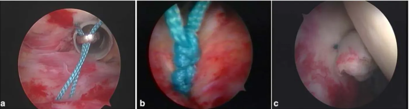
envelope the LHB tendon and the rotator cuff in a ‘‘U’’ shape. A bipolar electrocautery (Arthrocare) is intro-duced through the anterior cannula to release the LHB close its base while a mild tension force is applied on the sutures in order to protect them from potential damage and also to facilitate the release of tendon (Fig.3c). The residual stump of the LHB tendon is debrided to a stable margin. After bicipital release, the suture protruding from the skin is taut to evaluate the final effect that can be obtained with the knotting procedure. At this point the arm position is changed to approximately 20° of abduction to open the subacromial space. The arthro-scope is now inserted through the posterior portal into the subacromial space and a further arthroscopic examination is performed. Once the sutures are well visualized they are extracted through the anterior can-nula using a grabber and tied (Fig.4a). The knot can be a sliding one (we prefer the SMC knot specifically for this procedure), or non-sliding one, like the Revo knot according to the degree of friction produced by the soft tissues (Fig.4b). In order to promote adequate gliding and contact between the two tendon surfaces, we prefer to choose the posterior limb as the post (Fig.4c). Once the knot has been tied, the operation is completed in
accordance with the specific clinical situation present. Treatment consists of acromioplasty, tendon-to-bone or partial side-to-side repair or a combination of the three. In the case of partial side-to-side repair for massive rotator cuff tears, the bicipital tendon stump can effec-tively be used as additional tissue when the tendinous gap is very wide or the quality of the tissues found to be poor (Fig.5).
Discussion
Treatment of LHB tendon pathology has become an area of renewed interest and debate among orthopaedic sur-geons in recent years. Tenotomy or tenodesis of the LHB are undoubtedly the most favoured surgical techniques today [1]. In the 1972, Neer [14] changed the approach to shoulder pain, pointing out a close association between ruptures of LHB tendon and rotator cuff tears. He warned against the thoughtless tenodesis of the LHB tendon, as it ‘‘destroys its function as a head depressor and may precipitate an impingement problem’’. Fur-thermore, his work underline the decrement of medium and long-term results in direct relationship to the
Fig. 2 a Arthroscopic view of the first spinal needle transfixing the LHB tendon. b Shuttle relay derived through the spinal needle, pulled-out by a grasping clamp introduced through anterior portal.
c A no. 2, braided, non-absorbable polyester suture loaded in the eyelet of the shuttle is pulled through the anterior cannula and out of the anterior region of shoulder
Fig. 3 a Arthroscopic view of a second spinal needle with shuttle relay, transfixing the LHB tendon more proximal than the first spinal needle passage. b The limb of suture tied to the eyelet of the shuttle is pulled back through the anterior cannula, through the
biceps tendon to be recuperated out of the skin. c The two limbs of the suture are held by an assistant and release of LHB close to its base is made with a bipolar electrocautery introduced through the anterior cannula
progressive realignment of the humeral head. Recently, anatomic and biomechanical studies, have reconsidered previously theories of the functional role of the LHB tendon on glenohumeral stability and humeral head depression [4,10,14,17,18]. Characterization of the role of LHB tendon is very important to determine the indi-cations for tenodesis or tenotomy. Some authors have suggested a weak humeral head depressor role that in-creases in the presence of rotators cuff tears [11]. Today, we know that the painful shoulder caused by diseases of the LHB which is left undiagnosed and consequently untreated can constitute a common cause of persistent pain and malfunction after shoulder surgery. As recently reported by Gill [9], tenotomy or tenodesis of the LHB was employed as a technique for revision surgery as treatment for previous, failed surgery, in particular sub-acromial decompression for chronic rotator cuff ten-donitis or rotator cuff tear. So, although the prevailing operative strategy of preservation of the tendon and avoidance of tenodesis or tenotomy wherever possible remains, the fact that it can represent an important source of shoulder pain is now clear. A possible expla-nation could be found when one considers the close, anatomo-pathologic links between the LHB (tendonitis,
dislocation and partial rupture), the acromial hook and the rotator cuff disease. The sheath of the biceps tendon is an extension of the synovial lining of the glenohumeral joint and intimately related to the rotator cuff, so any inflammatory process affecting one of the structures can also potentially affect the other [16]. The LHB tendon is also susceptible to the same mechanical abutment with the impingement of rotator cuff tendon [16]. Dislocation of the LHB tendon is most commonly secondary to loss of the soft tissue restraints with degenerative rotator cuff tears [22]. It should not be forgotten, however, as recently reported [13], that it is not always easy to identify lesions involving the LHB by arthroscopic means since these lesions are macroscopically evident only in approxi-mately 50% of the cases. Nevertheless, arthroscopy remains undoubtedly the most specific and sensitive method of evaluation of the various pathological con-ditions of the LHB available today, and when patho-logical findings, also minimal, of LHB tendon such as
Fig. 4 a Arthroscopic view of subacromial space showing the sutures that are extracted through the anterior cannula using a grabber. b The sutures are tied using a knot pusher securing the LHB tendon to the rotator cuff. c Final aspect of anchorage tenodesis
Fig. 5 Arthroscopic view of bicipital tendon stump including as a patch in a partial side-to-side repair for massive rotator cuff tears
Fig. 6 The anchorage to rotator cuff allows a constant dynamic action of the tendon which depresses the head and impedes lateral translation
hyperaemic LHB tendon associated to weakening of the peritenon are found, LHB tendon should be treated. Recent reports [4,9,15], suggest a higher percentage of success rate in relation to tenodesis and tenotomy such as 80–90%, both for open surgery and arthroscopy. The idea to perform a tenodesis with a single suture including the LHB tendon is not new in literature. Sekiya et al. [20] proposed an arthroscopic tenodesis of LHB in which the tendon was secured to the transverse humeral ligament in the bicipital groove. Checchia et al. [6] has reported a series of arthroscopic LHB tenodesis in which the biceps tendon was included in the rotator cuff suture, but in his series, a complete cuff tear was present in all cases and the use of bone anchors was necessary. The technical variant that we propose in this paper can represent an onward step in the evolution of the concept of arthroscopic tenodesis. We think that this type of tenodesis can rep-resent an additional option in cases of rotator cuff tears with associated disease of LHB tendon requiring treat-ment. The rationale, although not supported by biome-chanical studies is to obtain a triple, biomebiome-chanical effect. The first of these biomechanical effects which we
try to promote through the procedure of transposition is the elimination of the deviation and oblique angle which occurs as the LHB completes its intra-articular course prior to reaching the bicipital groove. Tenodesis of the LHB to the rotator cuff can also ensure continual dynamic action of the tendon which depresses the head and impedes lateral translation (Fig.6). This technique is quite simple and shows a low learning curve and a low cost (one spinal needle and one suture). Furthermore, as previously showed by Checchia et al. [6] we have found this technique extremely useful in the presence of large ruptures of the rotator cuff with muscle retraction. In these cases, infraspinatus tenodesis allows it to shift in an anterior direction, thus facilitating the practice of side-to-side suturing and anchorage to the bone. The most common complication associated to this particular method, observed in less than 3%, is failed biological fixation which manifests as subsidence of the tenodesis and consequent descent of the tendon with evident aesthetic deformity; a very low percentage consider-ing that it is the expected final outcome of a simple tenotomy.
References
1. Ball C, Galatz LM, Yamaguchi K (2001) Tenodesis or tenotomy of the biceps tendon: why and when do it. Tech Shoulder Elbow Surg 2(3):140–152 2. Barber A, Byrd T, Wolf E, Burkhart S (2001) Point counterpoint: How would you treat the partially torn biceps ten-don? Arthroscopy 17:636–639 3. Becker DA, Cofield RH (1989)
Teno-desis of the long head of the biceps brachii for cronic bicipital tendinitis. J Bone Joint Surg Am 1(3):376–381 4. Berlemann U, Bayley I (1995) Tenodesis
of the long head of biceps brachii in the painful shoulder: improving results in the long term. J Shoulder Elbow Surg 4:429–435
5. Boileau P, Krishnan S, Coste J, Walch G (2002) Arthroscopic biceps tenodesis: a new technique using bioabsorbable interference screw fixation. Arthroscopy 18:1002–1012
6. Checchia SL, Doneux PS, Miyazaki AN, et al (2005) Biceps tenodesis asso-ciated with arthroscopic repair of rota-tors cuff tears. J Shoulder Elbow Surg 14(2):138–144
7. Dines D, Warren RF, Inglis AE (1982) Surgical treatment of lesions of the long head of the biceps. Clin Orthop 164:165–171
8. Edwards T, Walch G (2003) Biceps tendonitis: classification and treatment with tenotomy. Oper Tech Sports Med 11:2–5
9. Gill TJ, McIrvin E, Scott MD, Hawkins RJ (2001) Results of biceps tenotomy for treatment of pathology of the long head of the biceps brachii. J Shoulder Elbow Surg 10(3):247–249
10. Itoi E, Kuechle DK, Newman SR, et al (1993) Stabilising function of the biceps in stable and unstable shoulders. J Bone Joint Surg Br 75:546–550
11. Leffert RD, Rowe CR (1989) Tendon rupture. In: Rowe CR (eds) The shoul-der. Churchill Livingstone, New York, pp 131–163
12. Lippmann RK (1944) Bicipital teno-synovitis. NY State J Med 44:2235–2241 13. Murthi AM, Vasburgh CL, Neviaser TJ
(2000) The incidence of pathologic changes of the long head of the biceps tendon. J Shoulder Elbow Surg 9:382–385
14. Neer CS (1972) Anterior acromioplasty for the chronic impingement syndrome in the shoulder. A preliminary report. J Bone Joint Surg Am 54:41–50 15. Nevasier TJ (1987) Arthroscopy of the
shoulder. Orthop Clin North Am 18:361–372
16. Neviaser TJ (1987) The role of the biceps tendon in the impingement syndrome. Orthop Clin North Am 18:383–386
17. Pagnani MJ, Deng XH, Warren RF, Torzilli PA, O’Brien SJ (1996) Role of the long head of the biceps brachii in glenohumeral stability: a biomechanical study in cadavers. J Shoulder Elbow Surg 5:255–262
18. Rodosky MW, Harner CD, Fu FH (1994) The role of the long head of the biceps muscle and superior glenoid labrum in anterior stability of shoulder. Am J Sports Med 22:121–130
19. Rowe CR (1988) The shoulder. Chur-chill Livingstone, New York, NY, p 145 20. Sekiya LC, Elkousy HA, Rodosky MW
(2003) Arthroscopic biceps tenodesis using percutaneous intra-articular tran-stendon technique. Arthroscopy 19(10):1137–1141
21. Sethi N, Wright R, Yamaguchi K (1999) Disorders of the long head of the biceps tendon. J Shoulder Elbow Surg 8:644–654
22. Slatis P, Aalto K (1979) Medial dislo-cation of the tendon of the long head of the biceps brachii. Acta Orthop Scand 50:73
23. Walch G, Nove`-Josserand L, Boileau P, Levigne C (1998) Subluxations and dislocations of the tendon of the long head of the biceps. J Shoulder Elbow Surg 7:100–108