Design and Development of Calcium Sensitive Contrast
Agents for fMRI
MASSACHUSETTS IN
by OF TECHNOLO
Tatjana Atanasijevic
MAR
12 20
B.S. Physical Chemistry
University of Belgrade, Serbia, 2002
LIBRARIE
Submitted to the Department of Nuclear Science and Engineering the requirements for the degree of
in partial fulfillment of
Doctor of Philosophy in Nuclear Science and Engineering at the
MASSACHUSETTS INSTITUTE OF TECHNOLOGY
September 2009
@ Massachusetts Institute of Technology 2009. All rights reserved.
Author:
J
9
Certified by:
Associate Professor, Departments
Read by:
Accepted by:
Department of Nuclear science and Engineering Aygust 31, 2009
Alan P. Jasanoff
of Biological Engineering, Nuclear Science and Engineering, and Brain and Cognitive Sciences Thesis Supervisor
. I-- avid G. Cory Professor of Nvclear Science and Engineering Thesis reader
Jacquelin C. Yanch
Professor of Nuclear Science and Engineering ir, Department Committee on Graduate Students
STGfTE GY 10
Design and Development of Calcium Sensitive Contrast Agents for
fMRI
by
Tatjana Atanasijevic
Submitted to the Department of Nuclear Science and Engineering at MIT on August 31, 2009,
in partial fulfillment of the requirements for the degree of Doctor of Philosophy in Nuclear Science and Engineering
Abstract
There is a considerable interest in new technologies that allow noninvasive imaging of physiological parameters in living systems, especially in the neuroscience. Magnetic resonance imaging (MRI) is a very powerful tool for neuroimaging, because it can image the tissue noninvasively, in depth, at a good spatial (~100 pm) and temporal (~1s) resolution. In order to study neural activity at a cellular level, it would be of notable significance to combine MRI with calcium-sensitive contrast agents because of the important role of calcium as a second messenger in cellular signaling pathways. Here we describe a family of calcium sensors for MRI based on the conjugation of superparamagnetic iron oxide nanoparticles (SPIOS) to calcium sensing protein calmodulin and its target peptides. In the presence of calcium, interaction between the two protein domains drives the aggregation of SPIOs which results in up to four fold T2 changes. The calcium sensing is reversible and occurs at midpoint of roughly 1 ptM Ca , which makes these contrast agents suitable for imaging of the cytosolic calcium fluctuations in cells. We introduce two generations of these sensors: the first one based on commercial larger SPIOs, and the second one that uses small crosslinked lipid coated nanoparticles (xLCIOs) which have potential to overcome some of the limitations of the prototype sensor, such as inadequate diffusivity and relatively slow kinetic response. When combined with technologies for cellular deliveries of nanoparticles, these sensors and their derivatives may be useful for functional molecular imaging of biological signaling network in live, opaque specimens.
Thesis Supervisor: Alan Jasanoff
Title: Associate Professor, Departments of Biological Engineering, Nuclear Science and Engineering, and Brain and Cognitive Sciences
Acknowledgements
First and foremost, this thesis would not have been possible without my advisor, Alan Jasanoff, who has supported me throughout my thesis with his patience, knowledge and encouragement. I am honored to be his first student and be involved in his lab from the very beginning. One could simply not wish for a better advisor.
I would like to express my gratitude to my academic advisor, David Cory, who has been a wonderful teacher, tremendous resource and offered great advice throughout the whole grad school.
I am grateful to Numpon Insin for all the help with small nanoparticle project, espe-cially for provision of the iron oxide cores and assistance with transmission electron mi-croscopy.
I am indebted to all of my lab colleagues who have been amazing sources of invalu-able information, helped me learn many laboratory techniques and provided a wonderful and stimulating environment to work in.
This work would not have been possible without the financial support from Ray-mond & Beverely Sackler Foundation, the McKnight Endowment Fund for Neurosci-ence, and NIH grant R21-EB005723.
Finally, I am forever indebted to my parents, brother and friends for their under-standing, endless patience and encouragement when it was most needed. I could not pos-sibly imagine accomplishing this without their tremendous support and unconditional love.
Table of Contents
Abstract 3
Acknowledgements 4
Table of Contents 6
List of figures 8
1. Background and Introduction 10
- Molecular imaging: motivation and modalities 10
- Principles of nuclear magnetic resonance (NMR) 12
- Relaxation in NMR 16
- Relaxation in the presence of contrast agents 20
- Contrast agent classification 26
- MRI contrast agents for molecular imaging of brain activity 29
- Calcium-sensitive contrast agents for MRI 32
- Basis and potential advantages of calcium sensors based on aggregation of
superparamagnetic iron oxide nanoparticles 36
- Organization of thesis 38
- Figure Legends 39
- Figures 42
2. Calcium sensors for Magnetic Resonance Imaging Based on
Superparamagnetic Iron oxide Nanoparticles and Calmodulin 48
- Introduction 49
- Materials and methods 51
- Results and discussion 55
- Figure legends 67
- Figures 73
3. MRI Calcium Sensors Based on Crosslinked-Lipid Coated Iron
Oxide Nanoparticles 81
- Introduction 82
- Materials and methods 83
- Results and discussion 87
- Figure legends 91
- Figures 93
4. Conclusions and Future Directions 98
Appendix A - Dynamic Imaging with MRI Contrast Agents:
Quantitative Considerations 110
- Introduction I1I
- Materials and methods 112
- Results and discussion 116
- Figure legends 131
- Figures 131
List of Figures
1. Background and Introduction
- 1. Contrast mechanisms in molecular MRI 42
- 2. Classification of MRI contrast agents 43
- 3. Examples of responsive T1 and T2 contrast agents 44
- 4. Examples of previously developed T, contrast agents for metal ion sensing 45 - 5. Structure and calcium-dependent substrate binding of calmodulin 46 - 6. Design of a fluorescent calcium sensor based on CaM-M13 interaction 47
2. Calcium Sensors for Magnetic Resonance Imaging Based on Superparamagnetic Iron Oxide Nanoparticles and Calmodulin
- 1. Calcium sensor mechanism 73
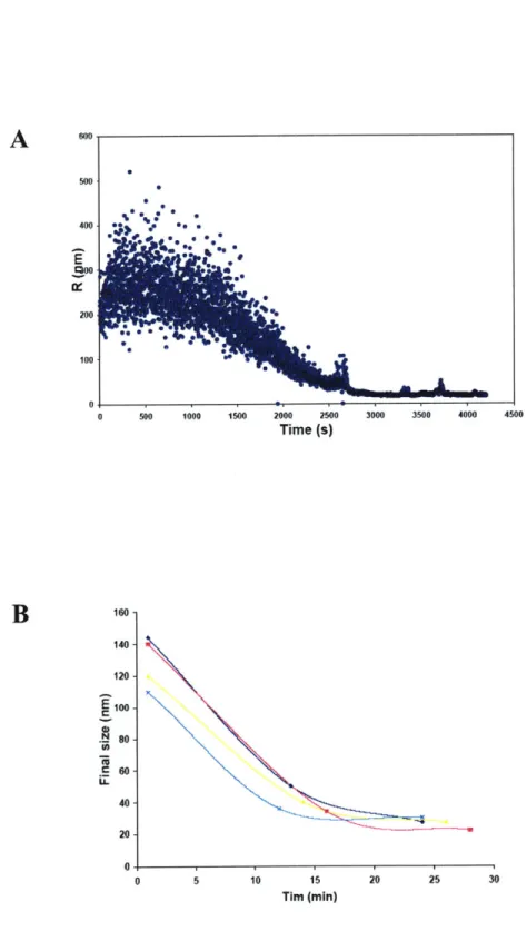
- 2. Light scattering and AFM analysis of calcium-dependent behavior 74
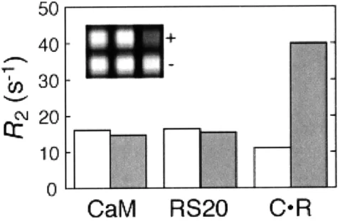
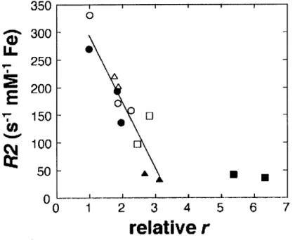
- 3. T2relaxation rate changes measured by MRI 75
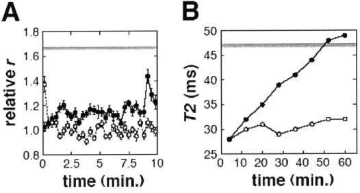
- 4. T2 relaxivity dependence on relative aggregate size 76 - 5. Time courses and reversibility of calcium-dependent changes 77 - 6. Calcium sensitivity range and tuning of calcium sensor variants 78 - 7. Equivalence of T2values measured at different positions in
a microtiter plate 79
- 8. T2* measurements obtained from SPIO solutions 80
3. MRI Calcium Sensors Based on Crosslinked-Lipid Coated Iron Oxide Nanoparticles
- 1. Size characterization of LCIOs 93
- 2. MRI characterization of LCIO-based calcium sensors 94
- 3. Instability of the LCIO aggregates 95
- 4. Crosslinked lipid coated iron oxide nanoparticles 96 - 5. Light scattering analysis of the calcium-dependent behavior of
xLCIOs conjugates 97
Appendix A - Dynamic Imaging with MRI Contrast Agents: Quantitative Considerations
- 1. Predicted characteristics of nanoparticle aggregation and disaggregation 132
1. Background and Introduction
1.1 Molecular imaging: motivation and modalities
Molecular imaging is a rapidly growing field of biomedical research that is focused on understanding biological phenomena on a molecular level. This growing scientific discipline aims to develop novel tools and methods to image specific molecular pathways in vivo (1).
Neuroscience is a research discipline that can especially benefit from development of molecular imaging technologies. A fundamental goal in this area is to gain a better understanding of the function of the neural circuits and signaling events in the brain. Electrophysiology is a standard method to study neuronal activity by using microelec-trodes to record the action potentials of individual neurons. This approach is invasive and can not record from the whole brain at once. On the other hand, information on a cellular level has historically been only available from in vitro assays, from the studies of cells or fixed tissues, which lead to information loss compared to studies in live speci-mens. Recently, various molecular probes have been developed to study molecular events in real time, using imaging modalities such as optical imaging, positron emission tomography (PET), and magnetic resonance imaging (MRI). Lately, probes that can be used in multi-modal imaging are being developed as well.
Optical imaging sensors are typically based on fluorescence or bioluminescence. A variety of fluorescent sensors has been developed to target enzyme activity, pH, DNA and RNA interactions, metal ions and various metabolites (2-4), which have great
sensi-tivity as well as spatial (< 1 pim) and temporal (1ims) resolution. However, these
meth-ods can only be applied to cells or surface tissues because of the tissue scattering of the light. This presents a particular issue for neuroscience studies, because the skull encasing the brain is opaque.
PET is based on the detection of y-photons produced from radioactive, positron-emitting probes after positron-electron annihilation. The most widely used PET probe is
18F-fluorodeoxyglucose (FDG), which accumulates in the areas of increased metabolic
activity, such as tumors (5). Other PET sensors have been developed to study the expres-sion and spatial distribution of receptor ligands (6). PET has exquisite sensitivity; it can detect the probes in picomolar range. Another advantage of PET as that it can be per-formed in a quantitative way. This method has a couple of limitations when it comes to molecular imaging: the spatial resolution is very low (1 mm or worse), the signal is al-ways "on" so it can not be made sensitive to a particular molecular event as it is
happen-ing, and the data interpretation is often based on kinetic modeling.
Magnetic resonance imaging (MRI) is a very powerful tool for neuroimaging, be-cause it can be done in vivo, deep into the tissue, with good resolution. Most of the func-tional MRI imaging is based on the blood oxygenation level dependence (BOLD) effect, which is a combination of changes in local blood oxygenation, flow and volume triggered by elevated neural activity. BOLD offers a noninvasive, though indirect readout of re-gional brain activity (7, 8). The resolution and specificity of this conventional fMRI technique are seriously limited by hemodynamics. A number of strategies have been proposed to sensitize MRI signal to the cellular level neuronal activity. The most promis-ing one may be the use of image-alterpromis-ing chemicals, called contrast agents, which change
the pattern of distribution of signal intensity in an MRI image. A variety of mechanisms are available by which MRI contrast agents can produce a change in image contrast as a response to interaction with a specific target. The physical origin of the signal in MR and the MRI contrast agents are discussed in the next couple of sections.
1.2 Principles of nuclear magnetic resonance (NMR)
Nuclear Magnetic Resonance is a physical phenomenon that was detected for the first time in 1946 by Bloch (9, 10) and Purcell (11) independently, for which they shared a Nobel Prize in physics 6 years later. NMR is concerned with the interaction of the nu-clear spin angular momentum of the sample, placed in static magnetic field, and an oscil-lating electromagnetic field (the radio frequency field). The theory of NMR can be
de-.1
scribed using both quantum-mechanical and classical way for spin - nuclei. Both will
2 be utilized here in this introduction.
Only isotopes with non-zero angular momentum can be observed by NMR. Such nuclei must have an odd number of protons or neutrons, and most commonly used ones for NMR are H, H, 13C, 19F, 31P etc. Nuclei with non-zero spin angular momentum, I,
have an associated magnetic momentum, p, which are related by gyromagnetic ratio:
For spin - nuclei, the nuclear charge distribution is spherically symmetric, so the
2
nucleus then has the properties of a magnetic dipole whose strength is described by the
above equation. Nuclei with spin - are most often studied by NMR. The most widely
2
studied nucleus is by far that of hydrogen 'H, which has greatest importance.
The energy of a nuclear magnetic dipole in an external magnetic field is given by the classical expression as:
E =-eBO
where BO is the strength of the external magnetic field. In NMR the typical system is the one where the applied magnetic field is oriented along the z-axis. Hydrogen can basi-cally have two different energy levels in the field, which correspond to the magnetic moment oriented along or against the external field. The signal that is measured in NMR is the result of the transition between those two energy levels, and the energy difference is dependent on the magnetic field strength:
AE = hyBo = hco
The resonance frequency is referred to as the Larmor frequency, OL:
In thermal equilibrium, the relative populations of spins in the two states (parallel and antiparallel to the field) are in accordance with Boltzman statistics:
N AE 40_
__=_ kBT =e kBT
N.,
At room temperature, the polarization is only about one part in a million. It is this excess polarization that leads to the NMR signal. The aligned nuclear magnetic moments, total number of which is denoted by N, provide a bulk magnetization to the sample which can be expressed as:
N72h2B0
M1 ~
4kBT
The dynamics of this magnetization in a magnetic field can be described by two types of motion (12):
1. The rotation of the magnetization about an applied magnetic field, or Larmor pre-cession, for which the classical torque equation applies:
dM
-= M x B
dt
If the bulk magnetization is along the field direction, there is no torque and hence no motion, as it is at equilibrium. If the system is away from the equilibrium state, that is, if the bulk magnetization vector is not oriented along the z-axis, then the magnetiza-tion vector precesses around the z-axis with the angular frequency equal to Larmor fre-quency, and there is also a relaxation process to bring the vector back to along the z-axis. The x- and y- components of the magnetization decay towards zero, and the z-component decays to the equilibrium value of Mo. The complete spin dynamics is described by the Bloch equations, which in the rotating frame, with a field rotating along the x-axis are given by: dMX M dt T2 dM ~M, d = AcoM, + OM T d~t T2 dM 1 =z-co My - (M - MO) d~t T,
where Ao is a small off-resonance term, the difference between the rotating frame quency and the Larmor frequency, and oi is the strength of the rotating field (radio fre-quency field), given with yB1. The time constants that appear in these equations describe
the relaxation of the system back to the equilibrium. T, is the spin-lattice relaxation con-stant or longitudinal relaxation time. T2 is the spin-spin relaxation constant or transverse relaxation time. These constants are phenomenlogically introduced into Bloch equations.
1.3 Relaxation in NMR
The equilibrium state of a spin system in a static magnetic field has two important features: there is a magnetization parallel to the applied field and there is zero magnetiza-tion in the plane perpendicular to the field (13). Once the system is perturbed with the application of the radio frequency (rf) pulses, it returns to thermal equilibrium. The physical cause of relaxation lies in the fact that not all the nuclei see exactly the same magnetic field, and moreover, these fields may change with time. The system does this through two types of relaxation processes, longitudinal and transverse, which are charac-terized by time constants Ti and T2, respectively. Longitudinal relaxation is the return of
the z-component of the magnetization to the equilibrium value, Mo. Immediately follow-ing the 900 rf pulse, the z-component of the magnetization is zero, and as time elapses, it
increases exponentially until the equilibrium value is restored:
t
M,(t)= Mo(1-e Ti)
Transverse relaxation involves the dephasing of the magnetization on the x-y plane. There are two main reasons for this net loss of coherence. A time constant T2 is used to describe the transverse relaxation due to processes that are intrinsic to the sample, such as dipolar interaction between the proton spins. However, there are other, external causes of the transverse relaxation, such as the inhomogeneity of the static field Bo, and the combi-nation of all of these contributions is described with the time constant T2*. Right after the
90' rf pulse, the transverse components of the magnetization will have the same ampli-tude as MO and with time will exponentially decay to zero with time constant T2*:
t
Ml,(t)=Moe T2
In NMR, relaxation of the magnetization back to the thermal equilibrium state is 1.
caused by the fluctuating local magnetic fields. Spin - is treated as magnetic dipole.
2
Since molecules are continually moving (translating, rotating, vibrating etc.), these mo-tions modulate the interacmo-tions between the nuclei. Therefore, molecular momo-tions can cause the local fields to fluctuate and allow the spins to relax via some interaction, such as dipolar interaction Spins can also relax via other mechanisms too, such as chemical shift anisotropy, scalar coupling and spin-rotation interactions.
From the equation of motion of magnetization in a magnetic field, one can conclude that change of the local fields in z-direction has no effect on T1, only on T2. Thus, Ti
depends only on changes of local magnetic fields in the transverse plane, which are af-fected by fast molecular motions. T2 is affected by both fast and slow molecular motions.
This is a very important difference between T1 and T2. T2 is always to shorter or equal to T1 because of the cylindrical symmetry of the Hamiltonian that describes these processes.
In order to look at the molecular motions in more detail, the concepts of correlation function and correlation times will be introduced. The correlation function describes the average behavior of a molecular motion in a system and is typically defined the correla-tion between random variables at two different points in space or time. If one considers
the correlation function between random variables at the same point but at two different times then one refers to this as the autocorrelation function. It can usually be approxi-mated by an exponential function:
G(t)= A2e ,
It has a characteristic time scale defined by the correlation time rc. This time is related to the variations of the local magnetic fields. For times much smaller than uc correlation function varies negligibly from its initial value, while for times significantly greater than ,c G(t) has decayed to zero. The Fourier transform of the correlation function is called spectral density function and it describes the range of frequencies at which the motion exists. If G(t) is described by an exponential function as above, than the spectral density function is Lorentzian:
J(co) oc A 2 2 2
1+ rc
1
The spectral density maximum occurs when rc = -, which is another way of stating that
CO)
efficient relaxation in NMR occurs when a motion has frequency components at the Lar-mor frequency. Experimentally, if we wish to increase the correlation time (slow down the system) one approach to do so would be to increase the viscosity of the solution by
lowering the temperature. Conversely, increasing the temperature will speed up the sys-tem (lower the correlation time).
The primary interaction that is responsible for the relaxation in the proton systems is the dipolar interaction. Using the expression for the dipolar Hamiltonian for the inter-action between two spins, the relaxation rates are:
1 y4
- OC' [J(coo) + J(2wo)] T r 6
1 y4
-- c -[J(O) + J(coo) + J(2coo)]
T2 r
The spectral density functions indicate the magnitude of a frequency component at a specified frequency. T1 relaxation depends on frequency components at Larmor
fre-quency and twice that value - fast dynamic processes. T2 has the same terms with the addition of the zero frequency term, which corresponds to slow dynamic processes.
The intrinsic T2 relaxation time is usually measured by a spin-echo type pulse se-quence, which consists of a 900 pulse followed by a 1800 pulse after a suitable time delay ,c . One can do several such experiments and vary the time delay, or the whole experi-ment can be done in one shot using multiple 1800 pulses. T2 can be extracted from the plot of the amplitudes of the echo signals vs. time, by the equation:
T
The most common way to measure T, relaxation time is the inversion recovery pulse sequence. It a contains a 1800 pulse, and a delay of some time 'U, followed by a 900 pulse. If the experiment is repeated a couple of times using different delay times, T1 can than be determined using the equation:
M,(r) = MO (1-2e )
1.4 Relaxation in the presence of contrast agents
Contrast agents for MRI are image-altering chemicals that modify patterns of con-trast in an MRI image, rather than being imaged directly. Most of the MRI concon-trast agents are paramagnetic substances whose large paramagnetic moment interacts with water spins that are being imaged, promoting their relaxation (Figure 1A, B). They shorten the longitudinal relaxation time TI, or the transverse relaxation time T2; this re-sults in image brightening in case of the Ti agents and image darkening in case of the T2 agents. T, contrast agents are typically complexes of paramagnetic metal ions, such as Gd"3 , whose unpaired electrons interact with the water spins. The most commonly used T2 contrast agents are superparamagnetic iron oxide nanoparticles (SPIOs), which consist
of the iron oxide core and a coating that makes them water soluble. They produce local magnetic field inhomogeneities, which promote the transverse relaxation of the water protons. There are other types of contrast agents which work through different mecha-nisms, such as chemical exchange saturation transfer (CEST) contrast agents (14). A
CEST contrast agent contains labile protons that exchange with bulk water, but have well resolved chemical shifts.. If the exchangeable protons are presaturated using an appro-priate rf pulse, the signal of the bulk water pool is reduced since these protons are ex-changing with the presaturated ones. This produces darkening in the MRI image. CEST contrast may be turned on and off, since it is dependent on the power and frequency of the presaturation rf pulse (Figure 1 C). Another way to produce MRI contrast is by imag-ing non-standard nuclei, such as 13C (15). The images are formed in an equivalent
man-ner to the proton imaging, but usually result in much lower signal-to-noise ratio. One of the ways to improve the MRI signal of 13C containing molecules is to hyperpolarize them
right before the imaging using techniques such as dynamic nuclear polarization (Figure ID).
In the presence of the relaxation promoting contrast agents, the observed relaxation rate will be the sum of the diamagnetic and paramagnetic contributions, given by:
( )ob= (I )d +() i = 1, 2
The diamagnetic contribution represents the relaxation rate of the solvent in the absence of the paramagnetic species. In the absence of the solute-solute interactions, a simple model for the dependence of the paramagnetic relaxation rate on the concentration of the paramagnetic species [C] is a linear one. Relaxivity (Ri) is the slope of this dependence, defined in the units of s-1 M-1 (more commonly s-1 mM') and it represents the strength of a contrast agent.
()b -I)d+R,[C] i=1,2
TO T
The relaxivity is also a function of the strength of the static magnetic field. The higher the agent's relaxivity is, the less of it needs to be used to produce a contrast change in an image.
The strength of the dipolar interaction between the water protons and paramagnetic species is influenced by the proximity of the water protons. According to the location of the water protons, the mechanisms of interaction and contribution to the relaxivity can be classified as inner-sphere, second-sphere or outer-sphere effects. Inner-sphere mecha-nism occurs when the water molecule binds in the primary coordination sphere of the metal ion and exchanges with the bulk solvent. If the hydrogen-bounded water is in the second coordination sphere and the lifetime of this interaction is long compared to the time required for the solvent molecule and paramagnetic agent to diffuse past each other, that is described as the second-sphere relaxation mechanism. Finally, outer-sphere re-laxation refers to the rere-laxation mechanism of a water molecule diffusing past a para-magnetic species. Due to the lack of deep understanding of the second-sphere coordina-tion interaccoordina-tions, it is often not distinguished form the outer-sphere interaccoordina-tions (16). For T, agents the inner-sphere mechanism is the most dominant contribution to the total re-laxivity, while for T2 contrast agent the outer-sphere contribution tends to be the most
significant one.
The exchange of the water molecules between the bulk solvent and the primary coordination sphere is the cause of the longitudinal contribution to the inner-sphere re-laxation mechanism, as described by the following equation:
( )is = 1 PPM M
T Tim +V-M
The variable Pm is the mole fraction of the metal ion, q is the number of the water mole-cules bound to a metal ion, Tim is the longitudinal relaxation time of the bound water protons and Tm is the lifetime of the water molecule in the complex. Tim is given by the
Solomon-Bloembergen equations (17, 18) which represent the sum of the two contribu-tions, dipolar and scalar:
1 1 1 TDD TSC TM I I 1 rCgp B S(S+1) 3rc 7rc DD 15 r1 + C21-2) (I+2 O)2 1 y(g22 4S( + A)[e 2 -sc 3 h (1+_ r2 1 _ 1 1 1 rC Te "M TR 1 1 1 Te Te 1M
The parameters here are: 71 is the proton gyromagnetic ratio, g is the electron g-factor, pB is the Bohr magneton, S is the electron spin quantum number of the metal ion, r is the distance from the center of the paramagnetic species to the center of the water proton
undergoing relaxation, ol and os are the Larmor frequencies for the nuclear and electron spin and A is the electron-nuclear hyperfine coupling constant. The correlation times for dipolar and scalar mechanism are rc and ue, T1e is the electron longitudinal spin relaxation
time and TR is the rotational correlation time of the whole metal-water complex. To
opti-mize the relaxivity, a couple of parameters from the above equations can be influenced by an appropriate agent design. Typically, when designing T, contrast agents, the most important parameters are the number of the coordinated water molecules to the central metal ion, the distance between the water molecules and the metal ion, and the rotational correlation time.
The outer sphere relaxation mechanism is a more complex problem centered on solvation dynamics and diffusion. If the system consists of a dilute solution of small iso-tropic paramagnetic species, than the contribution from each of them can be assumed to be independent and additive. The most common type of paramagnetic species is iron ox-ide particles. Many authors have addressed this problem (19), and the resulting expres-sions have some similarity with the Solomon-Bloembergen equations. If the concentra-tion of the paramagnetic particles is [C], their spin is S, gyromagnetic ratio ys and their diffusion constant relative to the water protons is D (for which the protons can not ap-proach closer than R, and for which the only contribution to the fluctuations in the local magnetic fields comes from the diffusion of the protons past paramagnetic species); then the paramagnetic contribution to the transverse relaxation is given by (20):
1 _16rc N [C]
-- - 051r yh 2S(S+1)
100
[3j(coSTD
3( 1 I )+ 4(0)]where TD, the diffusion correlation time, is the time needed to diffuse a distance on the
order of R:
D=R 2
D
The spectral density functions j(o) are given by:
j(w)
= Re 1+ -(icol-D) 2 4 1 4 1+(irD) I+(iwrD)± 2 9 1 9and the secular term
j
(0) =1. Diffusion coefficients can be estimated if the motion can be described by the diffusion of rigid spheres in a medium of viscosity q:D kBT
D = kB
6rcR rq
A condition for the validity of the theory is that
(o)'<1
where 6o is the change of the equatorial magnetic field at the surfaces of the particles. In NMR, this is typically referred to as the motional averaging regime. Under this condi-tion, the same amount of paramagnetic material is much more effective in promoting transverse relaxation of water protons when distributed as fewer larger particles than as a greater number of smaller ones. With the increase of the paramagnetic particle size, the T2 relaxation constant becomes smaller.
However, as the particle size increases, the basic assumptions for outer sphere re-laxation theory are no longer valid, since, among other factors, the local magnetic field contributions from paramagnetic particles start to overlap. Once (So)TD is greater then 1, which is referred to as the static dephasing regime, the relationship between the paramag-netic particle size and T2 relaxation rate becomes inverse: with increase of particle size the relaxation rates are slower. This has been extensively explained by the theoretical work of Gillis and colleagues (21-23). Another notable characteristic of the static dephasing regime is that T2* remains roughly constant with the increase of the particle
size, whereas it decreases in the motional averaging regime.
1.5. Contrast agent classification
MR contrast agents can be divided into three groups: nonspecific, targeted and re-sponsive (Figure 2) (24). Nonspecific contrast agents are exemplified by blood pool agents such as Gd-DTPA that show a relatively nonspecific distribution pattern. They are not used to access molecular targets. Targeted contrast agents are conjugated to affinity
ligands, such as peptides, antibodies or small molecules that impart molecular specificity to the probe and work in analogous way to PET probes. They are especially useful in detection of cell surface proteins, such as receptors expressed by cancer cells. Respon-sive contrast agents change their relaxivity upon interaction with a specific target. They are often assumed to exist in two states, "off' and "on", one which is of lower and the other one higher relaxivity. The switch from one to the other state occurs when the con-trast agent binds its target. This type of concon-trast agent is also referred to as bioactivated. Typical parameters to which smart contrast agents can be responsive to are pH, oxygen pressure, temperature, enzymatic activity, redox potential, as well concentration of spe-cific molecules such as DNA, proteins, small molecules, ions, etc.
Responsive T1 contrast agents are typically small chelates of paramagnetic metals,
for which some relaxivity determinant changes upon the interaction with specific molecu-lar target. One of the parameters that can be controlled in such a way is solvent accessi-bility. A typical design of such a responsive contrast agent involves a group that blocks water protons from exchanging with the paramagnetic metal ion, and once the target binds the contrast agent, intramolecular rearrangement occurs, that removes the blocking group away from the central metal ion allowing access to water molecules. This increases q, the total number of water molecules bound to the metal ion, which promotes the Ti relaxation rate. An example of such a contrast agent is "EgadMe" designed by Meade and colleagues (Figure 3A) (25). In this agent, Gd-ion is caged by an enzymatically cleavable substrate (galactopyranoside). In the presence of the target enzyme,
p-galactosidase, the substrate is cleaved, exposing the Gd-ion to the water and increasing q. This increases Ti relaxivity about three times. This agent can be used for monitoring
gene expression if
p-galactosidase
is used as a marker for gene expression from specific promoters, as was demonstrated in injected embryos of Xenopus laevis. Another parame-ter than can be controlled in responsive Ti contrast agents is the spin state of the para-magnetic atom. Hemoglobin, the iron-containing protein responsible for oxygen transfer in the blood works this way. In the deoxy state, the protein is paramagnetic since Fe2 ions have unpaired electrons. Once oxygen is bound, these electrons become paired which makes the oxy-hemoglobin diamagnetic. This makes hemoglobin an effective oxygen sensor, allowing it to sense regional oxygen tension (pO2) (26). This same effect is also the basis for BOLD effect, the main method used in modem fMRI. Yet another T1relaxivity determinant, rotational correlation time, increases if the target is a molecule that is much larger than the paramagnetic chelate. Binding of a protein molecule to a small paramagnetic chelate can result in relaxivity changes of more than one order of magnitude.
T2 contrast agents are typically superparamagnetic iron-oxide particles. Based on their size, they can be classified as ultra-small nanoparticles (r 50 nm) (USPIOs), small
nanoparticles (50 nm r 1 im) (SPIOs) or micron-sized (r > 1p im). These agents can be detected at much lower concentrations (submicromolar) than T, contrast agents due to their high T2 relaxivity, which is greatly influenced by their size and microscopic spatial distribution in the solution (27). Clustering of these particles is the basis for sensing a particular molecular target. Weissleder and colleagues (28, 29) designed the first re-sponsive contrast agents based on nanoparticle aggregation by conjugating the nanoparti-cles to a pair complimentary oligonucleotide sequences. A crosslinked network was formed, that disaggregated upon the addition of a restriction nuclease (Figure 3B). This
resulted in 5-fold change in T2 relaxivity. A similar mechanism was used to produce
ag-gregation-based contrast agents for sensing of other enzymes, protein-protein interac-tions, DNA, viruses etc. (30). These types of sensors are sometimes referred to as mag-netic relaxation switch. The interaction with the specific target causes the particle aggre-gation and concomitant large changes in T2 due to the changes in local magnetic field gradient that water protons diffusing past these agents are experiencing. Some of these sensors can be designed to be reversible, so that, once the target is removed from the sys-tem or its concentration drops below a certain critical value the particle clusters disaggre-gate back to the original, dispersed state.
Appendix A discusses key quantitative considerations connected to design, mecha-nism and application of smart MRI contrast agents (31).
1.6. MRI contrast agents for molecular imaging of brain activity
MRI continues to have a tremendous impact in neuroscience and in brain disease diagnostics and treatment. A whole array of methods has been developed for anatomical, functional and molecular imaging of the brain (32). The field continues to show rapid progress. Functional MRI methods have blossomed with the development of the BOLD technique. However, due to some inherent limitations of this method, it can not be ap-plied to study neural activity at the cellular level. For that, MRI should be performed in conjunction with contrast agents that can detect certain aspects of neural physiology. There has been a significant improvement in the development of such probes in the recent
years, although most have still not been shown in work in live, functioning animal brains. There are some limitations when it comes to use of these agents in vivo, such as toxicity and delivery. Many MRI contrast agents incorporate metals such as gadolinium and man-ganese which are extremely toxic even at very low concentrations, so they are usually complexed with organic molecules that make the concentration of the free metal much lower. Another significant challenge is delivering the agents through blood-brain barrier. MRI contrast agents have been developed that target metabolic activity, pH, gene expres-sion in the brain, as well as metal ions (calcium, zinc, copper) important in signaling of the neural networks. This section reviews the first three groups, while probes that target calcium are discussed in more details in the subsequent section.
Uptake and processing of some cerebral metabolites is closely correlated with neu-ronal activity. Hemoglobin has been used as an oxygen-responsive contrast agent, both as an endogenous protein (which underlies the BOLD effect) and as an exogenous contrast agent to detect local partial oxygen pressure in the tissue (26). Traditionally, PET has been widely used in conjunction with FDG to study cerebral metabolites. A somewhat similar approach with MRI has been demonstrated by Golman et al. (15) who used hyper-polarized 13C labeled pyruvate to study the kinetics of the pyruvate uptake as well as its
turnover to lactate. Since 1 3C MRI is usually not sensitive enough at the typical concen-trations of pyruvate in the tissues, dynamic nuclear polarization method has been used to significantly boost the 13C signal. The main drawback of this method is the short time
window available for imaging due to the T, relaxation of the hyperpolarized 13C, so the agent needs to be continuously administered during imaging studies longer than a minute.
Both relaxation and CEST-based MRI contrast agents can be designed to be pH-sensitive and therefore used to monitor neural activity since a slight acidification of the extracellular medium is characteristic of this activity (33). These changes are typically in the range from pH 7.2 to 7.4. A contrast agent based on phosphonated Gd3 has been used by Garcia-Martin et al. (34) to look at intravascular acidification in rat glioma. It could distinguish differences in pH on the order of one pH unit, and it could in principle be used for functional brain imaging, although it might not be sensitive enough under realistic conditions. A different approach for pH-imaging of the brain is to use the CEST mechanism of endogenous amide protons, which has been demonstrated in detection of focal ischemia in rats (35).
Biology has benefited tremendously from the discovery of green fluorescent protein (GFP) (36) and the subsequent development of smart fluorescent GFP-based probes such as chameleons (37). There have been significant efforts in the past decade towards engi-neering of equivalent reporter genes that modulate MRI contrast. There are couple of approaches that have been employed in this endeavor: (1) enzyme-based reporter genes, where enzymes cleave functional groups that block proton exchange or binding MRI trast agents; (2) expression of surface receptors that enable binding of specific MRI con-trast agents, (3) design of artificial CEST-based reporter gene and (4) expression of iron-containing metalloproteins such as ferritin and transferrin receptor, that promote T2 re-laxation (38-40). Previously described "EgadMe" contrast agent is an example of the first group. This sensor can detect the activity of the enzyme
p-gal
which is encoded by the LacZ gene. Transferrin conjugated to SPIOs, used to target the transferrin receptor in vivo (41), is a typical representative of the second approach. An artificial, lysine-richprotein (LRP), which contains a high density of amide protons, was cloned and expressed in rat glioma cells and detected in the mouse brain in xenografts of LRP-expressing cells (42). This CEST-based reporter gene has typical characteristic of CEST agent contrast agents: it is switchable, but suffers from low sensitivity. Finally, there has been consider-able interest in iron-based reporter genes such as ferritin. Genove et al. has demonstrated its use as an MRI reporter gene by injecting adenovirus encoding for human ferritin in mouse brain, which resulted in significant loss of signal in the T2-weighted image (43).
Coexpression of ferritin and transferring receptor in neuronal stem cells produced a simi-lar effect (44). Recently, Shapiro et al. demonstrated a smart, aggregation-based T2 con-trast agent based on ferritin as well (45). In that work, a system was developed to express self-assembled ferritin nanoparticles, incorporating multiple surface functionalities, which were subsequently used to produce a protein kinase A sensor. Mag A, a bacterial iron transporter from a type of magnetotactic bacteria, has been expressed in 293FT cells under the regulation of doxycycline (46). A significant signal loss in T2*-weighted
im-ages was shown upon the inoculation of these cells in a mouse brain.
1.7. Calcium-sensitive contrast agents for fMRI
Calcium ions have an important role as secondary messengers in cellular signaling pathways and many groups have focused on developing MRI contrast agents that would be responsive to calcium. The dominant isotope of calcium, 40Ca, accounts for 96.94 %
of the natural abundance and can not be directly imaged with MRI because it has zero nuclear spin.
There are a number of factors that make neuronal calcium ion concentration fluc-tuations an optimal target for fMRI imaging agents. First, calcium levels are closely cor-related with the firing rates of neurons, which would make calcium readout of neural ac-tivity much more direct that the readout based on BOLD effect. Second, calcium levels in the neurons usually fluctuate on a timescale that might be appropriate for detection by the fastest MRI methods (100-1000 ms). The concentrations of other important ions, as well as the voltage, fluctuate much more rapidly (on the order of 1-10 ms) and are not possible to detect with MRI in its current state of development. Third, the changes in neuronal calcium concentrations are quite dramatic, ranging from 0.1-1.0 pM in the cytoplasm and 0.1-100 gM near boutons during synaptic depolarization. Extracellular calcium concen-trations also fluctuate during neuronal activity, although the changes are less specific. The typical range of these changes is from 1.2 mM to 0.8 mM, although in some patho-logical cases the changes can be much more pronounced. These fluctuations could also be targeted by appropriately designed fMRI calcium sensors. Finally, there are a number of fluorescent calcium imaging agents that have successfully been used for optical imaging of neurons, which could be used as basis for the design of the MRI sensors (37).
Bis(aminophenoxy)ethane tetraacetic acid (BAPTA) is well known calcium chela-tor that is often used in the design of the calcium sensors. In 1983, a fluorinated deriva-tive of BAPTA was produced by Smith et al. for 19F magnetic resonance measurements (47). Years later, 19F-BAPTA was used for quantitative calcium measurements (48). However imaging with this molecule is not very practical because of its low
concentra-tion. Even at very high concentrations of this molecule, such as 1mM, the method is still
5 orders of magnitude less sensitive that standard proton imaging. The first relaxation
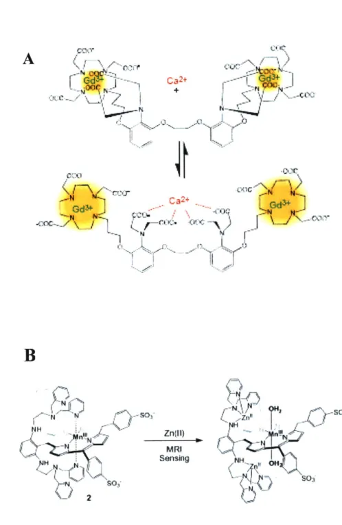
promoting MRI sensor for calcium developed by Li et al. also involves BAPTA molecule in its design (Figure 4A) (49). This paramagnetic T, contrast agent, Gd-DOPTA, consists of a derivative of the BAPTA molecule attached to two copies of Gd3*-1,4,7,10-tetraazacyclododecane-1,4,7,10-tetraaacetic acid (Gd-DOTA). This molecule has a Ca2+ dissociation constant of~ 1 pM and its relaxivity increases approximately 80 % (from 3.3 to 5.8 mM-I s.1 at 11.7 T) upon addition of the calcium ion to a calcium-free solution. In the absence of the calcium, the Gd3* coordination sphere is completed by two closest carboxylic groups of BAPTA. Once calcium is added, these groups now bind this ion, allowing two water molecules to complete the Gd3* coordination sphere, which promotes Ti relaxivity due to the increase in q. The response to calcium is fairly fast because it involves intermolecular rearrangement. This agent has yet to be used for functional imag-ing of neural activity in vivo. A different type of Ti contrast agent based on chelated Gd 3 that targets extracellular calcium has been introduced by Logothetis and colleagues (50). Zinc ions are also influenced by neuronal activity and a couple of contrast agents
for targeting of the zinc ions have been developed. Recently, a contrast agent similar to that of Li et al. has been introduced for zinc sensing (Figure 4B) (51). This contrast agent is based on manganese-containing porphyrin molecule. It has Ti relaxivity comparable to that of Gd3* agents, but a significant advantage of being cell permeable.
Zinc-dependent signal changes have been demonstrated in cells incubated with this contrast agent.
A different approach to imaging neural activity has been introduced by Koretsky and co-workers, using paramagnetic manganese ions as Ca2+ analog. Mn2+ ions produce signal enhancement in Ti-weighted images that is coupled to neuronal activity (52). Mn2+ can enter excitable cells using some of the same transport systems as Ca 2+ such as
voltage-gated calcium channels. Manganese ion can bind a number of intracellular sites because it has high affinity for Ca2+ and Mg2+ binding sites in nucleic acids and proteins (53). Activity dependent accumulation of Mn2+ has been demonstrated when it was ap-plied near receptory neurons in olfactory epithelium of a mouse (54). Once it was deliv-ered past the blood-brain barrier, it was observed that accumulation of Mn2+ in rat neu-rons is slow, on the order of minutes, and its excretion from cells is even slower, some-times on the order of days (55, 56). This means that Mn2+ can serve as a marker for neu-ral activity history, but that Mn2+ fluctuations in the brain are too slow for real time func-tional imaging. Mn2+ can therefore be used to label active neurons during sensory or be-havioral tasks performed outside the magnet. This technique is somewhat analogues to the use of 2-deoxyglucose for activity mapping by autoradiography. It was recently used for Ti-weighted mapping of auditory cortex in mice (57) and for functional brain studies
in developing insects (58). A major drawback to the use of Mn2+ is its cellular toxicity, so the dose used should be as low as possible.
1.8. Basis and potential advantages of calcium sensors based on aggregation of superparamagnetic iron oxide nanoparticles
The goal of this thesis work is to develop MRI contrast agents that can be used to image neuronal calcium in vivo at high temporal and spatial resolution. We believe that the best approach to do so is to design a calcium sensitive contrast agent based on the aggregation of SPIOs which have high T2 relaxivities. In order to make the SPIO aggre-gation sensitive to calcium, proteins that bind each other in a calcium-dependent manner are conjugated to nanoparticles. Many proteins associate and dissociate reversibly de-pending on the local calcium concentrations. Calmodulin (CaM) is a very well studied protein that binds a number of targets in a calcium-dependent fashion. The CaM struc-ture is characterized by a helix-loop-helix organization. The protein has four EF-hand structures than can bind calcium in a cooperative manner. Upon calcium binding, an intramolecular conformational change in the structure of the protein occurs, which allows CaM to bind its target (Figure 5). Most CaM binding partners are short basic peptide sequences. The peptide sequences chosen for this work are M13, which is derived rabbit skeletal muscle myosin light chain kinase, and RS20, derived from smooth muscle my-osin light chain kinase. Reversible binding of CaM and M13 dependent on calcium con-centration has been detected in cells using fluorescent methods (59). A fluorescent cal-cium sensor based on the CaM - M13 binding has previously been developed (Figure 6) (37). Our MRI calcium sensor consists of a binary mixture of SPIOs conjugated to the protein and to the peptide, which should form clusters if the calcium concentration is above a particular threshold.
There are numerous advantages of this type of sensor over previously described paramagnetic small molecules. SPIOs have relaxavities which are typically an order of magnitude greater than those of complexes of paramagnetic metals. Upon aggregation changes of relaxivities may be significantly greater than those achievable with T, contrast agents. This type of sensor may be used at significantly lower concentrations than the small molecule sensors. Therefore, it is unlikely that these sensors would buffer intercel-lular calcium. The timescale on which SPIOs influence T2 contrast is quite fast, so these
agents might be able to achieve a significantly better temporal resolution. Since SPIOs have been already used in different scientific fields, such as cell trafficking, there are already some published methods on cell delivery of these agents (60). Finally, because this sensor is part protein, tools of molecular biology may be used to control the interac-tion driving the aggregainterac-tion.
Despite these advantages, there are also a number of issues when it comes to appli-cation of this type of sensors in cells and organisms. Large SPIOs may diffuse infectively through the cytoplasm, especially if they are larger than 50 nm. Since the T2 relaxation of the water molecules depends on their diffusion in the vicinity of SPIOs, compartmen-talization of the water molecules and nanoparticles within the cells will generally make T2 values different from those measured in vitro. Toxicity and delivery to cells in the
brain might also present challenges for in vivo use of SPIO-based sensors.
This thesis has two main goals. The first is to develop and demonstrate in vi-tro a prototype calcium sensor based on SPIO aggregation that would be potent, reversi-ble, tunable and suitable for detection of typical intracellular fluctuations of calcium con-centrations. The second aim is to design the next generation of the sensor based on much
smaller nanoparticles that would address the issue of the diffusivity of the larger SPIOs. The two following chapters describe the work on both of these goals, respectively.
1.9. Organization of thesis
This thesis is organized in four chapters. The second chapter, titled "Calcium-sensitive MRI contrast agents based on superparamagnetic iron oxide nanoparticles and calmodulin", describes the work on our prototype sensor designed in above described manner using commercially available SPIOs. Chapter 3, titled "MRI calcium censors based on crosslinked lipid-coated iron oxide nanoparticles" describes our work on the second generation of the sensor based on much smaller crosslinked lipid-coated iron ox-ide nanoparticles (LCIOs). The final, fourth chapter provox-ides conclusions and future directions, and discussion of the limitations that need to be overcome for the in vivo use of this sensor. It is followed by an appendix titled "Dynamic imaging with MRI contrast agents: quantitative considerations" which discusses theoretical considerations important in design of the MRI sensors.
FIGURE LEGENDS
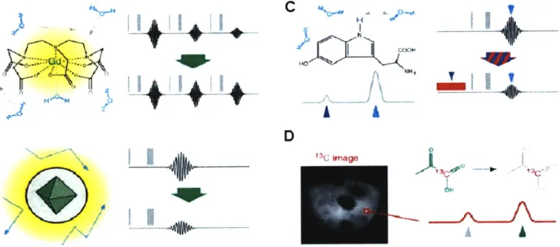
Figure 1. Contrast mechanisms in molecular MRI. Signal in MRI is proportional to the concentration of directly detected nuclei (usually protons in water molecules), their polarization, manipulations through pulse sequences and relaxation rates. (A) An exam-ple of Ti contrast agent, gadolinium-diehtylenetriaminepentaacetic acid (Gd-DTPA). Water molecules interact with paramagnetic Gd3 through space or through direct coordi-nation, which promotes Ti relaxivity. If MRI is performed using Ti-weighted pulse se-quences, this results in image brightening. (B) Superparamagnetic iron oxide nanoparti-cle (SPIO), a typical T2 contrast agent. SPIOs induce perturbations in the local magnetic fields that induce relaxation of water molecules diffusing in proximity. This results in reduction of MRI signal and image darkening. (C) An example of chemical exchange saturation transfer (CEST) contrast agent is 5-hydroxytriptofan which contains an indole nitrogen proton that exchanges with the bulk water. Application of the saturation pulse at the frequency of indole proton indirectly reduces the signal from water molecules, which leads to darkening of the MRI image. (D) Contrast agents incorporating nuclei such as
13C and 19F may be imaged directly by MRI. An image obtained by Goldman et al.
us-ing spectroscopic imagus-ing techniques to image hyperpolarized 13Cpyruvate and its meta-bolic product, 13C-lactate.
Figure 2. Classification of MRI contrast agents. (A) Nonspecific contrast agents such as blood pool agents, show a relatively nonspecific distribution pattern. (B) Targeted
con-trast agents, which bind by specific ligands to protein structures on cell surface. (C) Re-sponsive contrast agents which are activated by an interaction with specific target.
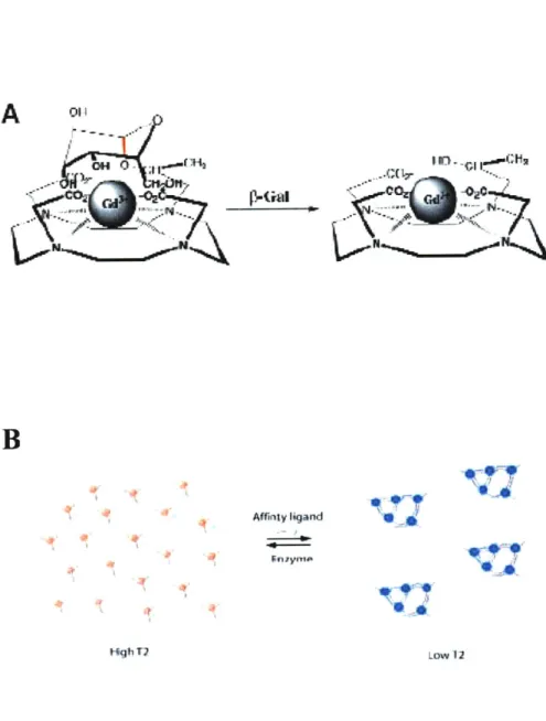
Figure 3. Examples of responsive T1 and T2 contrast agents. (A) A reporter for
p-galactosidase activity called EgadMe from Louie et al. Upon the cleavage of the sugar group by the enzyme at the red bond site, an inner sphere coordination site of the Gd 3 becomes more accessible to water molecules. (B) A T2 contrast agent based on SPIO ag-gregation from Perez et al. SPIOs cluster in the presence of a target which changes T2 relaxation time. The clusters of SPIOs can be dispersed by the action of the enzyme orother factors.
Figure 4. Examples of previously developed T1 contrast agents for metal ion sensing. (A) A Ca 2-sensitive contrast agent from Li et al., based on Ca2+ chelator BAPTA and Gd-DOTA. In the absence of Ca2+, carboxylic groups of BAPTA are coordinating para-magnetic Gd3* ion. Once Ca2+ binds, these ligands rearrange to bind this ion, increasing
inner sphere water accessibility to the complex which results in the increase of Ti relaxiv-ity. (B) A cell permeable, manganese-containing porphyrin molecule that senses Zn2+ ion from Zhang et al. Binding of the zinc causes intramolecular rearrangement which ex-poses central Mn3+ to water promoting the Ti relaxation rate of the complex.
Figure 5. Structure and calcium-dependent substrate binding of calmodulin. Calmodulin is a protein whose structure is characterized by four calcium binding motifs. These motifs have a helix-loop-helix structure (EF hands). CaM has two globular
do-mains linked by a flexible linker. Each domain consists of a pair of EF hands connected by a linker region and binds two Ca2+ with positive cooperavity. The binding of Ca2+ causes an intramolecular conformation change that allows the protein to bind its target.
Figure 6. Design of a fluorescent calcium sensor based on CaM-M13 interaction. Sensors called cameleons, from Miyawaki et al., consist of tandem fusions of blue- or cyan- emitting mutant of green fluorescent protein (GFP), calmodulin, the calmodulin-binding peptide M13 and an enhanced green- or yellow- emitting GFP. Binding of Ca2+
causes calmodulin to wrap around M13 which increases the fluorescent resonance energy transfer (FRET) between the flanking variants of GFP.
Figure 1
11+142+
COO" . 0 ... p H ... -IFigure 2
A
B
B
... . .. ... I
Figure 3 17' - ~ coz~
$
Affiny ligand Law 12 . ... ....Figure 4
A
( 1 +BI
I:fi Cl .C 0a2+ G3 N Zn(II) MRI Sensing ...Figure 5
EF3
a
helix-loop-helix
protein
binding of 4 Ca
Ca2+
allows calmodulin
to bind target
protein...
activating it.
EF1
Figure 6 370 or 440 nm 370 or 440 nm 440 or 480 nm CaM 13 -Ca 2+ +4 Ca 2 FRET 510 or 535 nm 4 2+
2. Calcium Sensors for Magnetic Resonance Imaging Based on
Super-paramagnetic Iron Oxide Nanoparticles and Calmodulin
The work described in here is the basis of the research paper that has been pub-lished in a peer reviewed journal (81). We introduce a new family of calcium indicators for magnetic resonance imaging (MRI), formed by combining a powerful iron oxide nanoparticle-based contrast mechanism with the versatile calcium sensing protein calmodulin and its targets. Calcium-dependent protein-protein interactions drive particle clustering and produce up to fivefold changes in T2 relaxivity, an indication of the sen-sors' potency. A variant based on conjugates of wild-type calmodulin and the peptide M13 reports concentration changes near 1 ptM Ca2+, suitable for detection of elevated
intracellular calcium levels; the midpoint and cooperativity of the response can be tuned by mutating the protein domains that actuate the sensor. Robust MRI signal changes are achieved even at nanomolar particle concentrations (less than 1 pM in calmodulin), that are unlikely to buffer calcium levels. When combined with technologies for cellular de-livery of nanoparticulate agents, the new sensors and their derivatives may be useful for functional molecular imaging of biological signaling networks in live, opaque specimens.
INTRODUCTION
Calcium ions (Ca2+) have been a favorite target in molecular imaging studies
be-cause of the important role of calcium as a second messenger in cellular signaling path-ways. Fluorescent calcium sensors are used widely in optical imaging, both at the cellu-lar level and at the cell population level. Calcium-sensitive dyes have recently been used in conjunction with laser scanning microscopy to follow neural network activity in small three-dimensional brain areas (61), and to characterize patterns of interaction among cells in developing vertebrate embryos (62). Due to the scattering properties of dense tissue, high resolution optical approaches like these are usually limited to superficial regions of specimens, and to restricted fields of view (63). To probe calcium dynamics more glob-ally in living systems, a different imaging modality must be used.
Magnetic resonance imaging (MRI) is an increasingly accessible technique for im-aging opaque subjects at fairly high spatial resolution, and MRI studies of calcium dy-namics could in principle complement optical approaches by offering both greatly ex-panded coverage and depth penetration in vivo (64). Calcium isotopes are unsuitable for direct imaging by magnetic resonance, so attempts to sensitize MRI to calcium have fo-cused around the use of molecular imaging agents. Fluorinated derivatives of the biva-lent cation chelator 1,2-bis(2-aminophenoxy)ethane-N,N,N',N'-tetraacetic acid (BAPTA) have allowed calcium measurements in vivo by 19F MRI, but only at approximately
10-the sensitivity of standard MRI methods (47, 48). Two potentially more powerful proton Ti relaxation-promoting contrast agents have been introduced subsequently. The para-magnetic ion manganese (Mn2+) mimics calcium by entering cells through calcium

![Figure 6 A 1.0" x 0.8 0.6 0.4 0.2 0 -8 -7 -6 -5 -4 -3 log([Ca2+ 1.2M 0 0 0.1 0.2 0.6 1.5 5.0 [Ca 2 +J (pM) C 70 50 --30 --00 -7 -6 -5 log([Ca2+]) D 12 1.00.8 -0.6 -0.4 0.2 -I I ,-8 -7 -6 -5 -4 -3EFx1m ,1 I IAM0 0.6 1.5](https://thumb-eu.123doks.com/thumbv2/123doknet/14678852.558719/78.918.187.720.369.914/figure-log-ca-ca-log-ca-efx-iam.webp)