HAL Id: hal-00356812
https://hal.archives-ouvertes.fr/hal-00356812
Submitted on 8 Jun 2021
HAL is a multi-disciplinary open access
archive for the deposit and dissemination of
sci-entific research documents, whether they are
pub-lished or not. The documents may come from
teaching and research institutions in France or
abroad, or from public or private research centers.
L’archive ouverte pluridisciplinaire HAL, est
destinée au dépôt et à la diffusion de documents
scientifiques de niveau recherche, publiés ou non,
émanant des établissements d’enseignement et de
recherche français ou étrangers, des laboratoires
publics ou privés.
Distributed under a Creative Commons Attribution| 4.0 International License
Virulent synergistic effect between Enterococcus faecalis
and Escherichia coli assayed by using the Caenorhabditis
elegans model.
Jean-Philippe Lavigne, Marie-Hélène Nicolas-Chanoine, Gisèle Bourg, Jérôme
Moreau, Albert Sotto
To cite this version:
Jean-Philippe Lavigne, Marie-Hélène Nicolas-Chanoine, Gisèle Bourg, Jérôme Moreau, Albert Sotto.
Virulent synergistic effect between Enterococcus faecalis and Escherichia coli assayed by using the
Caenorhabditis elegans model..
PLoS ONE, Public Library of Science, 2008, 3 (10), pp.e3370.
and
Escherichia coli
Assayed by Using the
Caenorhabditis elegans
Model
Jean-Philippe Lavigne1,2, Marie-He´le`ne Nicolas-Chanoine3,4, Gise`le Bourg1, Je´roˆme Moreau5, Albert Sotto1*
1 Institut National de la Sante´ et de la Recherche Me´dicale, ESPRI 26, Universite´ Montpellier 1, UFR de Me´decine, Nıˆmes, France, 2 Laboratoire de Bacte´riologie, Virologie, Parasitologie, CHU Caremeau, Nıˆmes, France,3 Service de Microbiologie, Hoˆpital AP-HP Beaujon, Clichy, France, 4 Institut National de la Sante´ et de la Recherche Me´dicale, U773, Universite´ Paris 7, Faculte´ de Me´decine D. Diderot, Paris, France, 5 Equipe Ecologie-Evolution UMR 5561 Bioge´osciences, Universite´ de Bourgogne, Dijon, France
Abstract
Background:The role of enterococci in the pathogenesis of polymicrobial infections is still debated. The purpose of this study was to evaluate the effect of virulent enterococci in the presence or absence of Escherichia coli strains in the in vivo Caenorhabditis elegans model.
Principal Findings: This study demonstrated that there was a synergistic effect on virulence when an association of enterococci and E. coli (LT50 = 1.6 days60.1 according to the tested strains and death of nematodes in 4 days60.5) was tested in comparison with enterococci alone (LT50 = 4.6 days60.1 and death in 10.4 days60.6) or E. coli alone (LT50 = 2.160.9 and deaths 6.660.6) (p,0.001). In addition, there was a relation between the virulence of E. faecalis strains alone and the virulence potential of the association with E. coli strains. Finally, in the presence of avirulent E. coli strains, enterococci have no effect (LT50 = 4.360.5 and deaths in 10.860.8), independently of the level of their own virulence, demonstrating that the ‘enterococci effect’ only occurred in the presence of virulent E. coli strains.
Conclusion:This study allows a better understanding of a bacterial cooperation. Moreover, it could help to optimize the antibiotic regimen during polymicrobial infections.
Citation: Lavigne JP, Nicolas-Chanoine MN, Bourg G, Moreau J, Sotto A (2008) Virulent Synergistic Effect between Enterococcus faecalis and Escherichia coli Assayed by Using the Caenorhabditis elegans Model. PLoS ONE 3(10): e3370. doi:10.1371/journal.pone.0003370
Editor: Frederick M. Ausubel, Massachusetts General Hospital, United States of America Received July 23, 2008; Accepted September 16, 2008; Published October 9, 2008
Copyright: ß 2008 Lavigne et al. This is an open-access article distributed under the terms of the Creative Commons Attribution License, which permits unrestricted use, distribution, and reproduction in any medium, provided the original author and source are credited.
Funding: INSERM Espri 26 is supported by INSERM, Universite´ de Montpellier 1, La Ville de Nıˆmes and La Re´gion Languedoc Roussillon. However, the funders had no role in study design, data collection and analysis, decision to publish, or preparation of the manuscript.
Competing Interests: The authors have declared that no competing interests exist. * E-mail: [email protected]
Introduction
The role played by enterococci in the pathogenesis of intra-abdominal infections is still debated because enterococci are frequently isolated from the polymicrobial flora [1–3]. In order to get insight into the role of enterococci in polymicrobial infections, Carbon et al. developed animal models and showed that enterococci might have a synergistic effect on the pathogenic traits of associated microorganisms, particularly Escherichia coli [4– 6]. In the present study, we used another in vivo model, namely the Caenorhabditis elegans model, which was previously used to explore virulence of various micro-organisms (Serratia marcescens, Salmonella enterica, Pseudomonas aeruginosa, Burkholderia cepacia, E. coli, Staphylococcus aureus and Streptococcus pyogenes [7–13]). Garsin et al. also used this model to investigate Enterococcus faecalis related virulence factors [14,15]. To our knowledge, the present study is the first one in which the C. elegans model was used to assess the virulence of associated microorganisms such as E. coli and E. faecalis.
Results
The virulence factor-encoding genes detected in the different strains of E. faecalis are listed in Table 1.
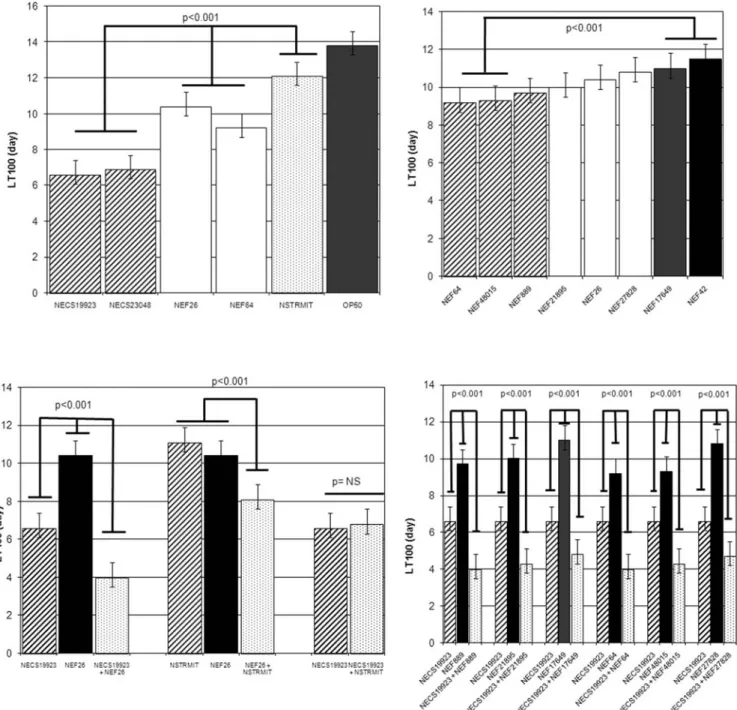
All E. faecalis, E. coli and S. mitis strains tested alone killed C. elegans (Fig. 1A). However, the two uropathogenic E. coli strains were significantly more virulent (LT50 of 2 days) than both E. faecalis strains and the S. mitis strain (LT50 varying from 4 to 5 days) (p,0.001). Interestingly, the bacteraemic E. faecalis strains (NEF64, NEF48015) were significantly more virulent (LT50 4.1 days60.1) than UTI strains (LT50 4.4 to 4.8 days60.1), even though they harboured no more VFs than UTI strains (p,0.001) (Fig. 1B).
When E. coli and E. faecalis strains were associated, a virulent synergistic effect was observed, irrespective at the specific uropathogenic E. coli and E. feacalis strains associated (Fig. 1C and 1D). The LT50s obtained with the strain associations varied between 1.5 and 1.7 days and were significantly shorter than the LT50s observed with E. coli strains alone (p,0.001). To demonstrate the role of enterocci in the increase in virulence,
the Enterococcus spp. strain was replaced by the S. mitis strain (which harboured ply gene). In the nematode killing model, E. faecalis and S. mitis strains presented a similar virulence (LT50NSTRMIT
5.4 days vs LT50NEF26 4.6 days; (p: not significant)) (Fig. 1A).
The association of E. coli NECS19923 and the S. mitis strain did not increase the virulence observed in the nematode model (LT50NECS19923+NSTRMIT 2.2 days) in comparison with the
virulence obtained with E. coli alone (LT50NECS19923 2.1 days)
(Fig. 1C). Conversely, the virulence of the association of the S. mitis strain with E. faecalis (LT50NEF26+NSTRMIT3.6 days) was
signifi-cantly higher than the virulence observed with either S. mitis alone (LT50NSTRMIT 5.4 days) and enterocci alone (LT50NEF26
4.6 days) (p,0.001) (Fig. 1C). All these findings strongly suggested the essential role of E. faecalis strains in the synergistic effect. However this synergistic effect of the association between E. coli and E. faecalis was only detected in presence of virulent E. coli strains. This was suggested by the LT50s observed when E. faecalis strains were associated with the avirulent E. coli OP50 strain (LT50NEF26 4.5 days vs LT50NEF26+OP50 4.3 and LT100NEF26
10.4 days vs LT100NEF26+OP5010.8; p: not significant).
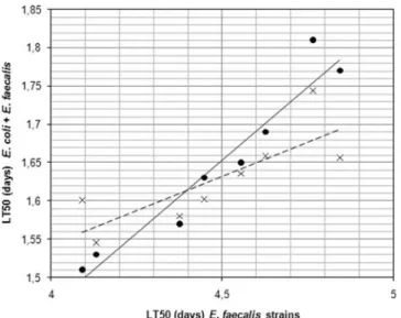
Moreover, as indicated in Fig. 2, a correlation was observed between the value of the LT50 of the E. faecalis strains tested alone and the value of the LT50 of the association of E. coli and E. faecalis strains. To verify the ingestion of E. coli, S. mitis and E. faecalis strains and the proliferation in the C. elegans intestine, L4 C. elegans were fed on a mixed lawn of E. faecalis and E. coli, E. faecalis and S. mitis or S. mitis and E. coli at a same ratio 1:1. At 72 h, the nematodes were washed to remove bacteria from their surfaces and then disrupted to recover bacteria from inside their digestive. The number of E. faecalis, S. mitis and E. coli CFU within the nematode gut varied between 105and 106bacteria per worm between each combination without statistical difference (Table 2).
Discussion
Both the clinical significance of enterococci in peritonitis and the use of empiric anti-enterococcal coverage have been the subject of a long-lasting and ongoing debate [16–19]. In a literature review recently published by Harbarth and Uckay, was presented evidence arguing in favour of using empirical therapy with enterococcal coverage for intra-abdominal infections among certain patients considered as high-risk [19].
The pathogenic role of enterococci has been studied in animal models. Onderdonk et al. showed in a model of peritonitis in rat
that enterococci inoculated alone induced neither early death nor later abscesses [20]. They demonstrated a synergic effect between enterococci and anaerobes but not between enterocci and E. coli. Later, Carbon et al. underlined several important points by using another model of experimental intra-abdominal infection. They studied the pathophysiological role of enterococci in a non fatal model of peritonitis in rats by implanting a gelatin capsule containing E. coli and Bacteroides fragilis with or without increasing concentrations of E. faecalis or heat-inactivated enterococci [6]. Effects were measured after several hours and several days following inoculation. Thus, they observed that the highest concentration of enterococci was correlated with an increase in E. coli and proinflammatory cytokines (tumor necrosis factor and interleukin) concentrations [21]. They suggested that enterococci might limit phagocytosis and intracellular death of other pathogens, particularly E. coli. In another study, the same authors evaluated the effects of E. faecalis strains containing various combinations of virulent factors in mouse and rat models of peritonitis. However, no conclusion was provided because of variable findings [4].
Currently, ten of enterococcal virulence factors are known. Their exact role in pathogenic mechanisms remains dubious and in several publications no conclusions were possible or were contradictory. In this study we screened for the six most studied virulence factors. Aggregation substance, encoded by asa1, is a pheromone-inducible protein that increases (i) bacterial adherence to renal tubular cells and heart endocardial cells, (ii) internalization by intestinal cells, and (iii) the valvular vegetation mass in an animal model of endocarditis. Gelatinase, encoded by gelE, is an extracellular zinc endopeptidase that hydrolyzes collagen, gelatin, and small peptides and that has been shown to exacerbate endocarditis in an animal model [22]. The production of cytolysin (encoded by cylA) has also been shown to significantly worsen the severity of endocarditis and endophthalmitis in animal models and is a virulence factor that affects C. elegans killing [14,22]. The enterococcal surface protein, encoded by esp, is associated with increased virulence, colonization and persistence in the urinary tract, and biofilm formation. Hyaluronidase, encoded by hyl, contributes to invasion of the nasopharynx and pneumococcal pneumonia [22]. Hemolysin, encoded by hlyB, is a cytotoxic protein that contributes to the destruction of red blood cells. Vergis et al. showed that the presence of gelatinase, hemolysin and enterococcal surface protein singly or in combination was not associated with mortality among patients with bacteremia due to Table 1. Detection of virulence factor (VF)-encoding genes (asa1, gelE, cylA, esp and hyl) and production of hemolysin (hlyB), gelatinase and aggregation substance among E. faecalis strains.
Strain Origin gelE cylA esp hyl asa1 VF score
Clumping factor hlyB NEF26 UTI + (+) 2 + 2 + 3 + (2) NEF42 UTI 2(2) + 2 2 2 1 NA (2) NEF889 UTI + (2) + + 2 + 4 + (2) NEF21895 UTI + (+) 2 + 2 2 2 NA (2) NEF17649 UTI 2(2) 2 + 2 2 1 NA (2) NEF64 Bacteraemia + (+) 2 2 + + 3 + (+) NEF48015 Bacteraemia + (+) 2 2 2 2 1 NA (2) NEF27828 UTI 2(2) 2 2 2 + 1 2 (2) Symbols in parentheses indicate expression of the gene:+, presence of virulence factor; 2, absence of virulence factor
NA, not applicable
doi:10.1371/journal.pone.0003370.t001
E. faecalis [23]. In this work, the most virulent E. faecalis strains did not necessarily contain the most number of virulence genes and no virulent ‘markers’ could be identified, including Cyl, that caused faster killing in the nematode model [14]. This confirms the difficulty in understanding the virulence of enterococci.
In the present study, using for the first time the C. elegans model to investigate microorganism association, we clearly demonstrated a synergistic interaction between E. coli and E. faecalis, two bacteria frequently isolated from peritonitis pus and UTI and also known to independently induce an intestinal infection in this nematode model [9,14]. The LT50s of the associations of E. faecalis and E. coli strains were definitively smaller than the LT50s observed with
each microorganism tested alone. Interestingly the higher the level of virulence of the E. faecalis strain, the more virulence was observed in the bacterial association with E. coli. However, the synergic effect did not seem to be influenced by the number of virulence factors of E. faecalis as described previously [24], but seemed to be influenced by the virulence of E. coli. Indeed no synergistic effect was observed between E. faecalis and E. coli OP50, a strain with low virulence potential. Such findings are in accordance with a recent data that have shown an association between the presence of numerous virulence factors, specific O types, B2 phylogenetic group and virulence in the C. elegans and murine models among uropathogenic E. coli strains [25,26].
Figure 1. In vivo kinetics of killing ofC. elegansinfected byE. coli, E. faecalisandS. mitisstrains. A. Comparison of LT100 between the different strains tested alone. B. Comparison of LT100 between E. faecalis strains tested alone. C. Comparison of LT100 with strains tested alone or in association. NEF26 was representative of the different E. faecalis strains. D. Comparison of LT100 between E. coli NECS19923 strain and the different E. faecalis strains tested in association. The results are representative of at least four independent trials for each group of strains. NS: non significant. doi:10.1371/journal.pone.0003370.g001
Finally the determination of the number of bacteria within the C. elegans digestive tract validated the presence and the overgrowth of E. faecalis, E. coli and S. mitis in the intestine of nematodes. This result demonstrates the establishment of a true infection validating our results. As previously demonstrated, E. faecalis not only accumulates in the intestinal lumen but proliferates in the gut,
resulting in a long-lasting infection that persists for the entire lifespan of the worms [14].
Many questions concerning the exact pathogenicity of entero-cocci remain unanswered. However, this model suggests the role of enterococci in increasing virulence in a polymicrobial infection. This is consistent with Carbon’s model. This study allows us to better understand bacterial cooperation and it could help to optimize the antibiotic regimen during polymicrobial infections.
Materials and Methods
Bacterial strains and growth conditions
Eight E. faecalis strains (NEF26, NEF42, NEF889, NEF21895, NEF17649, NEF64, NEF48015, NEF27828) isolated from the blood and urinary tract infections [27] (Table 1) were studied alone and combined with other microorganisms. These microor-ganisms, which also were studied alone, comprised Streptococcus mitis strain NSTRMIT isolated from the blood and E. coli strains NECS19923, NECS23048 both isolated from UTIs [9]. Two others strains were used as controls in the different experiments namely E. faecalis JH2-2 (control strain for expression of aggregation substance) and the avirulent E. coli OP50 (an international standard food for nematodes without known virulence factors). Bacteria were grown in trypticase soy broth or agar (TSA) or Columbia agar gelose with 5% fresh sheep blood (bioMerieux, Marnes La Coquette, France) at 37uC in 5% CO2.
Virulence genotyping
E. coli strains used in this study had been previously characterized for the virulence genotype [9]. NECS19923 and NECS23048 belonged to B2 group and presented virulence profiles: papGII, papA, papC, sfaS, focG, afa/draBC, fimH, hlyA, cnf1, iutA, kpsMTK1, kpsMTII, traT, iroN, malX, irp2 and papGIII, papA, papC, focG, afa/draBC, fimH, hlyA, cnf1, iutA, kpsMTK1, kpsMTII, traT, iroN, malX, irp2, respectively. E. faecalis and S. mitis isolates were tested by PCR for the presence of a panel of genes encoding known virulence factors (VFs). For E. faecalis, the multiplex PCR used the panel of 5 genes (asa1, gelE, cylA, esp and hyl) previously described [22]. For S. mitis, we used ply PCR to determine the presence of pneumolysin gene [28]. Production of gelatinase was determined by using TSA supplemented with 1.5% skimmed milk. The presence of a clear halo around colonies after overnight incubation at 37uC was considered to be a positive result [29]. Haemolysin production was evaluated on Columbia agar gelose with 5% fresh sheep blood. Zones of clearing around colonies after 18 h at 37uC indicated production of b-hemolysin [24]. Expression of aggregation substance was determined for PCR-positive strains by the clumping assay as previously described [21,30]. E. faecalis JH2-2 was used as control.
Nematode Killing Assay
The C. elegans infection assay was carried out as described by Kurz et al. [25] except that the Fer-15 mutant line, which has a temperature sensitive fertility defect, were used rather than wild type N2 worms. Fer-15 was provided by the Caenorhabditis Genetics Center, which is funded by the NIH National Center for Research Resources (NCRR). To synchronize the growth of worms, eggs were collected using the hypochlorite method. Overnight cultures in brain heart infusion (BHI) medium of E. coli, S. mitis and E. faecalis strains were harvested by centrifugation, washed once and suspended in phosphate buffered saline solution (PBS) at pH 7.0 at a concentration of 105CFU/ml. BHI agar plates were inoculated with 10 ml of each strain and incubated at 37uC for 8210 h. Plates were allowed to cool to room temperature and seeded with L4
Figure 2. Correlation between virulence of E. faecalis strains alone (expressed by LT50s in days) and in combination withE. colistrains. The linear regression model confirmed a linear relation. Dot corresponds to E. faecalis strains associated with NECS23048; cross corresponds to E. faecalis strains associated with NECS19923. The line and the dotted line established the linear regression of NECS23048 and NECS19923, respectively.
doi:10.1371/journal.pone.0003370.g002
Table 2. Evaluation of the number of bacteria within the C. elegans digestive tract.
Strains
Median CFU’s of strains/ nematode after 72 h Strain 1* Strain 2 E. coli NECS19923y 1.4 106[0.9–1.7 106] – E. faecalis NEF26y 6 105 [5.5–6.7 105 ] – E. faecalis NEF42y 4.5 105 [4.0–5.1 105 ] – S. mitis NSTRMITy 2.2 105[1.9–3.0 105] – NEF26y + NECS19923 5 105 [4.5–5.5 105 ] 8 105 [7.5 105 –106 ] NEF42y+ NECS19923 3.9 105 [3.0–4.6 105 ] 1.1 106 [0.8–1.3 106 ] NEF26y+ NSTRMIT 5.1 105[4.4–6.0 105] 2.7 105[1.9–3 105] NEF42y + NSTRMIT 4.3 105 [3.6–5.1 105 ] 3.2 105 [2.4–4.1 105 ] NECS19923y + NSTRMIT 106 [0.8–1.3 106 ] 1.9 105 [0.9–2.7 105 ] E. faecium, control strainy 101[0.5–1.3 101] –
After 72 hr of infection, the C. elegans were washed and ground, and dilutions of the resulting suspension were plated on selective media. The number of CFU per worm of the different strains alone or in combination was calculated. Three replicates were performed for each bacterial combination. E. faecium was used as negative control. No statistical difference could be demonstrated. Range of values are indicated into [square brackets]
*
Strain 1 corresponded to the first strain withy
; strain 2 corresponded to the second strain when associations were tested.
doi:10.1371/journal.pone.0003370.t002
stage worms (20–30 per plate). Plates were then incubated at 25uC and scored each day for live worms under a stereomicroscope (Leica MS5) [9]. At least three replicates repeated 5 times were performed for each selected clone. A worm was considered dead when it no longer responded to touch. Worms that died as a result of becoming stuck to the wall of the plate were excluded from the analysis. Lethal Time 50% (LT50) and death corresponded to time (in hours) required to kill 50% and 100% of the nematode population, respectively.
Measuring the number of bacteria within the C. elegans digestive tract
This assay was carried out as described by Garsin et al. [14]. Five C. elegans were picked at 72 h, and the surface bacteria were removed by washing the worms twice in 4 ml drops of M9 medium on a BHI agar plate containing 25 mg/ml gentamicin. The nematodes were placed in a 1.5 ml Eppendorf tube containing 20 ml of M9 medium with 1% Triton X-100 and were mechanically disrupted by using a pestle. The volume was adjusted to 50 ml with M9 medium containing 1% Triton X-100 which was diluted and plated on BHI agar containing 50 mg/ml ampicillin. At least three replicates were performed for each bacterial combination.
Statistical analysis
To compare the entire survival curves in nematode killing assays, we used a Cox regression. In order to perform pairwise comparison between two different strains, we used a log rank test. The analysis was carried out using SPSS 6.1.1 (SPSS Inc., Chicago, IL, USA).
Acknowledgments
This work was presented in part in the 27e`meRe´union Interdiciplinaire de
Chimiothe´rapie Anti-Infectieuse (RICAI), Paris, France, December 2007. We thank Dr J. Caillon (CHU Nantes) for the gift of E. faecalis JH2-2 strain. Fer-15 nematodes were provided by the Caenorhabditis Genetics Center, a foundation of the NIH National Center for Research Resources (NCRR).
Author Contributions
Conceived and designed the experiments: JPL MHNC JM AS. Performed the experiments: JPL GB. Analyzed the data: JPL MHNC JM AS. Contributed reagents/materials/analysis tools: JPL GB AS. Wrote the paper: JPL MHNC AS.
References
1. Dupont H, Vael C, Muller-Serieys C, Chosidow D, Mantz J, et al. (2007) Prospective evaluation of virulence factors of enterococci isolated from patients with peritonitis: impact on outcome. Diagn Microbiol Infect Dis 60: 247–253. 2. Murray BE (1990) The life and times of the enterococcus. Clin Microbiol Rev 3:
46–65.
3. Nichols RL, Muzik AC (1992) Enterococcal infections in surgical patients: the mystery continues. Clin Infect Dis 15: 72–76.
4. Dupont H, Montravers P, Mohler J, Carbon C (1998) Disparate findings on the role of virulence factors of Enterococcus faecalis in mouse and rat models of peritonitis. Infect Immun 66: 2570–2575.
5. Montravers P, Andremont A, Massias L, Carbon C (1994) Investigation of the potential role of Enterococcus faecalis in the pathophysiology of experimental peritonitis. J Infect Dis 169: 821–830.
6. Montravers P, Mohler J, Saint Julien L, Carbon C (1997) Evidence of the proinflammatory role of Enterococcus faecalis in polymicrobial peritonitis in rats. Infect Immun 65: 144–149.
7. Jansen WT, Bolm M, Balling R, Chhatwal GS, Schnabel R (2002) Hydrogen peroxide-mediated killing of Caenorhabditis elegans by Streptococcus pyogenes. Infect Immun 70: 5202–5207.
8. Kothe M, Antl M, Huber B, Stoecker K, Ebrecht D, et al. (2003) Killing of Caenorhabditis elegans by Burkholderia cepacia is controlled by the cep quorum-sensing system. Cell Microbiol 5: 343–351.
9. Lavigne JP, Blanc-Potard AB, Bourg G, Moreau J, Chanal C, et al. (2006) Virulence genotype and nematode-killing properties of extra-intestinal Escherichia coli producing CTX-M beta-lactamases. Clin Microbiol Infect 12: 1199–1206. 10. Schulenburg H, Ewbank JJ (2004) Diversity and specificity in the interaction
between Caenorhabditis elegans and the pathogen Serratia marcescens. BMC Evol Biol 4: 49.
11. Sifri CD, Begun J, Ausubel FM, Calderwood SB (2003) Caenorhabditis elegans as a model host for Staphylococcus aureus pathogenesis. Infect Immun 71: 2208–2217. 12. Tan MW, Ausubel FM (2000) Caenorhabditis elegans: a model genetic host to study
Pseudomonas aeruginosa pathogenesis. Curr Opin Microbiol 3: 29–34.
13. Tenor JL, McCormick BA, Ausubel FM, Aballay A (2004) Caenorhabditis elegans-based screen identifies Salmonella virulence factors required for conserved host-pathogen interactions. Curr Biol 14: 1018–1024.
14. Garsin DA, Sifri CD, Mylonakis E, Qin X, Singh KV, et al. (2001) A simple model host for identifying Gram-positive virulence factors. Proc Natl Acad Sci USA 98: 10892–10897.
15. Maadani A, Fox KA, Mylonakis E, Garsin DA (2007) Enterococcus faecalis mutations affecting virulence in the Caenorhabditis elegans model host. Infect Immun 75: 2634–2637.
16. Blot S, De Waele JJ (2005) Critical issues in the clinical management of complicated intra-abdominal infections. Drugs 65: 1611–1620.
17. Chatterjee I, Iredell JR, Woods M, Lipman J (2007) The implications of enterococci for the intensive care unit. Crit Care Resusc 9: 69–75.
18. Dupont H (2007) The empiric treatment of nosocomial intra-abdominal infections. Int J Infect Dis 11: S1–S6.
19. Harbarth S, Uckay I (2004) Are there patients with peritonitis who require empiric therapy for enterococcus? Eur J Clin Microbiol Infect Dis 23: 73–77. 20. Onderdonk AB, Bartlett JG, Louie T, Sullivan-Seigler N, Gorbach SL (1976)
Microbial synergy in experimental intra-abdominal abscess. Infect Immun 13: 22–26.
21. Dunny GM, Craig RA, Carron RL, Clewell DB (1979) Plasmid transfer in Streptococcus faecalis: production of multiple sex pheromones by recipients. Plasmid 2: 454–465.
22. Vankerckhoven V, Van Autgaerden T, Vael C, Lammens C, Chapelle S, et al. (2004) Development of a multiplex PCR for the detection of asa1, gelE, cylA, esp, and hyl genes in enterococci and survey for virulence determinants among European hospital isolates of Enterococcus faecium. J Clin Microbiol 42: 4473–4479.
23. Vergis EN, Shankar N, Chow JW, Hayden MK, Snydman DR, et al. (2002) Association between the presence of enterococcal virulence factors gelatinase, hemolysin, and enterococcal surface protein and mortality among patients with bacteremia due to Enterococcus faecalis. Clin Infect Dis 35: 570–575.
24. Franz CM, Muscholl-Silberhorn AB, Yousif NM, Vancanneyt M, Swings J, et al. (2001) Incidence of virulence factors and antibiotic resistance among Enterococci isolated from food. Appl Environ Microbiol 67: 4385–4389. 25. Kurz CL, Chauvet S, Andres E, Aurouze M, Vallet I, et al. (2003) Virulence
factors of the human opportunistic pathogen Serratia marcescens identified by in vivo screening. EMBO J 22: 1451–1460.
26. Diard M, Baeriswyl S, Clermont O, Gouriou S, Picard B, et al. (2007) Caenorhabditis elegans as a simple model to study phenotypic and genetic virulence determinants of extraintestinal pathogenic Escherichia coli. Microbes Infect 9: 214–223.
27. Lavigne JP, Marchandin H, Czarnecki E, Kaye C, Sotto A (2005) Enterococcal bacteremia at Nıˆmes university hospital. Pathol Biol 53: 539–545.
28. Neeleman C, Klaassen CH, Klomberg DM, de Valk HA, Mouton JW (2004) Pneumolysin is a key factor in misidentification of macrolide-resistant Streptococcus pneumoniae and is a putative virulence factor of S. mitis and other streptococci. J Clin Microbiol 42: 4355–4357.
29. Coque TM, Patterson JE, Steckelberg JM, Murray BE (1995) Incidence of hemolysin, gelatinase, and aggregation substance among enterococci isolated from patients with endocarditis and other infections and from feces of hospitalized and community-based persons. J Infect Dis 171: 1223–1229. 30. Creti R, Imperi M, Bertuccini L, Fabretti F, Orefici G, et al. (2004) Survey for
virulence determinants among Enterococcus faecalis isolated from different sources. J Med Microbiol 53: 13–20.