HAL Id: inserm-00807850
https://www.hal.inserm.fr/inserm-00807850
Submitted on 4 Apr 2013
HAL is a multi-disciplinary open access
archive for the deposit and dissemination of sci-entific research documents, whether they are pub-lished or not. The documents may come from teaching and research institutions in France or abroad, or from public or private research centers.
L’archive ouverte pluridisciplinaire HAL, est destinée au dépôt et à la diffusion de documents scientifiques de niveau recherche, publiés ou non, émanant des établissements d’enseignement et de recherche français ou étrangers, des laboratoires publics ou privés.
GATA-6 transcription factors.
Nicolas Jonckheere, Amélie Velghe, Marie-Paule Ducourouble, Marie-Christine
Copin, Ingrid Renes, Isabelle van Seuningen
To cite this version:
Nicolas Jonckheere, Amélie Velghe, Marie-Paule Ducourouble, Marie-Christine Copin, Ingrid Renes, et al.. The mouse Muc5b mucin gene is transcriptionally regulated by thyroid transcription factor-1 (TTF-1) and GATA-6 transcription factors.: Regulation of Muc5b mucin gene by TTF-1 and GATA factors. FEBS Journal, Wiley, 2011, 278 (2), pp.282-94. �10.1111/j.1742-4658.2010.07945.x�. �inserm-00807850�
For Review Only
The mouse Muc5b mucin gene is transcriptionally regulated by TTF-1 and GATA-6 transcription factors
Journal: FEBS Journal Manuscript ID: FJ-10-0798.R1 Manuscript Type: Regular Paper
Subdiscipline: Gene expression, transcription and translation Date Submitted by the
Author: 20-Oct-2010
Complete List of Authors: Jonckheere, Nicolas; INSERM U837/JPARC, Team 5 " mucins, epithelial differentiation and carcinogenesis"
Velghe, Amélie; INSERM U837/JPARC, Team 5 "mucins, epithelial differentiation and carcinogenesis"
Ducourouble, Marie-Paule; INSERM U837/JPARC, Team 5 "mucins, epithelial differentiation and carcinogenesis"
Copin, Marie-Christine; INSERM U837/JPARC, Team 5 " mucins, epithelial differentiation and carcinogenesis"
Renes, Ingrid; Erasmus MC and Sophia Children’s Hospital, Division of Neonatology, Department of Pediatrics
van Seuningen, Isabelle; INSERM U837/JPARC, Team 5 " mucins, epithelial differentiation and carcinogenesis"
Key Words: mucin, Muc5b, TTF1, GATA, lung, differentiation
For Review Only
The mouse Muc5b mucin gene is transcriptionally regulated by TTF-1 and GATA-6 transcription factors
Nicolas Jonckheere1,2, Amélie Velghe1, Marie-Paule Ducourouble1,2, Marie-Christine Copin1,
2, 3
, Ingrid B. Renes4, and Isabelle Van Seuningen1, 2
1
Inserm, U837, Team #5 “Mucins, epithelial differentiation and carcinogenesis”, rue Polonovski, 59045 Lille Cedex, France
2
Université Lille Nord de France, 1 Place de Verdun, 59045 Lille cedex, France
3
Centre de Biologie-Pathologie, Centre Hospitalier Régional et Universitaire de Lille, Boulevard du Professeur Jules Leclercq, 59000 Lille, France
4
Laboratory of Pediatrics, Division of Neonatology, Erasmus MC-Sophia Hospital, Rotterdam, The Netherlands
Running title: Regulation of Muc5b mucin gene by TTF-1 and GATA factors
Genbank accession number : AY744445
To whom correspondence should be sent: Nicolas Jonckheere, Ph.D.
1
Inserm, U837, Team #5, Rue Polonovski, 59045 Lille cedex, France Phone: 33.320.29.88.67 FAX: 33.320.53.85.62 E-mail: nicolas.jonckheere@inserm.fr 3 4 5 6 7 8 9 10 11 12 13 14 15 16 17 18 19 20 21 22 23 24 25 26 27 28 29 30 31 32 33 34 35 36 37 38 39 40 41 42 43 44 45 46 47 48 49 50 51 52 53 54 55 56 57 58 59 60
For Review Only
Abstract
MUC5B is one of the major mucin genes expressed in the respiratory tract. Previous
studies in our laboratory have demonstrated that MUC5B is expressed in human lung adenocarcinomas and during lung morphogenesis. Moreover, in human lung adenocarcinoma tissues, a converse correlation between MUC5B and TTF-1 expression, a lung-specific transcription factor, was established. However, the molecular mechanisms that govern the regulation of MUC5B expression in the lung are largely unknown. In order to better understand the biological role of MUC5B in lung pathophysiology, we report now the characterization of the promoter region of the mouse Muc5b mucin gene. The promoter is flanked by a TATA box (TACATAA) identical to that in the human gene. Human and murine promoters share 67.5% similarity over the first 170 nucleotides. By RT-PCR, co-transfection studies and gel-shift assays we show that Muc5b promoter activity is completely inhibited by TTF-1 whereas factors of the GATA family (GATA-4/-5/-6) are activators. Altogether, these results demonstrate for the first time that Muc5b is a target gene of transcription factors (TTF-1, GATA-6) involved in lung differentiation programs during development and carcinogenesis and identifies TTF-1 as a strong repressor of Muc5b. The characterization of the structural and functional features of Muc5b mucin gene will provide us with a strong base to develop studies in murine models aimed at identifying its biological role in lung pathophysiology.
Keywords: mucin, Muc5b, TTF1, GATA, lung, differentiation
3 4 5 6 7 8 9 10 11 12 13 14 15 16 17 18 19 20 21 22 23 24 25 26 27 28 29 30 31 32 33 34 35 36 37 38 39 40 41 42 43 44 45 46 47 48 49 50 51 52 53 54 55 56 57 58 59 60
For Review Only
Introduction
Mucins are high molecular weight glycoproteins that are synthesized by specialized epithelial cells and are thought to promote tumor cell invasion [1]. In the tracheobronchial tree, the main mucin genes are MUC5B and MUC5AC, that encode two secreted mucins, and
MUC4, that encodes a transmembrane mucin [2]. MUC5B and MUC5AC are expressed in
mucus-producing cells with MUC5AC in the surface goblet cells and MUC5B in the mucous cells of the submucosal gland whereas MUC4 is found in a wide array of epithelial cells [3-7].
MUC5B expression, in the developing lung, is seen as of 13 weeks of gestation in the
epithelial folds of the surface epithelium [8]. At a later stage, MUC5B is found in cells of the gland ducts and mucous glands [9]. In the adult lung, the expression of MUC5B follows a restricted pattern with a positive gradient from the surface to the glands and a decrease in intensity from the tracheo-bronchus toward the bronchioles, with no signal in small bronchioles and pneumocytes [10]. The murine Muc5b mucin gene was recently characterized in our laboratory and was shown to be expressed in mucous cells of the laryngeal glands [11]. In lung adenocarcinomas, MUC5B is frequently expressed in mucus-secreting carcinomatous cells [12]. In mucinous type of bronchioloalveolar carcinoma, MUC5B expression is the most intense along with that of MUC5AC. The expression of MUC5B is lost in poorly differentiated and non mucinous lung carcinomas [12]. From these studies, it appears that MUC5B may be used as a marker of cytodifferentiation in the lung associated with mucous differentiation [5].
The early expression of mucin genes before mucous cell differentiation or during the process of differentiation suggests that they may be the targets of transcription factors responsible for those programs [13]. Accordingly to that hypothesis, we recently showed that
MUC2 and MUC4 are transcriptionally regulated by Cdx homeodomain proteins and GATA
3 4 5 6 7 8 9 10 11 12 13 14 15 16 17 18 19 20 21 22 23 24 25 26 27 28 29 30 31 32 33 34 35 36 37 38 39 40 41 42 43 44 45 46 47 48 49 50 51 52 53 54 55 56 57 58 59 60
For Review Only
factors [14-16]. The thyroid transcription factor-1 (TTF-1) is an important factor during lung morphogenesis [17-20] and drives the expression of several lung-specific genes such as surfactant proteins [21], CCSP [22] and CC10 [23]. Moreover, we have recently shown a converse correlation between MUC5B and TTF-1 expression in human lung adenocarcinomatous tissues [24], suggesting a negative regulation of MUC5B by this transcription factor. GATA factors also possess a restricted pattern of expression during lung development [25]. GATA-6 seems to be involved during different phases of the development [26] whereas GATA-5 plays a role in transcriptional programs in the earliest steps of lung development [27]. Moreover, synergistic mechanisms between homeoprotein TTF-1 and zinc-finger GATA-6 have been recently described [21,28].
Having found (i) binding sites for these factors in both the human [29] and murine (this report) MUC5B mucin genes, (ii) a restricted pattern of MUC5B expression in the respiratory tract [5] and (iii) expression of MUC5B, TTF-1 and GATA-6 in lung adenocarcinomas [12,24,30,31], we undertook to study the regulation of the Muc5b promoter by TTF-1 and GATA factors. By this approach we aimed to show transcriptional regulation of Muc5b by these two transcription factors thereby providing a strong base for the development of studies aimed at identifying the biological role of Muc5b in the lung using mouse models.
3 4 5 6 7 8 9 10 11 12 13 14 15 16 17 18 19 20 21 22 23 24 25 26 27 28 29 30 31 32 33 34 35 36 37 38 39 40 41 42 43 44 45 46 47 48 49 50 51 52 53 54 55 56 57 58 59 60
For Review Only
Results
Characterization of the sequence of the promoter of the murine Muc5b mucin gene
The sequence covering 1210 nucleotides upstream of the transcription initiation site is shown in Fig. 1A. It is characterized by the presence of a TATA box (TACATAA) at -28/-22. The immediate sequence is GC-rich and contains a few putative binding sites for Sp1-like factors (GC-boxes and CACCC boxes). We also note the presence of putative binding sites for the lung-specific factor TTF-1 throughout the sequence. GATA putative binding sites are present both in the proximal and distal parts of the promoter.
Alignment of the human and mouse promoter sequences showed that there is a high homology (67.5 %) over the first 157 nucleotides flanking the TATA box (Fig. 1B) and that the sequence of the TATA box (TACATAA) is identical in the two species.
Characterization of Muc5b promoter activity
Mouse Muc5b transcriptional regulation at the promoter and mRNA levels was studied in the murine CMT-93 colorectal cancer cell line that is commonly used to study murine mucin gene regulation since it is known to express several mucin genes [15,32] and of interest in this study expresses Muc5b mRNA (Fig. 2A). Since no murine lung epithelial cell line expressing Muc5b is available at this time we also studied mMuc5b promoter regulation in the human lung NCI-H292 cell line that expresses MUC5B as we previously demonstrated [33]. To define essential regions that drive transcription of the Muc5b promoter, six deletion mutants, that cover 1.2 kb of the promoter, were constructed in the promoterless pGL3 basic vector (Fig. 2B). Data indicate that the promoter is active both in murine intestinal CMT-93 and human lung NCIH292 cell lines. The four deletion constructs tested (169/1, 478/1, -717/-1 and -1195/-1) have similar luciferase activities in each cell line, which suggests that
3 4 5 6 7 8 9 10 11 12 13 14 15 16 17 18 19 20 21 22 23 24 25 26 27 28 29 30 31 32 33 34 35 36 37 38 39 40 41 42 43 44 45 46 47 48 49 50 51 52 53 54 55 56 57 58 59 60
For Review Only
the proximal region -169/-1 is sufficient to drive maximal activity of the promoter in these cells (Fig. 2C). The influence of the 5’-UTR on the promoter activity was studied using the constructs -478/+47 and -717/+47. When the 5’-UTR region +1/+47 was included, the activity of the promoter remained similar (compare activities of 478/1 to 478/+47 and of -717/+47 to -747/-1).
TTF-1 is a strong repressor of Muc5b expression
Overexpression of TTF-1 in CMT-93 cells led to a strong decrease of the amount of
Muc5b mRNA (75 % loss, Fig. 3A). Co-transfection experiments in the presence of the
pCMV-TTF-1 expression vector showed that overexpression of TTF-1 also led to a dramatic decrease (60-75 %) of the activity of Muc5b promoter both in CMT-93 and NCI-H292 cells (Fig. 3B). The decrease was even more pronounced in NCI-H292 cells (80 % loss). The strong inhibition was seen with all the constructs tested in this work suggesting that the -477/-1 region is sufficient to convey the repression of Muc5b promoter by TTF--477/-1. TTF--477/-1 binds to the -CAAG- consensus sequence. Putative binding sites were found throughout the sequence of the Muc5b promoter (see Fig. 1A). EMSAs were performed with several probes containing TTF-1 consensus binding sites found in the murine promoter (Table 2) as well as with their mutated version (CAAG to GTAT). The probe T211 contains two putative TTF-1 binding sites at 358/355 and 353/350 and the probe T212 contains two sites at 709/706 and 700/697, respectively. The probes T213, T238 and T242 contained one predicted site at -112/-109, -325/-322, and -417/-414, respectively. Incubation of T211 and T212 radiolabeled probes with nuclear proteins from NCI-H292 cells produced one specific shifted band (Fig. 3C, lanes 2 and 8). Specificity of the complex was confirmed by the loss of the shifted band (indicated by an asterisk) when cold probes, in a 50x excess, were incubated with nuclear proteins before adding the radiolabeled probe (lanes 3 and 9). Moreover, no competition
3 4 5 6 7 8 9 10 11 12 13 14 15 16 17 18 19 20 21 22 23 24 25 26 27 28 29 30 31 32 33 34 35 36 37 38 39 40 41 42 43 44 45 46 47 48 49 50 51 52 53 54 55 56 57 58 59 60
For Review Only
could be observed when mutated probes were used in the competition (lanes 4 and 10). Implication of TTF-1 in the complex formation was further confirmed when mutated probes were radiolabeled and incubated with nuclear extracts. In that case, no binding was visualized (lanes 6 and 12). The probe T238 did not produce any shift and the probes T213 and T242 that contained a predicted TTF-1 site did not bind TTF-1 (not shown).
Role of GATA factors in the regulation of Muc5b expression
Besides TTF-1, GATA factors and especially GATA-6 are important factors in lung morphogenesis and are known to (i) regulate TTF-1 and (ii) synergize with TTF-1 to activate transcription of their target genes. Analysis of the sequence of the promoter of Muc5b showed that putative binding sites for GATA factors were present throughout the sequence (see Fig. 1A), which is in favor of a possible role in the regulation of Muc5b.
At the mRNA level, we observed an increase of Muc5b expression with GATA-5 (4 fold) and GATA-6 (14 fold) once we had overexpressed those transcription factors in CMT-93 cells (Fig. 4A). There was no effect visualized with GATA-4. To localize the GATA responsive elements we then performed co-transfection experiments both in the CMT-93 (Fig. 4B) and NCI-H292 (Fig. 4C) cell lines. Overexpression of GATA-5 in CMT-93 cells induced a strong activation of the three constructs of the Muc5b promoter (4-, 4- and 6-fold activation on -478/-1, -717/-1 and -1195/-1 constructs, respectively, p<0.05). Overexpression of GATA-4 and GATA-6 in these cells also induced transactivation of -717/-1 and -GATA-478/-1 Muc5b promoter constructs, respectively (2-4 fold activation, p<0.05) (Fig. 4B). In lung NCI-H292 cells, the profile was slightly different in that the strong transactivating effect of the three GATA factors on the -717/-1 region went down with the -1195/-1 deletion construct (Fig. 4C). This suggests that some inhibitory factors binding to the -1195/-718 region of the promoter may interfere with GATA function in these cells.
3 4 5 6 7 8 9 10 11 12 13 14 15 16 17 18 19 20 21 22 23 24 25 26 27 28 29 30 31 32 33 34 35 36 37 38 39 40 41 42 43 44 45 46 47 48 49 50 51 52 53 54 55 56 57 58 59 60
For Review Only
GATA cis-elements within the promoter of Muc5b were then identified by performing EMSA experiments with DNA probes containing GATA putative binding sites located at -411/-408 (T242), -454/-449 (T254) and -1143/-1140 (T84), respectively. As shown in Fig. 4D, incubation of these three probes with nuclear proteins from CMT-93 cells produced one specific shifted complex (GATA) (lanes 2, 9 and 15, respectively). Specificity was confirmed by complete inhibition of the complex formation when unlabelled competition was performed with a 50x excess of the cold probe (lanes 3, 10 and 16). GATA-4 and GATA-6 were both able to bind the GATA element present in T254 and T84 probes since a supershift was visualized upon addition of a GATA-4 (lanes 11 and 17) or GATA-6 (lanes 13 and 19) antibody in the mixture. GATA-4 is involved in the complex formation with the T242 probe since a supershift was observed upon addition of the anti-GATA-4 antibody in the reaction mixture (lane 5). No supershift was seen when anti-GATA-5 antibody was used (lanes 6, 12 and 18). However, we can not conclude that this factor does not bind to these sites since it also did not induce a supershift when a commercial consensus GATA probe was used (not shown). ChIP assay was carried out on -503/-261 region of Muc5b promoter containing notably T242 (-411/-408) and T254 (-454/-449) binding sites. Binding of GATA-4, GATA-5 and GATA-6 to the Muc5b promoter was observed in CMT-93 cells (Fig. 4E). Specificity of the binding was confirmed by complete absence of PCR amplification using IgGs.
In order to show a possible synergistic mechanism of regulation between TTF-1 and GATA-6, co-transfections with those two factors were carried out in CMT-93 cells with the -478/-1, -717/-1 and -1195/-1 Muc5b promoter constructs (Fig. 4E). As previously shown, overexpression of GATA-6 transactivated the three deletion mutants, whereas overexpression of TTF-1 strongly repressed the transcriptional activity of the three constructs. When co-transfected together, TTF-1 inhibited the transactivating effect of GATA-6, which led to a
3 4 5 6 7 8 9 10 11 12 13 14 15 16 17 18 19 20 21 22 23 24 25 26 27 28 29 30 31 32 33 34 35 36 37 38 39 40 41 42 43 44 45 46 47 48 49 50 51 52 53 54 55 56 57 58 59 60
For Review Only
loss of the transactivation of the Muc5b promoter. The same result was obtained in NCI-H292 cells (not shown).
Expression of MUC5B, TTF-1 and GATA-6 in well-differentiated mucus-secreting lung adenocarcinomas
Immunohistochemical analyses revealed that in well-differentiated mucus-secreting lung adenocarcinomas, MUC5B expression was intense and cytoplasmic (Fig. 5A), whereas there was no expression of TTF-1 in MUC5B-positive cells (Fig. 5B). In a papillary adenocarcinoma, MUC5B was not detected (Fig. 5D). On the contrary, TTF-1 was expressed in the nucleus of all these papillary adenocarcinomatous cells (Fig. 5E). In a non-mucinous type of bronchioloalveolar carcinoma, TTF-1 was expressed in the majority of the carcinomatous cells (Fig. 5H). In contrast, these TTF-1 positive cells did not express MUC5B (Fig. 5H, inset). Interestingly, in another region of the same bronchioloalveolar carcinoma, that was focally mucus-secreting, we found expression of MUC5B in a few mucus-secreting tumor cells (Fig. 5G). In these MUC5B-expressing cells, TTF-1 was not expressed (Fig. 5G, inset). Immunohistochemical studies on the same lung tumor tissues indicated that GATA-4 was not expressed in these samples (not shown) whereas GATA-6 was consistently expressed in the cytoplasm of MUC5B-expressing cells (Fig. 5C, F and I).
3 4 5 6 7 8 9 10 11 12 13 14 15 16 17 18 19 20 21 22 23 24 25 26 27 28 29 30 31 32 33 34 35 36 37 38 39 40 41 42 43 44 45 46 47 48 49 50 51 52 53 54 55 56 57 58 59 60
For Review Only
Discussion
Human MUC5B mucin gene is one of the main mucin genes expressed in the respiratory tract, in which it is mainly found in the mucous cells of the submucosal glands. Recently, we have characterized the human MUC5B promoter [29,34] and studied its expression both during lung development [8] and lung carcinogenesis [12]. From these studies it appears that the MUC5B promoter contains several putative binding sites for transcription factors playing critical roles in the formation, differentiation, and function of cells lining the respiratory tract such as TTF-1 and GATA factors [13]. Moreover, expression studies revealed a somewhat surprising early expression of MUC5B in the developing lung concomitant to mucous cell differentiation [8] and altered patterns of expression in lung adenocarcinomas [5,12,24].
The regulation of MUC5B by these transcription factors is however unknown and development of murine models of lung diseases are necessary to gain insight into, and understand, the regulation of the murine homologue of MUC5B. In the present study, we have isolated and characterized the promoter of the murine Muc5b mucin gene in order to study its transcriptional regulation by TTF-1 and GATA transcription factors. This approach will provide necessary knowledge to study Muc5b regulation in murine models and more particularly its biological role in lung pathophysiology.
Analysis of the promoter sequences of the murine Muc5b and the human MUC5B genes showed that they are highly similar over the first 170 nucleotides and more importantly that the TATA box is identical. This suggests that conserved regulatory mechanisms exist for these two genes throughout evolution and especially between mouse and human species.
Furthermore, in this report we demonstrate that TTF-1, which plays an important role in lung morphogenesis, lung repair after injury, or during carcinogenesis [20,35,36] and is a strong repressor of Muc5b expression at the promoter level. These results corroborate with our data in human tissues from different subsets of lung carcinoma in which we have also
3 4 5 6 7 8 9 10 11 12 13 14 15 16 17 18 19 20 21 22 23 24 25 26 27 28 29 30 31 32 33 34 35 36 37 38 39 40 41 42 43 44 45 46 47 48 49 50 51 52 53 54 55 56 57 58 59 60
For Review Only
shown a converse expression of TTF-1 and MUC5B proteins ([24] and this report) and with another study that showed that mucinous parts of lung carcinomas expressing MUC5B are TTF-1 negative [24,37]. Altogether, these results identify, for the first time, Muc5b as a direct target gene of TTF-1, which most likely is responsible for the repression of MUC5B in certain types of lung adenocarcinomas.
The main consequence of MUC5B repression by TTF-1 will be a modification in the composition of the respiratory mucus since most of the mucus secretion in the lung comes from mucous cells of the submucosal glands that secrete MUC5B [5,38]. The rheological properties of the mucus and its ability to maintain a normal defense line against bacterial infection, immune recognition of the cancer cell [1] or during development or repair [5] will then be greatly impaired. In future studies, it will be interesting to determine whether repression of MUC5B by TTF-1 represents a more general mechanism in lung diseases.
The GATA family of transcription factors is composed of several factors [25]. In the lung it has been shown that GATA-4/-5/-6 are expressed in a restricted manner. These factors participate in epithelial cell differentiation during embryonic development and establishment of cell lineages derived from primitive intestine [39]. Previous work in our laboratory has allowed identification of GATA factors as activators of mucin gene expression [15,16], such as GATA-4 for Muc2 in intestinal cells [15], with obvious association between mucin activation by GATA factors and terminal differentiation of the specialized epithelial cell in which the mucin expression is activated. In this work, it appears that GATA-5 and GATA-6 are also activators of Muc5b transcription. GATA-4 only had a moderate effect on the promoter activity. Previously, when we looked at GATA-4 expression in human lung tissues, we could not find any expression of GATA-4. This is in agreement with a recent report that showed that GATA-4 expression in lung carcinomas was repressed by hypermethylation of its promoter [40]. Thus GATA-4 does not appear to be a candidate for MUC5B regulation in the
3 4 5 6 7 8 9 10 11 12 13 14 15 16 17 18 19 20 21 22 23 24 25 26 27 28 29 30 31 32 33 34 35 36 37 38 39 40 41 42 43 44 45 46 47 48 49 50 51 52 53 54 55 56 57 58 59 60
For Review Only
lung. Moreover, we consistently found positive cytoplasmic staining of GATA-6 in MUC5B-expressing cells in the same sections used for TTF-1. A positive correlation was found between GATA-6 and MUC5B expression in the same cells. However, despite the fact that GATA-6 is a strong inducer of Muc5b transcription, its localization in the cytoplasm of MUC5B-expressing lung carcinoma cells underscores its role as a major regulator of MUC5B expression in the types of lung carcinomas studied in this report. Alteration of GATA-6 expression and aberrant cytoplasmic localization in ovarian cancer cells was recently proposed to contribute to dedifferentiation of the tumor cells seen in the process of adaptation to neoplastic progression [41].
Regulation of Muc5b expression by transcription factors expressed early during lung development, such as TTF-1 and GATA-6, may have a critical role both in normal and cancerous differentiation processes. From our data and others, the regulation of mucin genes by GATA factors seems to be more general and may affect the expression of other mucin genes, like MUC2, MUC3 and MUC4, since their promoters also contain cis-elements for these transcription factors [6,13,42,43]. Since GATA factors are expressed in endodermal tissues, we hypothesize that this mechanism of regulation will occur in tissues derived from endoderm and primitive gut including the lung but also the digestive tract as we have already shown for Muc2 expression by GATA-4 [15], MUC4 by GATA-4/-5/-6 [16] and MUC6 by GATA-5/-6 (I Van Seuningen, unpublished observations) in intestinal goblet cells. MUC4, encodes a membrane-bound mucin expressed as early as 6.5 weeks after gestation, by the primitive epithelial cells, and have the potential to differentiate in all the epithelial cell types of the conducting airways and alveolar epithelium. In the lung, we think that MUC4 may be a good candidate [8]. TTF-1 and GATA-6 are required for the formation and differentiation of distal epithelium [20,44,45]. In normal adult tissue, MUC4 is preferentially expressed by the
3 4 5 6 7 8 9 10 11 12 13 14 15 16 17 18 19 20 21 22 23 24 25 26 27 28 29 30 31 32 33 34 35 36 37 38 39 40 41 42 43 44 45 46 47 48 49 50 51 52 53 54 55 56 57 58 59 60
For Review Only
epithelium of the tracheobronchial tract and is probably downregulated in alveolar cells [10]. Future studies have to confirm this hypothesis.
In conclusion, we have characterized the 5’-flanking region of the murine Muc5b mucin gene and showed that the proximal part is highly homologous to its human counterpart. We also showed that Muc5b is a direct target of and is transcriptionally regulated by the TTF-1 (inhibitor) and GATA-6 (activator) transcription factors which are known to regulate cell fate during lung morphogenesis. Altogether, the characterization of these structural and functional features of the Muc5b mucin gene will allow studies in murine models (inflammatory or cancerous) to define the biological role of Muc5b in lung pathophysiology.
3 4 5 6 7 8 9 10 11 12 13 14 15 16 17 18 19 20 21 22 23 24 25 26 27 28 29 30 31 32 33 34 35 36 37 38 39 40 41 42 43 44 45 46 47 48 49 50 51 52 53 54 55 56 57 58 59 60
For Review Only
Material and methods
Construction of Muc5b-pGL3 deletion mutants
The murine Muc5b-pGL3 deletion mutants covering 1194 nucleotides upstream of the first ATG were constructed into pGL3 basic vector (Promega, Charbonnières-les-Bains, France) using a PCR-based method as previously described [29]. PCR reactions were carried out on Ali2 cosmid clone, previously used to isolate Muc5b 5’-flanking region [11]. Internal deletion mutants were generated by PCR using pairs of primers bearing specific restriction sites at their 5’ and 3’ ends (Table 1). PCR products were digested, gel-purified (QIAquick gel extraction kit, Qiagen, Courtaboeuf, France) and subcloned into the pGL3 Basic vector that had been previously cut with the same restriction enzymes. All clones were sequenced on both strands on an automatic LI-COR sequencer (ScienceTech, France) using infra-red labeled RV3 and GL2 primers (Promega). The promoter sequence was submitted to Genbank (accession number AY744445). Plasmids used for transfection studies were prepared using the Endofree plasmid Mega kit (Qiagen).
Cell culture
Murine rectal cancer cell line CMT-93 was a kind gift of Dr. D. Podolsky (Boston, USA). This cell line was cultured as previously described [15,32]. Human lung NCI-H292 cell line was cultured as previously described [33].
RT-PCR
Total RNAs from cultured cells were prepared using the QIAamp RNA blood mini-kit from Qiagen. Total RNA (1.5 µg) was used to prepare first strand cDNA (AdvantageTM
RT-3 4 5 6 7 8 9 10 11 12 13 14 15 16 17 18 19 20 21 22 23 24 25 26 27 28 29 30 31 32 33 34 35 36 37 38 39 40 41 42 43 44 45 46 47 48 49 50 51 52 53 54 55 56 57 58 59 60
For Review Only
for-PCR kit, BD Biosciences Clontech, Montigny-le-Bretonneux, France). PCR was performed on 2 µl of cDNA using specific pairs of primers as previously described [14]. The
annealing temperature was 58°C. Muc5b forward primer:
5’-GAGGTCAACATCACCTTCTGC-3’, Muc5b reverse primer:
5’-TCTCATGGTCAGTTGTGCAGG-3’. β-actin was used as an internal control, mouse β-actin
forward primer: 5’-TCACGCCATCCTGCGTCTGGACT-3’, mouse β-actin reverse primer:
5’-CCGGACTCATCGTACTCCT-3’. Muc5b [11] and β-actin PCR product sizes are 319 and
582 basepairs (bp), respectively. 100 bp DNA ladder was purchased from Amersham Bioscience (Orsay, France). Densitometric analyses of PCR band for mMuc5b and β-actin were performed using gel analyst software (Clara Vision, Paris, France).
Transfections
Transfection and co-transfection experiments were performed using Effectene® reagent (Qiagen) as previously described [34]. Total cell extracts were prepared after a 48h incubation at 37°C using 1X Reagent Lysis Buffer (Promega) as described in the manufacturer’s instruction manual. Luciferase activity (20 µl) was measured on a Turner Design 20/20 luminometer (Promega). Total protein content in the extract (4 µl) was measured using the bicinchoninic acid method in 96-well plates as described in the manufacturer’s instruction manual (Perbio Sciences, Brebieres, France). Relative luciferase activity was expressed as fold activation of luciferase activity by each deletion mutant compared with that of empty pGL3 basic vector. In co-transfection experiments, 1 µg of the deletion mutant of interest was transfected with 0.25 µg of the expression plasmid encoding the transcription factor of interest. Results were expressed as fold activation of luciferase activity of the transcription factor of interest compared to the co-transfection performed in the presence of the corresponding empty control vector. Each plasmid was assayed in triplicate in three separate
3 4 5 6 7 8 9 10 11 12 13 14 15 16 17 18 19 20 21 22 23 24 25 26 27 28 29 30 31 32 33 34 35 36 37 38 39 40 41 42 43 44 45 46 47 48 49 50 51 52 53 54 55 56 57 58 59 60
For Review Only
experiments. To study the effect of transcription factor overexpression on endogenous Muc5b mRNA level, cells (0.5 x 10⁶) were transfected as before [15] with 4 µg of the expression vectors of interest and cultured for 48h before being lysed and processed for total RNA preparation and RT-PCR analysis. These experiments were performed in triplicate in three independent series. The Muc5b/β-actin ratio was calculated by densitometric analysis of the DNA bands on the agarose gel by using the GelAnalyst-GelSmart software (Clara Vision).
Nuclear extract preparation
Nuclear extracts from the CMT-93 and NCI-H292 cells, that express the different transcription factors of interest, were prepared as described by Van Seuningen et al. [46], and kept at -80°C until use. Protein content (2 µl of cell extracts) was measured using the
bicinchoninic acid as described above.
Oligonucleotides and DNA probes
The sequences of the oligonucleotides used for electrophoretic mobility shift assays (EMSA) are indicated in table 2. They were synthesized by MWG-Biotech (Ebersberg, Germany). Putative binding sites were identified using MatInspector (www.genomatix.de) and Match and AliBaba 2.1 (www.gene-regulation.com) softwares. Consensus GATA probe was purchased from SantaCruz laboratories. Equimolar amounts of single-stranded oligonucleotides were annealed and radiolabeled using T4 polynucleotide kinase (Promega) and [γ32P]-dATP. Radiolabeled probes were purified by chromatography on a Bio-Gel P-6 column (Bio-Rad, Marnes-la-Coquette, France). The commercial GATA probe 5’-CAC TTG ATA ACA GAA AGT GAT AAC TCT-3’ was purchased from Santa Cruz Laboratory (sc-2531). 3 4 5 6 7 8 9 10 11 12 13 14 15 16 17 18 19 20 21 22 23 24 25 26 27 28 29 30 31 32 33 34 35 36 37 38 39 40 41 42 43 44 45 46 47 48 49 50 51 52 53 54 55 56 57 58 59 60
For Review Only
Electrophoretic mobility shift assayEMSA were carried out as before [14]. Briefly, nuclear proteins (8 µg) were
pre-incubated for 20 min on ice in 20 µl binding buffer with 1 µg of poly dI-dC (Sigma-Aldrich,
Saint-Quentin Fallavier, France) and 1 µg sonicated salmon sperm DNA. Radiolabeled DNA
probe was added (60,000 c.p.m.) and the reaction was left for another 20 min on ice. For super-shift analyses, 1 µl of the antibody of interest (GATA-4, GATA-5,
anti-GATA-6, 0.2 mg/ml, SantaCruz Biotechnology, Tebu-Bio, Le Perray en Yvelines, France) was added to the proteins and left for 30 min at RT before adding the radiolabeled probe. Cold competition was performed by preincubating the nuclear proteins with a 50x excess of the unlabeled probe before adding the radioactive probe. Reactions were stopped by adding 2
µl of loading buffer. The GATA consensus probe was purchased from Santa Cruz Laboratory
(sc-2531). Samples were loaded onto a 4% non-denaturing polyacrylamide gel and electrophoresis conditions were as described in [29]. Gels were vacuum-dried and autoradiographed overnight at -80°C.
Chromatin Immuno Precipitation (ChIP)
The Chromatin Immuno Precipitation (ChIP) assay was carried out as previously described [47] using 4 mg of anti-GATA-4, anti-GATA-5 (R&D) and anti-GATA-6 (N18 from Santa Cruz Biotechnology) antibodies or normal rabbit IgGs (Upstate, Millipore, St Quentin en Yvelines, France) with slight modifications. Immuno-precipitation was performed using Dynabeads® magnetic beads A and G (Invitrogen) with Dynabeads® rack (Invitrogen) following manufacturer’s protocol. For the PCR, primers were designed to selectively amplify a -503/-261 region of Muc5b promoter. Forward primer 5’-CAG ACC CTC AGA AGC TAC A-3’ and reverse primer 5’-CTA TGG GGT GGG TAT TTG-3’. PCR was carried out in a 30
3 4 5 6 7 8 9 10 11 12 13 14 15 16 17 18 19 20 21 22 23 24 25 26 27 28 29 30 31 32 33 34 35 36 37 38 39 40 41 42 43 44 45 46 47 48 49 50 51 52 53 54 55 56 57 58 59 60
For Review Only
µl volume containing 50 ng of DNA, 5U of AmpliTaq Gold (Applied Biosystems, Courtaboeuf, France), 0.5 mM of each primer, 2.5mM MgCl2 and 5% dimethylsulphoxide using the following protocol: 3 min at 95°C followed by ((95°C) 15 s, (55°C) 15 s, (72°C) 15 s) for 34 cycles, and 72°C for 5 min. 242 bp PCR products were analysed on a 1.2 % (w/v) agarose gel containing ethidium bromide.
Immunohistochemistry
Immunohistochemical studies for TTF-1 and MUC5B expression in lung adenocarcinomas were performed as previously described [12]. TTF-1 monoclonal antibody was purchased from DAKO (Trappes, France). MUC5B monoclonal antibody was provided by Dr. D Swallow (MRC, London). The antibodies were used as followed : 1:2500 dilution of goat anti-GATA-4 (R&D) or goat anti-GATA-6 (R&D) in PBS containing 1 % (w/v) bovine serum albumin and 0.1% (v/v) Triton X-100. The sections were incubated for 1 h with biotinylated horse anti-goat IgG (diluted 1:2000, Vector Laboratories, Biovalley, Marne la Vallée, France). Sections were counterstained with haematoxylin, dehydrated and mounted. A positive control for GATA-4 and GATA-6 immunostainings on human small intestine was included in each set of experiments.
Statistical analysis
Statistical analyses were performed using Graphpad Prism 4.0 software. Data are presented as mean±s.d. Differences in the mean of samples were analysed using ANOVA test with selected comparison. P<0.05 was considered significant and indicated with an *. *** indicates p<0.001 3 4 5 6 7 8 9 10 11 12 13 14 15 16 17 18 19 20 21 22 23 24 25 26 27 28 29 30 31 32 33 34 35 36 37 38 39 40 41 42 43 44 45 46 47 48 49 50 51 52 53 54 55 56 57 58 59 60
For Review Only
Acknowledgements
We thank Dr. M.-P. Buisine (Inserm U837, team 5) for the kind gift of Ali2 cosmid, Dr. S. Cereghini (Inserm U423, Paris, France) for the kind gift of the pMT2-GATA-4 expression vector, Dr. J. K. Divine (Washington University, St-Louis, MO, USA) for the kind gift of pSG5-GATA-5 and pSG5-GATA-6 expression vectors, and Dr. R. Di Lauro (Stazione Zoologica Anton Dohrn, Naples, Italy) for the kind gift of pCMV-TTF-1 expression vector. We are grateful to Dr. D. Podolsky (Massachusetts General Hospital, Boston, MA, USA) for providing us with the murine CMT-93 cell line. N. Jonckheere is the recipient of a Ligue Nationale contre le Cancer (LNCC) postdoctoral fellowship.
3 4 5 6 7 8 9 10 11 12 13 14 15 16 17 18 19 20 21 22 23 24 25 26 27 28 29 30 31 32 33 34 35 36 37 38 39 40 41 42 43 44 45 46 47 48 49 50 51 52 53 54 55 56 57 58 59 60
For Review Only
References
[1] Hollingsworth, MA and Swanson, BJ (2004) Mucins in cancer: protection and control of the cell surface, Nat Rev Cancer 4, 45-60.
[2] Dekker, J, Rossen, JW, Buller, HA and Einerhand, AW (2002) The MUC family: an obituary, Trends Biochem Sci 27, 126-31.
[3] Audie, JP, Janin, A, Porchet, N, Copin, MC, Gosselin, B and Aubert, JP (1993) Expression of human mucin genes in respiratory, digestive, and reproductive tracts ascertained by in situ hybridization, J Histochem Cytochem 41, 1479-85.
[4] Chen, Y, Zhao, YH, Di, YP and Wu, R (2001) Characterization of human mucin 5B gene expression in airway epithelium and the genomic clone of the amino-terminal and 5'-flanking region, Am J Respir Cell Mol Biol 25, 542-53.
[5] Copin, MC, Buisine, MP, Devisme, L, Leroy, X, Escande, F, Gosselin, B, Aubert, JP and Porchet, N (2001) Normal respiratory mucosa, precursor lesions and lung carcinomas: differential expression of human mucin genes, Front Biosci 6, D1264-75. [6] Jonckheere, N and Van Seuningen, I (2010) The membrane-bound mucins: From cell
signalling to transcriptional regulation and expression in epithelial cancers, Biochimie
92, 1-11.
[7] Rose, MC and Voynow, JA (2006) Respiratory tract mucin genes and mucin glycoproteins in health and disease, Physiol Rev 86, 245-78.
[8] Buisine, MP, Devisme, L, Copin, MC, Durand-Reville, M, Gosselin, B, Aubert, JP and Porchet, N (1999) Developmental mucin gene expression in the human respiratory tract, Am J Respir Cell Mol Biol 20, 209-18.
[9] Reid, CJ and Harris, A (1998) Developmental expression of mucin genes in the human gastrointestinal system, Gut 42, 220-6.
[10] Copin, MC, Devisme, L, Buisine, MP, Marquette, CH, Wurtz, A, Aubert, JP, Gosselin, B and Porchet, N (2000) From normal respiratory mucosa to epidermoid carcinoma: expression of human mucin genes, Int J Cancer 86, 162-8.
[11] Escande, F, Porchet, N, Aubert, JP and Buisine, MP (2002) The mouse Muc5b mucin gene: cDNA and genomic structures, chromosomal localization and expression, Biochem J 363, 589-98.
[12] Copin, MC, Buisine, MP, Leteurtre, E, Marquette, CH, Porte, H, Aubert, JP, Gosselin, B and Porchet, N (2001) Mucinous bronchioloalveolar carcinomas display a specific pattern of mucin gene expression among primary lung adenocarcinomas, Hum Pathol
32, 274-81.
[13] Van Seuningen, I, Pigny, P, Perrais, M, Porchet, N and Aubert, JP (2001) Transcriptional regulation of the 11p15 mucin genes. Towards new biological tools in human therapy, in inflammatory diseases and cancer?, Front Biosci 6, D1216-34. [14] Mesquita, P, Jonckheere, N, Almeida, R, Ducourouble, MP, Serpa, J, Silva, E, Pigny,
P, Silva, FS, Reis, C, Silberg, D, Van Seuningen, I and David, L (2003) Human MUC2 mucin gene is transcriptionally regulated by Cdx homeodomain proteins in gastrointestinal carcinoma cell lines, J Biol Chem 278, 51549-56.
[15] van der Sluis, M, Melis, MH, Jonckheere, N, Ducourouble, MP, Buller, HA, Renes, I, Einerhand, AW and Van Seuningen, I (2004) The murine Muc2 mucin gene is transcriptionally regulated by the zinc-finger GATA-4 transcription factor in intestinal cells, Biochem Biophys Res Commun 325, 952-60.
[16] Jonckheere, N, Vincent, A, Perrais, M, Ducourouble, MP, Male, AK, Aubert, JP, Pigny, P, Carraway, KL, Freund, JN, Renes, IB and Van Seuningen, I (2007) The human mucin MUC4 is transcriptionally regulated by caudal-related homeobox,
3 4 5 6 7 8 9 10 11 12 13 14 15 16 17 18 19 20 21 22 23 24 25 26 27 28 29 30 31 32 33 34 35 36 37 38 39 40 41 42 43 44 45 46 47 48 49 50 51 52 53 54 55 56 57 58 59 60
For Review Only
hepatocyte nuclear factors, forkhead box A, and GATA endodermal transcription factors in epithelial cancer cells, J Biol Chem 282, 22638-50.
[17] Cardoso, WV (1995) Transcription factors and pattern formation in the developing lung, Am J Physiol 269, L429-42.
[18] Costa, RH, Kalinichenko, VV and Lim, L (2001) Transcription factors in mouse lung development and function, Am J Physiol Lung Cell Mol Physiol 280, L823-38.
[19] Kimura, S, Hara, Y, Pineau, T, Fernandez-Salguero, P, Fox, CH, Ward, JM and Gonzalez, FJ (1996) The T/ebp null mouse: thyroid-specific enhancer-binding protein is essential for the organogenesis of the thyroid, lung, ventral forebrain, and pituitary, Genes Dev 10, 60-9.
[20] Maeda, Y, Dave, V and Whitsett, JA (2007) Transcriptional control of lung morphogenesis, Physiol Rev 87, 219-44.
[21] Liu, C, Glasser, SW, Wan, H and Whitsett, JA (2002) GATA-6 and thyroid transcription factor-1 directly interact and regulate surfactant protein-C gene expression, J Biol Chem 277, 4519-25.
[22] Zhang, L, Whitsett, JA and Stripp, BR (1997) Regulation of Clara cell secretory protein gene transcription by thyroid transcription factor-1, Biochim Biophys Acta
1350, 359-67.
[23] Ray, MK, Chen, CY, Schwartz, RJ and DeMayo, FJ (1996) Transcriptional regulation of a mouse Clara cell-specific protein (mCC10) gene by the NKx transcription factor family members thyroid transciption factor 1 and cardiac muscle-specific homeobox protein (CSX), Mol Cell Biol 16, 2056-64.
[24] Copin, MC, Perrais, M and Van Seuningen, I (2008) Mucins in the pathophysiological
lung. Expression and roles in immuno- and gene-based therapies in: The Epithelial Mucins: Structure/Function. Roles in Cancer and Inflammatory Diseases, pp. 109-124 (Van Seuningen, I., Ed.) Research Signpost, Kerala, India.
[25] Molkentin, JD (2000) The zinc finger-containing transcription factors GATA-4, -5, and -6. Ubiquitously expressed regulators of tissue-specific gene expression, J Biol Chem 275, 38949-52.
[26] Morrisey, EE, Ip, HS, Lu, MM and Parmacek, MS (1996) GATA-6: a zinc finger transcription factor that is expressed in multiple cell lineages derived from lateral mesoderm, Dev Biol 177, 309-22.
[27] Morrisey, EE, Ip, HS, Tang, Z, Lu, MM and Parmacek, MS (1997) GATA-5: a transcriptional activator expressed in a novel temporally and spatially-restricted pattern during embryonic development, Dev Biol 183, 21-36.
[28] Weidenfeld, J, Shu, W, Zhang, L, Millar, SE and Morrisey, EE (2002) The WNT7b promoter is regulated by TTF-1, GATA6, and Foxa2 in lung epithelium, J Biol Chem
277, 21061-70.
[29] Van Seuningen, I, Perrais, M, Pigny, P, Porchet, N and Aubert, JP (2000) Sequence of the 5'-flanking region and promoter activity of the human mucin gene MUC5B in different phenotypes of colon cancer cells, Biochem J 348 Pt 3, 675-86.
[30] Tan, D, Li, Q, Deeb, G, Ramnath, N, Slocum, HK, Brooks, J, Cheney, R, Wiseman, S, Anderson, T and Loewen, G (2003) Thyroid transcription factor-1 expression prevalence and its clinical implications in non-small cell lung cancer: a high-throughput tissue microarray and immunohistochemistry study, Hum Pathol 34, 597-604.
[31] Yamazaki, K (2003) Pulmonary well-differentiated fetal adenocarcinoma expressing lineage-specific transcription factors (TTF-1 and GATA-6) to respiratory epithelial
3 4 5 6 7 8 9 10 11 12 13 14 15 16 17 18 19 20 21 22 23 24 25 26 27 28 29 30 31 32 33 34 35 36 37 38 39 40 41 42 43 44 45 46 47 48 49 50 51 52 53 54 55 56 57 58 59 60
For Review Only
differentiation: an immunohistochemical and ultrastructural study, Virchows Arch
442, 393-9.
[32] Jonckheere, N, Van Der Sluis, M, Velghe, A, Buisine, MP, Sutmuller, M, Ducourouble, MP, Pigny, P, Buller, HA, Aubert, JP, Einerhand, AW and Van Seuningen, I (2004) Transcriptional activation of the murine Muc5ac mucin gene in epithelial cancer cells by TGF-beta/Smad4 signalling pathway is potentiated by Sp1, Biochem J 377, 797-808.
[33] Perrais, M, Pigny, P, Copin, MC, Aubert, JP and Van Seuningen, I (2002) Induction of MUC2 and MUC5AC mucins by factors of the epidermal growth factor (EGF) family is mediated by EGF receptor/Ras/Raf/extracellular signal-regulated kinase cascade and Sp1, J Biol Chem 277, 32258-67.
[34] Perrais, M, Pigny, P, Buisine, MP, Porchet, N, Aubert, JP and Van Seuningen-Lempire, I (2001) Aberrant expression of human mucin gene MUC5B in gastric carcinoma and cancer cells. Identification and regulation of a distal promoter, J Biol Chem 276, 15386-96.
[35] Hackett, B, Bingel, C and Gitlin, J (1996) Mechanisms of gene expression and cell fate determination in the developing pulmonary epithelium, Annu Rev Physiol 58, 51-71.
[36] Whitsett, JA (2002) Intrinsic and innate defenses in the lung: intersection of pathways regulating lung morphogenesis, host defense, and repair, J Clin Invest 109, 565-9. [37] Nakamura, N, Miyagi, E, Murata, S, Kawaoi, A and Katoh, R (2002) Expression of
thyroid transcription factor-1 in normal and neoplastic lung tissues, Mod Pathol 15, 1058-67.
[38] Hovenberg, HW, Davies, JR and Carlstedt, I (1996) Different mucins are produced by the surface epithelium and the submucosa in human trachea: identification of MUC5AC as a major mucin from the goblet cells, Biochem J 318 ( Pt 1), 319-24. [39] Zaret, K (1999) Developmental competence of the gut endoderm: genetic potentiation
by GATA and HNF3/fork head proteins, Dev Biol 209, 1-10.
[40] Guo, M, Akiyama, Y, House, MG, Hooker, CM, Heath, E, Gabrielson, E, Yang, SC, Han, Y, Baylin, SB, Herman, JG and Brock, MV (2004) Hypermethylation of the GATA genes in lung cancer, Clin Cancer Res 10, 7917-24.
[41] Capo-chichi, CD, Roland, IH, Vanderveer, L, Bao, R, Yamagata, T, Hirai, H, Cohen, C, Hamilton, TC, Godwin, AK and Xu, XX (2003) Anomalous expression of epithelial differentiation-determining GATA factors in ovarian tumorigenesis, Cancer Res 63, 4967-77.
[42] Ren, CY, Akiyama, Y, Miyake, S and Yuasa, Y (2004) Transcription factor GATA-5 selectively up-regulates mucin gene expression, J Cancer Res Clin Oncol 130, 245-52. [43] Buisine, MP, Porchet, N and Van Seuningen, I (2008) Mucin expression and
regulation during development and cell differentiation in: The Epithelial Mucins: Structure/Function. Roles in Cancer and Inflammatory Diseases, pp. 75-94 (Van Seuningen, I., Ed.) Research Signpost, Kerala, India.
[44] Naltner, A, Wert, S, Whitsett, JA and Yan, C (2000) Temporal/spatial expression of nuclear receptor coactivators in the mouse lung, Am J Physiol Lung Cell Mol Physiol
279, L1066-74.
[45] Yang, H, Lu, MM, Zhang, L, Whitsett, JA and Morrisey, EE (2002) GATA6 regulates differentiation of distal lung epithelium, Development 129, 2233-46.
[46] Van Seuningen, I, Ostrowski, J, Bustelo, XR, Sleath, PR and Bomsztyk, K (1995) The K protein domain that recruits the interleukin 1-responsive K protein kinase lies
3 4 5 6 7 8 9 10 11 12 13 14 15 16 17 18 19 20 21 22 23 24 25 26 27 28 29 30 31 32 33 34 35 36 37 38 39 40 41 42 43 44 45 46 47 48 49 50 51 52 53 54 55 56 57 58 59 60
For Review Only
adjacent to a cluster of c-Src and Vav SH3-binding sites. Implications that K protein acts as a docking platform, J Biol Chem 270, 26976-85.
[47] Piessen, G, Jonckheere, N, Vincent, A, Hemon, B, Ducourouble, MP, Copin, MC, Mariette, C and Van Seuningen, I (2007) Regulation of the human mucin MUC4 by taurodeoxycholic and taurochenodeoxycholic bile acids in oesophageal cancer cells is mediated by hepatocyte nuclear factor 1alpha, Biochem J 402, 81-91.
3 4 5 6 7 8 9 10 11 12 13 14 15 16 17 18 19 20 21 22 23 24 25 26 27 28 29 30 31 32 33 34 35 36 37 38 39 40 41 42 43 44 45 46 47 48 49 50 51 52 53 54 55 56 57 58 59 60
For Review Only
Figure legends
Figure 1: Sequence of the promoter of murine Muc5b mucin gene. (A): Muc5b promoter
is flanked by a TATA box (double-underlined). The broken arrow indicates the position of the transcription start site designated +1. The first ATG is bold and italicized. Gray boxes indicate putative binding sites for transcription factors and boxed sequences indicate the sequences of oligonucleotides used in EMSA. Identified transcription factors by EMSA are in bold. (B): Alignment of the proximal part of mouse Muc5b and human MUC5B promoters. Conserved nucleotides are in gray and conserved TATA box is bold and boxed.
Figure 2: Characterization of Muc5b promoter activity in CMT-93 and NCI-H292 cancer cell lines by transient transfection. (A): Expression of Muc5b by RT-PCR in
CMT-93 cells. 2 and 10 µl of β-actin (lane 2) and Muc5b (lane 3) PCR products were respectively
loaded on a 1.5% agarose gel containing ethidium bromide. 100 bp ladder (lane 1). (B): Schematic representation of the different deletion mutants used to study Muc5b promoter activity. Numbering refers to transcription initiation site designated as +1, (C): Luciferase activity diagram showing Muc5b promoter activity in CMT-93 (black bars) and in NCI-H292 (gray bars) cells. 1 µg of each pGL3-Muc5b deletion mutant was transfected as described in the materials and methods section. Results are expressed as fold activation of luciferase activity of the deletion mutant of interest compared to the activity of empty pGL3 basic vector (white bar). Standard deviation represents the means of values obtained in triplicate in three separate experiments.
Figure 3: Regulation of Muc5b promoter by TTF-1 transcription factor. Identification of TTF-1 cis-elements by EMSA. (A): Measurement of Muc5b mRNA level by RT-PCR in
3 4 5 6 7 8 9 10 11 12 13 14 15 16 17 18 19 20 21 22 23 24 25 26 27 28 29 30 31 32 33 34 35 36 37 38 39 40 41 42 43 44 45 46 47 48 49 50 51 52 53 54 55 56 57 58 59 60
For Review Only
CMT-93 cells transfected with either 4 µg of pCMV-TTF-1 (TTF-1) or 4 µg pCMV4 empty
vector (Ref.). The diagram represents the calculated ratio of Muc5b/β-actin. Standard
deviation represents the means of values obtained from three separate experiments. (B): Co-transfection experiments in CMT-93 (black bars) and in NCI-H292 (gray bars) cells were performed in the presence of 1 µg of Muc5b pGL3-deletion mutants and 0.25 µg of
pCMV-TTF-1 expression vector. Ref. refers to the normalized luciferase activity of the pGL3-deletion mutants of interest co-transfected with the empty expression vector pCMV4. Luciferase activity for each co-transfection is represented as fold activation compared to activity obtained with empty pCMV4 vector. Standard deviation represents the means of values obtained in triplicate in three separate experiments. (C): Identification of TTF-1 cis-elements by EMSA. 8 µg of nuclear extracts from NCI-H292 cells were incubated with the radiolabeled DNA probes as indicated. Lanes 1-4, T211, TTF-1 sites at -358/-355 and -353/-35; lanes 5-6, mutated T211; lanes 7-10, T212, TTF-1 sites at -709/-706 and -700/-697; lanes 11-12, mutated T212. Radiolabeled probe alone (lanes 1, 5, 7, and 11). Incubation of T211, mut. T211, T212, or mut. T212, probes with NCI-H292 nuclear proteins (lanes 2, 6, 8, and 12). Cold competition with 50x excess of cold T211 (lane 3), mutated cold T211 (lane 4), cold T212 (lane 9) or mutated cold T212 (lane 10) probes, respectively. DNA-protein complexes (TTF-1) are indicated by an arrow on both sides of the autoradiograms. The asterisk highlights the TTF-1 band decreased by T212 probe cold competition (lane 9).
Figure 4: Regulation of Muc5b promoter by GATA-4/-5/-6 transcription factors. Identification of a GATA cis-element by EMSA. (A): Measurement of Muc5b mRNA level
by RT-PCR in CMT-93 cells transfected with either 4 µg of pMT2-GATA-4 (GATA-4),
pSG5-GATA-5 (GATA-5), pSG5-GATA-6 (GATA-6) or 4 µg of the corresponding empty vector (Ref.). The diagram represents the calculated ratio of Muc5b/β-actin. Standard
3 4 5 6 7 8 9 10 11 12 13 14 15 16 17 18 19 20 21 22 23 24 25 26 27 28 29 30 31 32 33 34 35 36 37 38 39 40 41 42 43 44 45 46 47 48 49 50 51 52 53 54 55 56 57 58 59 60
For Review Only
deviation represents the means of values obtained from three separate experiments. (B): Co-transfection experiments in CMT-93 cells were performed in the presence of 1 µg of Muc5b
pGL3-deletion mutants and 0.25 µg of pMT2-GATA-4 (white bars), pSG5-GATA-5 (black
bars) or pSG5-GATA-6 (gray bars) expression vectors. Ref. refers to the normalized luciferase activity of the pGL3-deletion mutants of interest co-transfected with the corresponding empty vectors, respectively. Luciferase activity for each co-transfection is represented as fold activation compared to activity obtained with empty vector. Standard deviation represents the means of values obtained in triplicate in three separate experiments. Statistical analysis was performed using ANOVA test with selected comparisons. * indicates p<0.05. *** indicates p<0.001. (C): Co-transfection experiments in NCI-H292 cells were performed under the same conditions as in CMT-93 cells. (D): Identification of GATA cis-elements by EMSA. 8 µg of nuclear extracts from CMT-93 cells were incubated with the T242 (GATA at –411/-408), T254 (GATA at –454/-449) and T84 (GATA at -1143/-1140) radiolabeled DNA probes. Radiolabeled probes alone (lanes 1, 8 and 14), incubation of T242, T254 and T84 probes with CMT-93 nuclear proteins (lanes 2, 9 and 15), cold competition with 50x excess of cold T242 (lane 3), mutated cold T242 (lane 4), cold T254 (lane 10), and cold T84 (lane 16), supershift analysis with anti-GATA-4 (lanes 5, 11 and 17), anti-GATA-5 (lanes 6, 12 and 18), and anti-GATA-6 (lane 7, 13 and 19) antibodies, respectively. DNA-protein complex (GATA) and supershift (ss GATA-4, ss GATA-6) are indicated by an arrow on both sides of the autoradiograms. (E) In vivo binding of GATA-4, -5, and -6 to chromatin by ChIP in CMT-93 cells. PCRs were carried out with specific pairs of primers covering GATA sites. PCR products (10 µl) were analyzed on 1.2 % (w/v) agarose gels. IgGs, negative control with rabbit IgGs. (F): Study of synergistic activity between TTF-1 and GATA-6 on
Muc5b promoter. Co-transfection experiments were performed in CMT-93 cells in the
presence of 1 µg of Muc5b pGL3-deletion mutants as indicated and 0.25 µg of pCMV-TTF-1,
3 4 5 6 7 8 9 10 11 12 13 14 15 16 17 18 19 20 21 22 23 24 25 26 27 28 29 30 31 32 33 34 35 36 37 38 39 40 41 42 43 44 45 46 47 48 49 50 51 52 53 54 55 56 57 58 59 60
For Review Only
0.25 µg of pSG5-GATA-6 or both. Results are expressed as fold activation of luciferase
activity obtained in co-transfected with expression vector encoding the transcription factor of interest or both compared with cells transfected with corresponding empty vectors (Ref.). Standard deviation represents the means of values obtained in triplicate in three separate experiments.
Figure 5: Expression of MUC5B, TTF-1 and GATA-6 in several types of human lung adenocarcinomas. Immunohistochemistry was performed as described in the materials and
methods. Well-differentiated lung adenocarcinoma stained strongly for MUC5B (A), not for TTF-1 (B) and stained for GATA-6 (C), x100 magnification. Papillary lung adenocarcinoma stained for MUC5B (D), TTF-1 (E) and GATA-6 (F), x200 magnification. Focal mucinous area of a non mucinous bronchioalveolar carcinoma stained for MUC5B (G) and not for TTF-1 (inset), x400 magnification. (H): Same tumor as in (G), non mucinous bronchioalveolar carcinoma stained for TTF-1 and not for MUC5B (inset) and stained for GATA-6 (I), x200 magnification. 3 4 5 6 7 8 9 10 11 12 13 14 15 16 17 18 19 20 21 22 23 24 25 26 27 28 29 30 31 32 33 34 35 36 37 38 39 40 41 42 43 44 45 46 47 48 49 50 51 52 53 54 55 56 57 58 59 60
For Review Only
Position in the promoter Oligonucleotide sequences (5’ 3’) Orientation Muc5bCGC GAG CTC CAC ATA GAC TTT TCC CTT S -1195/-1
CGC ACG CGT GGC ACA GTG ATG TAA ATC AS
CGC GAG CTC CCA GGG CCC TTG AGA C S
-717/-1
CGC ACG CGT GGC ACA GTG ATG TAA ATC AS
CGC GAG CTC CAG GGA CCC TGC CAG S
-478/-1
CGC ACG CGT GGC ACA GTG ATG TAA ATC AS CGC GAG CTC TTG CTC CCT GGG GGC CTG S -169/-1
CGC ACG CGT GGC ACA GTG ATG TAA ATC AS
CGC GAG CTC CCA GGG CCC TTG AGA C S
-717/+47
CGC ACG CGT CCT GGG GGC AGT ACA AS
CGC GAG CTC CAG GGA CCC TGC CAG S
-478/+47
CGC ACG CGT CCT GGG GGC AGT ACA AS
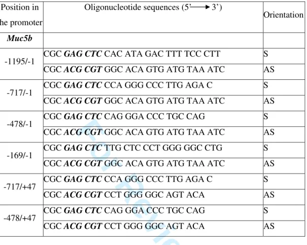
Table 1: Sequences of the pairs of oligonucleotides used in PCR to produce deletion mutants
covering murine Muc5b promoter. SacI (GAGCTC) and MluI (ACGCGT) sites (bold and italicized) are added at the end of the primers to direct subcloning into pGL3 basic vector. S: sense, AS: antisense.
3 4 5 6 7 8 9 10 11 12 13 14 15 16 17 18 19 20 21 22 23 24 25 26 27 28 29 30 31 32 33 34 35 36 37 38 39 40 41 42 43 44 45 46 47 48 49 50 51 52 53 54 55 56 57 58 59 60
For Review Only
Probe Putative binding site Sequence (5' 3')Muc5b T213 TTF-1 (-112/-109) CTGCCATGGCCCCTCCCCAAGAGCAAA T211 TTF-1 (-358/-355; -353/-350) CGGCAAACACAAGCCAAGGTTGTTGTC T240 Mutated T211 CGGCAAACAGTATCGTATGTTGTTGTC T212 TTF-1 (-709/-706; -700/-697) TCCAGGGCCCTTGAGACCCTTGGTCATTTC T241 Mutated T212 TCCAGGGCCAGTAAGACCAGTAGTCATTTC T238 TTF-1 (-325/-322) CCCCTGATCCTTGTAGTGTCTAGT T84 GATA (-1143/-1140) TCTCAGAAAGATAAGGATGGGGGC T242 TTF-1/GATA (-417/-414; -411/-408) TCACAGCCTTGTTGATACTTTGGGGAC T239 Mutated T242 TCACAGCCTTGTTCTTACTTTGGGGAC T254 GATA (-454/-449) GCCCATGACCATCTGGAGCATAAT consensus GATA
GATA CAC TTG ATA ACA GAA AGT GAT AAC TCT
Table 2: Sequences of the sense oligonucleotides used for EMSAs. Antisense
oligonucleotides were also synthesized and annealed to the sense oligonucleotides to produce double-stranded DNA. Positions of the putative binding sites are italicized and underlined. Mutated bases are bold and underlined.
3 4 5 6 7 8 9 10 11 12 13 14 15 16 17 18 19 20 21 22 23 24 25 26 27 28 29 30 31 32 33 34 35 36 37 38 39 40 41 42 43 44 45 46 47 48 49 50 51 52 53 54 55 56 57 58 59 60
For Review Only
-1210 A G T T T A C A G A C T T T T C A C A T A G A C T T T T C C C T T T C C T G T T C C A T C T C T A G G A A T T T A G T C GATA-4 -1150 T C A G A A A G A T A A G G A T G G G G G C A G A C A G G G A G G G G T T T T C G T G A T A C C A A A G T C A C C T G A -1090 A C A C T G T G C C A C T C C C T G A C A T G G C C A A C C C C T A G G A A C T T T C A G G G G T G G T C A G A T C T G -1030 G T G T C C A T T C C T G G T G G C T A T A A C A G A A G G A A T G T G A C T G T A T T C T G T A T G T A T C T A A C A -970 G G G A A G A G A A A C T T A G A C G T G T G C A C C C T G C T C T T T G T G G C A G T G T A G A A A C G G C G T T G G -910 T G G A T A T G T T C A T G G T T C A G G A A A T G G C T C C C C A G A A C C C T C A G G A A A C C C A A C C A A C A C -850 A A T T A A G A G A G C T G A G C C C C C G G G A C T C C C A T T T C A G G A T G G A C A G T G G G T C A G G A G A C T -790 C C C G G T T A T G C C C T T G C C C C A C T G T C C C G C A G T T T G G G G T T T A C A A G T T C C A G A G C T G A G TTF-1 TTF-1 -730 G C C T G A G G T A G G T C C A G G G C C C T T G A G A C C C T T G G T C A T T T C C T T T G T C A A G G C C C A G G G -670 T T A A T A A G C T G A C C C T G G G A A G A G G T G G A G T C C A A C T A G A G A A G C T C A T G G C C T G C C T T G -610 G C C A A A A C C A G T A C T G G G C C T C A T T T T C T C C A T G T G C G T G A G C T C T A C A G C A G A C A G G A G -550 C C C A G A G A C T C A T C C T T T C T C A G A T T C T G T G A G G G T T C C C C T T C T C C A C A G A C C C T C A G A GATA-4/-6 -490 A G C T A C A C A G A A C A G G G A C C C T G C C A G G C C C A T G A C C A T C T G G A G C A T A A T T T C C T G A G C TTF-1 GATA-4 -430 C C A C C C T C A C A G C C T T G T T G A T A C T T T G G G G A C C C A G T G C C T A G C A C C T G G G G T C A G C T T TTF-1 TTF-1 TTF-1 -370 C C C C G G C A A A C A C A A G C C A A G G T T G T T G T C C C T T T G C C C C T G A T C C T T G T A G T G T C T A G T Sp1 -310 C T C C T A G A G G A G C T G A C A C A G G G C C G C C T G G C C A A A T A C C C A C C C C A T A G G C C C A G G G C T -250 T C G A A T C C A G A G T A G T C A T C T C C C T A C C C T G T C C A C T C C C T C C C A G T T C T C A G A A G C C A C -190 A A G G G G T G G G A G C A C T C T G G T G G C A C A G T G A T G T A A A T C C T T C C T T C T C C A T G G C C T C T T TTF1 -130 C C T G C C A T G G C C C C T C C C C A A G A G C A A A C A C A C G T G G C T G T G C A G G G A G T A G A C C G T G C C -70 T T A C A T G G C C A G G G A A C T G G G C T C T G G G C C A A A G C C T T T G G C T A C A T A A G T C C A G G C C C C -10 C A G G G A G C A A G T G C C T T G T C T C A G T C C C T C C T G C C T G C C C T C T G T A C T G C C C C C A G G A T G Muc5b G G C A C A G T G A T G T A A A T C C T T C C T T C T C C A T G G C C T C T T C C T G C C A T G G C C C C T C C C C A A -110 MUC5B G G C A C A G A G C T G C A A A T C C T T C C T G A T C C A G G C C T C T C C C C T G C C A C A G C C C C T C C C C G A Muc5b G A G C A A A C A C A C G T G G C T G T G C A G G G A G T A G A C C G T G C C T T A C A T G G C C A G G G A A C T G G G -50 MUC5B G A G C A A A C A C A C G T G G C T G A G A G C G G G G A A G A G C A C G G T G C C C T G C G T G G C C T G G C C T G G Muc5b C T C T G G G C C A A A G C C T T T G G C T A C A T A A -22 MUC5B C T T G G G G C C A A G G C T C C C T G C T A C A T A A +1Figure 1 : Jonckheere et al.
A B 3 4 5 6 7 8 9 10 11 12 13 14 15 16 17 18 19 20 21 22 23 24 25 26 27 28 29 30 31 32 33 34 35 36 37 38 39 40 41 42 43 44 45 46 47 48 49 50 51 52 53 54 55 56 57 58 59 60