HAL Id: inserm-00188491
https://www.hal.inserm.fr/inserm-00188491
Submitted on 20 Nov 2007
HAL is a multi-disciplinary open access
archive for the deposit and dissemination of sci-entific research documents, whether they are pub-lished or not. The documents may come from teaching and research institutions in France or abroad, or from public or private research centers.
L’archive ouverte pluridisciplinaire HAL, est destinée au dépôt et à la diffusion de documents scientifiques de niveau recherche, publiés ou non, émanant des établissements d’enseignement et de recherche français ou étrangers, des laboratoires publics ou privés.
Modeling and interpretation of scalp-EEG and
depth-EEG signals during interictal activity.
Delphine Cosandier-Rimélé, Jean-Michel Badier, Patrick Chauvel, Fabrice
Wendling
To cite this version:
Delphine Cosandier-Rimélé, Jean-Michel Badier, Patrick Chauvel, Fabrice Wendling. Modeling and in-terpretation of scalp-EEG and depth-EEG signals during interictal activity.. Conference proceedings : .. Annual International Conference of the IEEE Engineering in Medicine and Biology Society. IEEE Engineering in Medicine and Biology Society. Annual Conference, Institute of Electrical and Elec-tronics Engineers (IEEE), 2007, 1, pp.4277-4280. �10.1109/IEMBS.2007.4353281�. �inserm-00188491�
Modeling and interpretation of scalp-EEG and depth-EEG signals
during interictal activity
D. Cosandier-Rimélé, J.M. Badier, P. Chauvel, F. Wendling
Abstract—In epileptic patients candidate to surgery, the
interpretation of electrophysiological signals recorded invasively (depth-EEG) and non-invasively (scalp-EEG) is a crucial issue to determine epileptogenic network and to define subsequent therapeutic strategy. This issue is addressed in this work through realistic modeling of both scalp-EEG and depth-EEG signals. The model allows for studying the influence, on signals, of source-related parameters leading to the generation of epileptic transient activity (interictal spikes). This parametric study is based on a variety of scenarios in which either spatial or temporal features of the sources of activity are modified. Statistical quantities measured on simulated signals allow for better understanding of the influence of source-related parameters on the information conveyed by these signals, collected from scalp or depth electrodes.
I. INTRODUCTION
E
lectroencephalography (EEG) and stereoelectroen-cephalography (SEEG or depth-EEG) are two electrophysio-logical investigation methods used during the presurgical evaluation of patients with drug-resistant partial epilepsies. They both provide real-time information about brain electrical activity: global information for EEG in which scalp electrodes are used to collect signals, and local information for SEEG in which intracerebral electrodes are used to directly record the activity from brain structures potentially involved in the generation of epileptic activity. At respective spatial scale, both methods provide convoluted signals (scalp-EEG and depth-EEG) that must be interpreted, prior to surgery, in order to i) localize the epileptogenic zone and ii) accurately determine its organization.However, the relationship between the properties of signals recorded by electrodes (surface and depth) and the spatio-temporal organization of the neuronal sources that produce the observed signals is highly complex, as it involves many nonlinear mechanisms, from subcellular to brain structure level. This issue, which relates to the broader
problem of physiological interpretation of biosignals, is addressed in this work through realistic modeling of both scalp-EEG and depth-EEG signals. The proposed model is based on a physiologically-relevant representation of the sources of brain electrical activity. The model was presented into details in a previous report [1] that showed excellent agreement between simulated signals and real depth-EEG signals (recorded along multi-contact intracerebral electrodes), not only in terms of morphological features but also in terms of amplitude gradients over space.
Manuscript received April 2, 2007. This work was supported by the French Ministry of Research (ACI Neurosciences Integratives et Computationelles 2002).
D. Cosandier-Rimélé and F. Wendling are with INSERM, U642, Rennes, F-35000, France; and with Université de Rennes 1, LTSI, Rennes, F-35000, France (e-mail: {delphine.cosandier-rimele,fabrice.wendling} @univ-rennes1.fr).
J.M. Badier and P. Chauvel are with INSERM, U751, Marseille, F-13000, France; and with Université de la Méditerranée, Marseille, F-F-13000, France (e-mail: jean-michel.badier @medecine.univ-mrs.fr).
In this paper, we go one step further. In the extended version of the model presented here, depth-EEGG and scalp-EEG signals can be generated simultaneously. Consequently, it allows for studying the influence, on signals, of source-related parameters leading to the generation of epileptic transient activity (interictal spikes). This parametric study is based on a variety of scenarios in which either spatial or temporal features of the sources of activity are modified. For each scenario, some statistical quantities measured on signals allow for better understanding of the influence of source-related parameters on the information conveyed by these signals, recorded from scalp or depth electrodes.
The model and the method used to simulate signals are briefly described in section II. Results obtained from the parametric study of the model are given in section III and discussed in section IV that also emphasizes the perspectives of this work.
II. SIMULATION MODEL FOR SCALP-EEG AND DEPTH-EEG
SIGNALS
At the cellular level, biochemical and electrical activity in each neuron leads to the generation of extracellular currents. Each neuron can be represented by an elementary current dipole which intensity is proportional to its synaptic activity. At the level of local neuronal assemblies (or populations), extracellular currents will sum up when cells are well organized in space. This is typically the case for the cerebral neocortex in which principal cells (pyramidal neurons) are organized in a parallel way (in « palissades ») [2]. Under this condition of spatial organization, local currents sum up and the activity of a population of neurons can be itself represented by an equivalent current dipole that is the vectorial sum of elementary dipoles. Moreover, extracellular currents spread inside a volume conductor (the head) which may be assumed to be essentially resistive. Consequently, there exists a difference of potential between any point of this volume and a reference electrode. The EEG signal corresponds to time-varying fluctuations of this difference
HAL author manuscript inserm-00188491, version 1
of potential. The properties of this signal (its amplitude in particular) will then depend on the properties of current sources (dipoles) as well as on those of the volume conductor (conductivity, homogeneity, isotropy) and on the features of electrodes (size, positioning).
These considerations directed our modeling approach. Indeed, the model we developed to simulate EEG signals (on scalp and depth electrodes) is based on the association of a biophysical representation of dipole sources and a biomathematical representation of neuronal populations (see figure 1). The former (dipole layer) accounts for the spatial properties of an extended source of activity. The latter (network of coupled neuronal populations) is used to simulate the temporal dynamics of the source.
a) b)
c)
Fig. 1. The source model starts from a realistic mesh of the neocortical surface obtained from MRI (a). Electrical contribution of the source is obtained from the association of a dipole layer model (b) and a macroscopic model of interconnected populations of neurons (c).
A. Spatial properties of the source model
We assumed that neocortex is composed of N populations of neurons which electrical contributions are represented by
N current dipoles (one dipole per population). Each dipole is
characterized by three parameters: its position, its orientation and its intensity.
In order to account for geometrical properties of the neocortex, we used a realistic mesh of the cortical surface, obtained from segmentation of anatomical MRI data (interface between grey and white matter). The complex geometry of the neocortex (folded surface) was described as accurately as possible using a high resolution mesh with an average triangle surface equal to 1 mm². A dipole is positioned at the barycenter of each triangle of the mesh. Its orientation is normal to the cortical surface (as pyramidal cells can be considered as being oriented perpendicularly to the neocortex surface).
In addition to position and orientation parameters, the dipole intensity was obtained by multiplying the corresponding triangle surface by a quantity related to the cortical current density (temporally invariant). The result is then weighted by a time-varying coefficient that reflects the temporal dynamics of the activity of the neuronal population associated to the triangle (see figure 1).
These temporal dynamics correspond to background activity for all neuronal populations over the cortical surface except in one or more regions considered as being extended sources of epileptic activity. In these “epileptic patches”, temporal dynamics defining the intensity of dipoles correspond to epileptiform activity. Both background and epileptiform activities are generated by a model of neuronal population described hereafter.
B. Temporal dynamics of the source model
The temporal dynamics of the activity produced by neuronal populations were simulated using a neurophysiologically-relevant model presented elsewhere [3]. In this model, a set of N interconnected populations of neurons is considered. Each population contains two subsets of neurons. The first subset is composed of the principal cells (i.e. pyramidal cells). The second subset is composed of the local interneurons (i.e. other non-pyramidal cells: stellate or basket cells, etc.). Pyramidal cells receive excitatory input from other pyramidal cells (collateral excitation) and inhibitory input from interneurons. These latter receive excitatory input from pyramidal cells only.
Each subset is characterized by two functions, often referred to as the “pulse-to-wave function” and the “wave-to-pulse function” in the literature. The former is a linear transfer function that changes presynaptic information (i.e. the average density of afferent action potentials) into postsynaptic information (i.e. an average excitatory or inhibitory postsynaptic membrane potential). The latter is a static nonlinear function that relates the average level of membrane potential of the neurons in the considered subset to an average pulse density of potentials fired by these neurons. This nonlinear function mimics the integrating action that takes place in the soma (threshold and saturation effects). The connection from population i to population j is characterized by two parameters, defining the degree of coupling and the time-delay associated to the connection.
Therefore, networks of coupled neuronal populations can be built from the model which produces vectorial signals (one signal per population). Besides, some model parameters (local balance between excitation and inhibition, degree of coupling between populations) can be modified to simulate epileptiform signals with dynamics comparable to those of local field potentials recorded during interictal or ictal periods [3].
C. From sources to signals collected on sensors
In order to compute EEG signals collected on scalp and intracerebral electrodes, we used a model for the head (volume conductor) in which we solved the forward problem (quasi-static case, Maxwell’s equations).
When the head is represented by a set of three concentric spheres (brain, skull, and scalp), the electrical potential created by a current dipole can be analytically computed at any point in the volume. We used this spherical head model to simulate depth-EEG signals recorded inside the brain [1].
For scalp-EEG, electrode positions strongly depend on the head shape. Therefore, we used a realistic head model with three homogenous compartments (brain, skull, and scalp). Boundaries between compartments were obtained from segmentation of 3D MRI data. With this type of volume conductor model, the forward problem can be solved numerically only. To proceed, we used the boundary element method (BEM) [4].
III. RESULTS
Model parameters may be divided into two categories.
Space-dependent parameters include the position and the spatial extent of patches that generate epileptic activity (interictal spikes). Time-related parameters include the degree of synchronization of local field potentials generated by neuronal populations within a given patch and between patches when two sources (at least) are considered.
In this paper, we chose to illustrate results in the case of a focal source (one patch only) of interictal epileptic activity. We studied the influence, on signals, of its spatial extent, its position and for its synchronization degree. In simulations, scalp EEG signals were computed over a 63 electrode standard montage, and depth-EEG signals were computed along a 15 contact intracerebral electrode (see figure 2).
Orthogonally-implanted intracerebral electrode (multiple lead: L 2 mm, Ø 0.8 mm, Δ 1.5 mm)
a) b)
Fig. 2. 63 scalp electrodes (10-10 system) (a) and 15-contact intracerebral electrode (b).
A. Influence of the spatial extent of the source
In this first example, the model was used to study the effect of the surface of the patch generating interictal spikes on scalp and depth EEG signals. In order to quantify this influence, we defined a criterion based on the amplitude of the epileptic spike observed in EEG signals with respect to background activity. This criterion is referred to as the “spike-to-background ratio” (SBR). It is analogous to the classical signal-to-noise ratio and is expressed in dB:
) / ( log 10 10 Pspike Pbkg SBR=
where Pspike and Pbkg denote the average power of the spike
and of the background activity, respectively.
Results are summarized in figure 3. The position (left occipital region) and the surface of the patch (from 1 to 50 cm²) are shown in figure 3-a. The two curves showing the evolution of the SBR (averaged over sensors for comparison) are displayed in figure 3-b. Three important remarks can be made on these results. First, in both cases, SBR increases as the patch surface grows, but the “parabolic” shape of the curve indicates that amplitude of the epileptic event observed in EEG signals does not increase anymore beyond a certain extent of the source. Second, the curve corresponding to depth-EEG signals is less smooth than the one corresponding to scalp-EEG signals, indicating that the depth-EEG method is more sensitive to the 3D geometry of the patch. Third, it is noteworthy that a 3dB SBR value is obtained for 3 cm² for depth-EEG and for 7 cm² for scalp EEG, indicating that the former is more sensitive due to the proximity to sources. For scalp-EEG, the order of magnitude of the patch surface
confirms that in previous studies [5] showing that epileptic spikes could be observed in EEG signals only when the cortical surface is higher than 6 cm². Finally, examples of simulated signals are given in figure 3-c for a patch surface equal to 18 cm². Spikes can be observed on scalp-EEG and depth-EEG signals. In the former, higher amplitude is observed in the parieto-occipital region.
Spatial extent of the source (cm²)
Av er ag e S B R (d B) Scalp-EEG 1 2 3 4 5 6 7 8 9 10 11 12 13 14 15 0 2 468 10 12 14 16 18 20 22 24 26 28 30 32 34 36 38 40 42 44 46 48 50 0 1 2 3 4 5 6 7 8 9 10 11 Scalp-EEG Depth-EEG Fp2 F4 F8 C4 T8 P4 P8 O2 Fz Cz Pz Fp1 F3 F7 C3 T7 P3 P7 O1 Depth-EEG 1 sec b) c) S = 1 cm² S = 50 cm² a)
Fig. 3. Influence of the spatial extent of the source of epileptic activity (see section III-A for details). To make visualization simpler, only 19 channels are represented in scalp-EEG signals (10-20 standard system).
B. Influence of the position of the source
In this second example, the objective was to study the sensitivity of scalp-EEG and depth-EEG techniques to variations of the source position. To perform this study, the position of the patch (7 cm²) was moved over an antero-posterior line going from the temporal pole up to the occipital region, as shown in figure 4. For each position defined in figure 4, the effect of the source position was quantified using a criterion based on the normalized mean square error (NMSE) between simulated signals produced for a given position of the source and those produced for the reference position:
(
)
(
)
2 1 2 1 ( ) ( ) ( ) = = − =∑
∑
T ref pos t T ref t V t V t NMSE V twhere Vref(t) and Vpos(t) denote the potential at time instant t,
respectively computed for the reference and for a given position, and T is the signal duration in samples.
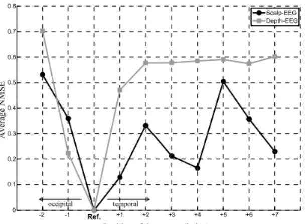
The obtained curves (average NMSE vs position) are shown in figure 5. They clearly show differences in the sensitivity between depth-EEG and scalp-EEG techniques to variations of the source position. In the former case, the NMSE dramatically increases as the source gets farther from the initial position. We interpreted this result as a strong dependence of depth-EEG technique on the 3D geometry of the source. Conversely, for scalp-EEG, the number of local
minima is greater than 1, indicating that the technique is less sensitive to position. This result also probably explains difficulties in localizing sources from non-invasive data.
0 -1 -2 +1 +2 +3 +4 +5 +6 +7 0 -1 -2 +1 +2 +3 +4 +5 +6 +7
Fig. 4. In order to study the influence of the position of the source of epileptic activity on scalp and depth-EEG signals, positions were arbitrarily defined over a temporo-occipital line. The reference position is indicated by the horizontal and vertical crossing lines.
Position of the source (index)
Ave rage NM SE -2 -1 0 +1 +2 +3 +4 +5 +6 +7 0 0.1 0.2 0.3 0.4 0.5 0.6 0.7 0.8 Scalp-EEG Depth-EEG Ref. temporal occipital
Fig. 5. Influence of the position of the source of epileptic activity on scalp-EEG and depth-scalp-EEG signals (see section III-B for details).
C. Influence of the synchronization degree of the source
In this third example, our goal was to get insight, from the model, on the influence of synchronization effects within the patch of epileptic activity (7 cm²). Therefore, the scenario consisted in mixing synchronous and non synchronous epileptic activities within the patch. To proceed, the same epileptiform activity was assigned to n randomly chosen neuronal populations inside the patch (composed of N populations) while independent activities were associated to the (N-n) remaining populations. This allowed us to define the percentage of synchronization as α = n/N x 100. The criterion used to study the effect of increasing α on scalp-EEG and depth-scalp-EEG signals was the same than that defined in section III-A, namely the spike-to-background ratio (SBR).
Results, displayed in figure 6, show, in both cases, increasing SBR values for increasing percentage of synchronization. However, epileptic spikes start to appear in scalp-EEG signals only when synchronization gets large: a 3dB SBR value is reached for α = 75%. Conversely, in depth-EEG signals, a 3dB SBR value is reached for α = 40%, indicating that epileptic spikes corresponding to low
synchronization level clearly appear in depth-EEG signals.
0 10 20 30 40 50 60 70 80 90 100 -1 0 1 2 3 4 5 6 7 8 Scalp-EEG Depth-EEG
Synchronization degree of the source (%)
Ave
ra
ge
S
BR (dB)
Fig. 6. Influence of the synchronization between local field potentials generated by neuronal populations within the patch of epileptic activity.
IV. DISCUSSION AND PERSPECTIVES
The interpretation of electrophysiological data recorded in epileptic patients during presurgical evaluation is a crucial but difficult issue. We think that progress can still be made in “decoding” the information conveyed by scalp-EEG and depth-EEG signals. This work is a first step into this direction. The model we propose not only allows for the simulation of signals collected on scalp and intracerebral electrodes but also allows for the analysis of simulated signals with respect to the configuration of epileptic sources. Although some issues like i) multiple patches of epileptic activity, ii) generation of ictal activity, and iii) signal-processing-based comparison between simulated and real data were not dealt with in this paper, we think that the approach has great potential value. In particular, we started to use the model in the evaluation of the numerous source localization techniques, as well as in the comparison between electroencephalographic data (EEG) and magnetoence-phalographic (MEG) data obtained for different probe configurations (gradiometer and/or magnetometer) [6].
REFERENCES
[1] Cosandier-Rimélé D., Badier J.M., Chauvel P. and Wendling F., A
physiologically plausible spatio-temporal model for EEG signals recorded with intracerebral electrodes in human partial epilepsy,
IEEE Trans. Biomed. Eng., vol. 54, pp 380-8, 2007.
[2] Lopes da Silva F., Electrical potentials, in Encyclopedia of the human
brain, vol. 2, V.S. Ramachandran, Ed. New York, pp 147-167, 2000.
[3] Wendling F., Bellanger J.J., Bartolomei F. and Chauvel P., Relevance
of nonlinear lumped-parameter models in the analysis of depth-EEG epileptic signals, Biol. Cybern., vol. 83, pp 367-378, 2000.
[4] Hämäläinen M.S. and Sarvas J., Realistic conductivity geometry
model of the human head for interpretation of neuromagnetic data,
IEEE Trans. Biomed. Eng., vol. 36, pp 165-171, 1989.
[5] Cooper R, Winter AL, Crow HJ, Walter WG, Comparison of subcortical, cortical and scalp activity using chronically indwelling electrodes in man, Electroenceph Clin Neurophysiol., vol. 18, pp 217-28, 1965.
[6] Badier JM, Cosandier-Rimélé D, Bénar CG, Schwartz D, Chauvel P, Wendling F, Realistic synthetic background neuronal activity for the
analysis of MEG probe configurations, EMBC 2007, Lyon, France.