Defined populations of inner ear progenitor cells show
limited and distinct capacities for differentiation into hair
cells, neurons, and glia
ARCHIES
MASSACHUSETTS INSTITUTE OF TEC
By
i2014
10r
Will McLean
SUBMITTED TO THE
HARVARD-MIT DIVISION OF HEALTH SCIENCE AND TECHNOLOGY
IN PARTIAL FULFILLMENT OF THE REQUIREMENTS FOR THE DEGREE OF
DOCTOR OF PHILOSOPHY IN HEALTH SCIENCE AND TECHNOLOGY
AT THE
MASSACHUSETTS INSTITUTE OF TECHNOLOGY
September 2014
Copyright 2014. Will J. McLean. All rights reserved.
The author hereby grants to MIT permission to reproduce and to distribute publicly paper and electronic copies of this thesis document in whole or in part in any medium now known or hereafter created.
Signature redacted
Signature of Author: re at
Department of Heal (ence and Technology Aug st 4, 2014
Certified by:
Signature redacted
Ruth Anne Eatock & Albert Edge
Professor Neurobiology, University of Chicago; Associate professor, Harv dical School Thesis Supervisors
Accepted by:
Signature redacted
mery N. Brown, MD, PhDDirector
Harvard-MIT Program in Health Sciences and Technology
Table of Contents
Abstract
Introduction
Background and significance
Research plan Methods Results Discussion Appendix Acknowledgments References 3 4 7 17 21 28 50 60 65 66
Defined populations of inner ear progenitor cells show limited and distinct
capacities for differentiation into hair cells, neurons, and glia
by
Will McLean
SUBMITTED TO THE HARVARD-MIT DIVISION OF HEALTH SCIENCE AND TECHNOLOGY ON SEPTEMBER 2,
2014 IN PARTIAL FULFILLMENT OF THE REQUIREMENTS FOR THE DEGREE OF DOCTOR OF PHILOSOPHY
IN HEALTH SCIENCE AND TECHNOLOGY
Abstract
Despite the fact that mammalian hair cells and neurons do not naturally regenerate in
vivo, progenitor cells exist within the postnatal inner ear that can be manipulated to generate
hair cells and neurons. This work reveals the differentiation capabilities of distinct inner ear progenitor populations and pinpoints cell types that can become cochlear hair cells, vestibular hair cells, neurons, and CNS glia.
We expanded and differentiated cochlear and vestibular progenitors from mice (postnatal days 1-3) and analyzed the cells for expression of mature properties by RT-PCR, immunostaining, and patch clamping. Whereas previous reports suggested that inner ear stem cells may be pluripotent and/or revert to a more neural stem cell fate, we find that cells from each organ type differentiated into cells with characteristics of the respective organ. Only cochlear-derived cells expressed the outer-hair-cell protein, prestin, while only vestibular-derived cells expressed the vestibular extracellular matrix marker, otopetrin. Since Atohi expression is consistently found in new hair cells, we used an Atohl-nGFP mouse line to identify
hair cell candidates. We find that cells expressing Atohl also expressed key transduction, hair bundle, and synaptic genes needed for proper function. Whole-cell patch clamp recordings showed that Atohl-nGFP+ cells derived from both cochlear and vestibular tissue had voltage-gated ion channels that were typical of postnatal hair cells. Only vestibular-derived
Atohi-nGFP+ cells, however, had Ih, a hyperpolarization-activated current typical of native vestibular hair cells but not native cochlear hair cells.
Lineage tracing studies with known supporting cell and glial cell markers showed that progenitor capacity of cochlear supporting cells positive for Lgr5 (Lgr5+ cells) was limited to differentiation into hair cell-like cells but not neuron-like cells. In contrast, glial cells positive for PLP (PLP1+ cells) from the auditory nerve differentiated into multiple cell types, with
properties of neurons, astrocytes, or mature oligodendrocytes but not hair cells. Thus, PLP+ progenitor cells within the auditory nerve are limited to neuronal or glial fates but have greater potency than Lgr5+ progenitors, which only formed hair cell-like cells.
In summary, this work identifies distinct populations of post-natal inner ear progenitors and delineates their capacity for differentiation and maturation.
Thesis Supervisors: Ruth Anne Eatock, Ph.D. & Albert Edge, Ph.D.
Title: Defined populations of progenitor cells show limited and distinct capacity for differentiation into hair cells, neurons, and glia
Introduction
In America alone, over 35 million people suffer from hearing loss (15% of the population) (Kochkin 2009). At some point in their lives, 35% of Americans suffer from vestibular/balance issues such as vertigo, dizziness, and disorientation, problems that are correlated with a risk of falling and severely reduced quality of life (Agrawal 2009). Over 90% of all hearing loss cases reflect hair cell or auditory nerve damage, which is typically caused by factors such as noise exposure, ototoxic drugs, viral/bacterial infections, and aging (NIDCD). Hair cell loss and noise exposure can also lead to auditory nerve degeneration (Spoendlin 1975, Kujawa et al. 2009). Cases of vestibular compromise are often treated with steroid injections or labyrinthectomies (inner ear removal), with the latter treatment always leading to hearing loss.
Although fish and birds regenerate hair cells lost to damage, mammals do not; thus, hair cell and neuronal death from disease or trauma leads to permanent loss of hearing or balance cues. Present remedies for hearing loss include hearing aids and cochlear implants, which have lirmitations. Both devices can only stimulate the residual sensory cells and/or neurons of the damaged inner ear, and because of electrical current spread, cochlear implants provide limited frequency resolution. Both remedies leave the patient with poor sound localization and musical perception, and improvements in speech perception vary widely (Eddington 2011). Thus, understanding biological mechanisms that could lead to hearing and balance restoration
would significantly advance therapy options. In recent years efforts have focused on
developing ways to regenerate the sensory cells and neurons of the inner ear. Despite the lack of hair cell regeneration, populations of progenitors have been demonstrated within the mammalian inner ear (Fig. 1). Prior work has suggested that inner ear stem cells are
pluripotent and can be isolated, cultured, and differentiated into hair cells and neural cells (Li et al. 2003, Oshima et al. 2007, Martinez-Monedero et al. 2008). Later work demonstrated that
Lgr5+ cells are the hair cell precursors within the cochlea (Chai et al. 2011, Shi et al. 2012). We hypothesize that, rather than a single pluripotent or multipotent stem cell type, there are separate pools of inner ear progenitors within the inner ear that give rise to vestibular hair cell-like cells, cochlear hair-cell cell-like cells, and neuron-cell-like cells.
Aim 1: Examine cochlear and vestibular stem cell's differentiation capabilities in vitro. Hair cells and supporting cells from each organ differ in their gene expression. For instance, outer hair cells of the cochlea express prestin, while supporting cells of the vestibular organs express otopetrin. Through the use of PCR and immunostaining, we can use these differences to determine which cells types are created from each organ's differentiated progenitors, and also determine if they express key genes that are needed for proper physiological function.
Hair cells commonly express large numbers of voltage-gated ion channels in particular families (e.g., inward and outwardly rectifying potassium (K) channels, sodium (Na) channels, and calcium (Ca) channels); the exact composition is specific to hair cell subtype and can be
used to recognize the cells' identity. With the patch clamp method, we recorded voltage-activated currents from differentiating progenitor cells and examined the data for similarities to whole-cell currents from native subpopulations of hair cells. With an Atohl-nGFP mouse line
(nuclear GFP), we record from Atohi-expressing cells to see if progenitors from each tissue give rise to particular hair cell subtypes based on accepted electrophysiological criteria.
Shi et al. (2012) determined that Lgr5+ cells in the mouse cochlea gave rise to hair cells, whereas Lgr5-negative (Lgr5-) cells did not. Since prior work suggested that inner ear cells when cultured as neurospheres (described in Methods) may revert back to a neural stem cell fate (Li et al. 2003, Oshima et al. 2007) or perhaps even a pluripotent state, we set out to
determine by lineage tracing if Lgr5+ cells were capable of becoming other cell types, such as neurons.
Previous work had also shown that a neural stem cell population arises from inner ear neurosphere culture (described in Methods) (Li et al. 2003, Oshima et al. 2007, Martinez-Monedero et al. 2008). We set out to identify which progenitor cells were responsible for forming neurons and glia. Work by Gomez-Casati et al. (2010) revealed that inner ear Schwann cells express PLP1. Using their PLP-Cre mouse line, we found that the Schwann cells of the inner ear generated both neurons and two types of CNS glia.
Background and Significance Stem cells and the inner ear
In the beginning of embryogenesis the fertilized egg goes through cell divisions to form the morula. These cells are totipotent because they can form all tissues of the organism and the placenta, which allow it to generate an intact animal. These cells then progress to the blastocyst stage, which contains the inner cell mass. The inner cell mass is the source of pluripotent embryonic stem cells, which can differentiate to form the specialized tissues of all three germ layers, but cannot generate a fertile adult mouse. A grown organism has adult stem cells which repair, replenish, and maintain normal turnover of regenerative organs, such as the complete regeneration that occurs every 2-5 days within the intestinal epithelium (Barker et al.
2007, Barker et al. 2008). These adult stem cells are called multipotent and are limited to
generating the cell types of a particular tissue.
The mammalian inner ear contains a population of stem cells that generate the highly specialized sensory and supporting cells of the vestibular and auditory sensory epithelia. The cochlea, however, lacks adult stem cells to allow regeneration and repair; adult mammalian vestibular epithelia may have some low-level regenerative capability (Burns et al. 2012) but it is inadequate to prevent significant hair cell loss with aging (Rauch et al 2001). We are interested in what gives various tissues different regenerative capabilities, and to what extent a specific progenitor cell can give rise to and repair diverse tissue types. The theory on stem cell potency has often been described by Conrad Waddington's "epigenetic landscape" theory (Fig. Bi) (Waddington 1957). The theory illustrates the successive restriction of initially totipotent cells to pluripotency, with further commitment to a certain lineage as the stem cell moves down the
path of differentiation. The inner ear provides a valuable tool to study this progression because isolated stem cells from the cochlea, vestibule, and ganglia are thought to be able to generate both hair cells and neurons (Li et al. 2003, Oshima et al. 2007, Martinez-Monedero et al. 2008) and to form all three germ layers (Li et al. 2003). In this research project we use a combination of molecular signaling manipulations, patch clamping, and detection of gene expression to analyze the ability of inner ear progenitor cells to differentiate into the specialized cellular fates of the various inner ear organs.
The inner ear has two parts, the cochlea (hearing) and vestibule (balance), both of which have hair cells, so named for their apical microvillar bundles ("hair bundles"). The cochlea and vestibular organs each have two different hair cell subtypes. In the cochlea, the signals we perceive as sound are transmitted by inner hair cells (IHCs), and the signal is amplified and tuned by outer hair cells (OHCs). The vestibular organs have type I and type II hair cells, which have different shapes, synaptic contacts, and microvillar bundle characteristics.
Electrophysiology experiments with the patch clamp method enable us to distinguish between these four hair cell subtypes based on their ion channels and transduction properties, and also how these properties are acquired in normal development. However, the developmental signals determining if an immature hair cell acquires a vestibular or cochlear fate remain elusive, and even less is known about how these subtypes emerge.
Hair cell differentiation and development depends on Atohi, a basic helix-loop-helix
(bHLH) transcription factor that is regulated through the Notch signaling pathway (Bermingham
et al. 1999; Zheng & Gao 2000; Woods et al. 2004, Izumikawa et al. 2005). Previous work has used the nuclear expression of the Atohl transcription factor to identify hair cells that have
differentiated from stem cells. In an analogous fashion, Atohl is necessary for intestinal progenitors to develop into epithelial secretory cells such as goblet, Paneth, and
enteroendocrine cells (Yang et al. 2001). In either case, when a cell expresses Atohi, it also expresses the DII1/Jagged2 ligand that binds to the Notch receptors of its neighboring cells, suppressing their Atohi expression. The resulting "lateral inhibition" mechanism specifies cells that express Atohl as hair cells (ear) or secretory cells (intestine), while the neighboring cells remain supporting cells (ear) or enterocytes (intestine) (Yang et al. 2001). This phenomenon is necessary to establish normal morphology in both of these organs.
Stem cell commitment
Totipotent
Pluripatent
-Lineage 3
committed
Figure B1: Waddington's "epigenetic landscape" theory suggests that stem cells behave like a marble rolling down a hill. As the marble proceeds down the hill it must commit to various troughs, with each trough representing another degree of commitment (i.e. decreased
potency). The further down the hill the marble rolls, the more difficult it is to change paths (represented by the blue arrows). This corresponds to the increased difficulty for a cell to change fates as it becomes more committed (Image modified by McLean; original from Waddington 1957).
Potential for therapy
Efforts to advance therapy for hearing loss and poor vestibular function have focused on developing ways to regenerate the hair cells of the inner ear so that the natural biological mechanisms of hearing can be restored. The hair bundles are moved by sound, in the case of the auditory organ, and head motions, in the case of vestibular organs. Motion of the bundle evokes an electrical signal in the hair cell, which is relayed by eighth-nerve fibers to the brain. Although hair cells regenerate after damage in fish and birds, hair cells do not regenerate in
mammals, and hair cell death from disease or trauma leads to permanent loss of hearing or balance cues.
Despite the fact that mammalian hair cells do not naturally regenerate in vivo,
populations of stem cells exist within the postnatal inner ear of mice (Fig. 1). Previous work has suggested that these inner ear stem cells may be pluripotent (Li et al. 2003), based on several lines of experimentation. Progenitors were isolated from the utricle and cultured in the
presence of the growth factors IGF and EGF to form neurospheres. The neurospheres were then differentiated, at which point cells with the properties of hair cells, neurons and glia were obtained. In addition, co-culturing neurospheres with a mesodermal cell line allowed them to differentiate into cells that stained positively for known muscle proteins. Furthermore, co-culturing the neurospheres with a totipotent cell line allowed the colonies to form cells that stained positively for myogenic markers. When lineage-tagged neurospheres were injected into stage 4 chicken embryos, which allows the cells to be distributed during gastrulation and exposes the cells to various inductive environments, the neurosphere cells were incorporated into all three germ layers.
Inner ear
e
Coch
Vestibular
Vestibular supporting
cells and hair cells Neuron
and glia
Modiolar cell types
C
s
lear cell types
b
ochlear supporting cells and hair cells
Figure 1: Analysis of inner ear progenitor cells
The potency and differentiation characteristics of 3 inner ear tissues are analyzed. Hair cell-like cells that are produced from cochlear and vestibular tissue are analyzed and compared. This work also analyzes the potency of Lgr5+ cells within the cochlea (green) and PLP+ glial cells within the auditory nerve (red).
Previous studies have shown that isolated cochlear, vestibular, and modiolar
progenitors can be cultured long term and survive through seven passages. Cochlear and
vestibular spheres gave rise to hair cell-like cells, supporting cells, and neurons in vitro, as
indicated by immunostaining for typical hair cell proteins such as Atohl and Myosin VIIA and neuronal genes such as Tuj and GFAP (Li et al. 2003, Oshima et al., 2007, Martinez-Monedero et al., 2008, Martinez-Monedero 2007). From the voltage dependence of whole-cell currents recorded from the generated hair cell-like cells, it was suggested that the cells most closely resembled embryonic hair cells (Oshima et al. 2007). Similarly, generated neurons were found to have similar, yet smaller, currents than neonatal neurons (Martinez-Monedero et al., 2008). Modiolar spheres were shown to produce a much higher yield of neural cell types when compared to the sensory organs, and although hair cell-like cells were reported, they occurred at an extremely low rate (<.1% of all cells)(Oshima et al 2007).
Thus, substantial progress has been made in discovering the underlying mechanisms of hair cell and neuronal development and in establishing reliable methods for their generation (though typically at low yields). These studies attempted to determine whether a latent capacity for regeneration was present in the inner ear and much of the work was done with unknown and possibly mixed types of progenitors. To identify regenerated hair cells, previous work used markers that are specific to hair cells within the native inner ear; however, many of these markers are expressed elsewhere in the body and are therefore not necessarily indicative of hair cells if newly created cells are in fact derived from pluripotent stem cells. Oshima et al.
(2007) described prestin expression via RT-PCR in differentiated cochlear spheres, but
otherwise previous work did not address whether specific subtypes of hair cells could be
developed, which can be detected by looking for specific gene expression (e.g. prestin for OHCs,
cells may exist within the mouse inner ear but did not address whether they comprise different populations or whether they differ in their potency and differentiation capabilities.
More specifically, previous studies used immunostaining against the proteins Atohi, espin, parvalbumin3, and myosin VIIA to identify hair cells; against Tuj to identify neurons; and against GFAP to identify glia. However, Atohl and Myosin VIIA are expressed in all hair cell subtypes and GFAP is expressed in some peripheral and CNS glia, as well as supporting cells (Rio et al. 2002). Furthermore, Atohl is known to be expressed in the Merkel cells of the skin, secretory cells of the gut, granular neurons, spinal cord interneurons, and, transiently, in
supporting cells of the ear (Lewis et al. 2012). Similarly, Myosin VIIA is known to be expressed in the secretory cells of the gut, testis, lung, kidneys, and retina. Thus, reliance on these proteins to identify progenitor-derived hair cells can be questioned because other cell types would be expected to express one or both of these proteins. Similarly, GFAP expression alone doesn't determine if cells are glia or supporting cells. This issue is further complicated if, as suggested
by Li et al. (2003), inner ear stem cells are pluripotent. If this is the case, it is possible that the
cells differentiated from inner ear stem cells could be any of the previously mentioned cell types. Therefore, using the current protein expression standards could lead to
misidentifications.
Thus, there is a need for a more extensive analysis of progenitor-derived hair cell-like cells and neural like cells. First, proper identification and understanding of what drives stem cells to a particular hair cell fate would move the field one step closer to being able to target specific hearing or balance ailments that are rooted in loss or damage of a certain hair cell subtype. In addition, a more thorough analysis is needed to determine if the newly created cells
possess all the machinery to function as legitimate hair cells, a step that would be necessary for proper hearing or balance restoration in future therapies.
Much is known about specific hair cell subtype characteristics, in terms of how function and gene expression progress through aging. Much of what is known about this particular area of hair cell function is owed to patch clamping experiments, in which a fine glass electrode is used to measure current through one or many ion channels of a cell. More specifically, one can measure a single transduction channel's response while deflecting the hair bundle with a probe (Beurg et al. 2006), or the current through thousands of channels activated by voltage.
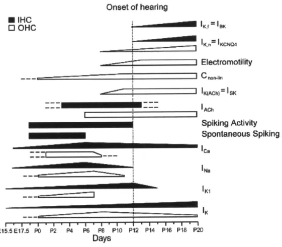
Furthermore, previous work had identified specific channel types that contribute to behavior of hair cell subtypes. For example, type I vestibular cells express the Nav1.5 sodium channel, and this expression changes with development (Wooltorton et al. 2007). Similarly, previous work has shown how each hair cell subtype follows a specific program of ion channel acquisition and expression, with the program and end product being unique to each subtype (RUsch et al. 1998, Marcotti et al 2003, Geleoc et al. 2004, Hurley et al. 2006). Electrophysiology can provide information that is not available from immunohistochemistry. In all, the patch clamp method provides a tool that can greatly aid in the characterization of hair cell-like cells. Figure B2 summarizes the dynamic physiological changes that cochlear hair cells undergo as they develop.
I __________________________________________________
I
* IHC 0 OHC I~ I I 1 4 1 1 E15.5E17.5 PO P2 P4 P6 PS P10 P12 P14 P16 P18 P20 DaysFigure B2: Extensive research into native hair cell development has revealed the time course for how hair cells acquire physiological characteristics.
Inner hair cells are indicated by white fill, while outer hair cells are indicated by black fill. Ramps show the increase or decrease of a current or other functional characteristic over time. Dashed lines indicate uncertainty or projections of behavior based on in vitro results. (Figure from: Goodyear et al. 2006)
Recent experiments in hair cell and glial gene expression have created new ways to identify specific cell types. For instance, the motor protein prestin, which is only found in OHCs, has a large rise in expression as the animal nears hearing (Belyantseva et al. 2000). Some other examples of protein markers that help distinguish hair cell subtypes include calretinin
(expressed in immature hair cells, mature inner hair cells (IHCs) and type 11 hair cells), VGLUT3
Onset of hearing ---* IKfIBI0( Z Electromotility lgAChf1 SK Spiking Activity Spontaneous Spiking le, 'Na IKI 11
(expressed in IHCs and vestibular hair cells), and alpha3 tubulin (found in kinocilia, which are present on immature hair cells and mature vestibular hair cells). In addition, the proteins 04 and 01 are selective markers for immature non-myelinating oligodendrocytes and for mature myelinating oligodendrocytes, respectively (Sommer and Schachner 1980, Sommer and Schachner 1982), and Aldhill is a specific astrocyte marker (Cahoy et al. 2008). These glial markers could help determine whether inner ear progenitors are capable of forming glia from outside their lineage. Furthermore, immunostaining and other molecular methods such as RT-PCR can help identify functional hair cells. Looking for expression of functional proteins such as
VGLUT3 in synaptic vesicles, Cav1.3 channels in the presynaptic membrane, Ctbp2 (in the ribbon
synapse), AChr9 (part of the acetylcholine receptor found on OHCs), and many others, can indicate whether the new hair cells have the potential to respond to stimuli in a similar manner to native hair cells. As a further example, we can use the PCR method to determine whether stem cell-derived hair cells express components of the transduction channel by taking
advantage of the recent identification of TMC1 and TMC2 as channel proteins (Kawashima et al. 2011, Pan et al. 2013). In order for newly created hair cells to function properly in stem cell therapies, these critical proteins must be generated.
By combining the techniques of molecular biology, biophysics, and lineage tracing, we
are able to examine the differentiation of inner ear progenitors. Building on previous work that showed how to generate hair cell-like cells and neural-like cells in vitro, our studies analyze the process of acquiring a certain cellular fate and whether these cells possess the cellular
machinery needed to be fully functional. The potency and differentiation characteristics of three inner ear tissues are analyzed: cochlea, vestibule, and the modiolus (auditory ganglion).
Hair cell-like cells from cochlear and vestibular tissue are compared, and the potencies of Lgr5+ cells within the cochlea and PLP+ glial cells within the spiral ganglion are investigated.
Research Plan
To test whether different populations of progenitors are responsible for creating vestibular hair cell-like cells, cochlear hair cell-like cells, and neural cell types, we separated and cultured inner ear tissues in parallel and analyzed their gene expression and function (Fig. P1).
Methods
Trypsin
postnatal mice isolate tissues
EGF IGF FGF Hps
Mechanical trituration to expand stem cells and kill native cells (x3)
Oshima et al. 2007
Figure P1: Our stem cell isolation protocol.
The inner ear organs are dissected from postnatal mice and cultured in parallel for the remainder of the experiments. The cells are cultured with growth factors that are known to expand stem cells. Spheres are triturated every few days to expand stem cells and kill native cells. Growth factors are then removed, which allows the stem cells to differentiate. After various time points we can analyze the cells that are present. Methods derived from Oshima et al. (2007).
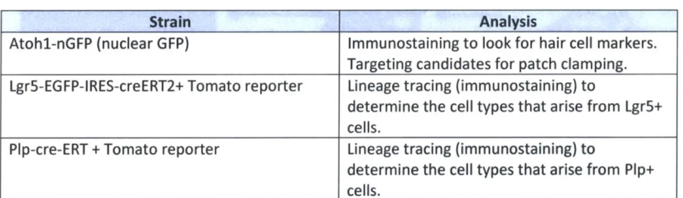
To test the differentiation capabilities of specific cellular populations, we took tissues from mouse lines that enable lineage tracing. Lgr5+ supporting cells were tested by crossing Lgr5-EGFP-IRES-creERT2 mice with td-tomato reporter mice and looking for tomato expression (red cells, Fig.
1) in hair cell-like or neuron-like cells following our usual neurosphere preparation and
differentiation. We also crossed Plp-cre/ERT2 and td-tomato reporter mice to determine if glial cells of the spiral ganglia are capable of producing hair cells or if they are limited to producing neurons or glia. The various strain crosses and the experiments we did with them are shown in Table P1.
Table P1: Mouse strains
Strain Analysis
Atohl-nGFP (nuclear GFP) Immunostaining to look for hair cell markers.
Targeting candidates for patch clamping. Lgr5-EGFP-IRES-creERT2+ Tomato reporter Lineage tracing (immunostaining) to
determine the cell types that arise from Lgr5+ cells.
Pip-cre-ERT + Tomato reporter Lineage tracing (immunostaining) to
determine the cell types that arise from Plp+
cells.
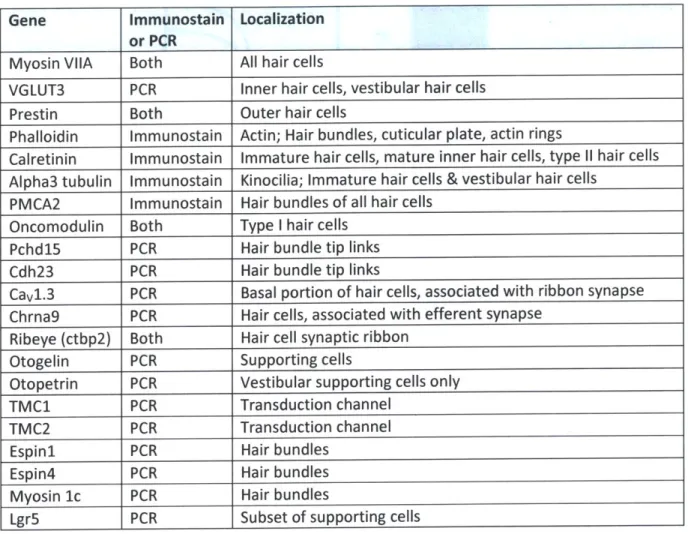
We analyzed the gene expression in the differentiated cells by RT-PCR and
immunostaining. In Table P2 we list the genes we examined, the method of detection, and the cell type and location of the expression.
Table P2: Genes analyzed
Gene Immunostain Localization
or PCR
Myosin VIIA Both All hair cells
VGLUT3 PCR Inner hair cells, vestibular hair cells
Prestin Both Outer hair cells
Phalloidin Immunostain Actin; Hair bundles, cuticular plate, actin rings
Calretinin Immunostain Immature hair cells, mature inner hair cells, type 11 hair cells
Alpha3 tubulin Immunostain Kinocilia; Immature hair cells & vestibular hair cells PMCA2 Immunostain Hair bundles of all hair cells
Oncomodulin Both Type I hair cells
Pchd15 PCR Hair bundle tip links
Cdh23 PCR Hair bundle tip links
Cavl.3 PCR Basal portion of hair cells, associated with ribbon synapse Chrna9 PCR Hair cells, associated with efferent synapse
Ribeye (ctbp2) Both Hair cell synaptic ribbon
Otogelin PCR Supporting cells
Otopetrin PCR Vestibular supporting cells only
TMC1 PCR Transduction channel
TMC2 PCR Transduction channel
Espini PCR Hair bundles
Espin4 PCR Hair bundles
Myosin 1c PCR Hair bundles
Lgr5 PCR Subset of supporting cells
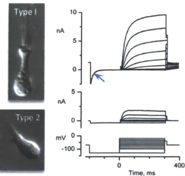
In addition, we used the patch clamp technique to identify currents that determine if the hair cells are functional and what subtype they may be. Examples of native hair cell currents from different hair cell-subtypes are shown in Figure P2. We selected hair cell candidates to patch by their Atohl-nGFP expression. Examples of electrophysiological characteristics looked for are listed in Table P3.
10-nA 5 nA 0 0 400 Time. ms
Figure P2: Examples of currents recorded from type I and type 11 vestibular hair cells.
A distinguishing characteristic is that type I hair cells have larger currents than type 11 hair cells. Type I hair cells also have the negatively-activating current GK,L (blue arrow). Image is courtesy of K.M. Hurley & R.A. Eatock, unpublished.
Table P3: Patch clamp experiments
Experiment Characteristic Identification
Differences in K+ currents Size and kinetics can help determine
maturation state and subtype - e.g., large
A-current in extrastriolar type 11 hair cells
K' current blockers (ex. TEA, IBTX) Can determine K+ current types present, which can indicate cell type and maturity Use large hyperpolarizing steps (from [I60mV Presence of Ih may indicate vestibular hair cell to l120mV) to look for slowly activating Ih type.
Current clamp recordings (to look at voltage Resting potential, which is well characterized
behavior) in developing hair cells and shifts with age.
Voltage responses to current steps can help identify cell type and maturation - e.g., young
By combining these genetic, molecular, and electrophysiology techniques, this body of
work should help determine the differentiation capabilities of cochlear and vestibular stem cells, and their functional state.
Methods
Isolation of stem cells from the inner ear
To gather progenitors for proliferation and hair cell-like cell generation, we extracted progenitor cells from the neonatal mouse inner ear. All animal studies were conducted under an approved institutional protocol according to National Institutes of Health guidelines. For each experiment, the cochleae and the vestibular organs of 6-8 CD1 or Atohl-nGFP pups (age pl-p4) were dissected in HBSS and kept separate from each other for the remainder of the protocol. This separation allows us to analyze each tissue's developmental properties separately. In the case of the cochleae, the organ of Corti (OC) was separated from the stria vascularis and the modiolus. The OC and vestibular tissues were transferred to the dissociating medium TrypLE (Life
Technologies) for 11-13 min at 37'C and then manually dissociated with a pipette. The triturated cells were then passed through a 70-ptm cell strainer to remove tissue and bone debris. Single cells were cultured in a 1:1 mixture of DMEM/high-glucose medium and F12, supplemented with N2,
B27 (Invitrogen), EGF (20 ng/mL; Chemicon), bFGF (10 ng/mL; Chemicon), IGF-1 (50 ng/mL;
Chemicon), and heparin sulfate (50 ng/mL; Sigma). Single cells were maintained in ultralow-cluster plates (Costar) for several days in culture to obtain floating spheres. The spheres were then
After passage, the cells were placed in fresh culture medium supplemented as described above. For each experiment, the cells were passaged three times to eliminate any hair cells or neurons that may have been carried over during the trituration process.
Mouse strains for lineage tracing
In addition to isolating each organ, cell type potency was analyzed for Lgr5+ cells of the cochlea and PLP+ cells of the ganglia. Male Lgr5-EGFP-IRES-creERT2
(http://jaxmice.jax.org/strain/008875.html) and male PLP-cre-ERT mice
(http://jaxmice.jax.org/strain/005975.html) were crossed with female td-tomato reporter mice (http://jaxmice.jax.org/strain/007909.html) in order to lineage trace the cells that resulted from each cell type. Mother mice were injected with 600 p tamoxifen (50mg/ml) on the day of birth and 1st day post birth (dO and dl). Pups were dissected at p3 and were identified as either
genetically positive or negative based on their fluorescence. Spheres were generated from the OC of Lgr5+ mice and the modiolar tissue of PLP+ mice.
Differentiation and treatment of spheres
To generate differentiated cells for future assays, 3rd generation spheres were plated in
4-well plates (Greiner) on round 10 mm glass coverslips coated with poly-L-lysine (Cultrex).
Attachment took place overnight in DMEM-high glucose/F12 (mixed 1:1, GIBCO) with N2 and B27 (Invitrogen). Spheres were differentiated in these conditions for 7-70 days, with fresh culture medium being applied every 2-3 days to maintain optimal culture conditions. Cells were then harvested for PCR analyses, immunostained for further gene expression analyses, or used for patch
clamping experiments. Control spheres
To ensure that the results obtained were not significantly affected by hair cells that were carried over during the trituration process that survived throughout culture, I counted the number of hair cells that were still present after the 3 rd passage (see appendix). For immunostaining
analyses, 3rd generation spheres were seeded in the same experimental culturing conditions as were used for experiments, but instead were only allowed to differentiate for 3 hours. This seeding time allows the spheres to adhere to the culturing surface, but should be insufficient to allow the cells to differentiate. All controls were analyzed for expression of Atohl-nGFP and myosin VIIA. Controls were viewed via confocal microscopy (Leica) over the entire seeding area. For PCR analyses, 3rd generation spheres were collected from floating cultures and Myosin VIIA expression was analyzed (n=3 cultures).
Electrophysiology
We performed patch-clamping experiments to test for functionality and to phenotypically assess between the cellular subtypes that are developed from the cochlea and vestibular tissue. Prior to recording, the cell culture solution was replaced with L-15 (Leibowitz 15) medium (supplemented with 10 mM HEPES, pH 7.3, ~320 mmol/kg osmolarity). In all experiments, we recorded from single cells using the whole-cell patch method at room temperature (22-250C). The
pipette solution contained (in mM): 135 KCI, 3.5 MgCl2, 5 Na2ATP, 10 HEPES, 10 EGTA, 0.1
Na-cAMP, 0.1 Li-GTP. The solution's pH was adjusted to 7.4 by adding 15 mM KOH. Osmolality of the solution was 280 5 mmol/kg. Recording pipettes were pulled from borosilicate glass and heat
polished to a resistance of 3-9 MO.
Currents were recorded with a patch-clamp amplifier (Axopatch 200B or the Multiclamp
700; Molecular Devices, Sunnyvale, CA). Series resistance (Rs) was estimated and compensated 20-90% with the intrinsic circuitry of the amplifier. Currents were filtered with an eight-pole low-pass
Bessel filter with a corner frequency of 2 kHz and sampled at more than twice the filter frequency with a Digidata 1440 board (Molecular Devices), controlled by Clampex software (version 10.1; Molecular Devices).
Analyses and fits were done with Origin software (version 9; OriginLab Software,
Northampton, MA), which uses a Levenberg-Marquardt least-squares fitting algorithm. All cells considered for analysis had a <100MO patch resistance. To obtain the activation curve for a current, we stepped to a series of test potentials and measured the "tail current" at -40 mV, that is, the current immediately upon stepping to -40 mV after each iterated test step. Plotting the tail currents against the test-step voltage produced sigmoidal "activation curves", which show how the conductance producing the current depends on voltage. Such curves could be fit with a Boltzmann function (Equation 1), where 1(V) is current at voltage V, Imin and Imax are minimum and maximum currents, V1/2 is voltage corresponding to half-maximal activation, and S is the voltage
corresponding to an e-fold increase in 1(V).
Eq. 1
I(V) = " '" +I I,
The time course of current activation was approximated by the fit of a monoexponential decay function (Origin 9.0).
Immunohistochemistry
Immunohistochemistry was performed In order to visualize and quantify the differentiated cells that express mature/functional genes. To accomplish this, differentiated spheres were fixed at room temperature in 4% paraformaldehyde/PBS for 15-20 min and then washed in PBS.
Permeabilization of the cellular membrane and blocking was performed with blocking solution
(0.3% Triton X-100, 15% heat inactivated goat or donkey serum in PBS) for 1 h. Diluted primary
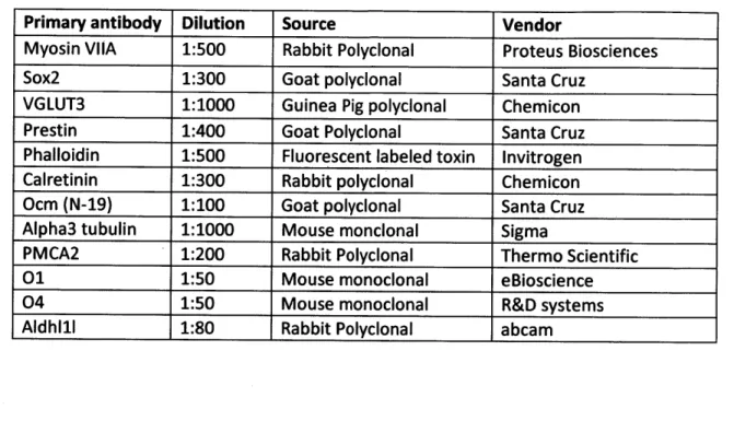
antibody (0.1% Triton X-100, and 10% heat inactivated goat or donkey serum in PBS) was applied overnight at 4' C. Primary antibody dilutions are listed in Table 1. Secondary antibodies (Alexafluor
488, 568, and 647-conjugated; Invitrogen) were used at 1:500 dilution for detection of primary
antibodies. Nuclei were visualized with 4,6-diamidino-2-phenylindole (Vector Laboratories).
Staining was visualized with confocal microscopy (TCD, Leica). All cellular counts of gene expression were performed manually.
Table 1. Antibodies
Primary antibody Dilution Source Vendor
Myosin VIIA 1:500 Rabbit Polyclonal Proteus Biosciences
Sox2 1:300 Goat polyclonal Santa Cruz
VGLUT3 1:1000 Guinea Pig polyclonal Chemicon
Prestin 1:400 Goat Polyclonal Santa Cruz
Phalloidin 1:500 Fluorescent labeled toxin Invitrogen
Calretinin 1:300 Rabbit polyclonal Chemicon
Ocm (N-19) 1:100 Goat polyclonal Santa Cruz
Alpha3 tubulin 1:1000 Mouse monclonal Sigma
PMCA2 1:200 Rabbit Polyclonal Thermo Scientific
01 1:50 Mouse monoclonal eBioscience
04 1:50 Mouse monoclonal R&D systems
Aldhl1l 1:80 Rabbit Polyclonal abcam
RT-PCR
We used RT-PCR to quickly and sensitively analyze gene expression in the various tissues. First, we extracted RNA from the inner ears of CD1 mice aged E13.5, P3, P16, or from differentiated spheres, using the RNeasy Maxi Kit (Qiagen) according to the manufacturer's instructions. RNA was denatured at 65'C for 5 min. For reverse transcription, ImProm 11
(Promega) was used with random hexamers. The reverse transcription conditions were 25*C for 5 min followed by 420 C for 60 min. The reaction was terminated at 700C for 15 min. To the
resulting cDNA we added primers for various inner ear proteins, which are listed below. The amplified products were then separated on a 2% agarose gel, stained with ethidium bromide, and visualized under a UV transilluminator.
Primers Cdhl5 CTGGGGCCAGTATGACGATG TGTACCGTTTCGACTCTCTTCA Cdh23 ACAGTCAATGCCACGGATCAA GGAGAGTGCTCGTAGATGTTAGT VGLUT3 CTCTTGTCAGCTGTCCCACA CAGTCCTG1TFTTCCCCAGA Prestin ACAGTGTGGATGTCGTTGGA CAGGTTGACGATCACAATGG Espini CCACAGGCTACCTCTCTGC AGCAGCCACTTCACCACATC Espin4 ATGGGCAATAGCTTGGAACAC GCGAGGACATACCCGAGTC Oncomodulin CTGACCACTGTTGGGGAGTT GCTCTGGAACCTCTGTAGG Otopetrin CTGCTCTGGATGCTGTGGTA CAAAACGATGGTGATGTTGC Gapdh AACACAGTCCATGCC TCCACCACCCTGTTGCTG Otogelin CCATGCAGGGTGTGTATGAG GGCATTGCAGTCTTCTGTCA CTBP2 A domain (Ribeye) TGCTGGACCTTTGCAGACAC CACCGTGTACGAGGTGGAGT Lgr5 CCTACTCGAAGACTTACCCAGT GCATTGGGGTGAATGATAGCA CaV1.3 AAGGGCTACCTGGACTGGAT CCACACACCACAAAGCAATC Myolc TCTTCATCCGATTTCCCAAG CCACACCATGTTCTTCATGC Alpha9(Chrna9) GGAACCAGGTGGACATATTCAAT GCAGCCGTAGGAGATGACG Tmcl CTGTCCCACCCTGTTTGACT TCACGAAACATGCTCTGAGG Tmc2 TGGCTACAGCTTGATGATCG GTTTCAAACAGAGGGGGACA
Results
In order to identify gene expression that may indicate the level of maturity, function, and which hair cell subtypes are being created from a given tissue source's progenitors, we performed immunostaining experiments, RT-PCR, and patch clamping assays. We first isolated progenitors from the cochlea or vestibule, allowed them to proliferate and form spheres, then differentiated the spheres for 14-70 days in order to allow significant maturation. We found that differentiated spheres contained cells that stained positively for hair cell markers, such as Atohi (Bermingham et al. 1999; Zheng & Gao 2000; Woods et al. 2004, Izumikawa et al. 2005), myosin VIIA (Hasson et al. 1995), and parvalbumin, a Ca 2-binding protein found in vestibular
type I hair cells (Dememes et al. 1993) and cochlear inner hair cells (Pack et al. 1995) (Table 2). In the results below, we focus on the other markers expressed by cells expressing either Atohl, myosin VIIA, or parvalbumin, which we refer to as "hair-cell like cells". We found that 51% of Atohl-nGFP+ cells (<1% of total cells in culture) double-stained for another hair cell marker such as myosin VIIA, which is similar to the 44% yield (<1% of total cells in culture) found by Oshima et al. (2007).
Hair Bundle proteins: 67% (285/423) of myosin VIIA-positive cells derived from vestibular progenitors had phalloidin-positive stereocilia protrusions; phalloidin stains filamentous actin, which is the major constituent protein in hair bundles (Fig. 2A; Table 2). Similarly, 81% (690/850) of myosin VIIA positive cells derived from cochlear progenitors had phalloidin-positive bundle-like protrusions (Fig. 2F; Table 2). Staining for a3 tubulin, which is expressed in the single kinocilium found on vestibular hair cells and immature cochlear hair
cells (Ogata et al. 1995), was much more rare than staining for actin: a3 tubulin antibody labeled no cochlear bundle-like structures (0/312), and 4% (9/289) of vestibular bundle-like structures showed evidence of a3 tubulin expression (Fig. 2B; Table 2). This suggests that bundle formation is imperfect, as is also suggested by the structures themselves.
Labeling for PMCA2 (Fig. 2C; Table 2), a Ca 2
+-ATPase that is enriched in postnatal hair
cell bundles (Dumont et al. 2001, Hill et al. 2006, Chen et al. 2012), was more frequent: 50% (27/54) of cochlear bundle-like structures and 73% (58/79) of vestibular bundle-like structures were PMCA2+ (Fig. 2H; Table 2). The PMCA2 immunoreactivity was strongest in the basal
portion of the stereocilia (Fig. 2C, 2H), PMCA2 antibody also labeled the basolateral cell membrane, a result that aligns with findings of Chen et al. (2012) (Fig. 2C, 2H).
Other markers: Actin rings near the Atohl-nGFP+ cells were reminiscent of the rings that typically surround supporting cells (Fig. 21). This observation suggests that the spheres form a partially organized epithelium during differentiation.
In addition, we stained for markers such as prestin, which is specifically expressed in the outer hair cells (OHCs) of the organ of Corti (OC), and shows increased expression at the onset of hearing (~P12 in mice). Organ of Corti-derived cells showed robust expression of prestin, while the vestibular-derived Atohl-nGFP+ cells did not (Table 2). 61% (58/95) of Atohl-nGFP+ cells from the OC were also prestin-positive. Of these prestin cells, 100% were also myosin
VIIA-positive (Fig. 2G). In contrast, 0% of the 1302 Atohl-nGFP+ cells generated from the vestibular organs expressed prestin (Table 2). These data suggest that each inner-ear stem cell population
is strongly biased to differentiate into a specific hair cell type - i.e., that the potency of each tissue is limited to cells specific to that tissue.
To test whether the vestibular spheres had a similar capacity for generating specific cell types, we stained for oncomodulin, a marker of type I hair cells in the vestibular organ (Fig. 2D; Simmons et al. 2010). 10% (8/83) of myosin VIIA+ cells derived from vestibular progenitors stained positive for oncomodulin; this is less than the expected fraction of type I hair cells in the total hair cell population of rodent inner ear organs (~60%, Rusch et al. 1998). (We did not test cochlear-derived cells for oncomodulin, which is selectively expressed by outer hair cells in cochlear tissue, because we used prestin instead.) We found that 33% (5/15) of Atohl-nGFP+ cells derived from vestibular tissue stained positively for calretinin, which is expressed in immature hair cells as well as mature type 11 hair cells (Figure 2E; Table 2; Desai et al. 2005, Li et al. 2008). No cochlear Atohl-nGFP+ cells stained positively for calretinin (0/26) (Table 2), suggesting that if IHCs were present, as indicated by parvalbumin staining, they were not expressing calretinin at a high enough level for detection (Dechesne et al. 1994).
Type I Type I
atub Cltub
hllidA Pmca2 PnCa2
B
C
D
E
.0
4)
E-IHC OHC
Prestin
Figure 2: Organ of Corti and vestibular derived stem cells are capable of differentiating to form cells that express hair cell proteins and possess typical hair cell characteristics A) Vestibular-derived hair cell-like cells possessed actin rich cuticular plates with bundle-like
structures emitting from the apical surface. Here, hair cells were recognized by their expression of parvalbumin, a Ca2+-binding protein found in type I hair cells within the vestibular organs.
B) Vestibular cells also possessed kinocilia-like structures, as indicated by a3 acetylated tubulin staining (white triangle).
C) PMCA2 labeled the cell membrane of vestibular cells, but was most robust at the bases portion of the bundle-like structures (arrow).
D) Vestibular-derived cells also expressed oncomodulin (arrowhead), which is found in type I
hair cells.
E) Some vestibular-derived hair cell-like cells (recognized by Atohi expression, green) also
expressed calretinin, a Ca2+-binding protein which is found in immature hair cells and type 11 hair cells.
F) Cochlear-derived hair cell-like cells (recognized by myosin VIIA expression and Atohi expression) had bundle-like structures emanating from the apical surface.
G) Many organ of Corti-derived cells that expressed myosin VIIA (a hair cell marker) also
expressed prestin, the electromotility motor specific to outer hair cells. Note the strong expression in the membrane, consistent with outer hair cell expression patterns.
H) PMCA2 antibody labeled the cell membrane of cochlear-derived cells and was robustly expressed at the basal portion of the bundle-like structure (arrow). The hair cells shown here also express parvalbumin, a Ca2+-binding protein found in IHCs within the cochlea.
1) Actin rings were also seen in cochlear-derived spheres. Here they are seen at the apices of a group of Atohl+ hair cell-like cells (green) and also on other cells (asterisk), indicating an epithelial like fate that is not hair cell.
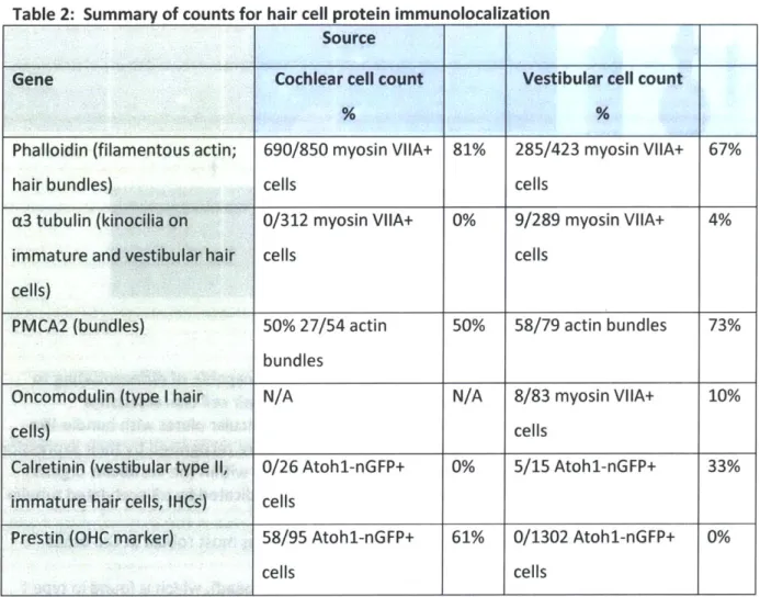
Table 2: Summary of counts for hair cell protein immunolocalization
Source
Gene Cochlear cell count Vestibular cell count
Phalloidin (filamentous actin; 690/850 myosin VIIA+ 81% 285/423 myosin VIIA+ 67%
hair bundles) cells cells
a3 tubulin (kinocilia on 0/312 myosin VIIA+ 0% 9/289 myosin VIIA+ 4%
immature and vestibular hair cells cells
cells)
PMCA2 (bundles) 50% 27/54 actin 50% 58/79 actin bundles 73%
bundles
Oncomodulin (type I hair N/A N/A 8/83 myosin VIIA+ 10%
cells) cells
Calretinin (vestibular type 11, 0/26 Atohl-nGFP+ 0% 5/15 Atohl-nGFP+ 33%
immature hair cells, IHCs) cells
Prestin (OHC marker) 58/95 Atohl-nGFP+ 61% 0/1302 Atohl-nGFP+ 0%
cells cells
In summary, immunolocalization showed that many cells in spheres differentiated into possible hair cell-like cells, as indicated by expression of Atohi, myosin VIIA, or parvalbumin. Of these cells, many had protrusions that express proteins that are prominent in hair bundles: f-actin and PMCA2, but few expressed the kinocilia marker, [3 tubulin. The expression of prestin
by a large fraction of hair cell-like cells derived from the organ of Corti but not by any cells
derived from vestibular tissue suggests that the progenitor cells in particular organs of the neonatal mouse are not fully competent to form hair cells of all types, but rather have fates
restricted to their inner ear compartment. This may indicate differentiation limitations in each tissue's progenitor cell population.
Expression of hair-cell specific genes
Because the newly generated cells expressed several key hair cell genes and possessed hair cell features, we next used RT-PCR to determine the presence or absence of genes that would provide key machinery for proper hair cell function. The genes we looked for are
indicated in Figure 3, where a band indicates that a given gene was being expressed in a particular tissue, and a lack of a band indicates that the gene was not expressed. We chose genes specific for mechanoelectrical transduction, for electromotility (prestin), the presynaptic
apparatus (e.g. proteins of synaptic ribbons and vesicles), and the postsynaptic apparatus (e.g. postsynaptic to cholinergic efferent input). We also tested for expression of genes related to supporting cell function, in particular secretion of the extracellular matrices (e.g. otogelin, otopetrin).
Transduction apparatus. We found that differentiated organ of Corti-derived
progenitors expressed the vital transduction channel component, Tmcl but not Tmc2, whereas the vestibular-derived cells expressed both Tmcl and Tmc2. Expression of these two proteins is
different in vivo, as TMC2 is initially expressed in both organs, but disappears in the cochlea near the onset of hearing (Kawashima et al. 2013). Differentiated progenitor cells from both organs also expressed Espnl and Espn4, the two ESPIN isoforms that are associated with
2006, Sekerkovd et al. 2006), consistent with the newly generating cells acquiring a certain level
of differentiation. Differentiated progenitors from both tissues also expressed myosin 1c, a gene that contributes to setting the resting current in hair cells and is critical to the adaptation response associated with hair bundle deflection (Fig. 2B) (Holt et al. 2002, Gillespie and Cyr 2004, Stauffer et al 2005). The differentiated progenitors from both tissues also expressed
Cdh23 and Pchd15, the two components of the hair cell tip-links that bind the stereocilia and
are critical to transduction (Fig. 3B)(Siemens et al. 2004, Kazmierczak et al. 2007).
Synaptic transmission. We also found that differentiated progenitors from both tissues expressed genes associated with both afferent and efferent synaptic function. The
differentiated cultures produced the cholinergic receptor, Chrna9, which is found in all hair cells and mediates the response to cholinergic terminals from efferent neurons in the brainstem (Elgoyhen et al. 1994, 2001, Luo et al. 1998, Zuo et al. 1999, Simmons and Morley 1998, Morley and Simmons 2002, Simmons 2002). They also produced the glutamate transporter VGLUT3, which fills synaptic vesicles with the excitatory neurotransmitter glutamate for afferent signaling (Fig. 3B) (Wang et al. 2007, Zhang et al. 2007, Seal et al. 2008, Peng et al. 2013). In addition, the differentiated cultures expressed Cav1.3 calcium channels, which admit calcium ions to initiate glutamate release from hair cells (Fig. 3B) (Platzer et al. 2000, Schnee and Ricci
2003, Brandt et al. 2003, Brandt et al. 2005), which ultimately leads to neurotransmitter
release. Similarly, the differentiated cultures expressed ribeye, the portion of the Ctbp2 protein that is highly specific to ribbon synapses (Schmitz et al. 2000). Both differentiated tissues also expressed oncomodulin, a calcium binding protein that is found in type I hair cells within vestibular tissue and outer hair cells of the cochlea (Fig. 3B) (Simmons et al. 2010).
Other. Both differentiated cultures expressed myosin VIIA, a hair cell marker protein. Similar to our staining results, we found that only differentiated cochlear cultures expressed the outer hair cell protein prestin (Fig. 3B). Whereas both differentiated tissues expressed the supporting cell genes otogelin and Lgr5, only vestibular-derived cultures expressed the supporting cell gene otopetrin, which is specific to the vestibular epithelia (Fig. 3B).
A
B
Type I
ITmc1l
Tmc2* Espnl Espn4 ILMyolc dh23 E Pcdh15 CU U) Cavl.3Ribeye Chma9 Cavi.3Ribeye
IHC
Tmc1 Espnl Espn4 Cdh23 Pcdhl5 CU U)0
Cavl Ribeye Chma9Type
11
Tmcl Tmc2* Espnl Espn4 Myolc Cdh23 Pcdh15 Chma9C
Tmcl Espn1 Espn4 MYolo Cdh23 Pcdhl5 Prestin* -/Chma9OH
I Cav Ribeye -14d 14d Native Native Vestibular Cochlear Vestibular Cochleargapdh Tmcl *Tmc2 Espnl Espn4 Myoic Cdh23 Pcdh15 Chrna9 Cavl.3 Ribeye Vglut3 MyoVila Prestin Ocm Otopetrin Otogelin Lgr5
Figure 3: Differentiated organ of Corti and vestibular stem cells express genes that are specific to their derived tissue and are required for proper physiological function
A) A schematic showing the genes that were analyzed and their location of expression within
each tissue and cell type.
B) Differentiated cells from both tissues expressed the hair cell gene myosin VIIA, hair bundle associated genes (Espnl, Espn4, Myo1c, Cdh23, Pchd15), genes associated with synaptic function (Chrna9, Cavl.3, ribeye, VGLUT3, Ocm), and the supporting cell genes Otogelin and
Lgr5. The differentiated cells from each tissue differed in that only cochlear derived cells expressed Prestin (*) and Tmcl alone, whereas only vestibular derived tissue expressed otopetrin (*) and the combination of Tmcl and Tmc2 (*). All results were confirmed with a
minimum of n=5 cultures. H -I IC
-'
C 010
1'
11
In summary, both cochlear and vestibular progenitor cells are capable of differentiating into populations of cells that express transduction channel components (TMC1 and TMC2), hair
bundle components (Espnl, Espn4, Myo1c, Cdh23, Pchd15), and genes associated with synaptic function (Chrna9, Cav1.3, ribeye, VGLUT3, Ocm), which may suggest that bonafide hair cells are being formed. Since only cochlear-derived progenitors differentiate to form prestin-expressing cell populations, and only vestibular-derived progenitors differentiate to form otopetrin-expressing cell populations, these results further suggest that progenitors from each tissue are limited to forming their own native cell types.
Voltage-gated currents
We conducted electrophysiological experiments to (1) further test the hypothesis that the newly generated cells form distinct cellular subtypes that are limited to those of their native organ; and (2) determine if these cells could respond to electrical stimuli in a manner similar to functional hair cells. With the patch clamp method, we recorded voltage-dependent whole-cell currents from hair cell-like cells, which we recognized in the recording dish by their expression of Atohl-nGFP. Most Atohl-nGFP+ cells produced large outward currents in response to depolarizations, with amplitudes and time courses that are qualitatively within the range of the
outward delayed rectifying K+ current (IKD) Of hair cells (Fig. 4A). We observed these currents in
91% (29/32) of Atohl-nGFP+ cells derived from the cochlea, and in 100% (34/34) of
Atohi-nGFP+ cells derived from vestibular organs (Fig. 5). In 7 Atohl-nGFP-negative cells tested (6 cochlear, 1 vestibular), none had hair cell-like currents (Figure 4B). In small samples of hair-cell like cells from each tissue, we obtained tail-current activation curves (see Eq. 1 in Methods),
which showed that for both tissues of origin, the outward currents activated around -60 mV and had activation midpoints (V1/2 values) that were typical of hair cells within their first postnatal week (Eatock and Hurley 2003). Cochlear V1/2 values ranged from -14 mV to -30 mV
(mean -21 3 mV, SE, n=3 cells) and vestibular V1/2 values ranged from -25 mV to -34 mV
(mean= -33 + 2 mV, n=5 cells). Figure 4C shows a cochlear-derived cell with a V1/2 value of
-20 mV and a vestibular-derived cell with a V1/2 value of -25 mV.
The vestibular-derived cells had currents similar to those found in previous
electrophysiological analyses of vestibular type 11 hair cells (Rusch & Eatock 1998, Holt et al.
1999) (Fig. 4E). Some cells resembled type 11 cells from the striolar and central zones of
vestibular epithelia in that the outward current did not show fast inactivation and they had a large lh current (28/32 cells =87%; Fig. 4A, top; Fig. 5). Other cells more closely resembled type
11 hair cells from the extrastriolar/peripheral epithelial zones in that they had inactivating
outward current that resembled A-current (IA)( 4/32 =12%) (Fig. 4D, bottom).
Outward currents in two cochlear cells had "times to half-maximum current" in the same range as previously recorded for neonatal cochlear hair cells (10-20 ms) by Marcotti and
Kros (1999, 2003). The outward currents of seven vestibular-derived cells were well fit with monoexponential decay functions with values that fell into two groups. At -45 mV, four cells had relatively slow time constants (49 15 ms; see Figure 4E top, Figure 4F), similar to the delayed rectifier of type I hair cells (IDR,I) after the first postnatal week (RUsch et al. 1998). The
other group of cells had a mean time constant of 14.2 1.9 ms (n=3)(Figure 4E bottom, Figure 4F), similar to the delayed rectifier in type I hair cells (IDR,II) in the utricular epithelium of
Vestibular hair cells acquire 'h current postnatally, and the size of the current
dramatically increases around P3 -P4 in mouse utricle (Rusch & Eatock 1998; Horwitz et al. 2010, Horwitz et al. 2011). In our voltage protocol, an ih -like current was recognized as a slowly increasing inward current during the 150-ms step to -125 mV at the start of each voltage protocol. We observed this lh -like current in 65% (22/34) of Atohl-nGFP+ cells derived from vestibular tissue but in 0 of 32 cochlear-derived cells (Fig. 4G; Fig. 5). This result suggests that only vestibular progenitors are capable of becoming vestibular hair cells.
Hair cells also possess an inward-rectifying potassium current known as Ii, which plays a large part in setting a hair cell's resting membrane potential during development (Rusch et al.
1998, Behrend 1997, Marcotti et al. 1999, 2003). Within the cochlea IHCs express this current
from E15-P14, while OHCs express this current from PO-P6 (Marcotti et al. 1999, 2003). Type I vestibular hair cells express Ki 1up to P4 and likely afterwards, while type II vestibular hair cells express Igi throughout the maturation process (Rusch et al. 1998). We found that 60% (19/32) Atohl-nGFP+ vestibular derived cells showed evidence of an IKl-like current, as indicated by the fast inward-rectifying current at the onset of the voltage step and its subsequent deactivation when voltage is stepped to more positive potentials (Fig. 4H; Fig. 5). All hair cell-like cells derived from cochlear tissue lacked evidence of lK1 in voltage clamp tests.
The time course of the inward currents evoked by large hyperpolarizing voltage steps could be fit with a double-exponential decay function. For hair-cell like cells from vestibular tissue, 150-ms steps from -65 mV to -125 mV typically elicited inward currents with fast and slow time constants (T) on the order of 1 ms and 100 ms, respectively: The mean slow T was 92 21 ms (n=6 cells) (Figure 4H). The mean fast T was 0.99 0.32 ms (n=5 cells) (Figure
4H). These numbers agree reasonably well with data from vestibular hair cells: The faster time constant we measured is comparable to values reported for IK, the fast inward rectifier, in early postnatal mouse utricular hair cells by Rusch et al. (1998). The slower time constant we
recorded is comparable to the faster of two time constants for lh activation in mouse utricular hair cells recorded with much longer voltage steps (Horwitz et al. 2011) - our brief voltage steps did not substantially activate the slower component of lh activation.
In small samples of the hair-cell like cells, we recorded membrane voltage in current-clamp mode to assess the resting potential and voltage responses to injected current steps for comparison with the literature on hair cell responses (Figure 41). The resting potentials were similar for Atohl-nGFP+ cochlear and vestibular cells: -53 8 mV (n=4 cochlear-derived cells)
and -53 6 mV (n=6 vestibular-derived cells). These values are set in large measure by the
developmental acquisition of K-selective inwardly-rectifying and outwardly-rectifying channels (e.g., Eatock and Hurley 2003; Goodyear et al. 2006) and are within the physiological range for postnatal hair cells.
Voltage clamp and current clamp recordings from the vestibular-derived cells lacked clear evidence of the type-I-specific current, IKL (RUsch et al. 1998, Holt et al. 1999).
Nevertheless, the activation time courses of the delayed rectifiers are consistent with
differentiation into type I and type 11 hair cells (Figure 4E). We also did not see the mixed Ca2
+_
Na' spikes that are typical of mouse inner hair cells in the first postnatal week (Marcotti et al.
2003). All Atohl-nGFP+ cells lacked the voltage-gated Na+ currents that are reported in
immature hair cell subtypes from both the cochlea and vestibular system (Witt et al. 2004, Oliver et al. 1997, Chabbert et al. 2003, Marcotti et al. 2003, Geleoc et al. 2004, Wooltorton et
al. 2007, Li et al. 2010, Eckrich et al. 2012). A possible explanation is that by 12 days of in vitro differentiation, the earliest time that we examined, the hair cell-like cells had advanced beyond the stage of significant Na channel expression.
In all, our results support our hypothesis that the early postnatal inner ear harbors different populations of progenitors with limited differentiation capabilities. We also show that organ-specific progenitors can differentiate to form Atohl-nGFP+ cells that have many of the
B
h N 1KOG
0o C_no
0 1A 41m AWNol 1K Iacbvs, 80 ffs P M IDR,1l ""I 30. -45 -5 - 0 E 100 _ _ _Figure 4: Differentiated organ of Corti and vestibular progenitor cells resemble the hair cells from their native tissues in certain electrophysiological properties.
A) Atohl-nGFP+ cells derived from both tissues had large voltage-dependent currents.
Vestibular-derived cells are in red (top panel) and cochlear-derived cells are in blue (bottom panel). The largest currents were outward currents evoked by depolarizing voltage steps and were reminiscent of the delayed rectifier potassium currents (IKD) found in native hair cells. No sodium currents were detected.
B) Atoh1-nGFP-negative cells did not have currents that resembled those found in native hair cells based on the size and time course of the currents.
C) Tail current activation curves taken at -40 mV. Smooth curves are single-Boltzmann fits (Methods) with midpoints (VI/2 values) as given. These are similar to those found in native hair cells within the first postnatal week.
D) Some but not all Atohl-nGFP+ cells derived from vestibular tissue had inactivating A-current
(A). This heterogeneity is seen in vivo in type I vestibular hair cells.
E) Outward currents from vestibular derived Atohl-nGFP+ had time courses that resembled the
A
F) Activation time courses in response to a series of voltage steps revealed that vestibular-derived Atohl-nGFP+ cells separated into two distinct groups that resembled the time courses seen in native type I and type 11 hair cells.
G) The vestibular-specific postnatal current lh was found in 65% of vestibular-derived
Atohi-nGFP+ cells and no cochlear-derived Atohl-Atohi-nGFP+ cells.
H) Vestibular hair cells showed evidence of the fast inward rectifier, IKI, in addition to lh, which
activated in response to voltage steps from -65 mV to -125 mV with time constants similar to those reported for native hair cells.
1) Both cochlear-derived and vestibular-derived Atohl-nGFP+ cells responded to current steps with voltage changes similar to those reported from postnatal hair cells.