HAL Id: inserm-02633099
https://www.hal.inserm.fr/inserm-02633099
Submitted on 27 May 2020
HAL is a multi-disciplinary open access
archive for the deposit and dissemination of
sci-entific research documents, whether they are
pub-lished or not. The documents may come from
teaching and research institutions in France or
abroad, or from public or private research centers.
L’archive ouverte pluridisciplinaire HAL, est
destinée au dépôt et à la diffusion de documents
scientifiques de niveau recherche, publiés ou non,
émanant des établissements d’enseignement et de
recherche français ou étrangers, des laboratoires
publics ou privés.
pathways recruitment drives basal extrusion in the
prostate-like gland of Drosophila
Amandine Rambur, Corinne Lours-Calet, Claude Beaudoin, Julio Bunay,
Marine Vialat, Vincent Mirouse, Amalia Trousson, Yoan Renaud, Jean-Marc
Lobaccaro, Silvère Baron, et al.
To cite this version:
Amandine Rambur, Corinne Lours-Calet, Claude Beaudoin, Julio Bunay, Marine Vialat, et al..
Se-quential Ras/MAPK and PI3K/AKT/mTOR pathways recruitment drives basal extrusion in the
prostate-like gland of Drosophila. Nature Communications, Nature Publishing Group, 2020, 11 (1),
pp.2300. �10.1038/s41467-020-16123-w�. �inserm-02633099�
Sequential Ras/MAPK and PI3K/AKT/mTOR
pathways recruitment drives basal extrusion
in the prostate-like gland of
Drosophila
Amandine Rambur
1,2
, Corinne Lours-Calet
1,2
, Claude Beaudoin
1,2
, Julio Buñay
1,2
, Marine Vialat
1,2
,
Vincent Mirouse
1
, Amalia Trousson
1,2
, Yoan Renaud
1
, Jean-Marc A. Lobaccaro
1,2
, Silvère Baron
1,2
,
Laurent Morel
1,2
& Cyrille de Joussineau
1,2
✉
One of the most important but less understood step of epithelial tumourigenesis occurs when
cells acquire the ability to leave their epithelial compartment. This phenomenon, described as
basal epithelial cell extrusion (basal extrusion), represents the
first step of tumour invasion.
However, due to lack of adequate in vivo model, implication of emblematic signalling
path-ways such as Ras/Mitogen-Activated Protein Kinase (MAPK) and phosphoinositide 3 kinase
(PI3K)/protein kinase B (AKT)/mammalian target of rapamycin (mTOR) signalling
path-ways, is scarcely described in this phenomenon. We have developed a unique model of basal
extrusion in the
Drosophila accessory gland. There, we demonstrate that both Ras/MAPK and
PI3K/AKT/mTOR pathways are necessary for basal extrusion. Furthermore, as in prostate
cancer, we show that these pathways are co-activated. This occurs through set up of
Epi-dermal Growth Factor Receptor (EGFR) and Insulin Receptor (InR) dependent autocrine
loops, a phenomenon that, considering human data, could be relevant for prostate cancer.
https://doi.org/10.1038/s41467-020-16123-w
OPEN
1Université Clermont Auvergne, GReD, CNRS UMR 6293, INSERM U1103, 28 place Henri Dunant, BP38, 63001 Clermont-Ferrand, France.2Centre de
Recherche en Nutrition Humaine d’Auvergne, 58 Boulevard Montalembert, 63009 Clermont-Ferrand, France. ✉email:cyrille.de_joussineau@uca.fr
123456789
W
orldwide, a large majority of cancers originates from
epithelial tissues such as lung, breast and prostate
1.
Despite reinforced prevention, most of the tumours
are detected at late stages, and patient care is centred on invasive
adenocarcinomas, resistant forms of these carcinomas and
metastatic carcinomas. As late stages of cancer progression have
been under intense scrutiny in the last decades, the molecular
mechanisms associated to such progression are largely described,
showing for example the major role of receptor tyrosine kinase
(RTK)-dependent signalling pathways in these mechanisms.
Typically, for prostate adenocarcinoma, the second most
com-mon cancer in men, both PI3K/AKT/mTOR pathway and Ras/
MAPK pathways are associated with tumour progression
2–4. In
the prostate adenocarcinoma, Ras/MAPK and PI3K/AKT/mTOR
pathways display activating genetic alterations in more than 40%
of primary tumours and in virtually all metastatic prostate
tumours
2, and phosphoproteomic studies confirmed a strong
correlation in the activation of these two pathways
5. Furthermore,
pre-clinical mouse models reproducing alteration of either one or
the other pathway in the prostate epithelium display
tumour-igenesis that mimics histopathological features of the human
adenocarcinoma
6,7. Moreover, advanced tumour progression is
obtained when combining alterations in both pathways
8–10.
These different data emphasise that in one hand, Ras/MAPK or
PI3K/AKT/mTOR pathways can initiate prostate tumour
devel-opment, and in the other hand that these pathways are implicated
in late phases of tumour progression. However, they explain
neither their respective or combined role in actual
adenocarci-noma formation nor the molecular mechanisms that could couple
these two pathways to promote this phenomenon in vivo.
Adenocarcinoma formation occurs when pre-invasive
epithe-lial cells acquire the ability to leave their epitheepithe-lial compartment.
This implicates that these cells are able to extrude from the
normal epithelium and to cross the basement membrane which is
the limit of the epithelial compartment. These phenomena can be
described as basal extrusion and are resulting in early invasion, as
opposed to late invasion associated to the metastatic process. Due
to the difficulty to precisely visualise basal extrusion in animals,
mechanistic associated to this phenomenon has been essentially
described in cellular models or in developing tissues such as
Drosophila imaginal disc of zebrafish embryo
11–13, even though
Hendley et al. showed the role of P120 catenin in basal extrusion
in a mouse model of pancreatic neoplasia
14. To the best of our
knowledge, role of Ras/MAPK pathway in basal extrusion has
only been described through the use of Ras
V12as an oncogenic
hit, and role of PI3K/AKT/mTOR pathway has never been
assessed.
To determine the role of Ras/MAPK and PI3K/AKT/mTOR
pathways in basal extrusion and understand the underlying
mechanisms that may coordinate their hyperactivation in
pros-tate cancer, we have developed a new model of in vivo early
invasive adenocarcinoma in the drosophila prostate-like accessory
gland. Drosophila is a powerful genetic model where more than
70% of genes implicated in human diseases display orthologs
15and where Ras/MAPK and PI3K/AKT/mTOR signalling
path-ways are well conserved
16,17. Drosophila has already proven its
pertinence as cancer model for brain
18,19, lung
20, and colon
21.
The Drosophila accessory gland is a functional equivalent for the
prostate, playing a role in fertility by secreting seminal
fluid
22,23.
Secretions come from a monolayer of epithelial cells that are well
differentiated and quiescent at the adult age, and there is no
evidence of stem cells in this tissue. Considering that a majority of
prostate adenocarcinoma is thought to originate from luminal
cells
24, epithelial cells from the accessory gland represent a
valuable model to study the mechanisms of epithelial prostate
tumourigenesis
25,26.
In this study, we describe this unique model of basal extrusion
and tumour formation in the accessory gland that recapitulates
most aspects of cancer development. We demonstrate that both
Ras/MAPK and PI3K/Akt/TOR pathways are overactivated in the
produced tumours, and that these pathways cooperate to induce
basal extrusion and subsequent tumour formation. Furthermore,
we describe the mechanism that allows the coactivation of these
pathways, which relies on the sequential recruitment of a double
autocrine feedback loop dependent on Epidermal (EGF/Spitz)
and Insulin-like (IGF/Ilp6) Growth Factors and their respective
receptors. Finally, using publicly available data of prostate cancer
samples
2and migration assay in human pre-tumoural prostate
epithelial cell line, we assess the possible role of these
findings in
the actual human pathology.
Results
Accessory gland recapitulates prostate microenvironment. To
realise our study, we
first determined whether accessory gland
represents a good model to study basal epithelial cell extrusion.
Prostate compartment is mostly composed of acini of epithelial
cells adjacent to a basement membrane and surrounded by a
fibromuscular stroma. It is already known that a layer of muscle
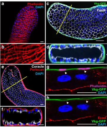
fibres completely encloses the accessory gland. Phalloïdin staining
reveals this well-organised muscle sheet that forms a continuous
layer at the surface of the gland (Fig.
1
a). Using higher
magni-fication, we further show that contiguous muscle fibres are linked
together by actin bridges, a phenomenon that could explain how
mostly unchanged distance between the muscle
fibres can be
conserved in spite of natural contraction of the
fibres (Fig.
1
b).
Furthermore, we show the presence of a basement membrane
along the epithelium, where collagen IV (Viking) and Perlecan
can be detected (Fig.
1
c–f). This basement membrane encloses the
muscle layer (arrow in Fig.
1
g, h) which must reinforce the
sta-bility of the accessory gland. We conclude that indeed, accessory
gland recapitulates classical epithelial microenvironnement as its
epithelial cells are adjacent to a basement membrane and closely
linked to a stromal-like compartment of muscle cells.
Oncogene induces epithelial tumourigenesis. We then set up
experimental conditions to induce epithelial tumourigenesis.
Tumourigenesis initiation is thought to start by alteration of one
single gene in one single cell. Thanks to genetic tools available in
Drosophila, we were able to precisely mimic this step by clonal
expression of known oncogene Ras
V12in a controlled number of
cells of the epithelium. Experiments were designed to induce an
average of ten clones per gland. Due to time of clonal induction,
before last cell division of accessory gland epithelial cells,
expression of GFP only (control, Fig.
2
a) typically produces
clones of two cells that are of same shape and size than
non-expressing neighbouring cells (Fig.
2
b, c). In contrast,
co-expression of GFP and Ras
V12was repeatedly correlated to the
presence of green masses composed of numerous cells, which
clearly appear outside the actual glands. This revealed that Ras
V12expression induced clonal epithelial cell hyperproliferation and
suggested that some of the clones were able to develop into
tumours (Fig.
2
d, e).
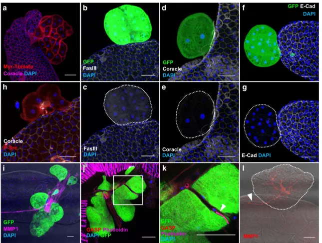
Then, we verified that these masses are actual tumours of
epithelial origin. Importantly, these masses are composed of
binucleated cells, a specific feature that confirms that they
originate from accessory gland epithelial compartment (Fig.
3
a).
However, these cells are mostly devoid of cell membrane staining
for all the epithelial markers we tested: Fasciclin III, Coracle and
E-cadherin (Fig.
3
b–g). As Fasciclin III and E-Cadherin are
adhesion molecules, their absence suggests that clonal cells lost
physical contact with surrounding epithelial cells, a phenomenon
associated with neoplastic growth in human
27and tumour
growth in Drosophila
28. Loss of epithelial identity also indicates
that Ras
V12expressing cells are able to initiate epithelium to
mesenchyme transition (EMT), which is considered as a key
tumourigenic event in the prostate
29. Src, a non-receptor kinase,
is associated with EMT and cell invasion in cancer
30, and notably
in metastatic progression of prostate adenocarcinoma
31. Its role
in tumourigenesis is assessed as well in Drosophila epithelium as
in mouse prostate epithelium
32,33. Phosphorylated Src is present
at the membrane of Ras
V12expressing cells, defining them as
tumour cells (Fig.
3
h). Src activation is also correlated to
neo-angiogenesis in cancer
31. In Drosophila, a tracheal system
provides oxygen to tissues, and growth of this system relies on
hypoxic activation by Drosophila HIF homolog sima
34. Therefore,
oncogenic expression has been associated to traces of
neo-tracheogenesis in this model, which have led to the conclusion
that neo-tracheogenesis is the counterpart of neo-angiogenesis in
cancer
35. Ras
V12clonal expression is correlated to the presence of
overgrown ectopic tracheas (Fig.
3
i). These tracheas can be
characterised by the use of two different antibodies directed
against Gasp and Matrix Metalloprotease 1 (MMP1) (Fig.
3
i–l).
Gasp, which is implicated in the opening of tracheas during their
growth
36, is strongly expressed in low diameter tracheas at the
surface of tumours (arrow in Fig.
3
k), but at weaker level in the
larger tracheas (Fig.
3
j). This shows that these ectopic tracheas are
almost entirely mature, i.e. functional, but also still growing, a
phenomenon that does not normally occur in adult
flies. MMP1
is detected at high levels in the tracheas (Fig.
3
i, l), as in embryo
and larva in which it is involved in tracheal development
37,38.
This MMP1 staining furthermore reveals that an arborescence of
tracheas grows inside the tumours from the tracheas that are
normally situated at the surface of the accessory glands (arrow in
Fig.
3
l). This shows that not only tracheas are growing actively,
but also in tight coordination with tumours. We conclude that
Ras
V12clonal expression induces neo-tracheogenesis in adult
accessory gland.
Then, we wanted to definitively confirm that we are in the
presence of a phenomenon of basal extrusion. First, we noticed
that the presence of tumours is linked to defects in the muscle
layer, reflecting the probable migration of the clonal epithelial
cells through this muscle sheet (arrow in Fig.
4
a), especially as
tumours frequently stay attached to the normal epithelium (arrow
in Fig.
4
b). Then, we revealed in the same experiments tumours in
red (RFP), basement membrane (viking-GFP in green
39) and the
stromal compartment of muscle cells (phalloïdin staining in
magenta) (Fig.
4
c–j). Tumours appear outside both the epithelial
and the stromal-like compartment of muscle cells (yellow arrows
in c–f, i, j). Surface of the tumours display no evident basement
membrane (yellow arrows in Fig.
4
c–h) and only basement
membrane within these masses corresponds to tracheas that grew
inside the tumours (white arrows in Fig.
4
c–h).
Together, our results demonstrate that expression of a known
oncogene in clones of accessory gland epithelial cells induces
epithelial cell extrusion and early invasion resulting in the
formation of adenocarcinoma-like tumours. Due to the shape and
structure of accessory gland, it is also particularly easy to visualise
the tumours directly at the dissection, allowing easy
quantifica-tion of this basal extrusion phenomenon.
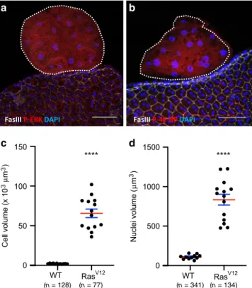
Invasive tumours display RTK-dependent pathways
coactiva-tion. Our aim was then to determine if accessory glands tumours
were able to reproduce in Drosophila this specific feature of
prostate adenocarcinoma which is the co-deregulation of Ras/
MAPK and PI3K/AKT/mTOR pathways
2,5. To test the activity of
the two considered pathways, the phosphorylation of two
down-stream targets, ERK (Rolled) and 4E-BP, was assessed. In response
to Ras
V12expression, P-ERK is logically detected in tumours
(Fig.
5
a) which indicates canonical Ras/MAPK pathway activation.
In the tumours, cells display up to 40 times their normal size (see
Fig.
3
a for membrane staining and Fig.
5
c for quantification),
showing that their growth is abnormally activated. Correlatively,
they display oversized nuclei (Fig.
5
d), indicating a probable
phenomenon of endoreplication. In Drosophila, cell size is tightly
controlled by TOR activity
17,40. Indeed, 4E-BP phosphorylation is
detected in tumour cells (Fig.
5
b), indicating a co-recruitment of
the PI3K/AKT/TOR pathway in these cells.
RTK-dependent pathways coactivation drives basal extrusion.
We then wanted to determine whether the activated pathways
were implicated in the process of invasion itself. So, we quantified
invasive tumour frequency for different combinations of
activa-tion/inactivation of the two considered pathways. Expression of
a
c
b
d
e
g
f
h
Fig. 1Drosophila accessory gland recapitulates prostate epithelial micro-environment. a, b alexa568-phalloidin (Phalloidin) staining reveals actin accumulation in the well-organised muscle sheet around the gland epithelium. b Higher magnification reveals actin bridges between the muscle fibres. This muscle sheet defines a stromal compartment in the gland. c–f Viking-GFP (GFP tagged Collagen IV, Vkg-GFP in (c, d) or Perlecan-RFP (Perlecan in (e, f)) expression reveals the presence of a basement membrane, and Fasciclin III (c, d) or Coracle (e, f) staining reveals the apico-lateral domain of the epithelial cells. Transversal optical section (along the yellow line in (c) and (e)) confirms deposition of Viking-GFP and Perlecan-RFP at the basal pole of the epithelial compartment.g, h Transversal optical section of the gland at high magnification. Viking-GFP expression reveals the presence of the basement membrane, E-Cadherin (Ecad) staining reveals the apico-lateral domain of one epithelial cell and alexa633- phalloidin (Phalloidin) staining reveals actin accumulation in the musclefibres. Arrows indicate where the basement membrane encloses the musclefibres. DAPI (blue) reveals nuclei in (a) and (b)–(h), mostly from binucleated epithelial cells (see g–h). Representative images in (a–d, g, h) from three or more experiments and in (e–f) from two. Scale bars: 50 μm in (a–f) and 10 μm in (g, h).
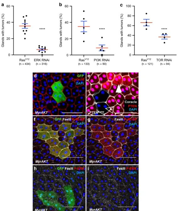
ERK RNAi in the Ras
V12expressing cells significantly decreases
invasive tumour frequency (i.e. clones that left the epithelium
compartment to form external masses) (Fig.
6
a), showing that
Ras
V12oncogenic activity depends on the canonical Ras/
MAPK pathway. Similarly, expression of PI3K RNAi or TOR
RNAi in the Ras
V12expressing cells significantly decreases
invasive tumour frequency (Fig.
6
b, c), showing that Ras
V12oncogenic activity depends also on the PI3K/AKT/TOR pathway
in the accessory gland.
PI3K/AKT/TOR pathway can be directly activated either by
phosphatase and tensin homolog (pten) deletion or by expression
of a myristoylated form of Akt (myr-Akt). The use of one or the
other of these genetic modifications does not induce tumour
development, but only mild cell hypertrophy is observed (Fig.
6
d,
e). Furthermore, myr-Akt expressing clones display 4E-BP
phosphorylation (Fig.
6
f, g), but no ERK phosphorylation (Fig.
6
h,
i), showing that PI3K/AKT/TOR overactivation does not induce
Ras/MAPK pathway. Thus, it indicates that a failure to co-recruit
the two pathways correlates with a lack of basal extrusion in the
accessory gland.
From our data, we conclude that both the canonical Ras/
MAPK pathway and the PI3K/AKT/TOR pathway are necessary
for cells to evade the epithelial compartment, and that lack of
recruitment of one or the other pathway impairs the basal
extrusion and subsequent tumour formation.
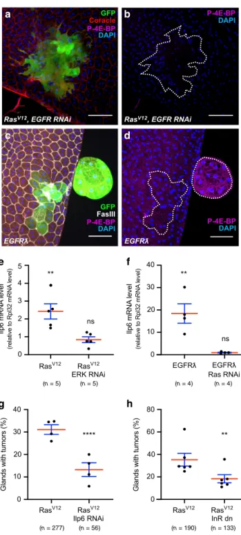
Tumour invasion depends on an EGF/EFGR autocrine loop.
Then, we decided to determine how the pathways are recruited
during tumourigenesis in the accessory gland. Considering that,
in prostate cancer, a majority of primary tumours display no
activating mutation in the considered pathways
2, we searched for
an alternative mechanism. Classical activation of the PI3K/AKT/
mTOR and the Ras/MAPK pathways is thought to mostly rely on
growth factors. Interestingly, Ras
V12clones overexpress EGF/
Spitz, an EGF/TGFα homolog and ligand of Drosophila EGFR
41(Fig.
7
a, b). EGFR is upstream of both Ras/MAPK and PI3K/
AKT/TOR pathways in mammals
42, but could be more specific to
Ras/MAPK pathway in Drosophila
43. Strikingly, suppression of
EGF/Spitz by RNAi expression in Ras
V12clones strongly reduces
invasive tumour frequency (Fig.
7
c), showing that expression of
this ligand sustains Ras
V12-driven basal extrusion in the accessory
gland. Moreover, if clonal overexpression of a secreted form of
EGF/Spitz (Spitz
sc)
44in epithelial cells mostly induces a high rate
of mortality during pupal stage, the remaining adult
flies can
display tumours exhibiting the same hallmarks of cell overgrowth
and loss of epithelial markers as in Ras
V12induced tumours
(Fig.
7
d). These results agree with the existence of an
EGFR-dependent autocrine feedback loop that is necessary for Ras
V12oncogenic transformation. Logically, co-expression of an EGFR
RNAi in Ras
V12clones strongly decreases invasive properties of
developed tumour (Fig.
7
e), confirming the role of EGFR in
Ras
V12-driven tumourigenesis. Furthermore, expression of a
constitutively active form of EGFR, EGFRλ, leads to the
forma-tion of tumours that display the same hallmarks as in Ras
V12induced tumours (Fig.
7
f), showing that overactivated EGFR can
induce basal extrusion of epithelial cell in the accessory gland.
Finally, co-expression of a Ras RNAi in EGFRλ clones strongly
decreases their invasive properties (Fig.
7
g), confirming the role
of Ras/MAPK signalling pathway in the EGFR-dependent
tumourigenic process. Interestingly, observation of epithelial
clones co-expressing EGFR RNAi and Ras
V12reveals that they
can be composed of many cells (observe the extension of the
clone in Fig.
8
a), suggesting that EGFR recruitment is less
required for cell division than for cell extrusion.
We conclude that upon Ras
V12dependent initial activation,
Ras/MAPK pathway must be re-activated by an EGF/EGFR
autocrine loop to induce cell invasive capacities, definitively
showing that Ras/MAPK pathway is necessary for basal extrusion
independently of the mechanism of oncogenic initiation.
a
d
e
b
c
µ (n = 8) Clones (n = 12)Fig. 2Drosophila accessory gland is subjected to oncogene-dependent epithelial cells division and extrusion. (a, b) and (d, e) Green fluorescence reveals clones of GFP expressing cells (WT). a–c Clonal cells expressing GFP/nlsGFP only. d–e Clonal cells co-expressing GFP and RasV12(RasV12). a, b
Clonal induction is revealed by multifocal localisation of GFP expressing cells (white arrows). Due to time of induction, clones in accessory gland epithelium are composed of two neighbouring cells. These cells do not display phenotype of hypertrophy, as quantified in (c) (Mann–Witney test; P = 0,3431). Data are represented as mean values ± SEM (n = 8 and n = 12 accessory glands Control and GFP analysed respectively, from two experiments). d, e GFP-RasV12
expressing clones are composed of numerous cells and are present outside the gland. DAPI (blue) reveals nuclei in (a, b) and (e). Representative images in (a–e) from three or more experiments. Scale bars: 200 μm in (a) and (d), 50 μm in (b, e).
Tumour invasion depends on an IGF/InR autocrine loop. We
then wanted to determine how PI3K/AKT/TOR pathway is
recruited. At
first, we observed that P-4E-BP staining is never
detected in Ras
V12/EGFR RNAi clones, and cells stay smaller in
this condition compared to Ras
V12expressing cells (Fig.
8
a, b),
indicating a lack of recruitment of the PI3K/AKT/TOR pathway
in these clones and suggesting that EGFR could be implicated in
the recruitment of this pathway. However, we also observed that
clonal expression of EGFRλ induces PI3K/AKT/TOR pathway
recruitment, as for Ras
V12expression, but this recruitment and
associated cellular hypertrophy occurs only in external cell masses
(Fig.
8
c, d). This suggests that PI3K/AKT/TOR pathway is not
recruited directly by EGFR, but via a molecular intermediate. In
Drosophila, PI3K/AKT/TOR pathway is mainly recruited by the
Insulin Receptor (InR)
45, and seven ligands are known to activate
InR. We then searched for their expression. Both Ras
V12and
EGFRλ clonal expression in the accessory gland induces mRNA
overexpression of one ligand, IGF/Ilp6 (left columns in Fig.
8
e, f).
Indeed, we decided to determine the potential role of IGF/Ilp6
overexpression in Ras
V12-driven tumourigenesis. Strikingly,
downregulation of IGF/Ilp6 (Ilp6 RNAi) in Ras
V12clones
sig-nificantly decreases invasive tumour frequency (Fig.
8
g).
Logi-cally, co-expression of a dominant-negative form of InR (InR dn)
in Ras
V12clones also decreases invasive tumour frequency
(Fig.
8
h), showing that this receptor is also implicated in Ras
V12oncogenic activity, and then defining a second autocrine loop that
participates to accessory gland invasive tumour formation.
Next, we decided to precise which molecular actors are
necessary for this IGF/Ilp6 overexpression. First, we
co-expressed Ras RNAi with EGFRλ. It completely abolishes IGF/
Ilp6 mRNA overexpression compared to EGFRλ expression
alone, showing that this overexpression depends on Ras
activation (right column in Fig.
8
f). Second, we co-expressed
ERK RNAi with Ras
V12. This also completely abolishes IGF/Ilp6
mRNA overexpression compared to Ras
V12expression alone,
demonstrating that IGF/Ilp6 overexpression depends also on ERK
activation (right column in Fig.
8
e). We conclude that IGF/Ilp6
mRNA overexpression exclusively depends on the activation of
the classical EGFR/Ras/MAPK pathway.
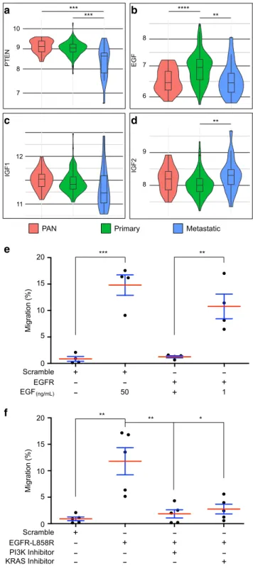
EGF is specifically overexpressed in primary prostate cancer.
Finally, to determine whether our
findings could be relevant to
prostate cancer, we decided to examine EGF and IGFs expression
in human samples. We selected data where mRNA levels were
available in normal, primary and metastatic samples
2. First, we
a
b
d
f
h
c
e
g
i
j
k
l
Fig. 3 Oncogene expression induces epithelial tumourigenesis in the accessory gland. a–l GFP-RasV12expressing clones display hallmarks of cancer.
a Visualisation of the tumour cell membranes with a myristoylated form of Tomato (Myr-Tomato) indicates that the cells conserve their typical binucleation.b–g Staining reveals that tumour-like GFP-RasV12expressing clones lost expression of the epithelial markers. Epithelial markers visualised by
staining are: (b) Fasciclin III (FasIII), (d) Coracle, (f) E-Cadherin (E-Cad). h Phosphorylated Src (P-Src) is detected at in GFP-RasV12expressing clones, a
feature of mesenchymal cells and/or transformed epithelial cells.i–l Tracheal system is revealed by Matrix Metalloprotease 1 (MMP1, i, l) or Gasp (j, k) staining.i Presence of tumour-like GFP-RasV12expressing clones is correlated to strong hypertrophy of tracheal system.j–k Gasp staining reveals trachea
network associated with tumour-like GFP- RasV12expressing clones.k Zoom of the boxed section in (j); white arrowhead points at a strong gasp
expression in a low diameter trachea, a feature of developing trachea.l MMP1 staining reveals an arborescence of trachea into the tumour and from accessory gland tissue (white arrowhead). DAPI (blue) reveals nuclei in (a–k). Representative images in (b–i, l) from three or more experiments, in (j)–(k) from two experiments and in (a) from one. Scale bars: 50μm.
determined whether these mRNA could reflect alterations that are
hallmarks of tumour progression. Typically, advanced prostate
cancers are associated to loss of PTEN expression
46. In the used
cohort, PTEN mRNA expression tends to decrease in primary
tumours when compared to normal tissue (p
= 0.07) and
sig-nificantly decreases in metastatic samples compared to normal
tissue (p
= 0.00047) and primary tumours (p = 0.002) (Fig.
9
a).
Thus, PTEN expression is, as expected, inversely correlated with
tumour grade. We then considered the expression of EGF, IGF1
and IGF2 (Fig.
9
b–d). EGF mRNA expression presents a
bell-shaped pattern (Fig.
9
b): it is significantly increased in primary
tumours compared to both normal (p
= 2 × 10
−5) and metastatic
(p
= 0.0089) tissues which are expressing very similar levels of
EGF (p
= 0.92). Furthermore, this expression increases even in
primary tumours with the lowest available Gleason score
(Glea-son score
= 6, p = 0.0012). In contrast, IGF1 expression does not
significantly change between the considered conditions (Fig.
9
c),
and IGF2 expression tends to decrease in primary tumours
compared to normal tissue (p
= 0.05) and significantly increases
in metastatic samples compared to primary tumours (p
= 0.0036)
(Fig.
9
d). Together, these results indicate that EGF mRNA is
specifically overexpressed during early carcinogenesis, suggesting
that EGF could play a role in this particular stage of cancer.
Then, we decided to check the role of the EGFR signalling
pathway in P69, a cell line derived from immortalisation of
human normal prostate epithelial cells
47. Using transwell
chamber assay, we show that supraphysiological doses of EGF,
or lower doses in a context of EGFR overexpression, significantly
increase P69 cells migration (Fig.
9
e). Furthermore, expression of
a
b
c
d
µ
µ
(n = 128) (n = 77) (n = 341) (n = 134)
Fig. 5 Ras/MAPK and PI3K/AKT/TOR pathways are concomitantly activated in RasV12expressing tumours. GFP-RasV12expressing tumours
are encircled by dotted lines. Fasciclin III (FasIII) staining reveals normal epithelial cells.a GFP-RasV12expressing tumours display phosphorylation
of ERK, a downstream target of the Ras/MAPK pathway.b GFP-RasV12
expressing tumours display phosphorylation of 4E-BP, a downstream target of the PI3//AKT/mTOR pathway.c, d Comparison of cell (c) and nuclei (d) volumes of tumour cells with wild-type cells (WT) (**** Unpairedt test; P < 0.0001) reveals strong hypertrophy of GFP-RasV12expressing cells
(RasV12). Data are represented as mean values±SEM (cell volume:n = 12
andn = 14 accessory glands Control and Rasv12analysed, respectively, from
seven experiments: nuclei volume:n = 11 and n = 14 accessory glands Control and Rasv12analysed respectively, from seven experiments).
Representative images in (a, b) from three or more experiments. DAPI (blue) reveals nuclei in (a, b). Scale bars: 50μm.
a
b
c
e
d
f
g
i
h
j
Fig. 4 Oncogene expression induces basal epithelial cell extrusion in the accessory gland. a, b GFP-RasV12and (c–f) RFP-RasV12expressing clones
are able to migrate from the epithelial compartment.a Alexa633-phalloidin (Phalloidin) staining reveals disorganisation of the musclefibres at the site of extrusion of GFP-RasV12expressing clones (white arrow).b Zoomed
image of (a) reveals that the tumour is still anchored to the epithelium (white arrow).c, j Alexa633-phalloidin (Phalloidin) staining reveals the musclefibres (c–f, i, j) as Viking-GFP staining reveals basement membranes (c–h). (d, f, h, j): optical cross-sections of top panels (respectively,c, e, g, i). Staining reveals that RFP-RasV12tumour develops
outside the epithelial compartment, as it is not enclosed by a basement membrane or a muscle layer (yellow arrows). Basement membrane staining reveals tracheal branches inside the tumour (white arrows). DAPI (blue) reveals nuclei in (a–j). Representative images from two experiments. Scale bars: 50μm.
a constitutively active form of EGFR (EGFR-L858R
48) induces
similar migration of P69 cells in the absence of EGF, and this
migration is impaired by treatment with specific inhibitors of
either KRAS
49or PI3K (Fig.
9
f). We conclude that, as
demonstrated in Drosophila, migration of human pre-tumoural
prostate epithelial cells depends on EGFR signalling and
down-stream activation of both Ras/MAPK and PI3K/Akt/mTOR
pathways.
So, collectively, our data show that accessory gland
tumour-igenesis, and more precisely basal extrusion, relies on a double
autocrine loop: the
first one depends on EGF/Spitz and its
receptor EGFR to amplify Ras/MAPK pathway activation; the
second one depends on Ras/MAPK-dependent production of
IGF/Ilp6 to recruit the PI3K/AKT/TOR pathway via InR
activation. And this coactivation is necessary in a
cell-autonomous manner for the epithelial cells to be able to leave
the epithelial compartment to form adenocarcinoma-like
tumours (Fig.
10
).
Discussion
More than 95% of prostate cancers are of epithelial origin and are
thought to mainly arise from a single cell
24. To faithfully
repro-duce what is thought to happen in the earliest stages of tumour
formation in patients, we have decided to produce a single genetic
alteration in few clones of randomly selected and mostly
a
d
e
f
g
h
i
b
c
(n = 434) (n = 316) (n = 133) (n = 90) (n = 121) (n = 94)Fig. 6 Ras/MAPK and PI3K/AKT/TOR pathways are both necessary for tumour formation. a–c Invasive tumour frequency of GFP-RasV12expressing
clones (RasV12) is impaired by downregulation of ERK (ERK RNAi in (a)), of PI3K(PI3K RNAi in (b)) and of TOR (TOR RNAi in (c)) (Chi2test; ****i <
0.0001 in (a) and (b), ***P = 0.0023 in (c)). Data are represented as mean values ± SEM. (d) Overactivation of PI3K/AKT/TOR pathway in GFP-myristoylated-Akt (MyrAkt) expressing clones induces mild hypertrophy compared to surrounding cells but no tumour formation. (e) Generation ofPTEN +/+ cells (white arrow, high expression of nlsRFP) and PTEN−/− cells (empty arrow, no nlsRFP) in PTEN+/− accessory gland reveals a correlation betweenPTEN copy number and cell size. f–i GFP-MyrAkt expressing clones display phosphorylation of 4E-BP (f, g) but not of ERK (h, i), revealing a lack of recruitment of the Ras/MAPK pathway in these cells. DAPI (blue) reveals nuclei in (d–i). Representative images in (d–g) from three or more experiments, in (h, i) from two. Scale bars: 50μm.
differentiated cells. Furthermore, we show that accessory gland
epithelium is adjacent to a basement membrane, is surrounded by
a stromal-like sheet of muscle
fibres, and that oncogene-induced
epithelial cells are able to cross both layers to form external
tumours (Figs.
1
and
4
). This recapitulates the phenomenon of
basal epithelial cell extrusion, which is thought to be central to
cell invasion
11,50. Basal extrusion has been described in cell
cul-ture
50, in Drosophila imaginal disc
51, in zebrafish embryo
13and
once in mouse
14. However, to the best of our knwowledge,
implication of Ras/MAPK and PI3K/AKT/mTOR pathways has
never been assessed in this phenomenon, despite the fact that
these pathways are among the most deregulated in cancers
52,53,
and especially in epithelial cancers
54–57such as prostate
adeno-carcinoma
58. Here, we show in a new model of accessory gland
tumourigenesis that both pathways are implicated in basal
extrusion, indicating that this step demands a particular state
of activation for the cell that undergoes this basal extrusion.
Furthermore, this
finding correlates with the fact that the two
considered pathways are already frequently co-deregulated in
a
b
c
e
f
g
h
d
(n = 5) (n = 5) (n = 4) (n = 4) (n = 190) (n = 133) (n = 277) (n = 56)a
b
c
d
e
g
f
(n = 79) (n = 55) (n = 348) (n = 254) (n = 323) (n = 340)Fig. 7 RasV12-driven tumour formation depends on the setting of an EGF/
EFGR autocrine activation loop. a, b Immunostaining reveals that EGF/ Spitz (Spitz) is expressed in GFP-RasV12expressing clones.c Invasive
tumour frequency of GFP-RasV12expressing clones (RasV12) is impaired by
downregulation ofEGF/Spitz (Spitz RNAi) (Chi2test;P < 0.0001). d Clonal
expression of a secreted form of EGF/Spitz (Spisc) leads to tumour
formation.e Invasive tumour frequency of GFP-RasV12expressing clones
(RasV12) is impaired by downregulation of EGFR (EGFR RNAi) (Chi2test;
P < 0.0001). f Clonal expression of a constitutively active form of EGFR (EGFRƛ) induces the formation of tumours. g Invasive tumour frequency of GFP- EGFRƛ expressing clones (EGFRƛ) is strongly impaired by
downregulation of Ras (Ras RNAi) (Chi2test;P < 0.0001). Data are
represented as mean values±SEM. Representative images in (a, b, d, f) from three or more experiments. ****P < 0.0001. Scale bars: 50 μm.
primary tumours
2. From our experiments, where oncogene
expression is restricted to few cells and intra-tumoural inhibition
of the pathways decreases invasion, we can infer that the
mechanisms of basal cell extrusion are cell autonomous, as
pre-viously shown in cell lines
50. Indeed, we show that this
cell-autonomous mechanism relies on the production of two growth
factors, and subsequent activation of two autocrine loops
(Fig.
10
). Role of autocrine loops has been hypothetized in late
tumourigenesis, as higher levels of growth factors have been
found in tumoural tissues
59,60, and has been studied in cell
models where inhibition of these loops decreases tumourigenic
features such as migration or proliferation capacity
61,62as their
activation have been linked to transformation of various epithelial
cells (reviewed in ref.
63). However, to the best of our knowledge,
role of autocrine loops has never been demonstrated for basal
extrusion in vivo. If these loops seem implicated in tumour late
progression, so could they be more important for early human
tumour development. In fact, many strategies have been
attempted to treat cancer patients especially by blocking EGF/
EGFR autocrine loop. However, for advanced prostate cancer,
these
strategies
have
shown
poor
results,
as
well
for
monotherapies
64,65as for combined treatments with classical
anti-prostate cancer agents
66,67. It could be logical if autocrine
loops are less implicated in late stages of cancer but more in the
capacity for tumour cells to leave the epithelial compartment. In
later stages, higher rates of activating mutations in the Ras/MAPK
and PI3K/AKT/mTOR pathways could suppress the need for
RTK-driven activation. In contrast, in early tumourigenesis, as
fewer genetic alterations are present, activation of signalling
pathways must rely on different mechanisms. As we show it in
the accessory gland, this recruitment could be efficiently done in
tumour cells by autocrine production of growth factors, autocrine
activation of their RTK and subsequent activation of the
path-ways necessary for the tumour development. In a human cohort
of prostate cancer samples, we found that EGF is more expressed
in primary tumours than either in normal tissue or in metastases.
This could correlate with an early requirement for such growth
factor in the formation of adenocarcinoma. Contrary to our
observations in Drosophila, no early overexpression of IGFs can
be detected in human samples. However, in human, EGFR is able
to recruit both Ras/MAPK and PI3K/AKT/mTOR pathway
42,
a
b
c
e
f
d
Fig. 9EGF expression is specifically increased in primary prostate cancer. a–d Violin plots showing mRNA expression data for five genes in normal prostate tissues (PAN, red), primary prostate tumours (PRIMARY, green) and metastatic prostate tumours (Metastatic, blue). Expression data werefirst published by Taylor et al.2. Unpairedt test, (a), P < 0.001 (PAN vs metastasis),
P = 0.0022 (primary vs metastasis); (b) P < 0.001 (PAN vs primary), P = 0.0089 (primary vs metastasis); (d)P = 0.0364 (primary vs metastasis). e Migration of P69 cells is promoted by EGF dependently of EGFR. Data are represented as mean values± SD, unpaired t test, n = 4 independent experiments. **P = 0.0066; ***P = 0.0004. f Migration of P69 cells is induced by expression of constitutive form of EGFR (EGFR-L858R) and this migration is impaired by specific inhibition of either PI3K or KRAS activity. Data are represented as mean values± SD, unpaired t test, n = 5 independent experiments. *P = 0.0104; **P = 0.0058. *P < 0.05; **P < 0.01; ***P < 0.001; ****P < 0.0001.
Fig. 8 RasV12-driven tumourigenesis depends on the setting of an IGF/
InR autocrine activation loop. a, b Phosphorylation of 4E-BP, a downstream target of the PI3//AKT/mTOR pathway, is not detected in RasV12, EGFR
RNAi expressing clones.c, d Phosphorylation of 4E-BP is detected in tumours induced by clonal expression of a constitutively active form of EGFR (EGFRl).e qPCR quantification reveals that IGF/Ilp6 (Ilp6) is overexpressed in glands displaying GFP-RasV12expressing clones compared to control
gland (left column, Two tailed Mann–Whitney test: P = 0,0079. RasV12-dependent overexpression of IGF/Ilp6 is suppressed by downregulation of ERK (ERK RNAi, right column, Two tailed Mann–Whitney test: P = 0,68). (f) qPCR quantification reveals that IGF/Ilp6 (Ilp6) is overexpressed in glands displaying GFP-EGFRl expressing clones compared to control gland (left column, Two tailed Mann–Whitney test: P = 0,0079. EGFRl-dependent overexpression ofIGF/Ilp6 is suppressed by downregulation of Ras (Ras RNAi, right column, Two tailed Mann–Whitney test: P = 0,21). g Invasive tumour frequency of GFP-RasV12expressing clones (RasV12) is
impaired by downregulation of IGF/Ilp6 (Ilp6 RNAi) (Chi2test;P < 0.0001).
h Invasive tumour frequency of GFP-RasV12expressing clones (RasV12) is
impaired by co-expression of a dominant-negative form of InR (InR dn) (Chi2test;P < 0.0001). Data are represented as mean values ± SEM.
Representative images in (a–d) from three or more experiments. **P < 0.01; ****P < 0.0001. Scale bars: 50 μm.
and EGF overexpression could drive their activation and act in
the same way as EGF/Spitz and IGF/Ilp6 in Drosophila.
To study early phases of tumourigenesis remains difficult
in vivo, especially for epithelial cells that can develop into benign
tumours still in the epithelial compartment such as benign
pro-static intraepithelial neoplasia, or into adenocarcinoma that are
characterised by an expansion out of the epithelial
compart-ment
68. The model we developed in the Drosophila accessory
gland represents a unique in vivo model to explore basal
extru-sion and early invaextru-sion. We were able to show that two major
pathways of cancer progression are implicated in this basal
extrusion, and to demonstrate that these two pathways are
co-recruited by autocrine loops (Fig.
10
). Further investigation will
be necessary to test whether other pathways implicated in late
tumourigenesis are important in this phenomenon. Furthermore,
it will also be important to determine which genes are activated or
inhibited by these pathways and which mechanisms are recruited
to promote the actual extrusion.
Methods
Fly stocks and experimental crosses. y,w,HS:flp122/+;Act:FRTstopFRTGal4, UAS:GFP/CyOflies allowed conditional clonal expression of GFP. When combined to UAS:RasV12flies (4847) or UAS:EgfrλTop (59843), they allowed conditional
clonal co-expression of GFP and RasV12(GFP-RasV12flies) or Egfrλtop (GFP-Egfrλ
flies). GFP-RasV12and GFP-Egfrλ flies were then crossed with following stocks to
realise experiments: UAS-GFP.nls (4775), UAS-TorRNAi (35578), UAS-SpiRNAi (34645), UAS-Pi3kRNAi (61182), UAS-MyrTomato (32221), UAS-RolledRNAi (34855), UAS-Egfrλtop (59843), UAS-RasRNAi (106642) UAS-InRK1409ADN
(8252), UAS-Ilp6RNAi (33684), UAS:s-Spi (58436) from the Bloomington Stock Center and UAS-EgfrRNAi (43267) from Vienna Drosophila Resource Center). y,w, HS:flp122/+;Act:FRTstopFRTGal4,UAS:GFP/CyO;UAS-myr-AKT/TM3Sb were also used to induce AKT/TOR pathway activation (GFP-MyrAktflies). Other stocks used: HS:flp122/+;FRT40A,PTENC494/FRT40A,Ubi:nlsRFP.
Conditional expression induction. Briefly, condition of use of the flippase (flp)/ FRT system was determined to produce an average of 4–6 clones per accessory gland (≈1% of total number of epithelial cells). Flippase-dependent recombination was induced during pupal stage by 12 min (for GFP-RasV12and GFP-EGFRλ flies)
or 20 min (for GFP-MyrAktflies) heat-shock at 37 °C. Flies were then kept at 25 °C until the end of pupal stage. Males were collected at emergence from pupae 3 to 3.5 days after heat shock and kept for another 3 days at 25 °C before dissection. Immunohistochemistry and imaging. Accessory glands were dissected in 1X PBS or 1X PBS containing phosphatase inhibitors (orthovanadate 1 mM, β-glycerophosphate 20 mM and NaF 1 mM) for the detection of phosphorylated proteins. They werefixed for 10 min in 4% paraformaldehyde, washed once with 1X PBS and three times with 1X PBS containing 0.2% Triton (PBS-T) for per-meabilization. The samples were blocked for 10 min with 0.5% of bovine serum
albumin in PBS-T and incubated overnight at 4°C in primary antibodies diluted in the same blocking solution. The tissues were then washed twice for 5 min with PBS-T and incubated for 1 h at room temperature in secondary antibody diluted 1:1000 in blocking solution. Added with secondary antibodies, DAPI (Di Amini-doPhenylIndol, D8417, Sigma) 1:1000 was used to stain DNA and 1/5000 Alexa568-phalloidin (A12380, Life Technology) or Alexa633-phalloidin (A22284, Life Technology) were used to reveal F-actin. Following two washes in PBS-T, the samples were then mounted in Vectashield (#-1000, Vector Laboratories) and visualised using a Leica SP5 or SP8 confocal microscope. Image stacks were pro-cessed in ImageJ or Imaris software.
We used the following antibodies: Mouse Coracle (1:300, #C566.9 DHSB), mouse Fasciclin III (1:400, #7610 DHSB), rat E-Cadherin (1:1000, #DCAD2 DHSB), rabbit P-4E-BP1 (1:200, 2855S Cell Signalling), rabbit P-ERK (1:500, 4370S Cell Signalling), goat GFP (1:1000, #5450 Abcam), mouse GASP (1/5, 2A12 DHSB), mouse MMP1 (1/100, 14A3D2 DHSB; 1:10 #3A6B4 #3B8D12 #5H7B11 DHSB), rat Spitz (1:100, DHSB), rabbit P-Src (1:500, #44-660G Invitrogen), secondary antibodies coupled to differentfluorophores 488 (1:1000, A11055 Invitrogen), Cy5 or Cy3 (1:1000, 711-165-152, 711-175-152, 712-165-153, 712-175-153, 715-165-150, 715-165-151, 715-175-150, 715-175-151, Jackson Immunology). RNA extraction, cDNA synthesis and Real-time Quantitative PCR. RNAs were isolated from accessory glands dissection, by Trizol-extracted total accessory glands (Invitrogen). Reverse transcription was performed by using SuperScript IV Reverse Transcriptase kit (ThermoFisher Scientific). Quantitative PCR was performed using on the LC480. rpl32 was used for the normalisation.
Primers for qRT-PCR analysis were. Dilp669: forward: 5′
TGCTAGTCCTGGCCACCTTGTTCG-3′; reverse: 5′ GGAAATACATCGCCAA GGGCCACC-3′
Rpl3270: forward: 5′-TTGGCTTCGGTTTCCGGCAAG-3′; reverse: 5′-ATCGA
TCCGACTGGTGGCGGAT3′
Cells and nuclei size. Nuclei and nucleus volume were determined from 3D reconstruction with Imaris software. Cells area was determined with direct measure of contour cells in Image J Software.
Invasive tumour frequency. During dissection, direct observation under binocular microscope allowed to visualise the presence of tumours at the surface of the accessory glands. Tumour frequency was determined as the percentage offlies that displayed at least one tumour on their accessory glands. Following preliminary tests, it is important to note that we designed the experiments to obtain a similar number of UAS in all theflies we tested to avoid Gal4 titration effect; to avoid a bias in clonal induction, for each repeat of each experiment, all procedures were done in the same time for all the genotypes compared in the experiment (as visualised by identical number of dots for each condition in the different panels). Cell culture, transfection and migration assay. Pre-tumoral human prostate epithelial cells (P69) were cultured in RPMI-1640 medium (Invitrogen, USA) with penicillin/streptomycin (100 mg/ml) (Invitrogen, USA), l-glutamine (2 mM) (Invitrogen, USA) and supplemented with 10% of foetal bovine serum (Eurobio, France). All cells were grown at 37 °C in a humified chamber with 5% CO2.
Transient plasmid DNA incorporation was performed on P69 cells. pGFP (scramble), pEGFR and pGFP-EGFR-L858R vectors were obtained from Addgene and transfection were performed using jetPEI®transfection reagent (Polyplus transfection, France), according to manufacturer´s protocol. After transfection and serum starvation overnight, cell migration ability was challenged in transwell chambers of 8-mm pores (Corning, USA), followed by addition in the lower chamber of recombinant human-EGF (Gibco, USA) at 1 or 50 ng/ml in serum-free RPM1 medium or in some experiments in the upper chamber with PI3K inhibitor LY294002 (2 µM) (Cell signalling technology) or KRAS inhibitor SAH-SOS1A (10 µM) (EMD Millipore, USA) in serum-free RPM1 medium.
RNA-seq data. We retrieved processed RNA-seq data from the websitehttp://cbio. mskcc.org/cancergenomics/prostate/data/.
We only considered already treated and normalised log2 expression data. Violin Plots. Violin plot were made using R package“ggplot2” applying “geom-violin()” function. “geomboxplot()” function was used to add boxplot inside violin. Statistical analyses. All experiments were done a minimum of four times (independent experiments, values represented as dots in the graphs) on numerous glands, or cells for Figs.2b and5c, d (total numbers indicated in thefigures as n). Statistical analyses (n= 4 or more) were performed using GraphPad Prism 5 or 6. Data are shown as means±SEM (±SD for Fig.9f–g). For cells and nuclei volumes the two-tailed Mann–Witney test (for non-Gaussian data) or the two-sided unpaired t test (for Gaussian data). For the invasive tumour frequency, Chi2test
was used. Unpaired t test was used to compare human mRNA levels and Two-Fig. 10 Ras/MAPK and PI3K/AKT co-recruitment is necessary for
tumour extrusion. Initiation by a oncogenic hit (red) allows ERK activation in rare epithelial cells. This induces production of two growth factors in these cells, EGF/Spitz and IGF-Ilp6 (violet). EGF/Spitz re-activates ERK in an autocrine manner, via EGFR activation (amplification loop, blue). This sustains an increase in the growth factors production. Subsequently, EGFR/ InR autocrine activation induces a coordinated recruitment of the Ras/ MAPK and PI3K/AKT/TOR pathways. Thisfinally allows formation of invasive tumours outside the epithelial compartment.
tailed Mann–Witney test was used for Drosophila mRNA levels. P69 migration tests were analysed with unpaired t test.
Reporting summary. Further information on research design is available in the Nature Research Reporting Summary linked to this article.
Data availability
The human prostate tissues data referenced during the study are available in a public repository from thehttp://cbio.mskcc.org/cancergenomics/prostate/data/website. Raw data corresponding to nucleus and cell size, tumour counts, qPCR, human mRNA, eukaryotic cell migration are available as source data. All the other data (imaging) supporting thefindings of this study are available within the article from the corresponding author upon reasonable request. A reporting summary for this article is available as a Supplementary Informationfile.
Received: 29 March 2016; Accepted: 16 April 2020;
References
1. Bray, F. et al. Global cancer statistics 2018: GLOBOCAN estimates of incidence and mortality worldwide for 36 cancers in 185 countries. Ca. Cancer J. Clin. 68, 394–424 (2018).
2. Taylor, B. S. et al. Integrative genomic profiling of human prostate cancer. Cancer Cell 18, 11–22 (2010).
3. Robinson, D. et al. Integrative clinical genomics of advanced prostate cancer. Cell 161, 1215–1228 (2015).
4. Faltermeier, C. M. et al. Functional screen identifies kinases driving prostate cancer visceral and bone metastasis. Proc. Natl Acad. Sci. 113, E172–E181 (2016).
5. Drake, J. M. et al. Phosphoproteome integration reveals patient-specific networks in prostate cancer. Cell 166, 1041–1054 (2016).
6. Scherl, A., Li, J.-F., Cardiff, R. D. & Schreiber-Agus, N. Prostatic intraepithelial neoplasia and intestinal metaplasia in prostates of probasin-RAS transgenic mice. Prostate 59, 448–459 (2004).
7. Wang, S. et al. Prostate-specific deletion of the murine Pten tumor suppressor gene leads to metastatic prostate cancer. Cancer Cell 4, 209–221 (2003). 8. Mulholland, D. J. et al. Pten loss and RAS/MAPK activation cooperate to
promote EMT and metastasis initiated from prostate cancer stem/progenitor cells. Cancer Res. 72, 1878–1889 (2012).
9. Wang, J. et al. B-Raf activation cooperates with PTEN loss to drive c-Myc expression in advanced prostate cancer. Cancer Res. 72, 4765–4776 (2012). 10. Jefferies, M. T. et al. PTEN loss and activation of K-RAS and beta-catenin
cooperate to accelerate prostate tumourigenesis. J. Pathol. 243, 442–456 (2017).
11. Slattum, G. M. & Rosenblatt, J. Tumour cell invasion: an emerging role for basal epithelial cell extrusion. Nat. Rev. Cancer 14, 495–501 (2014). 12. Fadul, J. & Rosenblatt, J. The forces and fates of extruding cells. Curr. Opin.
Cell Biol. 54, 66–71 (2018).
13. Anton, K. A., Kajita, M., Narumi, R., Fujita, Y. & Tada, M. Src-transformed cells hijack mitosis to extrude from the epithelium. Nat. Commun. 9, 4695 (2018). 14. Hendley, A. M. et al. p120 catenin suppresses basal epithelial cell extrusion in
invasive pancreatic neoplasia. Cancer Res. 76, 3351–3363 (2016).
15. Bier, E. Drosophila, the golden bug, emerges as a tool for human genetics. Nat. Rev. Genet. 6, 9–23 (2005).
16. Shilo, B. Z. Signaling by the Drosophila epidermal growth factor receptor pathway during development. Exp. Cell Res. 284, 140–149 (2003).
17. Scanga, S. E. et al. The conserved PI3′K/PTEN/Akt signaling pathway regulates both cell size and survival in Drosophila. Oncogene 19, 3971–3977 (2000). 18. Read, R. D., Cavenee, W. K., Furnari, F. B. & Thomas, J. B. A Drosophila model for EGFR-Ras and PI3K-dependent human glioma. PLoS Genet. 5, e1000374 (2009).
19. Read, R. D. Drosophila melanogaster as a model system for human brain cancers. Glia 59, 1364–1376 (2011).
20. Levine, B. D. & Cagan, R. L. Drosophila lung cancer models identify trametinib plus statin as candidate therapeutic. Cell Rep. 14, 1477–1487 (2016). 21. Bangi, E., Murgia, C., Teague, A. G. S., Sansom, O. J. & Cagan, R. L.
Functional exploration of colorectal cancer genomes using Drosophila. Nat. Commun. 7, 13615 (2016).
22. Xue, L. & Noll, M. Dual role of the Pax gene paired in accessory gland development of Drosophila. Development 129, 339–346 (2002).
23. Taniguchi, K., Kokuryo, A., Imano, T., Nakagoshi, H. & Adachi-Yamada, T. Binucleation of accessory gland lobe contributes to effective ejection of seminalfluid in Drosophila melanogaster. Zool. Sci. 35, 446–458 (2018).
24. Humphrey, P. A. Histological variants of prostatic carcinoma and their significance. Histopathology 60, 59–74 (2012).
25. Ito, S. et al. A genetic screen in Drosophila for regulators of human prostate cancer progression. Biochem. Biophys. Res. Commun. 451, 548–555 (2014). 26. Wilson, C., Leiblich, A., Goberdhan, D. C. I. & Hamdy, F. The Drosophila accessory gland as a model for prostate cancer and other pathologies. in. Curr. Top. developmental Biol. 121, 339–375 (2017).
27. Fawcett, J. & Harris, A. L. Cell adhesion molecules and cancer. Curr. Opin. Oncol. 4, 142–8 (1992).
28. Pagliarini, R. A. & Xu, T. A genetic screen in drosophila for metastatic behavior. Science 302, 1227–1231 (2003).
29. Nauseef, J. T. & Henry, M. D. Epithelial-to-mesenchymal transition in prostate cancer: paradigm or puzzle? Nat. Rev. Urol. 8, 428–439 (2011). 30. Baumgartner, M., Radziwill, G., Lorger, M., Weiss, A. & Moelling, K.
c-Src-mediated epithelial cell migration and invasion regulated by PDZ binding site. Mol. Cell. Biol. 28, 642–655 (2008).
31. Varkaris, A., Katsiampoura, A. D., Araujo, J. C., Gallick, G. E. & Corn, P. G. Src signaling pathways in prostate cancer. Cancer Metastasis Rev. 33, 595–606 (2014).
32. Poon, C. L. C., Brumby, A. M. & Richardson, H. E. Src cooperates with oncogenic Ras in Ttumourigenesis via the JNK and PI3K pathways in Drosophila epithelial tissue. Int. J. Mol. Sci. 19, 1585 (2018). 33. Cai, H., Babic, I., Wei, X., Huang, J. & Witte, O. N. Invasive prostate
carcinoma driven by c-Src and androgen receptor synergy. Cancer Res. 71, 862–872 (2011).
34. Centanin, L. et al. Cell autonomy of HIF effects in Drosophila: tracheal cells sense hypoxia and induce terminal branch sprouting. Dev. Cell 14, 547–558 (2008).
35. Grifoni, D., Sollazzo, M., Fontana, E., Froldi, F. & Pession, A. Multiple strategies of oxygen supply in Drosophila malignancies identify tracheogenesis as a novel cancer hallmark. Sci. Rep. 5, 9061 (2015).
36. Tiklová, K., Tsarouhas, V. & Samakovlis, C. Control of airway tube diameter and integrity by secreted chitin-binding proteins in Drosophila. PLoS One 8, e67415 (2013).
37. Page-McCaw, A., Serano, J., Santé, J. M. & Rubin, G. M. Drosophila matrix metalloproteinases are required for tissue remodeling, but not embryonic development. Dev. Cell 4, 95–106 (2003).
38. Glasheen, B. M., Robbins, R. M., Piette, C., Beitel, G. J. & Page-McCaw, A. A matrix metalloproteinase mediates airway remodeling in Drosophila. Dev. Biol. 344, 772–783 (2010).
39. Morin, X., Daneman, R., Zavortink, M. & Chia, W. A protein trap strategy to detect GFP-tagged proteins expressed from their endogenous loci in Drosophila. Proc. Natl Acad. Sci. U. S. A. 98, 15050–15055 (2001). 40. Zhang, H., Stallock, J. P., Ng, J. C., Reinhard, C. & Neufeld, T. P. Regulation of
cellular growth by the Drosophila target of rapamycin dTOR. Genes Dev. 14, 2712–2724 (2000).
41. Rutledge, B. J., Zhang, K., Bier, E., Jan, Y. N. & Perrimon, N. The Drosophila spitz gene encodes a putative EGF-like growth factor involved in dorsal-ventral axis formation and neurogenesis. Genes Dev. 6, 1503–1517 (1992). 42. Jorissen, R. N. et al. Epidermal growth factor receptor: mechanisms of
activation and signalling. Exp. Cell Res. 284, 31–53 (2003).
43. Brown, K. E., Kerr, M. & Freeman, M. The EGFR ligands Spitz and Keren act cooperatively in the Drosophila eye. Dev. Biol. 307, 105–113 (2007). 44. Duchek, P. & Rørth, P. Guidance of cell migration by EGF receptor signaling
during Drosophila oogenesis. Science 291, 131–133 (2001).
45. Brogiolo, W. et al. An evolutionarily conserved function of the Drosophila insulin receptor and insulin-like peptides in growth control. Curr. Biol. 11, 213–221 (2001).
46. McMenamin, M. E. et al. Loss of PTEN expression in paraffin-embedded primary prostate cancer correlates with high Gleason score and advanced stage. Cancer Res. 59, 4291–4296 (1999).
47. Plymate, S. R. et al. The effect on the insulin-like growth factor system in human prostate epithelial cells of immortalization and transformation by simian virus-40 T antigen. J. Clin. Endocrinol. Metab. 81, 3709–3716 (1996).
48. Ng, P. K.-S. et al. Systematic functional annotation of somatic mutations in cancer. Cancer Cell 33, 450–462.e10 (2018).
49. Leshchiner, E. S. et al. Direct inhibition of oncogenic KRAS by hydrocarbon-stapled SOS1 helices. Proc. Natl Acad. Sci. Usa. 112, 1761–1766 (2015). 50. Slattum, G., Gu, Y., Sabbadini, R. & Rosenblatt, J. Autophagy in oncogenic
K-Ras promotes basal extrusion of epithelial cells by degrading S1P. Curr. Biol. 24, 19–28 (2014).
51. Shen, J. et al. The orthologous Tbx transcription factors Omb and TBX2 induce epithelial cell migration and extrusion in vivo without involvement of matrix metalloproteinases. Oncotarget 5, 11998–12015 (2014).
52. Chappell, W. H. et al. Ras/Raf/MEK/ERK and PI3K/PTEN/Akt/mTOR inhibitors: rationale and importance to inhibiting these pathways in human health. Oncotarget 2, 135–164 (2011).
53. McCubrey, J. A. et al. Roles of the RAF/MEK/ERK and PI3K/PTEN/AKT pathways in malignant transformation and drug resistance. Adv. Enzym. Regul. 46, 249–279 (2006).
54. Papadimitrakopoulou, V. & Adjei, A. A. The Akt/mTOR and mitogen-activated protein kinase pathways in lung cancer therapy. J. Thorac. Oncol. 1, 749–751 (2006).
55. Temraz, S., Mukherji, D. & Shamseddine, A. Dual inhibition of MEK and PI3K pathway in KRAS and BRAF mutated colorectal cancers. Int. J. Mol. Sci. 16, 22976–22988 (2015).
56. Hugen, N. et al. The molecular background of mucinous carcinoma beyond MUC2. J. Pathol. Clin. Res. 1, 3–17 (2015).
57. Saini, K. S. et al. Targeting the PI3K/AKT/mTOR and Raf/MEK/ERK pathways in the treatment of breast cancer. Cancer Treatment Rev.https://doi. org/10.1016/j.ctrv.2013.03.009(2013).
58. Cancer Genome Atlas Research Network, T. C. G. A. R. The molecular taxonomy of primary prostate cancer. Cell 163, 1011–1025 (2015). 59. Glynne-Jones, E., Goddard, L. & Harper, M. E. Comparative analysis of
mRNA and protein expression for epidermal growth factor receptor and ligands relative to the proliferative index in human prostate tissue. Hum. Pathol. 27, 688–694 (1996).
60. Sherwood, E. R. & Lee, C. Epidermal growth factor-related peptides and the epidermal growth factor receptor in normal and malignant prostate. World J. Urol. 13, 290–296 (1995).
61. Tillotson, J. K. & Rose, D. P. Endogenous secretion of epidermal growth factor peptides stimulates growth of DU145 prostate cancer cells. Cancer Lett. 60, 109–112 (1991).
62. Putz, T. et al. Epidermal growth factor (EGF) receptor blockade inhibits the action of EGF, insulin-like growth factor I, and a protein kinase A activator on the mitogen-activated protein kinase pathway in prostate cancer cell lines. Cancer Res. 59, 227–233 (1999).
63. Normanno, N., Bianco, C., De Luca, A. & Salomon, D. S. The role of EGF-related peptides in tumor growth. Front. Biosci. 6, D685–707 (2001). 64. Small, E. J. et al. A phase II trial of gefitinib in patients with non-metastatic
hormone-refractory prostate cancer. BJU Int. 100, 765–769 (2007). 65. Sridhar, S. S. et al. A multicenter phase II clinical trial of lapatinib
(GW572016) in hormonally untreated advanced prostate cancer. Am. J. Clin. Oncol. 33, 609–613 (2010).
66. Gross, M. et al. A phase II trial of docetaxel and erlotinib asfirst-line therapy for elderly patients with androgen-independent prostate cancer. BMC Cancer 7, 142 (2007).
67. Azad, A. A. et al. A randomized phase II efficacy and safety study of vandetanib (ZD6474) in combination with bicalutamide versus bicalutamide alone in patients with chemotherapy naive castration-resistant prostate cancer. Invest. N. Drugs 32, 746–752 (2014).
68. Abate-Shen, C. & Shen, M. M. Molecular genetics of prostate cancer. Genes Dev. 14, 2410–2434 (2000).
69. Okamoto, N. et al. A fat body-derived IGF-like peptide regulates postfeeding growth in Drosophila. Dev. Cell 17, 885–891 (2009).
70. Stephano, F. et al. Impaired Wnt signaling in dopamine containing neurons is associated with pathogenesis in a rotenone triggered Drosophila Parkinson’s disease model. Sci. Rep. 8, 2372 (2018).
Acknowledgements
The authors thank N. Anglaret, L. Babkina, H. Gauchez, E. Gomez, A. de Haze, S. de Joussineau, F. Pellissier and J.-P. Saru for technical help; N. Goué for statistics; P. Pouchin and C. Vachias for imaging; CLIC and Fly facility platforms for imaging and fly care; Developmental Studies Hybridoma Bank, Bloomington Drosophila Stock Center and Vienna Drosophila RNAi Center for providing antibodies andfly stocks; I. Mangin and J.-L. Couderc for scientific help. This work was supported by CNRS, INSERM, Université Clermont Auvergne, GReD, La Ligue contre le cancer (C.J.) and Région Auvergne (Cancer Auvergne Prostate).
Author contributions
The project was conceived by A.R., C.L.C., C.B., L.M. and C.J. and developed by A.R., C.L.C., C.B., V.M., A.T., J.M.A.L., S.B., L.M. and C.J. A.R. and C.J contributed most Figs., with contributions of C.L.C. for Figs. 1-3, 5-6 and 8, of J.B. for Fig 9e-f, of M.V. for Fig 4c-i, of Y.R. for Fig 9a-d. A.R. and C.J. wrote and prepared the paper. C.L.C., C.B., J.B., V.M., A.T., J.M.A.L., S.B. and L.M. edited the paper.
Competing interests
The authors declare no competing interests.
Additional information
Supplementary informationis available for this paper at https://doi.org/10.1038/s41467-020-16123-w.
Correspondenceand requests for materials should be addressed to C.Joussineau. Peer review informationNature Communications thanks Nicola Normanno and the other, anonymous, reviewer(s) for their contribution to the peer review of this work. Reprints and permission informationis available athttp://www.nature.com/reprints
Publisher’s note Springer Nature remains neutral with regard to jurisdictional claims in published maps and institutional affiliations.
Open Access This article is licensed under a Creative Commons Attribution 4.0 International License, which permits use, sharing, adaptation, distribution and reproduction in any medium or format, as long as you give appropriate credit to the original author(s) and the source, provide a link to the Creative Commons license, and indicate if changes were made. The images or other third party material in this article are included in the article’s Creative Commons license, unless indicated otherwise in a credit line to the material. If material is not included in the article’s Creative Commons license and your intended use is not permitted by statutory regulation or exceeds the permitted use, you will need to obtain permission directly from the copyright holder. To view a copy of this license, visithttp://creativecommons.org/ licenses/by/4.0/.