HAL Id: inserm-02912259
https://www.hal.inserm.fr/inserm-02912259
Submitted on 5 Aug 2020
HAL is a multi-disciplinary open access archive for the deposit and dissemination of sci-entific research documents, whether they are pub-lished or not. The documents may come from teaching and research institutions in France or abroad, or from public or private research centers.
L’archive ouverte pluridisciplinaire HAL, est destinée au dépôt et à la diffusion de documents scientifiques de niveau recherche, publiés ou non, émanant des établissements d’enseignement et de recherche français ou étrangers, des laboratoires publics ou privés.
in Vietnam
Chaima Gzara, Monica Dallmann-Sauer, Marianna Orlova, Nguyen van Thuc,
Vu Hong Thai, Vinicius Fava, Marie-Thérèse Bihoreau, Anne Boland, Laurent
Abel, Alexandre Alcaïs, et al.
To cite this version:
Chaima Gzara, Monica Dallmann-Sauer, Marianna Orlova, Nguyen van Thuc, Vu Hong Thai, et al.. Family-based genome-wide association study of leprosy in Vietnam. PLoS Pathogens, Public Library of Science, 2020, 16 (5), pp.e1008565. �10.1371/journal.ppat.1008565�. �inserm-02912259�
RESEARCH ARTICLE
Family-based genome-wide association study
of leprosy in Vietnam
Chaima GzaraID1,2, Monica Dallmann-SauerID3,4,5☯, Marianna Orlova3,4,5☯, Nguyen Van Thuc6, Vu Hong Thai6, Vinicius M. FavaID3,4, Marie-The´rèse Bihoreau7, Anne BolandID7, Laurent Abel1,2,8, Alexandre Alcaïs1,2‡, Erwin Schurr3,4,5‡, Aure´lie Cobat
ID1,2‡*
1 Laboratory of Human Genetics of Infectious Diseases, Necker Branch, INSERM UMR1163, Paris, France, 2 Universite´ de Paris, Imagine Institute, Paris, France, 3 McGill International TB Centre, Montreal, QC, Canada, 4 Program in Infectious Diseases and Immunity in Global Health, The Research Institute of the McGill University Health Centre, Montreal, QC, Canada, 5 Department of Medicine and Human Genetics, Faculty of Medicine, McGill University, Montreal, QC, Canada, 6 Hospital for Dermato-Venereology, District, Ho Chi Minh City, Vietnam, 7 Universite´ Paris-Saclay, CEA, Centre National de Recherche en Ge´nomique Humaine, Evry, France, 8 St. Giles Laboratory of Human Genetics of Infectious Diseases, Rockefeller Branch, The Rockefeller University, New York, United States of America
☯These authors contributed equally to this work. ‡ These authors are joint senior authors on this work *aurelie.cobat@inserm.fr
Abstract
Leprosy is a chronic infectious disease of the skin and peripheral nerves with a strong genetic predisposition. Recent genome-wide approaches have identified numerous com-mon variants associated with leprosy, almost all in the Chinese population. We conducted the first family-based genome-wide association study of leprosy in 622 affected offspring from Vietnam, followed by replication in an independent sample of 1181 leprosy cases and 668 controls of the same ethnic origin. The most significant results were observed within the HLA region, in which six SNPs displayed genome-wide significant associations, all of which were replicated in the independent case/control sample. We investigated the signal in the HLA region in more detail, by conducting a multivariate analysis on the case/control sample of 319 GWAS-suggestive HLA hits for which evidence for replication was obtained. We iden-tified three independently associated SNPs, two located in the HLA class I region
(rs1265048: OR = 0.69 [0.58–0.80], combined p-value = 5.53x10⁻¹¹; and rs114598080: OR = 1.47 [1.46–1.48], combined p-value = 8.77x10⁻¹³), and one located in the HLA class II region (rs3187964 (OR = 1.67 [1.55–1.80], combined p-value = 8.35x10⁻¹⁶). We also vali-dated two previously identified risk factors for leprosy: the missense variant rs3764147 in the LACC1 gene (OR = 1.52 [1.41–1.63], combined p-value = 5.06x10⁻¹⁴), and the inter-genic variant rs6871626 located close to the IL12B gene (OR = 0.73 [0.61–0.84], combined
p-value = 6.44x10⁻⁸). These results shed new light on the genetic control of leprosy, by dis-secting the influence of HLA SNPs, and validating the independent role of two additional var-iants in a large Vietnamese sample.
a1111111111 a1111111111 a1111111111 a1111111111 a1111111111 OPEN ACCESS
Citation: Gzara C, Dallmann-Sauer M, Orlova M,
Van Thuc N, Thai VH, Fava VM, et al. (2020) Family-based genome-wide association study of leprosy in Vietnam. PLoS Pathog 16(5): e1008565.
https://doi.org/10.1371/journal.ppat.1008565
Editor: Thomas R. Hawn, University of
Washington, UNITED STATES
Received: February 4, 2020 Accepted: April 20, 2020 Published: May 18, 2020
Copyright:© 2020 Gzara et al. This is an open access article distributed under the terms of the
Creative Commons Attribution License, which permits unrestricted use, distribution, and reproduction in any medium, provided the original author and source are credited.
Data Availability Statement: All relevant data are
within the manuscript and its Supporting Information files.
Funding: This work was supported by grants from
the Canadian Institutes of Health Research (CIHR; FDN-143332) to ES, MALTALEP from l’Ordre de Malte to AA and ES, and the Agence Nationale de la Recherche (ANR) to AA. The funders had no role in study design, data collection and analysis, decision to publish, or preparation of the manuscript.
Competing interests: The authors have declared
Author summary
Due to its extreme reductive evolutionMycobacterium leprae is a rare example of an
essentially clonal bacterial pathogen that affects humans. However, exposed individuals display a wide diversity of symptoms reflecting the interactions between the host immune response and the bacterium. There is now accumulating evidence, in particular from genome-wide association study (GWAS), that common genetic variants play a role explaining this variability. Previous GWAS were based on case control design and almost all of them were conducted in the Chinese population. We conducted the first family-based GWAS of leprosy in the Vietnamese population and identified several genome-wide significant association signals within the HLA region. By performing a multivariate analysis in an independent case-control sample of the same ethnic origin we were able to decipher the HLA association signal and to reduce it to three independent SNPs, two in the class I and one in the class II region. We also validated two previously identified risk factors for leprosy, a missense variant in theLACC1 gene, and an intergenic variant
located close to theIL12B gene. These results shed new light on the genetic control of
lep-rosy and, in particular, on the influence of HLA SNPs in a large Vietnamese sample.
Introduction
Leprosy is a chronic infectious disease caused by eitherMycobacterium leprae or Mycobacte-rium lepromatosis. It primarily affects the skin and peripheral nerves, and can cause an
irre-versible impairment of nerve function, often leading to severe disabilities and social stigma. Despite a decrease in its prevalence over the last two decades, leprosy remains a major public health problem in regions of endemic countries, with over 200,000 new cases detected in 2018 (https://www.who.int/gho/neglected_diseases/leprosy/en/). However, this number of cases is probably a severe underestimate of the true incidence [1]. The clinical disease develops in a minority of exposed individuals, manifesting as a spectrum of disease symptoms reflecting the interactions between the host immune response and the bacterium [2]. Tuberculoid and lepro-matous leprosy are at opposite ends of the clinical spectrum and are associated with a relatively stable host immune status while borderline categories of the disease are associated with an unstable immune response to the bacilli.
Only a subset of individuals develops clinical leprosy after sustained exposure toM. leprae.
Due to the extreme reductive evolution of theM. leprae genome [3], it is highly unlikely that differences in susceptibility or clinical manifestations are governed by theM. leprae strain or
by intrastrain variation [4]. From the early observations of familial clusters of leprosy cases to recent whole-exome sequencing studies identifying genetic variants associated with leprosy, there is strong evidence to suggest that the development of this disease is under tight human genetic control [2,5]. Genetic studies, including positional cloning analyses [6–8], genome-wide association studies [9–15], and, more recently, whole-exome sequencing [16] have identi-fied several susceptibility loci for leprosy (reviewed in [17] and [5]), and have demonstrated the involvement of both innate and adaptive immune responses in this disease [18]. Almost all the susceptibility loci identified in genome-wide studies to date were found in the Chinese population. The association with leprosy has been replicated in the Vietnamese population for some of these loci [19,20], but others were found to be associated with type-1 reactions (T1Rs), a pathological inflammatory response afflicting a subgroup of leprosy patients and resulting in peripheral nerve damage [21–23], rather than with leprosy itself. We previously investigated susceptibility factors for T1Rs in Vietnamese families by GWAS and identified an
eQTL (rs1875147) for a lncRNA gene as a global risk-factor for T1R but not for leprosy [24]. Here, we performed a two-stage family-based genome-wide association study on leprosyper se, i.e. leprosy independent of its clinical subtype, in a large collection of Vietnamese families
including 622 affected offspring, followed by a replication study in an independent sample of 1181 leprosy cases and 668 controls.
Materials and methods
Ethics statement
The study was conducted in accordance with the Declaration of Helsinki. Written informed consent was obtained from all adult subjects participating in the study. All minors agreed to take part, and a parent or guardian provided informed consent on their behalf. The study was approved by the Research Ethics Board at the Research Institute at McGill University Health Centre in Montreal (REC98-041) and the regulatory authorities and ethics committees of Ho Chi Minh City in Vietnam (So3813/UB-VX and 4933/UBND-VX).
Samples and study design
Over twenty-five years (1990–2015), we have, in close collaboration with the Dermatology and Venereal Diseases Hospital of Ho-Chi-Minh City (Vietnam), recruited a large sample of nuclear families with at least one child diagnosed with leprosy and including either both parents or unaffected sibling(s) if one of the parents was unavailable. Leprosy was diagnosed based on a committee decision involving at least two independent and experienced physicians and was classified into multibacillary (MB) or paucibacillary (PB) subtype according to the operational WHO-96 definition based solely on the number of lesions [2]. All patients responded to therapy which was a strong argument in favor of the leprosy diagnosis. For repli-cation purposes, we collected an independent case/control sample of Vietnamese origin. The same criteria were used for leprosy diagnosis as in the family-based discovery study. The con-trols had no personal or family (among first-, second- or third-degree relatives) history of lep-rosy or tuberculosis.
GWAS genotyping and imputation
The genotypes of all children and parents (or siblings) were determined with the Illumina Human 660w Quad v1 bead chip containing 592,633 single-nucleotide polymorphisms (SNPs) during the discovery phase. Genotypes were called with the Illuminus algorithm [25]. Quality
control (QC) was performed on genotype data with PLINK software [26] and the “GASTON”
package in R (https://CRAN.R-project.org/package=gaston). SNPs with a call-rate < 0.95, more than 10 Mendelian errors, a minor allele frequency (MAF) < 0.05 or displaying signifi-cant departure from Hardy-Weinberg equilibrium (p < 10−5) were removed from the analyses, resulting in a final set of of 422,546 high-quality SNPs, including 9,907 SNPs on the X chromo-some. Individuals with a call-rate < 0.95, more than 10 Mendelian errors and a heterozygosity rate more than three standard deviations on either side of the mean were excluded. In total, 1850 individuals were genotyped and 1749 fulfilled the quality control criteria, including 622 leprosy-affected offspring, from 481 informative families (S1 Table).
Before imputation in the discovery sample, A/T or G/C SNPs were removed to prevent strand mismatches between the study sample and the reference sample used for imputation. The remaining high-quality SNPs were pre-phased with SHAPEIT [27] v2 software using to the duoHMM method, to incorporate the known pedigree information and improve phasing [28], except for the X chromosome for which duoHMM is not applicable. Genotypes from the
1000 Genomes Project phase 3 haplotype reference panel released in October 2014 were fur-ther imputed with IMPUTE2 software [29]. An information imputation threshold (Info) > 0.9 was applied to capture most of the common variants (MAF > 5%) with reasonable confidence, leading to the retention of ~5 million additional variants (SNPs and INDELs).
Replication strategy
We used three different strategies to select SNPs for genotyping in the replication sample, according to the type of variant. First, for genome-wide suggestive (p-value < 10−5) hits located outside the HLA region (non-HLA SNPs), we used a linkage disequilibrium (LD)-based clumping procedure, implemented in PLINK 1.9 [26,30], to identify independent loci. This procedure considers all SNPs withp-values for association below 10−5, and forms clumps of these ‘index’ SNPs, together with all other SNPs in LD with them at an r2threshold of 0.5 and in close physical proximity (less than 5 Mb away from the index SNPs). Only SNPs with a
p-value below a secondary significance threshold of 10−4are clumped. As input for the clump-ing procedure, we used associationp-values from the discovery GWAS and LD patterns
esti-mated from the Kinh in Ho Chi Minh City, Vietnam (KHV) population of the 1000 Genomes phase 3 reference set. We selected one SNP from each clump for genotyping in the replication sample, based on technical considerations. We applied a second strategy specifically to the non-HLA SNPs previously associated with leprosy at a genome-wide level of significance (S2 Table). For these SNPs, we used a 10−2p-value threshold to select the SNPs for the replication
study. Finally, for SNPs located within the HLA region, we used a third strategy, because there were large numbers of SNPs displaying suggestive evidence of association, including genome-wide significant (p-value < 5x10⁻⁸) results, and complex LD patterns. We selected a set of 88 SNPs located at positions from 30 Mb to 33 Mb on chromosome 6, on the basis of preliminary association results, LD structure and technical considerations for genotyping, and we geno-typed these SNPs in the replication sample. We then used these SNPs to impute, in the replica-tion sample, all the variants for which there was genome-wide suggestive evidence of
association.
For the replication sample, genotypes were determined by FLUIDIGM targeted sequencing or with SEQUENOM technology. For FLUIDIGM data, we used the Genome Analysis Toolkit [31] (GATK v3.3) to process the bam files. Individual genomic variant call files (gVCF) were generated with GATK HaplotypeCaller, and joint genotyping was performed with GATK Gen-otypeGVCFs. SNPs with a call-rate < 0.95 or significant departure from Hardy-Weinberg equilibrium (p < 0.001) were removed from the analyses. Individuals with a call-rate < 0.95
were excluded. Imputation in the MHC region was performed with SHAPEIT v2 [27] and
IMPUTE2 [29] software and the 1000 Genomes Project phase 3 haplotype reference panel.
Statistical methods
In the discovery phase, a family-based association test was used to estimate the non-random transmission of alleles from heterozygous parents to leprosy-affected offspring. For autosomal SNPs the analysis was carried out under the additive model, with FBATdosage v2.6 to account for the post-imputation genotype uncertainty [19]. For SNPs on the X chromosome, the asso-ciation with leprosy was carried out under the additive model with FBAT v2.0.4 [32]. Prior to association testing with FBAT, the imputed genotype probabilities for the X chromosomal SNPs were converted into best-guess genotypes using a probability threshold of 0.9. Imputed SNPs with call-rate below 99% or showing departure from Hardy Weinberg equilibrium (p < 10−3) after best-guess transformation were excluded. Alleles for which some evidence of association was obtained were also analyzed by conditional logistic regression, as previously
described [33–35]. Briefly, for each affected child, up to three matched unaffected pseudosibs were formed by all other possible combinations of parental alleles. The real affected offspring were compared with the pseudosibs in a matched case-control design. This approach gener-ated odds ratio estimates and made it possible to perform multivariate stepwise regression. Conditional logistic regression was also used for a combined analysis of the discovery family and replication case-control samples. In this combined analysis, all cases and controls from the case-control study were grouped together in a stratum for analysis, in combination with the strata consisting of the affected offspring and their pseudosibs, as previously described [34,
35]. Conditional logistic analysis was performed with the LOGISTIC procedure implemented in SAS software v. 9.3 (SAS Institute, Cary, North Carolina, USA).
Case/control association analyses of genotyped and imputed SNPs in the replication sample were performed by logistic regression analysis, under the additive model, with SNPTEST soft-ware [29,36]. A one-tailed test was performed with the alternative hypothesis that the leprosy susceptibility alleles of the discovery sample were also susceptibility factors in the replication sample, and ap-value threshold of 0.01 was used in this one-tailed test for the detection of
sig-nificant replication. We also performed a multivariable analysis of the HLA SNPs replicated in the case/control replication sample, using the best subset selection method of the SAS 9.3 logis-tic regression procedure.
Results
Genome-Wide Analyses in the Primary Cohort
We carried out a family-based GWAS on leprosyper se in 481 informative families consisting
of 1749 individuals, including 622 offspring with leprosy (S1 Table). For the discovery phase, 5,607,170 high-quality genotyped and imputed autosomal variants were analyzed. Principal component analysis of our samples with data for individuals from the 1000 Genomes database revealed clustering with the 1000 Genomes KHV population (S1 Fig). A Manhattan plot of the association with leprosyper se is shown inFig 1. The most significant results were obtained for the HLA region, in which genome-wide significant association with leprosy was detected for six SNPs (p-value < 5x10⁻⁸) (Table 1), and genome-wide suggestive association with leprosy was detected for 358 SNPs (p-value < 10−5). Outside the HLA region, there was no significant deviation from expectations on the quantile-quantile plot of the GWAS results (S2 Fig). In total, 21 SNPs from eight independent clusters (S2 Table) on chromosomes 4, 5, 9, 10, 11, 14 and 21 (2 clusters) displayed genome-wide suggestive association with leprosy (p-value < 10−5). Finally, among the 33 genome-wide significant previously published non-HLA hits, evidence of association (p < 10−2) with the same risk allele as in the original study was found for five of these SNPs in the Vietnamese discovery sample (S3 Table).
Replication study
Outside the MHC region. Within each of the 8 loci for which suggestive association with leprosy was detected, we genotyped one SNP in an independent replication cohort of 1189 lep-rosy cases and 674 controls. We also genotyped the 5 previously published non-HLA hits repli-cated in the discovery cohort with ap-value threshold of 10−2. In total, 10 of the 13 SNPs satisfying the quality control filters in the replication cohort were tested for association in the 1181 leprosy cases and 668 controls. No evidence of replication was obtained for any of the suggestive GWAS hits that could be tested in the replication sample (S2 Table). For two sug-gestive GWAS hits, on chromosomal region 4q26 and 9q31.1, the genotyping failed in the rep-lication sample. Reprep-lication was observed for two previously published hits fulfilling the genotyping quality criteria (Table 1andS3 Table). The first, rs3764147, located on
chromosome 13, is a missense variant (p.Ile254Val) of theLACC1 gene with a highly
signifi-cant genome-widep-value in the combined analysis of the discovery family and the replication
case/control samples (p = 5.1x10⁻¹⁴). The odds ratio (OR) for developing leprosy for AG sub-jects vs. GG subsub-jects (or AA vs. AG) was 1.52 [95% confidence interval: 1.41–1.63] (Table 2). This SNP belongs to a bin of 10 SNPs in strong LD (r2> 0.8) in the 1000 Genomes KHV
popu-lation that are located either within or just downstream of theLACC1 gene (S4 Table). The
Fig 1. Manhattan plot of the results for association with leprosyper se in the family-based GWAS. Manhattan plot
showing results of the family-based genome-wide association study of leprosyper se in 622 affected offspring, for
5,607,170 variants. The–log10(p-value) for each variant (y-axis) is plotted according to its chromosomal position
(x-axis, build hg19). The red and blue lines indicate the genome-wide significance (5.0x10⁻⁸) and suggestive (10−5) thresholds, respectively.
https://doi.org/10.1371/journal.ppat.1008565.g001
Table 1. Association statistics in the discovery and replication samples for the six genome-wide significant HLA SNPs and the five previously published non-HLA SNPs replicated in the discovery GWAS.
Discovery Replication
SNP Chr Position$ m/M Type MAF OR�[95% CI] P MAF P# OR*#[95% CI]
Significant HLA hits
rs3095309 6 31091475 T/C imputed 0.476 1.6 [1.43–1.77] 8.2x10-09 0.44 9.46x10-05 1.30 [1.16 –Inf]
rs3094194 6 31136240 G/A imputed 0.46 1.71 [1.53–1.88] 1.4x10-08 0.44 5.03x10-06 1.36 [1.21 –Inf]
rs2844633 6 31096189 T/C imputed 0.269 0.56 [0.38–0.75] 1.7x10-08 0.30 4.20x10-04 0.79] 0–0.89]
rs2844634 6 31096184 C/G imputed 0.269 0.56 [0.38–0.75] 1.7x10-08 0.30 4.65x10-04 0.79] 0–0.89] rs2394945 6 31220971 G/C imputed 0.353 1.73 [1.54–1.93] 4.2x10-08 0.35 9.20x10-07 1.42 [1.25 –Inf]
rs3094196 6 31093947 G/A imputed 0.269 0.57 [0.38–0.75] 4.9x10-08 0.30 4.70x10-04 0.79 [0–0.89] Previously published non-HLA hits replicated in the discovery GWAS
rs3762318 1 67597119 G/A genotyped 0.05 0.52 [0.12–0.93] 4.41x10-03 0.06 1.57x10-01 0.86 [0–1.10] rs2221593 1 212873431 T/C genotyped 0.229 1.33 [1.13–1.53] 1.94x10-04
Failed call rate < 95%
rs2058660 2 103054449 G/A imputed 0.414 0.75 [0.57–0.92] 5.66x10-03 0.40 6.46x10-02 0.90 [0–1.01]
rs6871626 5 158826792 A/C imputed£ 0.324 0.71 [0.58–0.86] 5.12x10-04 0.32 1.37x10-05 0.74 [0–0.83]
rs3764147 13 44457925 G/A genotyped 0.396 1.41 [1.18–1.67] 6.56x10-05 0.39 2.36x10⁻¹¹ 1.61 [1.43 –Inf]
m/M = minor/Major alleles; MAF = minor allele frequency; OR = odds ratio; CI = confidence interval
$
positions are given according to the hg19 genome build
�with respect to the minor allele
#
one-tailed test
£
imputed using the 1000 Genomes phase 1 reference panel Bold typeface indicates SNPs significant in the replication sample
second replicated hit, rs6871626, is located on chromosome 5, 69 kb 5’ toIL12B. A borderline
non-significant genome-widep-value of 6.4x10⁻⁸was obtained for this SNP, with an OR of developing leprosy for AC vs. AA (or CC vs. AC) of 0.73 [0.61–0.84]. This SNP is not part of the 1000 Genomes phase 3 reference panel and no LD information was available for the KHV population, but it was successfully sequenced in the genome part of The Genome Aggregation Database (gnomAD) [37].
Within the MHC region. We genotyped 88 SNPs within the MHC region, and used them to impute the 364 SNPs with a discoveryp-value < 10−5. More than 98% of these SNPs (358/ 364) were imputed with an information score > 0.8. The six HLA SNPs with genome-wide sig-nificance were replicated, all with a one-tailedp-value < 5x10⁻⁴(Table 1). In addition, 319 of the 358 genome-wide suggestive HLA SNPs were replicated, with a one-tailedp-value
thresh-old of 0.01. They clustered in two intervals, from 31 Mb to 31.5 Mb and from 32.5 Mb to 32.7 Mb (Fig 2,S5 Table). We further investigated the number of SNPs in the HLA region required to capture the full association signal, by performing a multivariable analysis. This analysis was conducted in the replication sample, which provided the most reliable set of SNPs and facili-tated the analysis because of its case/control nature. Before performing this multivariable anal-ysis, we clumped the SNPs, as described in the methods section, on the basis of their
genotyping status in the replication sample (genotyped SNPs were favored over imputed ones), their one-tailed replicationp-value and a LD r2threshold of 0.5.
The clumping procedure identified 18 index SNPs with one-tailedp-value < 0.01, which
were then tested in the multivariable model with the best subset method (SCORE) imple-mented in the logistic regression procedure of the SAS software (S6 Table). We found that three SNPs (rs3187964, rs114598080 and rs1265048) were required to capture the full associa-tion signal in the HLA region (S7 Table). The addition of a fourth SNP did not significantly improve the multivariable model (p-value = 0.23). The most significant SNP in the
multivari-able model was rs3187964 (multivarimultivari-ablep-value = 7.8x10⁻⁸). The strength of association was much lower for rs114598080 (multivariablep-value = 3.75x10⁻⁵), and even lower for
rs1265048, (multivariablep-value = 0.011). These three SNPs were sufficient to account for the
HLA association signal found in our GWAS.
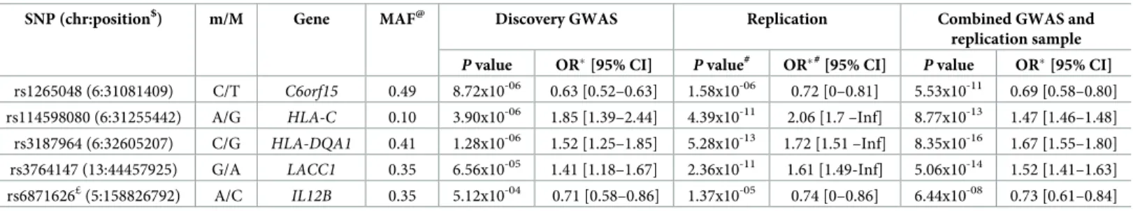
We then investigated in more detail the location, LD in the 1000 Genome KHV population and univariate results in the combined sample of these three HLA SNPs (Table 2). The
Table 2. Association statistics in the discovery GWAS, the case/control replication and the combined GWAS and replication samples for the five independent SNPs identified by multivariate analysis.
SNP (chr:position$) m/M Gene MAF@ Discovery GWAS Replication Combined GWAS and replication sample
P value OR�[95% CI] P value#
OR�#
[95% CI] P value OR�[95% CI]
rs1265048 (6:31081409) C/T C6orf15 0.49 8.72x10-06 0.63 [0.52–0.63] 1.58x10-06 0.72 [0–0.81] 5.53x10⁻¹¹ 0.69 [0.58–0.80] rs114598080 (6:31255442) A/G HLA-C 0.10 3.90x10-06 1.85 [1.39–2.44] 4.39x10⁻¹¹ 2.06 [1.7 –Inf] 8.77x10⁻¹³ 1.47 [1.46–1.48]
rs3187964 (6:32605207) C/G HLA-DQA1 0.41 1.28x10-06 1.52 [1.25–1.85] 5.28x10⁻¹³ 1.72 [1.51 –Inf] 8.35x10⁻¹⁶ 1.67 [1.55–1.80]
rs3764147 (13:44457925) G/A LACC1 0.35 6.56x10-05 1.41 [1.18–1.67] 2.36x10⁻¹¹ 1.61 [1.49-Inf] 5.06x10⁻¹⁴ 1.52 [1.41–1.63] rs6871626£(5:158826792) A/C IL12B 0.35 5.12x10-04 0.71 [0.58–0.86] 1.37x10-05 0.74 [0–0.86] 6.44x10-08 0.73 [0.61–0.84] m/M = minor/Major alleles; MAF = minor allele frequency; OR = odds ratio; CI = confidence interval
$
positions are given according to the hg19 genome build
@
MAF was estimated from the 939 leprosy unaffected parents of the discovery GWAS and 668 controls of the replication sample
�with respect to the minor allele
#
one-tailed test
£
imputed using the 1000 Genomes phase 1 reference panel
rs3187964 SNP belongs to the HLA interval corresponding to the HLA class II region (Fig 2B). In the combined analysis of the discovery and replication samples, it gave a univariatep-value
of 8.3x10⁻¹⁶with an OR for developing leprosy for CG vs. GG (or CC vs. CG) of 1.67 [1.55– 1.80]. This SNP is located in the 5’ untranslated region of theHLA-DQA1 gene and was not in
strong LD (r2> 0.8) with other SNPs in the 1000 Genomes KHV population in an LDproxy
analysis [38]. The other two SNPs are in the first HLA interval, which includes HLA class I genes, together with many other genes (Fig 2A). The rs114598080 SNP, located in a CTCF binding site (ENSR00000262841) 15.6 kb upstream of theHLA-C gene, gave a univariate
p-value of 8.8x10⁻¹³in the combined analysis, with an OR for developing leprosy for AG vs. GG (or AA vs. AG) of 1.47 [1.46–1.48]. This SNP belongs to a bin of 245 SNPs in strong LD that span almost 200 kb (S8 Table). The third SNP, rs1265048, is located near theC6orf15 (1.07
kb),PSORS1C1 (1.12 kb), and CDSN (1.2 kb) genes and it gave a univariate p-value of 5.5x10 -11
in the combined analysis, with an OR for developing leprosy for CT vs. TT (or CC vs. CT) of 0.69 [0.58–0.80]. It was not in strong LD with other SNPs in the 1000 Genomes KHV popula-tion in LDproxy analysis.
Discussion
We performed the first family-based GWAS for leprosy in a Vietnamese population. The main susceptibility loci identified were in the HLA region. The most significant SNP, rs3187964, is located in the MHC class II region, in the 5’ untranslated region ofHLA-DQA1 and did not
display strong LD (r2> 0.8) with other SNPs in the region in the 1000 Genomes KHV
Fig 2. Regional association plot for HLA class I (A) and class II (B) SNPs in the case/control replication sample. Locus zoom plot showing one-tailedp-values for
association within the HLA region. The three independent SNPs identified by multivariate analysis, rs1265048 (A), rs114598080 (A) and rs3187964 (B), are represented as blue, red and purple diamonds, respectively. The colors indicate the pairwise linkage disequilibrium (r2) with the three independent SNPs, as calculated in the 1000
Genomes KHV population. SNPs not found in the reference populations are shown in gray.
population. Several studies have consistently reported the involvement of HLA class II alleles, mostlyHLA-DRB1 alleles, as key genetic factors controlling susceptibility to various forms of
leprosy [11,18,39–41]. A recent HLA imputation-based meta-analysis in the Chinese
popula-tion identifiedHLA-DRB1 and HLA-DQA1 alleles and specific amino acids of HLA-DRβ1 as
independent protective factors for leprosy [39]. Interestingly, in the GTex project V7 (https:// gtexportal.org/home/), rs3187964 was identified as a strong eQTL for several HLA class II
genes withp-values below 10−50forHLA-DQB1, HLA-DQB2, HLA-DQA1 and HLA-DQA2 in
multiple tissues, including skin and nerves (S9 Table). It was also found to be a splice QTL for
HLA-DQA1 (nominal p-value = 3.3x10⁻⁷, FDR = 0.000155). HLA-DQ genes have been impli-cated in several autoimmune and inflammatory diseases [42] and, recently, in susceptibility to streptococcal disease [43]. Leprosy patients are prone to develop acute inflammatory reaction, T1R, and we have recently shown a striking overlap between T1R and inflammatory bowel dis-ease (IBD) risk alleles [24]. However, SNPs of the HLA region were not major risk loci for T1R suggesting an effect of HLA SNPs on leprosyper se susceptibility and not on T1R.
Interest-ingly, there are several examples of loci with mirror genetic effects between infectious diseases and inflammatory disorders, where the allele protective against infection is deleterious for inflammatory disorders, consistent with the view that the current high frequency of inflamma-tory/autoimmune diseases may reflect past selection for strong immune responses to combat infection [44,45]. Additional studies are needed to precise the role of HLA-DQ genes in lep-rosy susceptibility with respect to T1R and inflammatory diseases.
The other two independent SNPs associated with leprosy in the HLA region are located in the class I region. The most significant, rs114598080, belongs to a large bin of SNPs in strong
LD spanning the HLA class I genesHLA-C and HLA-B and the non-HLA genes CCHCR1,
TCF19, POU5F1, PSORS1C3, and HCG27. The complex LD structure makes it difficult to
identify the gene related to this association hit. Nevertheless, one SNP of the bin, rs2394885, was shown to tag theHLA-C�15:05 allele in the Vietnamese population [46]. This SNP was also validated as a leprosy susceptibility locus in an Indian population [46]. In the Chinese
HLA imputation-based meta-analysis,HLA-C�08:01 and specific HLA-C amino acids were
found to be independent protective and risk factors for leprosy [39]. The rs114598080 SNP is
also in strong LD with a possibly damaging missense variant of theCCHCR1 gene. CCHCR1 is
a protein of unknown function that was recently found to have properties typical of P body-resident proteins [47]. P bodies are evolutionary conserved cytoplasmic components that con-tain proteins involved in mRNA degradation, translation repression, and mRNA storage [48]. Interestingly, P body core components were recently shown to positively regulate plant pat-tern-triggered innate immunity [49]. In addition, rs114598080 is a strong eQTL in sun-exposed skin and tibial artery forPOU5F1 (p < 10−10), also known asOCT4, which encodes a
master transcription factor for pluripotent cell self-renewal [50] (S10 Table). The second SNP of the HLA class I region, rs1265048, is located 1.1 kb upstream ofC6orf15, 1.1 kb upstream of PSORS1C1 and 1.2 kb upstream of CDSN. Interestingly, the SNP correlates with the expression
of several genes, includingHLA-C (minimum p-value = 1.3x10⁻⁵
), andPOU5F1 (minimum
p-value = 2.8x10⁻⁵) in particular, sporadically in some tissues (S11 Table).
In addition to the HLA locus, we were able to consistently replicate theLACC1 and IL12B
loci previously associated with leprosy in the Chinese population both in the discovery GWAS and the replication sample. TheLACC1 leprosy susceptibility SNP rs3764147 is a missense
var-iant (p.Ile254Val) first identified by a leprosy GWAS in the Han Chinese population [9] and then replicated in Indian, African [51], Vietnamese [20], Yi [52], Wenshan and Yuxi [16] Chi-nese populations. The leprosy susceptibility allele was also shown to be associated with an increased susceptibility to Crohn’s [53,54] and inflammatory bowel diseases [55,56]. SNP rs3764147 has a PHRED-scaled CADD score [57,58] of 16, placing it in the top 2.5% of
deleterious variants relative to all possible reference genome single-nucleotide variants. Inter-estingly, recent functional studies have shown thatLACC1 encodes a central regulator of the
metabolic function and bioenergetic state of macrophages [59]. Furthermore, the rs3764147 G risk allele is associated with lower levels of innate receptor-induced activity in primary human monocyte-derived macrophages [60], potentially accounting for the higher susceptibility to intestinal inflammatory diseases and leprosy.
SNP rs6871626 is located in an intergenic region on chromosome 5. In a previous genome-wide meta-analysis study of ulcerative colitis, the most likely candidate gene assigned to this SNP by Gene Relationship Across Implicated Loci (GRAIL) pathway analysis wasIL12B [61]. GRAIL is a statistical tool that assesses the degree of relatedness of a list of disease regions by text mining PubMed abstracts, for the annotation of candidate genes for disease risk [62].
IL12B encodes IL-12p40, which is common to two interleukins, IL-12 and IL-23. Both
IL-12-and IL-23-dependent signaling pathways play critical roles in human antimycobacterial immunity, through the induction of IFN-γ [63,64]. Autosomal recessive completeIL12B
defi-ciency is a genetic etiology of Mendelian susceptibility to mycobacterial diseases (MSMD), a rare condition characterized by predisposition to clinical disease caused by weakly virulent mycobacteria, such as BCG vaccines and environmental mycobacteria, in otherwise healthy individuals [65]. The rs6871626 C allele is associated with a risk of leprosy, but has been shown to be protective against ulcerative colitis [61], ankylosing spondylitis and Crohn’s disease [66], inflammatory bowel disease [55] and Takayasu arteritis [67]. Interestingly, in patients with Takayasu arteritis, the rs6871626 C allele, which confers susceptibility to leprosy, was associ-ated with abnormally low levels of IL-12 (composed of IL-12p40 and IL-12p35) in plasma and low levels of both IL-12p40 and IL12 in the culture supernatants of patient monocytes/macro-phages stimulated with LPS [68].
Our study had some limitations. First, our sample size was limited with 622 affected off-spring. Assuming 622 trios, the power of the transmission disequilibrium test was below 80% for SNPs with a MAF of 0.05 with a relative risk below 2 or for SNPs with a MAF of 0.3 with a relative risk below 1.5, and we may have missed novel associations and failed to replicate some of the previously identified loci with modest genetic effect size. Second, as in all leprosy studies, the case definition was purely clinical. Although leprosy was diagnosed by two independent and experienced physicians, we cannot exclude some misdiagnosis which may affect the power of the study. Third, two out of eight of the non-HLA suggestive hits and one out of five of the pre-viously published non-HLA hits could not be tested for association in the replication sample because of genotyping failure. Finally, the investigation of the identified loci was based solely on in silico analysis using publicly available resources and not on functional studies. In conclusion, we performed the first family-based GWAS of leprosyper se in the Vietnamese population. The
strongest association signals were observed for SNPs within the HLA region. We also validated the independent impact of two additional variants, one inLACC1 and the second close to IL12B, recently reported to play functional roles. We further broke down the association signal
within the HLA region into three independent signals, two mapping to the HLA class I region, and one, the leading signal, located in the class II region close toHLA-DQA1. More refined
analyses based on direct HLA typing, which is lacking so far, are required to characterize further the precise contribution of HLA class I and class II alleles to leprosy susceptibility.
Supporting information
S1 Table. Clinical characteristics of study populations from the Vietnamese family-based and case–control studies.
S2 Table. LD-based clumping of genome-wide suggestive non-HLA hits. (XLSX)
S3 Table. Previously published genome-wide significant SNPs outside the HLA region. (XLSX)
S4 Table. SNPs in high linkage disequilibrium (r2>0.8) with rs3764147 in the 1000
Genomes KHV population. (XLSX)
S5 Table. Association statistics of genome-wide suggestive HLA SNPs in the discovery cohort and the replication cohort.
(XLSX)
S6 Table. LD-based clumping of replicated SNPs within the HLA region in the case/control replication sample.
(XLSX)
S7 Table. Results of the multivariate analysis within the HLA region in the case/control replication sample.
(XLSX)
S8 Table. SNPs in high linkage disequilibrium (r2>0.8) with rs114598080 in the 1000
genomes KHV population. (XLSX)
S9 Table. Single-Tissue eQTL for rs3187964 in the GTEx project. (XLSX)
S10 Table. Single-Tissue eQTL for ts114598080 in the GTEx project. (XLSX)
S11 Table. Single-Tissue eQTL for rs1265048 in the GTEx project. (XLSX)
S1 Fig. Principal component analysis of the GWAS discovery sample with all 1000 genomes phase 3 samples (A) and with the East-Asian 1000 genomes samples (B) AFR = African; AMR = American; EAS = East Asian; EUR = European; GWAS = Genome-wide association study samples; SAS = South Asian; CDX = Chinese Dai in Xishuangbanna, China; CHB = Han Chinese in Beijing, China; CHS = Southern Han Chinese; JPT = Japanese in Tokyo, Japan; KHV = Kinh in Ho Chi Minh City, Vietnam.
(TIF)
S2 Fig. Quantile-quantile plot of the association results with leprosyper se in the family-based GWAS for all SNPs (black) and all SNPs outside the HLA region (grey).
(TIF)
Acknowledgments
We thank all leprosy patients and their family who participated in this study. We thank the staff of leprosy control program in Southern Vie
˙
ˆt Nam and the members of leprosy control program at Hospital of D-V of Ho Chi Minh City, Vie
˙
ˆt Nam. We thank members of the labo-ratory of human genetics of infectious diseases and the Erwin Schurr labolabo-ratory for helpful dis-cussions. We thank the French National Centre of Human Genomics Research (CNRGH) and Genome Quebec for performing genotyping.
Author Contributions
Conceptualization: Alexandre Alcaïs, Erwin Schurr. Data curation: Chaima Gzara, Marianna Orlova.
Formal analysis: Chaima Gzara, Monica Dallmann-Sauer. Funding acquisition: Alexandre Alcaïs, Erwin Schurr. Methodology: Chaima Gzara, Aure´lie Cobat.
Resources: Nguyen Van Thuc, Vu Hong Thai, Vinicius M. Fava, Marie-The´rèse Bihoreau, Anne Boland.
Supervision: Laurent Abel, Alexandre Alcaïs, Aure´lie Cobat. Validation: Aure´lie Cobat.
Visualization: Chaima Gzara.
Writing – original draft: Chaima Gzara, Laurent Abel, Aure´lie Cobat.
Writing – review & editing: Chaima Gzara, Monica Dallmann-Sauer, Marianna Orlova, Vini-cius M. Fava, Alexandre Alcaïs, Erwin Schurr.
References
1. Smith WC, van Brakel W, Gillis T, Saunderson P, Richardus JH. The missing millions: a threat to the elimination of leprosy. PLoS Negl Trop Dis. 2015; 9(4):e0003658. Epub 2015/04/24.https://doi.org/10. 1371/journal.pntd.0003658PMID:25905706; PubMed Central PMCID: PMC4408099.
2. Gaschignard J, Grant AV, Thuc NV, Orlova M, Cobat A, Huong NT, et al. Pauci- and Multibacillary Lep-rosy: Two Distinct, Genetically Neglected Diseases. PLoS Negl Trop Dis. 2016; 10(5):e0004345. https://doi.org/10.1371/journal.pntd.0004345PMID:27219008; PubMed Central PMCID: PMC4878860.
3. Cole ST, Eiglmeier K, Parkhill J, James KD, Thomson NR, Wheeler PR, et al. Massive gene decay in the leprosy bacillus. Nature. 2001; 409(6823):1007–11. Epub 2001/03/10.https://doi.org/10.1038/ 35059006PMID:11234002.
4. Monot M, Honore N, Garnier T, Araoz R, Coppee JY, Lacroix C, et al. On the origin of leprosy. Science. 2005; 308(5724):1040–2. Epub 2005/05/17.https://doi.org/10.1126/science/1109759PMID:
15894530.
5. Fava VM, Dallmann-Sauer M, Schurr E. Genetics of leprosy: today and beyond. Hum Genet. 2019. Epub 2019/11/13.https://doi.org/10.1007/s00439-019-02087-5PMID:31713021.
6. Mira MT, Alcais A, Van Thuc N, Thai VH, Huong NT, Ba NN, et al. Chromosome 6q25 is linked to sus-ceptibility to leprosy in a Vietnamese population. Nat Genet. 2003; 33(3):412–5. Epub 2003/02/11. https://doi.org/10.1038/ng1096PMID:12577057.
7. Mira MT, Alcais A, Nguyen VT, Moraes MO, Di Flumeri C, Vu HT, et al. Susceptibility to leprosy is asso-ciated with PARK2 and PACRG. Nature. 2004; 427(6975):636–40. Epub 2004/01/23.https://doi.org/10. 1038/nature02326PMID:14737177.
8. Alcais A, Alter A, Antoni G, Orlova M, Nguyen VT, Singh M, et al. Stepwise replication identifies a low-producing lymphotoxin-alpha allele as a major risk factor for early-onset leprosy. Nat Genet. 2007; 39 (4):517–22. Epub 2007/03/14.https://doi.org/10.1038/ng2000PMID:17353895.
9. Zhang FR, Huang W, Chen SM, Sun LD, Liu H, Li Y, et al. Genomewide association study of leprosy. N Engl J Med. 2009; 361(27):2609–18. Epub 2009/12/19.https://doi.org/10.1056/NEJMoa0903753 PMID:20018961.
10. Zhang F, Liu H, Chen S, Low H, Sun L, Cui Y, et al. Identification of two new loci at IL23R and RAB32 that influence susceptibility to leprosy. Nat Genet. 2011; 43(12):1247–51. Epub 2011/10/25.https://doi. org/10.1038/ng.973PMID:22019778.
11. Liu H, Irwanto A, Fu X, Yu G, Yu Y, Sun Y, et al. Discovery of six new susceptibility loci and analysis of pleiotropic effects in leprosy. Nat Genet. 2015; 47(3):267–71. Epub 2015/02/03.https://doi.org/10. 1038/ng.3212PMID:25642632.
12. Wang Z, Sun Y, Fu X, Yu G, Wang C, Bao F, et al. A large-scale genome-wide association and meta-analysis identified four novel susceptibility loci for leprosy. Nat Commun. 2016; 7:13760. Epub 2016/12/ 16.https://doi.org/10.1038/ncomms13760PMID:27976721; PubMed Central PMCID: PMC5172377.
13. Wong SH, Gochhait S, Malhotra D, Pettersson FH, Teo YY, Khor CC, et al. Leprosy and the adaptation of human toll-like receptor 1. PLoS Pathog. 2010; 6:e1000979. Epub 2010/07/10.https://doi.org/10. 1371/journal.ppat.1000979PMID:20617178; PubMed Central PMCID: PMC2895660.
14. Liu H, Wang Z, Li Y, Yu G, Fu X, Wang C, et al. Genome-Wide Analysis of Protein-Coding Variants in Leprosy. J Invest Dermatol. 2017; 137(12):2544–51. Epub 2017/08/27.https://doi.org/10.1016/j.jid. 2017.08.004PMID:28842327.
15. Wang Z, Mi Z, Wang H, Sun L, Yu G, Fu X, et al. Discovery of 4 exonic and 1 intergenic novel suscepti-bility loci for leprosy. Clin Genet. 2018; 94(2):259–63. Epub 2018/05/04.https://doi.org/10.1111/cge. 13376PMID:29722023.
16. Wang D, Fan Y, Malhi M, Bi R, Wu Y, Xu M, et al. Missense Variants in HIF1A and LACC1 Contribute to Leprosy Risk in Han Chinese. Am J Hum Genet. 2018; 102(5):794–805. Epub 2018/05/01.https://doi. org/10.1016/j.ajhg.2018.03.006PMID:29706348; PubMed Central PMCID: PMC5986702.
17. Dallmann-Sauer M, Correa-Macedo W, Schurr E. Human genetics of mycobacterial disease. Mamm Genome. 2018; 29(7–8):523–38. Epub 2018/08/18.https://doi.org/10.1007/s00335-018-9765-4PMID: 30116885; PubMed Central PMCID: PMC6132723.
18. Cambri G, Mira MT. Genetic Susceptibility to Leprosy-From Classic Immune-Related Candidate Genes to Hypothesis-Free, Whole Genome Approaches. Front Immunol. 2018; 9:1674. Epub 2018/08/07. https://doi.org/10.3389/fimmu.2018.01674PMID:30079069; PubMed Central PMCID: PMC6062607.
19. Cobat A, Abel L, Alcais A, Schurr E. A general efficient and flexible approach for genome-wide associa-tion analyses of imputed genotypes in family-based designs. Genet Epidemiol. 2014; 38(6):560–71. Epub 2014/07/22.https://doi.org/10.1002/gepi.21842PMID:25044438.
20. Grant AV, Alter A, Huong NT, Orlova M, Van Thuc N, Ba NN, et al. Crohn’s disease susceptibility genes are associated with leprosy in the Vietnamese population. J Infect Dis. 2012; 206(11):1763–7. Epub 2012/09/18.https://doi.org/10.1093/infdis/jis588PMID:22984114.
21. Fava VM, Cobat A, Van Thuc N, Latini AC, Stefani MM, Belone AF, et al. Association of TNFSF8 regula-tory variants with excessive inflammaregula-tory responses but not leprosy per se. J Infect Dis. 2015; 211 (6):968–77. Epub 2014/10/17.https://doi.org/10.1093/infdis/jiu566PMID:25320285.
22. Fava VM, Manry J, Cobat A, Orlova M, Van Thuc N, Ba NN, et al. A Missense LRRK2 Variant Is a Risk Factor for Excessive Inflammatory Responses in Leprosy. PLoS Negl Trop Dis. 2016; 10(2):e0004412. https://doi.org/10.1371/journal.pntd.0004412PMID:26844546; PubMed Central PMCID:
PMC4742274.
23. Fava VM, Xu YZ, Lettre G, Van Thuc N, Orlova M, Thai VH, et al. Pleiotropic effects for Parkin and LRRK2 in leprosy type-1 reactions and Parkinson’s disease. Proc Natl Acad Sci U S A. 2019; 116 (31):15616–24. Epub 2019/07/17.https://doi.org/10.1073/pnas.1901805116PMID:31308240; PubMed Central PMCID: PMC6681704.
24. Fava VM, Manry J, Cobat A, Orlova M, Van Thuc N, Moraes MO, et al. A genome wide association study identifies a lncRna as risk factor for pathological inflammatory responses in leprosy. PLoS Genet. 2017; 13(2):e1006637.https://doi.org/10.1371/journal.pgen.1006637PMID:28222097; PubMed Cen-tral PMCID: PMC5340414.
25. Teo YY, Inouye M, Small KS, Gwilliam R, Deloukas P, Kwiatkowski DP, et al. A genotype calling algo-rithm for the Illumina BeadArray platform. Bioinformatics. 2007; 23(20):2741–6. Epub 2007/09/12. https://doi.org/10.1093/bioinformatics/btm443PMID:17846035; PubMed Central PMCID: PMC2666488.
26. Purcell S, Chang C. PLINK 1.9. Available from:www.cog-genomics.org/plink/1.9/.
27. Delaneau O, Marchini J, Zagury JF. A linear complexity phasing method for thousands of genomes. Nat Methods. 2011; 9(2):179–81. Epub 2011/12/06.https://doi.org/10.1038/nmeth.1785PMID:22138821.
28. O’Connell J, Gurdasani D, Delaneau O, Pirastu N, Ulivi S, Cocca M, et al. A general approach for haplo-type phasing across the full spectrum of relatedness. PLoS Genet. 2014; 10(4):e1004234. Epub 2014/ 04/20.https://doi.org/10.1371/journal.pgen.1004234PMID:24743097; PubMed Central PMCID: PMC3990520.
29. Marchini J, Howie B, Myers S, McVean G, Donnelly P. A new multipoint method for genome-wide asso-ciation studies by imputation of genotypes. Nat Genet. 2007; 39(7):906–13. Epub 2007/06/19.https:// doi.org/10.1038/ng2088PMID:17572673.
30. Chang CC, Chow CC, Tellier LC, Vattikuti S, Purcell SM, Lee JJ. Second-generation PLINK: rising to the challenge of larger and richer datasets. Gigascience. 2015; 4:7. Epub 2015/02/28.https://doi.org/ 10.1186/s13742-015-0047-8PMID:25722852; PubMed Central PMCID: PMC4342193.
31. DePristo MA, Banks E, Poplin R, Garimella KV, Maguire JR, Hartl C, et al. A framework for variation dis-covery and genotyping using next-generation DNA sequencing data. Nat Genet. 2011; 43(5):491–8. Epub 2011/04/12.https://doi.org/10.1038/ng.806PMID:21478889; PubMed Central PMCID: PMC3083463.
32. Laird NM, Horvath S, Xu X. Implementing a unified approach to family-based tests of association. Genet Epidemiol. 2000; 19 Suppl 1:S36–42.https://doi.org/10.1002/1098-2272(2000)19:1+< ::AID-GEPI6>3.0.CO;2-MPMID:11055368.
33. Cordell HJ, Barratt BJ, Clayton DG. Case/pseudocontrol analysis in genetic association studies: A uni-fied framework for detection of genotype and haplotype associations, gene-gene and gene-environment interactions, and parent-of-origin effects. Genet Epidemiol. 2004; 26(3):167–85. Epub 2004/03/17. https://doi.org/10.1002/gepi.10307PMID:15022205.
34. Grant AV, El Baghdadi J, Sabri A, El Azbaoui S, Alaoui-Tahiri K, Abderrahmani Rhorfi I, et al. Age-dependent association between pulmonary tuberculosis and common TOX variants in the 8q12-13 link-age region. Am J Hum Genet. 2013; 92(3):407–14. Epub 2013/02/19.https://doi.org/10.1016/j.ajhg. 2013.01.013PMID:23415668; PubMed Central PMCID: PMC3591857.
35. Grant AV, Sabri A, Abid A, Abderrahmani Rhorfi I, Benkirane M, Souhi H, et al. A genome-wide associa-tion study of pulmonary tuberculosis in Morocco. Hum Genet. 2016; 135(3):299–307. Epub 2016/01/16. https://doi.org/10.1007/s00439-016-1633-2PMID:26767831; PubMed Central PMCID: PMC5042142.
36. Wellcome Trust Case Control C. Genome-wide association study of 14,000 cases of seven common diseases and 3,000 shared controls. Nature. 2007; 447(7145):661–78. Epub 2007/06/08.https://doi. org/10.1038/nature05911PMID:17554300; PubMed Central PMCID: PMC2719288.
37. Karczewski Konrad J., Francioli Laurent C., Tiao Grace, Cummings Beryl B., Jessica Alfo¨ldi Qingbo Wang, et al. Variation across 141,456 human exomes and genomes reveals the spectrum of loss-of-function intolerance across human protein-coding genes. bioRXiv. 2019.https://doi.org/10.1101/ 531210.
38. Machiela MJ, Chanock SJ. LDlink: a web-based application for exploring population-specific haplotype structure and linking correlated alleles of possible functional variants. Bioinformatics. 2015; 31 (21):3555–7. Epub 2015/07/04.https://doi.org/10.1093/bioinformatics/btv402PMID:26139635; PubMed Central PMCID: PMC4626747.
39. Zhang X, Cheng Y, Zhang Q, Wang X, Lin Y, Yang C, et al. Meta-Analysis Identifies Major Histocompa-tiblity Complex Loci in or Near HLA-DRB1, HLA-DQA1, HLA-C as Associated with Leprosy in Chinese Han Population. J Invest Dermatol. 2019; 139(4):957–60. Epub 2018/11/06.https://doi.org/10.1016/j. jid.2018.09.029PMID:30389493.
40. Jarduli LR, Sell AM, Reis PG, Sippert EA, Ayo CM, Mazini PS, et al. Role of HLA, KIR, MICA, and cyto-kines genes in leprosy. Biomed Res Int. 2013; 2013:989837. Epub 2013/08/13.https://doi.org/10.1155/ 2013/989837PMID:23936864; PubMed Central PMCID: PMC3722889.
41. Krause-Kyora B, Nutsua M, Boehme L, Pierini F, Pedersen DD, Kornell SC, et al. Ancient DNA study reveals HLA susceptibility locus for leprosy in medieval Europeans. Nat Commun. 2018; 9(1):1569. Epub 2018/05/03.https://doi.org/10.1038/s41467-018-03857-xPMID:29717136; PubMed Central PMCID: PMC5931558.
42. Matzaraki V, Kumar V, Wijmenga C, Zhernakova A. The MHC locus and genetic susceptibility to auto-immune and infectious diseases. Genome Biol. 2017; 18(1):76. Epub 2017/04/30.https://doi.org/10. 1186/s13059-017-1207-1PMID:28449694; PubMed Central PMCID: PMC5406920.
43. Parks T, Elliott K, Lamagni T, Auckland K, Mentzer AJ, Guy R, et al. Elevated risk of invasive group A streptococcal disease and host genetic variation in the human leucocyte antigen locus. Genes Immun. 2019. Epub 2019/08/30.https://doi.org/10.1038/s41435-019-0082-zPMID:31462703.
44. Barreiro LB, Quintana-Murci L. From evolutionary genetics to human immunology: how selection shapes host defence genes. Nat Rev Genet. 2010; 11(1):17–30.https://doi.org/10.1038/nrg2698 PMID:19953080.
45. Manry J, Vincent QB, Johnson C, Chrabieh M, Lorenzo L, Theodorou I, et al. Genome-wide association study of Buruli ulcer in rural Benin highlights role of two LncRNAs and the autophagy pathway. Commun Biol. 2020; 3(1):177.
46. Alter A, Huong NT, Singh M, Orlova M, Van Thuc N, Katoch K, et al. Human leukocyte antigen class I region single-nucleotide polymorphisms are associated with leprosy susceptibility in Vietnam and India. J Infect Dis. 2011; 203(9):1274–81. Epub 2011/04/05.https://doi.org/10.1093/infdis/jir024PMID: 21459816; PubMed Central PMCID: PMC3069725.
47. Luo Y, Na Z, Slavoff SA. P-Bodies: Composition, Properties, and Functions. Biochemistry. 2018; 57 (17):2424–31. Epub 2018/01/31.https://doi.org/10.1021/acs.biochem.7b01162PMID:29381060; PubMed Central PMCID: PMC6296482.
48. Parker R, Sheth U. P bodies and the control of mRNA translation and degradation. Mol Cell. 2007; 25 (5):635–46.https://doi.org/10.1016/j.molcel.2007.02.011PMID:17349952.
49. Yu X, Li B, Jang GJ, Jiang S, Jiang D, Jang JC, et al. Orchestration of Processing Body Dynamics and mRNA Decay in Arabidopsis Immunity. Cell Rep. 2019; 28(8):2194–205 e6.https://doi.org/10.1016/j. celrep.2019.07.054PMID:31433992; PubMed Central PMCID: PMC6716526.
50. Radzisheuskaya A, Chia Gle B, dos Santos RL, Theunissen TW, Castro LF, Nichols J, et al. A defined Oct4 level governs cell state transitions of pluripotency entry and differentiation into all embryonic line-ages. Nat Cell Biol. 2013; 15(6):579–90. Epub 2013/05/01.https://doi.org/10.1038/ncb2742PMID: 23629142; PubMed Central PMCID: PMC3671976.
51. Wong SH, Hill AV, Vannberg FO, India-Africa-United Kingdom Leprosy Genetics C. Genomewide asso-ciation study of leprosy. N Engl J Med. 2010; 362(15):1446–7; author reply 7–8. Epub 2010/04/16. https://doi.org/10.1056/NEJMc1001451PMID:20393182.
52. Xiong JH, Mao C, Sha XW, Jin Z, Wang H, Liu YY, et al. Association between genetic variants in NOD2, C13orf31, and CCDC122 genes and leprosy among the Chinese Yi population. Int J Dermatol. 2016; 55 (1):65–9. Epub 2015/08/04.https://doi.org/10.1111/ijd.12981PMID:26235265.
53. Barrett JC, Hansoul S, Nicolae DL, Cho JH, Duerr RH, Rioux JD, et al. Genome-wide association defines more than 30 distinct susceptibility loci for Crohn’s disease. Nat Genet. 2008; 40(8):955–62. Epub 2008/07/01.https://doi.org/10.1038/ng.175PMID:18587394; PubMed Central PMCID: PMC2574810.
54. Franke A, McGovern DP, Barrett JC, Wang K, Radford-Smith GL, Ahmad T, et al. Genome-wide meta-analysis increases to 71 the number of confirmed Crohn’s disease susceptibility loci. Nat Genet. 2010; 42(12):1118–25. Epub 2010/11/26.https://doi.org/10.1038/ng.717PMID:21102463; PubMed Central PMCID: PMC3299551.
55. Jostins L, Ripke S, Weersma RK, Duerr RH, McGovern DP, Hui KY, et al. Host-microbe interactions have shaped the genetic architecture of inflammatory bowel disease. Nature. 2012; 491(7422):119–24. Epub 2012/11/07.https://doi.org/10.1038/nature11582PMID:23128233; PubMed Central PMCID: PMC3491803.
56. Liu JZ, van Sommeren S, Huang H, Ng SC, Alberts R, Takahashi A, et al. Association analyses identify 38 susceptibility loci for inflammatory bowel disease and highlight shared genetic risk across popula-tions. Nat Genet. 2015; 47(9):979–86. Epub 2015/07/21.https://doi.org/10.1038/ng.3359PMID: 26192919; PubMed Central PMCID: PMC4881818.
57. Kircher M, Witten DM, Jain P, O’Roak BJ, Cooper GM, Shendure J. A general framework for estimating the relative pathogenicity of human genetic variants. Nat Genet. 2014; 46(3):310–5. Epub 2014/02/04. https://doi.org/10.1038/ng.2892PMID:24487276; PubMed Central PMCID: PMC3992975.
58. Rentzsch P, Witten D, Cooper GM, Shendure J, Kircher M. CADD: predicting the deleteriousness of variants throughout the human genome. Nucleic Acids Res. 2019; 47(D1):D886–D94. Epub 2018/10/ 30.https://doi.org/10.1093/nar/gky1016PMID:30371827; PubMed Central PMCID: PMC6323892.
59. Cader MZ, Boroviak K, Zhang Q, Assadi G, Kempster SL, Sewell GW, et al. C13orf31 (FAMIN) is a cen-tral regulator of immunometabolic function. Nat Immunol. 2016; 17(9):1046–56. Epub 2016/08/02. https://doi.org/10.1038/ni.3532PMID:27478939; PubMed Central PMCID: PMC6581540.
60. Lahiri A, Hedl M, Yan J, Abraham C. Human LACC1 increases innate receptor-induced responses and a LACC1 disease-risk variant modulates these outcomes. Nat Commun. 2017; 8:15614. Epub 2017/06/ 09.https://doi.org/10.1038/ncomms15614PMID:28593945; PubMed Central PMCID: PMC5472760.
61. Anderson CA, Boucher G, Lees CW, Franke A, D’Amato M, Taylor KD, et al. Meta-analysis identifies 29 additional ulcerative colitis risk loci, increasing the number of confirmed associations to 47. Nat Genet. 2011; 43(3):246–52. Epub 2011/02/08.https://doi.org/10.1038/ng.764PMID:21297633; PubMed Cen-tral PMCID: PMC3084597.
62. Raychaudhuri S, Plenge RM, Rossin EJ, Ng AC, International Schizophrenia C, Purcell SM, et al. Identi-fying relationships among genomic disease regions: predicting genes at pathogenic SNP associations and rare deletions. PLoS Genet. 2009; 5(6):e1000534. Epub 2009/06/27.https://doi.org/10.1371/ journal.pgen.1000534PMID:19557189; PubMed Central PMCID: PMC2694358.
63. Boisson-Dupuis S, Ramirez-Alejo N, Li Z, Patin E, Rao G, Kerner G, et al. Tuberculosis and impaired IL-23-dependent IFN-gamma immunity in humans homozygous for a common TYK2 missense variant. Sci Immunol. 2018; 3(30). Epub 2018/12/24.https://doi.org/10.1126/sciimmunol.aau8714PMID: 30578352; PubMed Central PMCID: PMC6341984.
64. Martinez-Barricarte R, Markle JG, Ma CS, Deenick EK, Ramirez-Alejo N, Mele F, et al. Human IFN-gamma immunity to mycobacteria is governed by both IL-12 and IL-23. Sci Immunol. 2018; 3(30). Epub 2018/12/24.https://doi.org/10.1126/sciimmunol.aau6759PMID:30578351; PubMed Central PMCID: PMC6380365.
65. Bustamante J, Boisson-Dupuis S, Abel L, Casanova JL. Mendelian susceptibility to mycobacterial dis-ease: genetic, immunological, and clinical features of inborn errors of IFN-gamma immunity. Semin Immunol. 2014; 26(6):454–70. Epub 2014/12/03.https://doi.org/10.1016/j.smim.2014.09.008PMID: 25453225; PubMed Central PMCID: PMC4357480.
66. International Genetics of Ankylosing Spondylitis C, Cortes A, Hadler J, Pointon JP, Robinson PC, Kara-deri T, et al. Identification of multiple risk variants for ankylosing spondylitis through high-density geno-typing of immune-related loci. Nat Genet. 2013; 45(7):730–8. Epub 2013/06/12.https://doi.org/10.1038/ ng.2667PMID:23749187; PubMed Central PMCID: PMC3757343.
67. Terao C, Yoshifuji H, Kimura A, Matsumura T, Ohmura K, Takahashi M, et al. Two susceptibility loci to Takayasu arteritis reveal a synergistic role of the IL12B and HLA-B regions in a Japanese population. Am J Hum Genet. 2013; 93(2):289–97. Epub 2013/07/09.https://doi.org/10.1016/j.ajhg.2013.05.024 PMID:23830516; PubMed Central PMCID: PMC3738822.
68. Nakajima T, Yoshifuji H, Shimizu M, Kitagori K, Murakami K, Nakashima R, et al. A novel susceptibility locus in the IL12B region is associated with the pathophysiology of Takayasu arteritis through IL-12p40 and IL-12p70 production. Arthritis Res Ther. 2017; 19(1):197. Epub 2017/09/07.https://doi.org/10. 1186/s13075-017-1408-8PMID:28874185; PubMed Central PMCID: PMC5585951.