HAL Id: tel-01142116
https://tel.archives-ouvertes.fr/tel-01142116
Submitted on 14 Apr 2015HAL is a multi-disciplinary open access archive for the deposit and dissemination of sci-entific research documents, whether they are pub-lished or not. The documents may come from teaching and research institutions in France or abroad, or from public or private research centers.
L’archive ouverte pluridisciplinaire HAL, est destinée au dépôt et à la diffusion de documents scientifiques de niveau recherche, publiés ou non, émanant des établissements d’enseignement et de recherche français ou étrangers, des laboratoires publics ou privés.
biomolecular helical assemblies with an application to
RecA fillaments
Benjamin Boyer
To cite this version:
Benjamin Boyer. Heligeom : a multiscale approach to studying biomolecular helical assemblies with an application to RecA fillaments. Biomolecules [q-bio.BM]. Université Pierre et Marie Curie - Paris VI, 2014. English. �NNT : 2014PA066579�. �tel-01142116�
Laboratoire de Biochimie Th´eorique
Heligeom
A multiscale approach to studying biomolecular
helical assemblies with an application to RecA
filaments
Th`ese de doctorat de l’Universit´e Pierre et Marie Curie Benjamin Boyer
Under the supervision of Chantal PREVOST
20 june 2014 Jury committee Pr Tˆap Ha Duong Rapporteur Dr Juan Cort´es Rapporteur Pr Martin Zacharias Examinateur Pr Germain Trugnan Examinateur Dr Jean-Marc Victor Examinateur Dr Chantal Pr´evost Directrice de th`ese
Note:
Pictures on the title page were made by David Goodsell. They represent from left to rigth microtubule filaments, recombinase filaments and actin filaments. More information can be found at http://mgl.scripps.edu/ people/goodsell/.
Contents
I Introduction 1
1 General introduction 3
1.1 The cell . . . 3
1.2 General aim of the study. . . 4
1.3 Story of this work . . . 6
2 Macromolecular assemblies 8 2.1 Strength of association . . . 11
2.2 Stabilizing interactions . . . 12
2.3 Characteristics of macromolecular complexes . . . 14
2.4 Protein-DNA complexes . . . 20
2.5 Oligomers and polymers . . . 23
2.6 Towards higher level superstructures . . . 28
3 Homologous Recombination 30 3.1 Homologous recombination . . . 31
3.2 The RecA nucleoprotein filament . . . 32
3.3 ATP hydrolysis . . . 39
3.4 Conclusion . . . 43
II Methods 44 4 Introduction 46 4.1 Methods and representation in PTools . . . 46
4.1.1 Macromolecular docking . . . 46
4.1.2 Coarse-grained models . . . 47
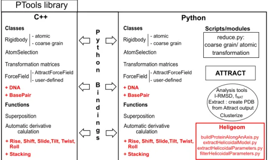
5 PTools: overview and developments 50 5.1 PTools . . . 50
5.1.1 Design . . . 50
5.1.2 PTools Objects and Functions . . . 52
5.1.3 Modules and Scripts . . . 55
5.2 PyAttract . . . 56
6 Manipulation of flexible DNA in PTools 59
6.1 DNA in PTools . . . 59
6.1.1 Editing DNA structures . . . 61
6.1.2 Manipulating DNA structures . . . 63
6.1.3 Deforming DNA structures . . . 63
6.2 First application . . . 65
7 Burdock 69 7.1 Overall strategy. . . 70
7.1.1 Preliminary docking and pruning . . . 71
7.1.2 From prediction to dot . . . 72
7.1.3 Clustering . . . 73 7.1.4 Axis construction . . . 74 7.1.5 DNA construction . . . 75 7.1.6 Optimization . . . 76 7.2 Conclusion . . . 76 8 Heligeom 77 8.1 Screw movement . . . 77 8.2 Construction : 2GLS . . . 79
8.3 Construction around a curved axis . . . 81
8.4 Coupling Heligeom with PyAttract . . . 83
8.5 Optimizing ring geometries . . . 83
8.6 Measuring protein filament groove width . . . 84
III Applications 87 9 Heligeom application : Construction 89 9.1 Extending modeling scales . . . 89
9.2 Interactions between topologically linear assemblies. . . 93
9.3 Structural interpretation of low resolution data . . . 94
9.4 Transferring information from the local to the global levels . 96 9.5 Beyond the topologically linear forms. . . 96
10 Exploring protein filaments 98 10.1 Abstract . . . 101
10.2 Author Summary . . . 102
10.3 Introduction. . . 102
10.4 Results. . . 105
10.4.1 Characterizing known RecA assembly modes . . . 105
10.4.2 Investigating geometries of RecA autoassembly . . . . 108
10.4.3 Variability within selected families of binding modes . 111 10.4.4 Binding mode variations in a single fiber. . . 113
CONTENTS v
10.5 Discussion . . . 115
10.6 Methods . . . 118
10.6.1 Overall approach . . . 118
10.6.2 Screw transformations with Heligeom . . . 119
10.6.3 PDB files . . . 120
10.6.4 Calculating interface area contributions . . . 121
10.6.5 Sampling protein modes of interaction . . . 121
10.6.6 Processing and filtering the sampling results. . . 122
10.6.7 Exploring the variability of binding modes . . . 123
10.7 Acknowledgments . . . 124
10.8 Additional results. . . 126
10.8.1 Flexible RecA fragments . . . 126
10.8.2 Near-cyclic to cyclic adjustment . . . 128
10.8.3 Results from docking simulation on RecA, RadA and Dmc1 . . . 129
10.8.4 Groove width . . . 131
IV Conclusion 132 11 Discussion and conclusion 134 11.1 Discussion . . . 134 V Appendix 138 A R´esum´e 140 A.1 Introduction . . . 140 A.2 Heligeom . . . 141 A.3 Application . . . 142
A.4 Application `a l’´etude de RecA . . . 145
A.5 Conclusion . . . 149
B Exploring RecA interfaces with PTools/Heligeom 150 B.1 Docking procedure . . . 150
B.2 Heligeom Analysis . . . 152
1.1 Development of computing power of the most powerful
com-puters. . . 5
2.1 Lac repressor binding to DNA. . . 10
2.2 Interfacial water molecules. . . 13
2.3 Definition of the buried surface area. . . 14
2.4 Interface topology. . . 16
2.5 Surface remodeling: flexible loops. . . 18
2.6 Proposed scheme for the thermodynamics of flexible protein-protein association. . . 19
2.7 Examples of protein-DNA complexes. . . 21
2.8 Actin network in a keratocyte. . . 25
2.9 Changes in the microtubule network morphology during the cell cycle. . . 26
2.10 Two assembly modes of DnaB cyclic hexamers. . . 27
3.1 Scheme of the strand exchange process. . . 31
3.2 ATP and its homologs. . . 33
3.3 Pitch variations of RecA-ATP and RecA-ADP filaments. . . . 33
3.4 Model of LexA binding to RecA. . . 34
3.5 Crystal structures of the RecA filament. . . 35
3.6 Atomic force microscopy images of hexameric RecA rings. . . 36
3.7 Filament forms of recombinases from higher organisms. . . . 36
3.8 Variety in the oligomeric forms of recombinase proteins. . . . 37
3.9 Structure of RecA filaments with bound DNA. . . 37
3.10 RecA-bound DNA structure is intermediate between B and S DNA forms. . . 38
3.11 3D-SIM imaging of RecA bundles. . . 39
3.12 Cox’s model of hydrolytic waves in a RecA filament. . . 40
3.13 Transitions between stretched and compressed forms of Rad51. 41 3.14 Electron micrograph of mixed RecA filaments. . . 41
5.1 Overview of the PTools library. . . 51
5.2 The force field equation used in pyAttract.. . . 56
LIST OF FIGURES vii
5.3 Coarse-grained representation in PyAttract. . . 56
5.4 The ATTRACT docking approach. . . 58
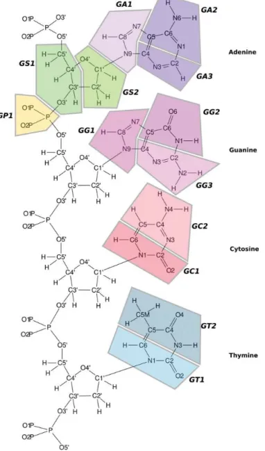
6.1 Coarse-grained model model of DNA. . . 60
6.2 Construction steps of a DNA oligomer. . . 61
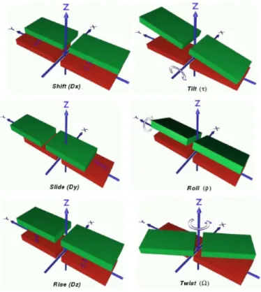
6.3 The six inter base pair translation and orientation parameters. 64 6.4 Geometric criteria for stacking interactions. . . 66
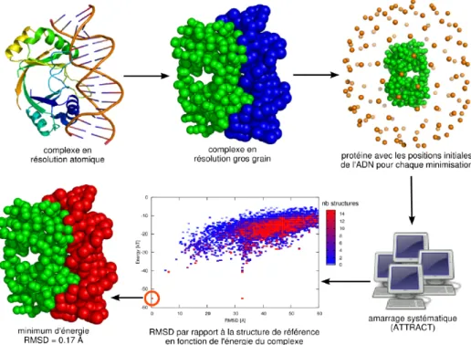
6.5 Results of the Monte Carlo flexible docking procedure. . . 67
7.1 Schematic illustration of the Burdock approach.. . . 70
7.2 Burdock step 1: Pruning. . . 71
7.3 Burdock step 2: Shaping clouds. . . 72
7.4 Burdock step 3: Clustering. . . 73
7.5 Burdock step 4: Axis extraction. . . 74
7.6 Burdock step 5: DNA reconstruction. . . 75
8.1 Scheme of the screw transformation between two monomers A and B. . . 78
8.2 Circular filament of 2GLS reconstructed with Heligeom. . . . 81
8.3 Heligeom construction around a curved axis.. . . 82
8.4 Optimizing near-cyclic geometries. . . 84
8.5 Groove width measurement . . . 86
9.1 Overlapping helical and cyclic forms of Dmc1 . . . 90
9.2 Changing scales: Models of RecA filament with alternating ATP- and ADP-form interfaces. . . 91
9.3 Hyperstructure variability. . . 92
9.4 Small size complex betwen DNA and RecA filament. . . 93
9.5 Large size complex betwen DNA and RecA filament. . . 94
9.6 View of one turn of a RecA negatively supercoiled filament at atomic resolution. . . 95
9.7 Electrostatic potential around an actin filament. . . 96
9.8 Construction steps of a virus capsid. . . 97
10.1 Overview of the Heligeom/PyAttract integrated approach. . . 99
10.2 Scheme of geometric filtering . . . 100
10.3 RecA-RecA docking. . . 106
10.4 RecA auto-assemblage . . . 108
10.5 Exploration of the RecA-ADP and RecA-ATP structural fam-ilies. . . 112
10.6 View of one turn of a RecA negatively supercoiled filament at atomic resolution. . . 114
10.7 Models of RecA filaments with alternating binding modes. . . 115
10.8 Accommodation of flexible regions in predicted RecA fiber forms. . . 125
10.9 Variability of the ADP and ATP fiber forms in the presence
of flexible/mobile interaction component. . . 127
10.10Helical parameters from Heligeom for regular assemblies ob-tained from ATTRACT docking simulations in RecA, RadA, and Dmc1. . . 130
10.11Variation of the groove width along one filament turn. . . 131
A.1 Sch´ema d’une transformation de vissage entre deux monom`eres A et B. . . 141
A.2 Illustration de la construction d’une h´elice par transforma-tions de vissage. . . 142
A.3 Constructions sur un axe arbitraire. . . 143
A.4 Formes “voisines” en h´elice et en anneau de Dmc1. . . 144
A.5 Potentiel ´electrostatique autour d’un filament d’actine. . . 145
A.6 Vue d’un tour de filament de RecA superenroul´e, en r´esolution atomique. . . 146
A.7 Etapes de construction d’une capside de virus `a partir de quatre unit´es en interaction. . . 147
A.8 Illustration de la diversit´e des formes possibles pour les oligom`eres de RecA. . . 148
Part I
Introduction
Chapter 1
General introduction
1.1
The cell
The name cell was proposed by R.Hooke in 1665 [1], following observa-tions on cork conducted with a microscope of his own design. This first observation was soon followed by many others and eventually a theory was developed in 1939 [2], based on the observation of Matthias Jakob Schleiden on plants [3] and Theodor Schwann on notochord. They formulated the idea that the cell is a basic biological unit of life that can exist as an independent organism or as a brick for larger organisms.
The theory was soon completed by the idea that all living cells arise from pre-existing cells by division. This idea was proposed by Rudolf Virchow in 1858 [4], influenced by the work of Robert Remak [5] on embryology in 1855. By a famous series of experiments in 1861, Louis Pasteur [6,7] refuted the concurrent theory of spontaneous generation, giving cell theory considerable momentum.
Since then, the study of the cell has been a corner stone of biology research.
The general organization of a cell is relatively simple : a plasma mem-brane mainly composed of a lipid bilayer separates the protoplasm from the outside environment. The protoplasm contains many biomolecules such as proteins and nucleic acids. Two types of cells exist : eukaryotic cells pos-sess a nucleus (first observed by Antonie van Leeuwenhoek in 1722 [8]) that contains most of the genetic material of the cell, while in prokaryotic cells the genetic material is by contrast directly found in the protoplasm. This proposition of a fundamental division in the realm of life was formulated by Roger Stanier and C. B. van Niel in 1962 [9]. Further distinction, proposed by Carl Richard Woese [10], separates prokaryotes into the Bacteria and Archaea based on differences in the cell wall structure and genetics.
Most of the mechanical properties of the cell depend on the
ton1, an ensemble of large protein filaments that structure and organize the cell. First thought unique to the eukaryotes, the presence of the cytoskele-ton was later demonstrated in prokaryotes in 1991 [12]. The cytoskeleton is composed of actin and microtubules in addition to diverse intermediate filaments in metazoan cells2. Actin plays a role in muscle contraction, cell motility, cell division and cytokinesis and cell signalling. Microtubules play a role in the movement of secretory vesicles, organelles and intracellular substances, the internal structure of cilia and flagella and the cell division. Other filaments can play an important role in decisive functions of the cell, such as the mechanical support ensured by trichocytic keratins that make up hair or nails, or mechanisms fundamental for the cell survival such as homologous recombination performed by the RecA filaments.
The study of protein filamentous assemblies appears to be a key step in the understanding of the cell.
1.2
General aim of the study
These last years have testified quick progress in so-called “low resolution” tech-niques such as electron microscopy (EM) or small angle neutron or X-ray scattering (SAS), both for data generation and interpretation and for the range of biological systems that can be studied.
Impressive progress is also underway in the field of 3D cell imaging, notably using fluorescent proteins and high resolution apparatus (e.g. 3D super-resolution microscopy and time lapse analysis), which permits for ex-ample to visualize the formation of supramolecular assemblies and their evolution along the cell cycle [13]. Although still unsuited to detailed study at the atomic or even molecular level, these techniques offer a wealth of information at the level of supramolecular assembly.
Molecular modeling is one of the fields that can contribute to associ-ating the observed global shapes with detailed structural, mechanical and dynamical characteristics at the atomic level, in an integrated vision of the cell.
However, molecular modeling only begins to take advantage of such im-portant data mainly because the principal techniques, such as Molecular Dynamics (MD) or Normal Modes (NM), are bound by computing power dependent performance. As illustrated in fig. 1.1, the computing power of computers doubled every one year and a half since the early 1969’s and is predicted to continue in the same fashion for the foreseeable future, a prediction commonly known as the Moore law3.
1
Term first proposed by Paul Wintrebert in 1931 [11].
2
The Metazoa subkingdom is formed by all animals whose bodies are made of differ-entiated cells arranged in tissues and organs.
3The Moore law actually predicts that the number of transistors on computers
1.2. GENERAL AIM OF THE STUDY 5 1960 1970 1980 1990 2000 2010 year 104 105 106 107 108 109 1010 1011 1012 1013 1014 1015 FL O PS ( flo at in g po in t o pe ra tio ns p er s ec on d) IBM 7090 CDC 6600
Cray I Cray II Fujitsu VP 200
Fujitsu Wind Tunnel Nat Aerospace Lab, Tokio Sandia Nat Lab, Intel
ASCI White, SP Power3 Earth Simulator CDC 7600 System X Columbia Blue Gene /L folding@home
Figure 1.1: Development of computing power of the most powerful com-puters [15].
Although the size of protein filaments is very variable, one can roughly evaluate the average size of a RecA or an actin filament to 103 to 104 monomers, which in turn represents 105 to 106 residues, to finally end with
a model of 106 to 107 atoms, reaching 108 atoms with the water. In ad-dition, as will be seen in the next chapter, the time scale of dynamic vari-ations within polymeric suprastructures such as assembly/disassembly or structural reorganization, which are essential for many of the cell functions, extends beyond the second.
As a comparison, recent work [16] on the ribosome (∼=106 atoms) re-ported MD simulation of a microsecond. As the cost of computing bigger systems roughly scales to O(NlogN), where N is the number of atoms, we should need 1000 times more power for a filament with 108 atoms than for the ribosome. Extension of the MD run from a microsecond to a full second would further necessitate 106 time more power for a full second finally total-izing a 109 ratio between the recent ribosome simulations and the planned one. We would thus need the power to double roughly 30 times (230 ∼= 109), which means we should wait forty five years before we have sufficient computer power, taking into account the Moore law.
As this rough calculation shows, it would be quite unreasonable to try
to directly tackle the study of molecular assembly with today computing capacity using classical MD simulations. It appears therefore that there is a need for methods allowing the tools at the molecular level such as classical molecular dynamics and the data at the molecular assembly level to communicate.
The elaboration of such methods and associated tools was the object of my thesis.
1.3
Story of this work
When I started my thesis work, my main focus was on flexible docking, precisely on developping a docking method for DNA-protein complexes with a flexible representation of the DNA, based on the previous work of Pierre Poulain in the laboratory [17]. To develop such a method, I elaborated and implemented new tools in the PTools library (the PTools library and the improvements added during this thesis are presented in chapter 5, the early work on DNA-protein docking simulations in chapter 6). Eventually, these developments led me to propose a promising approach (chapter7) but unfortunately, a very similar method [18] was published in the middle of its implementation.
Fortunately, the application part of the thesis was on the RecA filament, a nucleoproteic filament responsible for homologous recombination (chap-ter 3). This complex, formed by RecA polymerization on single-stranded DNA (ssDNA) resulting from a double strand break (DSB) lesion, promotes the search through the whole genome of a sequence identical to that of the ssDNA, then promotes the repair of the DSB via DNA strand exchange. In the nucleoprotein filament, the protein and the DNA share a common helical organization, which is thought to vary during the still unknown mechanism of sequence recognition and strand exchange. As we were stuck on the dock-ing front, it appeared that many tools I had developed for the manipulation and simulation of the DNA helix were generic enough to be used on protein filaments. The results we obtained using this principle were very promis-ing. For example, we found that the approach allows taking information obtained at the molecular or atomic level and projecting it to the level of protein filaments. We could also explore the relationship between local in-teraction geometries at the monomer-monomer level and global architecture of the resulting supra-assemblies. We decided to reorient the thesis towards this multiscale approach.
Chapter 8 will present the tools we developed (Heligeom suite) and chapter 9will present the possibilities of Heligeom alone for the construction of various types of large macromolecular assemblies. Chapter 10 reports the coupling of Heligeom with docking simulations for the study of the RecA forms of self-association, which is the object of an article currently in the
1.3. STORY OF THIS WORK 7
submission process. Finally, chapter 11 will present a discussion on the possibilities opened by this method and some conclusion about this thesis work.
But first, the next chapter will present some information on our biological subject of interest, molecular assembly.
Macromolecular assemblies
To exert their function in the cell, proteins as well as nucleic acids interact with other macromolecules. Interactions can be strong or weak, transient or obligate (section 2.3), fortuitous or productive. They can involve different types of macromolecules, for example within the huge molecular machineries responsible for replication, transcription, or signal transduction. They can also involve from two to thousands monomers of a same protein, notably to form the cytoskeleton responsible for vesicle transport and for the mechan-ical support of cell membranes. Macromolecular interactions are required to transmit information, to activate, control or inhibit biological events or to form quaternary superstructures with defined mechanical characteristics. Goodsell and Olson have quantified the occurrence of protein-protein com-plexes with strong enough interaction for the assembly to be detected by biophysical or biochemical methods [19]. Interestingly, these authors found that the majority of proteins in the cell are found in the form of oligomeric assemblies. Table2.1reproduces the data they extracted from the SwissProt databank for Escherichia coli.
Macromolecular association often contributes to exerting a particular function. An example of functional protein-DNA association is the binding of repressor proteins to selected DNA sequences in order to repress the tra-duction of genes. These proteins generally function as dimers or tetramers (fig. 2.1), which facilitates a large coverage of the double helix surface. Al-ternatively, some proteins mediate global structural transitions in the double helix. For example, spiral or helical oligomers of DnaA [20], DnaB [21,22] and DnaC proteins [23] unwind and separate DNA strands to intiate and translocate replication forks.
Morphology is a crucial issue when the objective is the construction of mechanical support. In the cytoskeleton, the proteins that form microfil-aments (actin), intermediate filmicrofil-aments (neurofilmicrofil-aments, keratin) or micro-tubules assemble to form long fibers. These fibers may in turn organize into higher order structures, either regular association, bundles or networks,
9
Table 2.1: Occurrence of oligomeric proteins in E. coli (data from SwissProt Databank, reproduced from the work of Goodsell and Olson, 2000 [19])
.
Oligomeric Number of Number of Percent
state homooligomers heterooligomers
Monomer 72 19.4 Dimer 115 27 38.2 Trimer 15 5 5.4 Tetramer 62 16 21.0 Pentamer 1 1 0.1 Hexamer 20 1 5.6 Heptamer 1 1 0.1 Octamer 3 6 2.4 Nonamer 0 0 0.0 Decamer 1 0 0.0 Undecamer 0 1 0.0 Dodecamer 4 2 1.6 Higher oligomers 8 2.2 Polymers 10 2.7
which increases their mechanical resistance. When proteins form filaments on DNA, like in the case of recombination, the shape of these filaments directly influences the structural characteristics of the bound DNA. The overall morphology and the global properties of the DNA molecule, itself a thin fiber, can also be regulated by the punctual association of proteins or protein assemblies, locally interrupting the persistence of its orienta-tion (architectural proteins such as HU, IHF, TBP) or enabling its com-pression and storage (nucleosome). More generally, association/dissociation processes regulate the fiber length, modulation of the mode of association regulates the fiber architecture and these processes control the mechanical and dynamical fiber properties.
The three-dimensional structures of macromolecular assemblies have lit-tle been studied up to now due to difficulties in their experimental determi-nation at the atomic level. The structures of complexes only occupy a small part of the Protein Data Bank. Filaments for example are too big to be stud-ied by NMR and do not easily form crystals. Actin, like intermediate fila-ments, has only been crystallized in the form of monomers or dimers [24–26], tubulin as heterodimers of α, β tubulin or dimers of α, β dimers in interaction
Figure 2.1: Lac repressor binding to DNA.The lac repressor is formed by a the association of two dimers, with each monomer being alternatively colored in dark green and light green. Binding of the lac repressor to the DNA in or-ange induced the formation of a large DNA loop together with local deformations characterized by the widening of consecutive grooves and the formation of bends. Representation by David Goodsell, Protein data bank, Molecule of the Month, http://www.rcsb.org/pdb/101/motm.do?momID=39
with drugs [27,28]. Information on the fibers mainly comes from low resolu-tion structure reconstrucresolu-tion based on electron or atomic force microscopy or from models built from the structure of monomers, which makes this information partial.
An important issue is to gain access to the shape variations of oligomeric assemblies : sixteen years of enduring efforts were necessary to solve the ac-tive form of the RecA filament after the structure of the inacac-tive form was published [29,30] (see chapter 3). In this context, theoretical approaches can offer complementary insights into the variability of structural assem-blies. One important issue of my thesis work has been to develop flexible modelling methods specifically adapted to this exploration.
I will concentrate on two classes of macromolecular assemblies, the com-plexes between proteins or protein dimers and DNA and the oligomeric as-semblies of protein monomers (as opposed to asas-semblies formed by a variety of proteins or nucleic acids such as the ribosome). Table2.1has shown that the homooligomeric forms of association are largely represented in the E. coli cell. Both assembly classes feature strongly interacting molecules, therefore factors that increase the binding strength will be of particular importance to determine the possible binding modes within these assemblies.
interac-2.1. STRENGTH OF ASSOCIATION 11
tions and focusses on the factors that favor strong association. Part of the chapter is reproduced from the review “Flexible interfaces and macromolec-ular assemblies” by Boyer and Pr´evost, accepted in the journal “Interdisci-plinary Sciences: Computational Life Sciences”.
First, it is useful to recall some thermodynamic principles related to binding strength and its experimental measurement.
2.1
Strength of association
Cells present a highly crowded environment, where macromolecules are sub-ject to numerous encounters. These encounters are principally diffusion driven, and many are non specific and do not result in the formation of com-plexes, with the partners quickly separating. Two types of driving forces can strengthen the association between macromolecules : long distance electro-static interactions and short distance surface complementarity. The balance between these forces depends on the system and varies along the path of association.
Experimental characterization
The quantity that precisely measures the stability of a complex is the Gibbs free energy. This is the quantity of energy that is liberated during asso-ciation performed at constant pressure and temperature. The free energy variation ∆Gbindingduring association can be decomposed into an enthalpic
component ∆H, which measures the internal energy of the system and its stability, and an entropic component T ∆S that increases with the number of conformational substates available to the system.
∆Gbinding= ∆H − T ∆S
Enthalpic and entropic components of the activation energy of com-plex formation have been measured or calculated for the comcom-plex barnase-bastar, which presents a dominant electrostatic contribution, or for the lysozyme monoclonal antibodies HyHEL-5-HEL and HyHEL-10-HEL, which do not [31–33]. These studies showed a small entropic contribution in both cases. However, mutations of interface residues did not change the entropic contribution for barnase-bastar, while larger variations were observed for mutants of lysozyme antibodies. Entropic changes may result from different factors, such as desolvation (favorable contribution, see below) but also the decrease of the rotational and translational degrees of freedom together with restriction of side chain movement in the two macromolecular components (unfavorable contribution) [34].
The variation of the Gibbs free energy during complex formation can also be directly related to the kinetic constants of association konand dissociation
∆Gbinding= −RT lnKeq
= −RT ln kon
kof f
The constants konand koffcan be determined experimentally by
measur-ing the time evolution of the components and complex concentrations durmeasur-ing complex formation (radio-labelling, spectroscopy, polarimetry, ...). The kon
constant is a good indicator of the importance of long distance electrostatics as a driving force for association. For systems highly dependent on long dis-tance electrostatics, the association rate kon exhibits variations with the ion
concentration that can reach five orders of magnitude while for systems that little rely on electrostatics, the rate changes by less than one order [35,36]. In fact, the kon value itself is indicative of the electrostatic dependency.
Ca-macho et al. [35] have shown that fast binding systems (kon > 108 M−1.s−1)
are strongly dependent on long distance electrostatics while slow binding systems (kon < 107 M−1.s−1) are not.
The role of long distance electrostatics primarily concerns the phase of approach between the two entities, where it participates in orienting the two partners and accelerating their approach. Its effect is mostly kinetic [33]. Once the two components have encountered, either following electrostatic steering or as a result of diffusion followed by microcollisions, the complex will stabilize only if the macromolecules can optimize their short range in-teractions and their surface complementarity.
2.2
Stabilizing interactions
DesolvationBefore they interact with their association partner, macromolecules are sol-vated by the surrounding water molecules. Polar and charged surface groups form hydrogen bonds or salt bridges with water molecules or ions, while the presence of hydrophobic groups restricts the movement of nearby water molecules. Upon formation of the complex, part of the solvent is released. Desolvation of charged and polar residues is energetically costly. However, the entropy of the solvent increases, which is particularly favorable when the solvated region was hydrophobic in the separate molecules. The hydrophobic effect has been proposed to be a major factor of complex stabilization.
Non-bonded interactions
In the process of complex stabilization, new hydrogen bonds or salt bridges form between the residues of each partner situated at the interface, which compensates in part the desolvation of the charged and polar residues. When they remain partly solvated, these interactions do not contribute strongly to the complex stability. For example, previous exploration in the laboratory
2.2. STABILIZING INTERACTIONS 13
Figure 2.2: Interfacial water molecules. Representation of protein-DNA (A,B) or protein-protein (C) interfaces with water molecules represented as spheres. (A) human TBP bound to the TATA-box, PDB code 1CDW; (B) dimeric lambda repressor with its DNA target, PDB code 1LMB; (C) complex between the hen egg white lysozyme (gray ribbon) and the Fv fragment of antibody D1.3 (red and blue ribbons), PDB code 1C08. In A and B, the protein surface is colored according to its electrostatic potential; in C, the interface is represented as a Voronoi model [37]; from figures 4 and 3 of references [38] and [39], respectively.
of the driving forces for the association of specific protein-DNA complexes indicated that steric complementarity is sufficient to unambiguously distin-guish a correctly assembled from a non-correctly assembled complex [17]. When they are buried in the interface and surrounded by hydrophobic in-teractions, interactions between polar and charged residues contribute much more strongly to the stability. The van der Waals interactions are important stabilizing interaction, provided that the interface atoms are densely packed. This requires high quality steric complementarity (see 2.3).
Small molecules and cofactors
Individual water molecules, ions or small molecules such as nucleotide triphos-phate (NTP) may fully participate to the complex stabilization. In the ex-ample of the dimeric lambda repressor bound to its target DNA (fig.2.2B), water molecules buried in the interface bridge interactions between polar or charged protein residues and DNA phosphates as well as DNA bases [38]. In the protein-protein complex represented in fig. 2.2C, a few water molecules are buried in the interface [39]. In addition, strongly bound water molecules commonly accumulate at the interface edges, where they may bridge inter-actions between polar or charged residues of each partner [39]. The inter-face between the TBP protein and its partner TATA-box is particularly hy-drophobic, even at the sugar-binding locations (fig.2.2A) [38]. In that case, strongly bound water molecules almost continuously line the DNA-binding region. Metal ions have also been reported in some structures, generally Mg2+ or other divalent cation for protein-nucleic acid complexes. NTPs (either adenine triphosphate ATP or guanine triphosphate GTP) are other molecules of great interest that participate to interface strengthening. In ad-dition to their high negative charge, NTP molecules store chemical energy in the form of a highly energetic bond between phosphate groups (see chapter3 for a description of the ATP hydrolysis reaction). In cellular processes the chemical energy brought by NTP hydrolysis can be converted to mechanical energy, and this often involves allosteric mechanisms (described below). In some cases such as microtubules or the recombination filaments, NTPs bind at the interface between two consecutive protein monomers (tubulin for the
microtubule, RecA or Rad51 for recombination) and may stabilize the asso-ciation. We will come back in more detail to this aspect of protein-protein association.
2.3
Characteristics of macromolecular complexes
Beyond the process of association itself, what keeps macromolecules strongly bound together is the complementarity of their interfaces [40]. This implies steric as well as electrostatic complementarity, and involves properties of the interfaces that will be described in this section.
Buried Surface Area
The degree of surface complementarity is often characterized by the buried surface area (BSA). The BSA is defined as the sum of the solvent accessible surface area (ASA) of the two macromolecules in their free form minus the solvent accessible surface area of the complex,
BSAAB= ASAA+ ASAB− ASAAB
where A and B are the molecular components of the complex AB.
In fig. 2.3, this represents the cumulated area of the shaded regions on each schematically represented protein (for certain purposes, it is more convenient to define the BSA per protein, which is the area of the shaded region of one protein, see chapter10).
Figure 2.3: Definition of the buried surface area (BSA).The solvent accessible surface area is delimited by the center of a sphere of typical radius 1.4 ˚A rolling over the surface [41] (thin black line). The BSA is the portion of the acces-sible surface area that becomes buried due to the formation of the complex (grey regions).
In a review on macromolecular complexes, Janin and collaborators have reported the range of BSA values obtained for different sets of non-obligate protein-protein complexes [39,42]. Non-obligate complexes are formed by proteins that independently fold then assemble to perform a particular task, contrarily to proteins that fold only in the presence of their partner. These complexes can be considered as transient, yet their lifetime can vary from less than a second (redox proteins, Kd ≈ 10−6M, with Kd = 1/Keq) to
days (barnase-bastar, Kd ≈ 10−14M). Similar ranges of lifetimes can be
found for protein-nucleic acids complexes [39]. In both cases, even the short lifetimes remain superior to the association lifetime resulting from random collisions between macromolecules that do not play a biological role together. The authors noted that contacts that form between macromolecules densely packed in a crystal are representative of random contacts resulting from
2.3. CHARACTERISTICS OF MACROMOLECULAR COMPLEXES 15
productive collision. Therefore, they compared the BSA values of non-obligate complexes to those characteristic of non-specific crystal contacts.
The majority of protein-protein complexes in a set of 70 protein-protein complexes examined by Lo Conte et al [42], then Chakrabarti and Janin [43], had a BSA comprised between 1100 ˚A2 and 2000 ˚A2, with typical interface
size of 1600 ± 400 ˚A2. This range of values is referred to as standard-size values as opposed to the small-size interfaces below 1200 ˚A2 and the large-size interfaces beyond 2000 ˚A2. Most of the antibody-antigen complexes, as
well as many enzyme-inhibitors complexes, exhibit standard-size interfaces. The typical standard interface contains 57 amino acids (28 for each partner) but 16 interfacial amino acid per protein are sufficient to make a stable interface of about 1200 ˚A2 [39]. More recently, analysis of a benchmark of non-redundant protein-protein complexes by Hwang and collaborators revealed complexes with interface size as low as 810 ˚A2 [44].
Complexes with small-size interface, which constitute 7% of the Hwang benchmark, mostly correspond to very short-lived complexes such as redox complexes involved in electron transfer (half-lives of redox proteins are lower than 1s) or ubiquitin-bound protein. Nevertheless, the size of these interfaces is generally greater than that of non-specific crystal contacts.
Complexes with large-size interface are more abundant in the Hwang benchmark where they represent 38%. Homodimers for example contain representative complexes of these type, and a set of homodimers assembled by Bahadur et al. [45] showed an average BSA value of 3900 ˚A2 (standart
deviation 2200 ˚A2). Other members of this class are found among the sig-nal transducing complexes. Large-size interfaces are usually associated to surface remodelling (see below).
BSA values have also been calculated for interfaces between proteins and nucleic acids [38,39]. The values are larger in average than for protein-protein complexes : 2530± 1210 ˚A2for protein-RNA and 3100± 1050 ˚A2for protein-DNA. The range of values is very extended, the smallest interfaces being found for protein-RNA complexes. Proteins often bind to DNA in the form of multimers, which creates multiple interfaces with very large total buried surfaces. Even single proteins can interact with DNA via multiple interfaces. For example, the complex between the integration host factor and its DNA target structure (fig. 2.7, PDB code 1ihf) is characterized by a BSA value of 5120 ˚A2.
Interface topology
Interfaces are composed by residues that contact the partner of association in the complex. The topology of the interface has been shown to play a crucial role for the affinity. In proteins, the interface residues are not nec-essarily consecutive within the polypeptide chains, but can provide from distant regions in the sequence, even from different domains. These residues
are spatially grouped to form patches on the protein surface. For nucleic acids, interfaces are formed by one or several patches of 6 to 15 base pairs in contact with 15 to up to 75 amino acids of the partner protein [17,38]. For a given value of the surface area buried by the association, the spatial organization of interface residues generally distinguishes interfaces charac-terizing good affinity complexes from crystal packing interfaces, typically composed of small, non contiguous contact patches [39]. The packing in the good affinity interfaces is high, with no internal cavity. The interface has been shown to divide into two regions, a core region where the atoms are completely buried, exceeding no more than 50% of the interface, sur-rounded by a rim region where the atoms are only partially buried. Several descriptors distinguish these two regions : the amino acid propensity (the core being enriched in aromatic and long aliphatic residues), the conserva-tion in evoluconserva-tion (maximum conservaconserva-tion for the core residues), the effect of site-directed mutagenesis (stronger effect for core residues) [39]. Small and standard-size interfaces exhibit a unique interface patch. To the contrary, large-size interfaces can be formed by the union of separate patches, each of them displaying the core/rim organization (fig.2.4).
Figure 2.4: Interface topology. (left panel) The interface between the G−α subunit of the heterotrimeric G-protein transducin (surface representation, PDB code 1GOT [46]) and the Gβ, Gγ subunits (tube representation) can be divided
between two separate patches, respectively colored in blue and red on the Gα
sur-face; (middle panel) the same interface is shown as a Voronoi model, with strongly bound water molecules represented as spheres that surround the two patches [37] (from figure 4 of reference [39]); (right panel) similar representation for RecA-RecA interface ( [30]). One monomer is represented in ribbon (dark grey), and the other in surface mode (light grey), with two distinct patches represented in blue and red. The red patch corresponds to the interface with a mobile N-terminal helix colored in green. ATP is in purple.
Surface remodeling
Association with other macromolecules can induce structural modifications in protein main chains, resulting in surface remodeling. This process is gen-erally involved in the formation of large-size interfaces. Notably, flexibility may be useful to assemble multiple-patched interfaces. In such cases, one patch may serve as an anchor to keep the partners in contact during adapta-tion of the rest of the protein and optimizaadapta-tion of the whole contact surface. For example, in the transducin system shown in fig.2.4A, the patch colored in red is situated on a flexible helical domain of the Gαsubunit. This domain
is unstructured in the absence of the partner protein and only folds when the three subunits associate. For the RecA proteins (fig.2.4B), a standart-size
2.3. CHARACTERISTICS OF MACROMOLECULAR COMPLEXES 17
binding patch on the surface of the protein central core is completed by a second patch, of almost identical size (938 ˚A2 for the central core, 868 ˚A2 for the second patch) [47], located on a terminal helix attached to the core domain by a very flexible linker. This same pattern is found in both known modes of RecA oligomeric association (see fig.10.9 in chapter101.
In addition to domain movements, surface remodeling frequently involves loop movements or loop refolding. In the protein-protein benchmark that gathers non-redundant complex structures for which the structure of iso-lated association partner is available [48], important structural differences between bound and unbound proteins forms mainly involve loops, some-times accompanied by domain movements. These cases represent 40 com-plexes among the 176 comcom-plexes of the benchmark. Loop refolding can completely remodel the surface of the protein accessible to partner binding, as seen in fig.2.5A when comparing the free form of actin (top) to the form bound to the DNase-I protein (bottom). The presence of flexible loop at the protein-protein interface does not always lead to the formation of large-size interfaces. During her Master project in the laboratory, Justine Houndekon has analyzed the contribution of flexible interfacial loops to the characteris-tics of protein-protein complexes [49]. She identified at least two complexes with standard to small-size interface where a flexible loop contributes to almost 100% of the interface (fig. 2.5B). More than half of the complexes with flexible loops in the Hwang benchmark present large-size interfaces, with BSA values per monomer comprised between 1000 and 1800 ˚A2. Cases were identified where several loops contribute to very large interfaces (up to 3000 ˚A2 per monomer) with moderate individual contribution.
Finally, the whole tridimensional structure of a protein can be implied in the conformational change that leads to interface reshaping. One striking case can be found within the G-Protein Coupled Receptors (GPCR) mem-brane protein family. The seven helices of these proteins, which cross the plasma membrane, undergo a concerted rotation when a ligand binds out-side the cell. This modifies the opposite surface of the protein inout-side the cell in such a way that binding G-proteins to this surface becomes favor-able [50,51]. This allosteric process implies concerted torsions and transla-tions of the seven helices, resulting in a wide opening of the GPCR accessible surface inside the cell.
Situations where association partners modify their conformation upon binding another partner, in order to obtain a perfect steric fit, have been identified as early as 1958 by Koshland [52] and referred to as induced-fit. Koshland attributed the conformational change to a response to the
exposi-1
Several oligomeric helicase proteins present the same type of “hooking helix ”charac-teristic and this seems to be an essential feature of their multimodal way of association.
Figure 2.5: Surface remodeling: flexible loops. (A) cartoon represen-tation of the unbound (top) and the bound (bottom) structures of actin (PDB codes 1IJJ and 1ATN, respectively), with the flexible interface represented as a transparent surface; (B) superposition of the bound (blue and pink) and free (grey and white) forms of the protein components of PDB structure 1AK4; a 14 residue flexible loop (orange, Cα-RMSD = 4.0 ˚A between the free and the bound forms)
contributes to 100% to the small-sized protein-protein interface (after figure 6 of reference [49]).
tion of each partner to the field of the other partner during the process of as-sociation. An alternative interpretation, which originated from the Monod-Wyman-Changeux model of allostery in 1963, propose that the bound state of each partner exists among substates accessible to the molecule even be-fore association takes place [53]. This conformational selection scheme builds upon the fact that molecules exist in solution as an ensemble where diversely populated conformational substates coexist [54]. In this scheme, confor-mational change results from the selection of substates of each association partner that best fit each other. While the latter is presently the favored interpretation [55], both processes probably take place during association. Fig. 2.6 summarizes the model proposed by Grunberg and collaborators based on their explorative work on the substate selection model [56].
A combined process probably takes place in the case of multi-patch, large-size protein-protein interfaces, as discussed above, or for protein-DNA complexes that bury a large surface area. The helical character of the DNA helix sometimes makes the protein wrap around the DNA (fig.2.7). In such cases, formation of a complex between the DNA and a protein already in the
2.3. CHARACTERISTICS OF MACROMOLECULAR COMPLEXES 19
Figure 2.6: Proposed scheme for the thermodynamics of flexible protein-protein association. Protein-protein association is governed by dif-fusion, selection of adequate substates and induced fit. Rf et Lf represent the ensemble of receptor and ligand substates in a free state. R∗f et L∗f are the bound states of the receptor and the ligand (recognition states). The height and lengths of the bars are only indicative and do not reflect real proportions. The middle and bottom panels indicate the thermodynamic and kinetic factors involved in the dif-ferent steps of association (middle) and the resulting energy profile (bottom) (after reference [56]).
bound form would be sterically difficult, hence the necessity of coordinated steps of flexible association such as loose association followed by protein rearrangement.
Control of association
In the GPCR case, the conformational change is induced by the binding of a small molecule, e.g. hormone or neurotransmitter, to a specific site in the extracellular part of the protein. In that way, the ligand binding indirectly controls G-protein association. Other examples of controlled as-sociation mediated by conformational changes are commonly found in the process of formation or dissociation of protein fibers such as actin, tubulin or the nucleofilaments of homologous recombination (RecA or Rad51 proteins). Each monomer of these filaments binds a nucleotide triphosphate (NTP) as a cofactor. Hydrolysis of the NTP cofactor results in domain movements or in loop refolding, with consequences on the monomer/monomer contact surface that trigger association or dissociation events. For example, in the case of actin filaments, it has been suggested that the domain rotation that accompanies phosphate release and that closes an internal cleft in actin monomers [57] liberates the access for depolymerizing proteins like cofilin, ADF or destrin binding to actin [58,59]. In microtubules, structural change of an interfacial loop following the hydrolysis of the GTP cofactor has been proposed to modify the tubulin/tubulin geometry of association in such a way that it becomes incompatible with the maintenance of the microtubule quaternary structure [60]. While the mechanism of depolymerization re-mains to be determined for RecA nucleofilaments, it has been established that depolymerization in the presence of double stranded DNA requires the hydrolysis of the ATP cofactor and that the mode of association between RecA/ATP and RecA/ADP monomers is modified upon ATP hydrolysis (chapter3).
Finally, surface remodeling can be directly controlled by the association of a binding partner. The DNase I-binding loop of actin is situated in a region of the protein that is poorly structured [58] and the loop modifies its
structure upon binding the DNase I protein (fig.2.5A).
2.4
Protein-DNA complexes
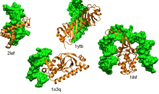
As shown in Fig.2.7, proteins associate to DNA in a variety of ways. Com-monly found binding patterns have been classified by Luscombe and collab-orators [61]. Several of these patterns feature a α-helix binding the DNA major groove where it probes the DNA sequence. In this category are found the helix-turn-helix motif (see for example complex 1o3q in Fig. 2.7), the helix-loop-helix motif, the zinc finger motif, where Zn2+ ions forms four bounds with cystein or histidine residues or the leucine zipper. α-helices within High-Mobility-Group (HMG) domains (groups of three L-shaped he-lices, see Fig. 2.7, complex 2lef) can also interact with the DNA minor groove. Alternatively, the binding domain can be composed of β-sheets. In the 1ytb complex (Fig.2.7), a β-sheet extensively interacts with the minor groove of the DNA. Finally, the binding motif is not always rigid, it can be formed for example by long, flexible loops such as the long loop of the integration host factor in 1ihf (Fig.2.7).
Factors that favor protein-DNA association
As noticed earlier, protein-DNA interfaces generally bury more surface area than protein-protein complexes. Moreover, the DNA surface is particularly contrasted, with an alternation of deep grooves and protruding phosphodi-ester backbones. These two elements indicate that the steric complemen-tarity between the protein and the DNA surfaces may play a particularly important role in the strength of association. Indeed, Poulain and collabora-tors [17] have observed during theoretical docking studies2 that the correct geometry of association of a large range of specific protein-DNA complexes could be unambiguously identified on the sole basis of steric complementar-ity when the electrostatic interactions were artificially set to zero.
Electrostatics can however prove decisive for non-specific protein-DNA complexes. The small DNase I protein binds any DNA sequence and induces a cleavage of the DNA backbone. Gu´eroult and collaborators have demon-strated the importance of two calcium and two magnesium ions to assist bovine pancreatic DNase I in binding DNA [62]. These ions strongly bind to four sites on the protein, three of them being situated on flexible loops distant from the DNA binding site. In this case, association and dissociation are directly regulated by the presence of the cations, the control that they exert is collective and it exclusively concerns the electrostatic fit.
2
The principles of macromolecular docking methods, which are developed to predict-ing the 3D structure of macromolecular complexes startpredict-ing from the structure of their
2.4. PROTEIN-DNA COMPLEXES 21
Figure 2.7: Examples of protein-DNA complexes. The proteins, which present various DNA-binding motifs (see text), are shown in ribbon representation (orange); from left to right, lymphoid enhancer-binding factor (2lef), catabolite gene activator (1o3q), TATA-box-binding protein (1ytb), integration host factor (1ihf). The DNA is shown in surface representation (green).
Mechanism of protein-DNA recognition: role of DNA flexi-bility
In many cases, the protein binds to specific DNA sequences. It has been established that in those cases, the fidelity of recognition results from the combination of two fundamentally different mechanisms [63,64]. One is the direct or base readout mechanism. In this mode of recognition, specific amino acids are put in registration with DNA functional groups using a rigid support such as the α-helices that are present in several major groove-binding motifs. This fingertip reading scheme involves the formation of hydrogen bonds or water-mediated hydrogen bonds and mainly takes place in the DNA major groove, where patterns of hydrogen bond donors and ac-ceptors associated to a given sequence can unambiguously be distinguished. The second mechanism, either called indirect or shape readout, takes advan-tage of spontaneous or protein-induced sequence specific shape modulation. This can go from minor groove narrowing, which exacerbates the electro-static field near the groove [64], to the creation of large deformations such as bends (Fig. 2.7, 1ihf) or local transitions to different substates of the double-helix [65,66] (Fig. 2.7, 1ytb). The protein-DNA interaction is then optimized by a combination of modifications of physical (e.g. electrostatic field) and mechanical (torsion or bending rigidity, ...) properties of the dou-ble helix, as well as modifications of its shape. For example in the complex between the TATA-Box and the TBP protein shown in Fig.2.7 (1ytb), the local transition of the DNA region contacted by the protein towards the TA-DNA form, an unwound and stretch form with A-form character [65], results in a wide opening of the minor groove and offers a large and flat surface for protein binding. In the case of indirect readout, sequence discrimination re-sults from the sequence-dependent energetic cost of the shape modulation. Both mechanisms generally contribute to some extent to the specificity of association and can be combined to reinforce the selectivity [66]. For ex-ample, optimal fit of the protein on a tailored DNA surface may position a readout motif in the exact position for directly probing nucleotide functional groups.
Prediction of the geometry of protein-DNA association
The characteristics of protein-DNA complexes, with often large size and con-trasted interfaces, make them easier targets for theoretical structure predic-tion than protein-protein complexes. Indeed, the binding site on the protein side can be predicted unambiguously in numerous cases just by consider-ing the steric adjustment, which does not even need to be strictly accurate. Poulain et al. [17] have shown that the protein interface can be predicted even when the DNA structure in the complex differs from canonical B-DNA,
2.5. OLIGOMERS AND POLYMERS 23
as far as the structural deviation is not too much important (less than 20◦ curvature).
When the DNA distortion is important, it is necessary to explicitly pre-dict the induced DNA deformation simultaneously to prepre-dicting the relative position/orientation of the two macromolecules. Solutions for this problem have been first proposed by the group of Bonvin, based on the information-driven program HADDOCK [67]. In a first step, directions for internal DNA deformation are extracted from semi-rigid data-driven docking simulations starting from B-DNA. The detected deformations are then amplified using the 3D-DART DNA modelling server [68] and the distorted structures are used as new starting point for HADDOCK docking simulations. Another algorithm has recently been proposed by Banitt and Wolfson [18]. In this method, the protein surface is screened by rigid DNA fragments and a curved DNA structure is reconstructed on the identified consensus surface patches. This method only considers curvature for possible DNA deformation and does not support unwinding or stretching deformation nor possible local transitions to different substates but it is efficient as a first approximation treatment of shape deformation. In chapters 6 and 7, I will present the strategies I have developed to address the variety of possible DNA deforma-tions that can be encountered in protein-DNA complexes.
In addition to detecting the DNA binding site on the protein and pre-dicting the deformation of the bound DNA, complete prediction of the asso-ciation geometry requires the phasing of the DNA sequence with respect to the protein. For van Dijk and collaborators [67], this question is inseparable from the detection of the overall geometry of association since the assembly is driven by experimental information on protein and DNA residues involved in the complex formation. Ab initio prediction is more tricky. In case the sequence is known, threading the sequence on a pre-deformed DNA struc-ture should enable both the detection of sequence regions that minimize the deformation energy and sequence regions prone to direct recognition by DNA-binding motifs. One may also resort to the use of the ADAPT methodology [69,70], which was exactly developed to optimize the sequence of oligonucleotides given their structure.
2.5
Oligomers and polymers
At larger length scales, protein can self-associate as closed forms, such as rings or polyhedral envelopes (capsids), or as open forms like spiral fila-ments [19]. Polyhedral envelops enclose the genetic material of viruses. Rings are commonly found in processes involving processive DNA manipu-lation such as replication, repair or recombination [71]. As discussed earlier, filaments and filament assemblies (fibers) are the essential component of the cytoskeleton, responsible for the cell shape as well as for its mechanical
properties and also used as support for vesicle transport. Filaments can be used also to process DNA, in which case there is a coupling between the shape of the protein filament and the DNA form (chapter3).
Due to their limited size, ring-shaped assemblies are often accessible to atomic structure determination. All forms can be observed using low reso-lution techniques such as EM, AFM or SAS. When combined with advanced image treatment, EM can determine the structure of regular forms at sub-nanometer resolutions.
Dynamics of filament association/dissociation
Open forms resulting from protein self-association need to be regulated to avoid ever-growing filaments. More fundamentally, the dynamic interplay between growth and dissociation is an essential element of the filament func-tion in the cytoskeleton: it enables the cytoskeleton to exert mechanical forces on the cell membrane that are directly responsible for the cell shape; it is fundamental for the microtubule role in chromosomal segregation dur-ing mitosis. Particularly, the microtubule undergoes alternation of rapid growth and sudden disassembly events (known as “catastrophe”) that are inherent to its function. Antitumoral drugs such as taxol, colchicine or vinblastine that disregulate the microtubule assembly/disassembly cycle by artificially inhibiting its formation (colchicine, vinblastine) or overstabilizing the assembled microtubule (taxol), provoke an arrest of the cell division.
Several factors contribute to this regulation.
Accessory proteins can reinforce the stability of filaments or mote their dissociation. This is the case of microtubule-binding pro-teins (MAPs), which initiate (γ-tubulin) or stabilize (Tau, MAP2, MAP4) the microtubule association, or promote (stathmin) its dis-sociation [60].
The growth, stability and dissociation of actin filaments is also regu-lated by proteins such as Arp2/3 (initiation), capping proteins (growth termination), ADF/cofilin (promote the dissociation) or profilin (re-store the growth) [72].
NTP cofactors are another central factor that regulates filament self-assembly. As discussed earlier (section2.3), the chemical energy stored by NTP molecules can be transmitted to the protein as a mechanical energy, enabling the transition between internal states of the protein and modifying the interface accessible to association partners. These partners whose association is controlled by the state of NTP hydrolysis can be other monomers, but they can as well be the accessory proteins described above. For example, cofilin associates along the sides of actin filaments or to actin monomers where ATP has been hydrolyzed [72].
2.5. OLIGOMERS AND POLYMERS 25
Figure 2.8: Actin network in a keratocyte. (left) Electron micrograph of a keratocyte; detail of region delimited by a white square is shown in the right panel; (right) three zones of actin filament organization are shown; the schematic diagram below indicates the location of key proteins and the curves indicate actin filament assembly (red, actin subunits per unit time) and disassembly (blue). After figure 1 of [72].
In certain cases, the NTP molecule is located at the interface be-tween two monomers. The presence of NTP or the products of its hydrolysis then chemically modifies the interface properties, notably its electrostatic properties. For example, in the protofilaments consti-tutive of the microtubules, the interface between the β-subunit of a α, β-tubulin dimeric unit and the α-subunit of the neighboring dimer hosts a GTP molecule [73], whose hydrolysis destabilizes the micro-tubule construction. In the active form of recombination filaments, an ATP molecule is sandwiched between each two consecutive monomers (fig. 2.4, right) [30]. Its presence is necessary for the ATPase activity as well as the recombinase activity, and its absence or its hydrolysis destabilizes the interface, leading to an alternative assembly mode for the filament [29]. The exact way these interfacial cofactors influence the stability of the assembly mode remains to be established. For mi-crotubules, it has been proposed that hydrolysis of the GTP may lead to a preferentially curved form of the protofilament, analogous to the colchicine or stathmin-bound crystal form [28,73]. This hypothesis has been questioned by the observation of lateral loop conformational change consecutive to GTP hydrolysis and by a recent study based on theoretical calculations and EM observations [74], which indicates that GTP hydrolysis would primarily affect the lateral contacts between protomers. For RecA, the question is still awaiting further studies. In these cases where the NTP molecule is situated at the interface, some residues from the partner monomer participate to the hydrol-ysis reaction. This leaves the possibility of a coordinated interplay between assembly and hydrolysis3 that may fully take part in the fila-ment growth control, albeit in a process that still has to be elucidated. Super structuralization of the fiber network has also been asso-ciated to the dynamics of association/dissociation. Reymann et al. [76] reported that the structural organization of the network of actin fibers influences the dynamics of polymerization/depolymerization.
3
In the case of actin, it has been proposed that the conformational change induced by longitudinal monomer binding would position a key glutamine amino-acid close to the ATP in a way that may enhance the rate of ATP hydrolysis [75].
The fibers can be organized as branched entangled and dense net-works, as antiparallel contractile bundles linked to the myosin motor, or as parallel bundles (fig.2.8). The branch networks are associated to strong dynamics of actin polymerization/depolymerization.
Microtubules are formed by the lateral assembly of between 11 and 16 quasi-straight protofilaments, formed by longitudinally assembled α, β dimers, which results in a tubular geometry. The dynamic turnover of microtubule association/dissociation varies by a factor of 4-100 de-pending on the phase of the cell cycle [77] [fig.2.9. This change is associated with the formation of different spatial and temporal or-ganization of the microtubules, such as spindle microtubules during mitosis.
Finally, the recombination filaments also have been shown to assemble in dynamic superstructures such as the RecA bundles recently ob-served by Lesterlin and collaborators [13] during the initiation phase of recombination (chapter3, fig.3.11). No accessory protein has been associated to this organization, yet ATP hydrolysis is required for the bundles to form.
Figure 2.9: Changes in the microtubule network morphology dur-ing the cell cycle. Microtubules are in red in metaphase and anaphase and in green in telophase and interphase; the chromosomal DNA is in blue. After Figure 2 of [77].
Beyond their apparent link with filament dynamics, variations in super-structure probably reflect varying states of the protein organization within the filaments, modulated by the presence of bound accessory proteins or with the state of ATP hydrolysis.
Multiple modes of association and filament morphology As noted by Goodsell and Olson [19] and later theoretically justified by Andr´e and collaborators [78], the basal state of filamentous cellular struc-tures such as protein filaments but also nucleic acids consists in a regular helical or cyclic organization4. It is also under this basal state that the corre-sponding structures are more easily elucidated, based on crystal information on the symmetry state, on crystal structures of dimeric complexes or EM observations. Nevertheless, as discussed in the previous section, filament are also highly dynamic and subject to a high degree of variability. These properties are essential to their function, yet they are difficult to apprehend in a structural point of view.
4
2.5. OLIGOMERS AND POLYMERS 27
X-ray or EM observations of DNA processing proteins such as the T7 gp4 helicase, the replication protein DnaB or the recombination proteins RecA, RadA, Rad51 or Dmc1 have revealed several forms for self-assembly, ranging from rings to helices with various morphologies. For example DnaB has been crystallized both as a helical ring [79] and as a spiral form [22], proposed to correspond to a translocation state of this helicase. The large diversity of the modes of recombinase self-organization will be detailed in chapter 10(see for example fig. 3.8of that chapter).
In these cases, the observed assembly forms were homogeneous, which means that the geometry was regular along the whole length of the oligomer. This is not always the case and non homogeneous oligomeric forms have been identified for the T7 gp4 and the DnaB helicases, both at low resolu-tion [80,81] and in crystal structures [82,83]. In both cases, the hexamers could be observed as 6-fold symmetry rings with homogeneous modes of as-sociation, or as trimers of dimers (3-fold symmetry) combining two binding modes. The two symmetry states, either 6-fold or 3-fold, have been found to coexist in samples of DnaB hexamers analyzed by EM [80](Fig. 2.10. The cyclic structure of DnaB is a double-layered ring, with an upper ring containing the six C-terminal domains and a lower ring formed by the six N-terminal domains (Fig.2.10). The two binding modes differ by the contacts between the upper and lower rings (stars in Fig.2.10), with each N-terminal domain contacting either the C-terminal domain of its own monomer or that of the following monomer (arrows in Fig. 2.10). Heterogeneous structures have been proposed to help breaking the helicase ring when clamping the processed DNA. They have also been proposed to play a role as intermedi-ates in the catalytic pathway leading to helicase unwinding and processive translocation [82,83], together with the recently detected spiral assembly mode of DnaB [22]. Moreover, the simultaneous presence of different bind-ing modes within the T7 gp4 helicase has been attributed to different states of hydrolysis of the nucleotide cofactor [82].
Figure 2.10: Two assembly modes of DnaB cyclic hexamers. Anal-ysis of EM micrographs by Yang et al. [80] has revealed two symmetry forms for the cyclic self-assembly of DnaB hexamers in the presence of the AMP-PNP cofactor (a non-hydrolyzable ATP homolog) : a three-fold symmetry (a,c,e) and a six-fold symmetry (b,d,f). (a,b) and (e,f) are seen along the axis and are related by a 180
◦rotation; (c,d) represent a view perpendicular to the axis. The six monomers are
labelled from 1 to 6, the helicase domain is labelled with a H and the N-terminal domain with a N; reproduced from [80], figure 7.
Helicases can therefore combine alternative modes of association with-out loosing their ring-shaped geometry, at the unique topology cost of a change in symmetry characteristics. Coexistence of alternative modes of
binding has also been reported in spiral oligomers (chapter3, fig.3.14), and it can reasonably be supposed that alternation of binding modes occurs in the course of their processive function. This may have implications in the filament morphology and the overall fiber organization.
2.6
Towards higher level superstructures
Oligomers tend to self-assemble as regular helical or cyclic structures. The presence at a given time of NTP cofactors in several hydrolysis states along the filament or the binding of auxiliary proteins can disrupt this regular-ity and introduce defects in the structure, with dynamic evolution. These irregularities are necessary to control the association/dissociation process in spiral filaments, they may also be necessary to perform processive tasks such as DNA unwinding or DNA strand exchange. They can result from internal structural variations of the monomers and subsequent modification of the surface accessible for binding a partner, like in actin fibers, they can also result from alterations of the binding surface or from association via completely different binding modes, like in recombinases. However, due to the difficulty of experimentally characterizing irregular and dynamically changing oligomers, there are presently few documented examples of these variations, which makes them poorly characterized. The principal observa-tion here is that the quality of the interface and the strength of associaobserva-tion locally associated to a given binding mode may play a role in determining the architecture of the whole oligomer.
The oligomers in turn participate in higher level structures, examples of which are shown in fig. 2.8 of this chapter or fig. 3.11 of chapter 3. These higher level structures are diverse, with distinct properties in terms for ex-ample of association/dissociation turnover, and they are the hallmarks of specific functions. In a 2007 review, Norris and collaborators [84] described and classified what they call “hyperstructures ”, defined as the intermediate level of organization between the macromolecule and the cell. These hy-perstructures are generally defined as heteromeric protein associations, yet several hyperstructure classes as defined by Norris and coll. are centered on oligomeric structures, for example the cytoskeletal hyperstructures or the cell cycle hyperstructures responsible for DNA replication, sequestration of newly replicated origins, segregation, compaction, and division.
Experimental studies of these defined and specialized hyperstructures are rapidly progressing, with the development of new techniques, apparatus and protocols adapted to their size and dynamics and dedicated to their study within the cells [13] or in vitro [76]. There is a growing need to understanding the physical rules governing these assemblies, as they are the key towards an integrated vision of the cell functioning.
2.6. TOWARDS HIGHER LEVEL SUPERSTRUCTURES 29
dynamic gaps in the experimental study of biomacromolecules Expanding these methods to studying cellular super or hyperstructures should help rationalize the new observations. This is inherently a multiscale problem that requires considering the detail of interfaces together with large scale protein superstructure organisation. A great part of my work presented in chapters 8, 10 has been devoted to developing computational tools and modeling strategies to this aim.
![Figure 1.1: Development of computing power of the most powerful com- com-puters [15].](https://thumb-eu.123doks.com/thumbv2/123doknet/15015528.680938/14.892.158.667.206.555/figure-development-computing-power-powerful-com-com-puters.webp)
![Table 2.1: Occurrence of oligomeric proteins in E. coli (data from SwissProt Databank, reproduced from the work of Goodsell and Olson, 2000 [19])](https://thumb-eu.123doks.com/thumbv2/123doknet/15015528.680938/18.892.144.659.294.634/table-occurrence-oligomeric-proteins-swissprot-databank-reproduced-goodsell.webp)
![Figure 3.5: Crystal structures of the RecA filament. The crystal structures of the RecA-ADP (left, PDB ID 2REB) [29] and RecA-ATP (right, PDB ID 3CMW) [30] filaments are shown in surface representation](https://thumb-eu.123doks.com/thumbv2/123doknet/15015528.680938/44.892.212.579.324.628/figure-crystal-structures-filament-crystal-structures-filaments-representation.webp)