Cell biology: Death drags down the neighbourhood
The MIT Faculty has made this article openly available.
Please share
how this access benefits you. Your story matters.
Citation
Vasquez, Claudia G., and Adam C. Martin. "Cell biology: Death
drags down the neighbourhood." Nature 518 (12 February 2015),
pp.171-173.
As Published
http://dx.doi.org/10.1038/nature14198
Publisher
Springer Nature
Version
Original manuscript
Citable link
http://hdl.handle.net/1721.1/106810
Terms of Use
Creative Commons Attribution-Noncommercial-Share Alike
Detailed Terms
http://creativecommons.org/licenses/by-nc-sa/4.0/
Cell biology
Death drags down the neighbourhood
An analysis of dying cells reveals that they play an active role in shaping tissues, where by dying, a cell pulls on its neighbours, inducing the neighbouring cells to contract their apices and fold the tissue. See Letter p.XXX
Claudia G. Vasquez & Adam C. Martin
Apoptotic cell death is a normal and essential part of tissue development, maintenance, and repair1. Apoptosis has a key role in sculpting tissue morphology, removing cells from between
forming digits and eliminating vestigial structures. However, this is often regarded as a passive elimination of unwanted cells. In a paper published on Nature’s website today,Gettings et al.2
report a surprising finding — that apoptosis can trigger contractions that fold tissues. Rather than being inert, apoptotic cells actively affect their surroundings, causing lasting changes in tissue form and structure.
Cells undergoing apoptosis shrink and fragment into membrane-bound structures called
apoptotic bodies that are engulfed by phagocytic cells. Epithelial tissues line the body’s cavities, creating boundaries between different extracellular environments. Accordingly, epithelial cells are organized along the depth of the tissue layer, generating apical and basal domains of the cell (Fig. 1). In epithelial tissues, apoptosis induces apical contractility in neighbouring cells. The result is a purse-string-like contraction in surrounding cells that pushes the dying cell from the epithelium3. Furthermore, epithelial cells remain attached to their neighbours, via adherens
junctions, while they undergo apoptosis, inducing neighbouring cells to stretch and elongate towards the apoptotic cell4. While the active requirement of apoptosis to generate tissue-scale
shape changes has also been shown5 the link between the two is poorly understood.
Cells generate contractile forces through the actin and myosin proteins that make up their cytoskeleton. Actin filaments assemble into meshes and bundles that underlie cell membranes, whereas myosin is a motor protein that forms bipolar minifilaments that connect actin filaments and walk directionally along actin filaments. Myosin minifilaments can contract the actin network
to generate cellular tension. In response to signalling molecules, contractility can be increased and transmitted between cells via adherens junctions to promote dramatic changes in tissue shape.
The generation of cellular tension is essential for the generation and maintenance of specific tissue architectures. One well-characterized example of how cell contractility and the resulting cell shape change influences tissue architecture occurs in the fruit fly Drosophila melanogaster. During early embryonic development, a signalling pathway activates the actin–myosin
cytoskeleton, constricting the apices of a specific set of cells, and so folding the epithelial sheet and moving muscle cell precursors inside the embryo6,7. By contrast, the cytoskeletons of cells
in a mature, static epithelium constantly tug on neighbouring cells. This state of tension is required to maintain an ordered hexagonal cell array in epithelial tissues8. Thus, a fundamental
property of epithelial tissues is that cells continually exert pulling forces on each other.
The leg joints of fruit flies form in the third larval stage of their development, when folds in this epithelial tissue cause successive rounds of leg subdivision. Fold formation requires apoptosis of cells at the centre of the fold5 and the apical side of the tissue contracting and folding inward
towards the basal side. Gettings and colleagues set out to determine the link between apoptosis and fold formation in these leg joints. Using live imaging, they studied a fluorescent version of myosin in cells undergoing apoptosis, and report that the protein accumulated along the apical-basal axis of the apoptotic cell. They also observed that as the apoptotic cell contracts and shrinks into the epithelium, it pulls on neighbouring cells. Consequently, myosin levels in
neighbouring cells increase, which in turn causes these cells to constrict their apices and form a fold (Fig. 1)2.
How does apoptosis cause neighbouring cells to constrict? The authors show that inhibiting myosin function in the apoptotic cell suppresses both the accumulation of myosin at the apical edges of surrounding cells and fold formation2. This suggests that the mechanical pulling of
neighbouring cells by the dying cell could trigger myosin activity in neighbouring cells.
Studies have demonstrated that applying mechanical forces to tissues can elevate myosin activity 9,10. But the mechanism through which a pulling force exerted by an apoptotic cell could
recruit apical myosin in the surrounding tissue remains unknown. One possibility is that the myosin motor itself might respond to tension, with tension increasing the length of time that
myosin remains bound to actin filaments11. Alternatively, tension might affect signalling factors
that regulate activity of the Rho-kinase enzyme, as has been proposed for the folding of embryonic epithelia10, because myosin dynamics and spatial organization are regulated by a
balance between myosin phosphorylation by Rho kinase and dephosphorylation by
phosphatase enzymes12. It is also possible that contractile activity in the apoptotic cell could
compromise membrane integrity in this cell, leading to the release of a chemical signal, such as ATP, that induces contraction in neighbouring cells13. An experiment that would directly test the
requirement of physically transmitting tensile force from the apoptotic cell to the neighbouring tissue would be to disrupt adherens junction proteins, which are required to mechanically couple epithelial cells during apoptosis4.
How widespread is the phenomenon of apoptosis triggering or contributing to morphogenetic movements? Apoptotic cells contribute to generating the epithelial tension that drives epithelial-sheet movement in an epithelial tissue of fruit flies called the amnioserosa14. Gettings and
co-workers’ study demonstrates that apoptosis — and possibly the resulting pulling force —
induces myosin accumulation and fold formation in three other epithelial tissues. Thus, apoptotic cells may have a more active role in shaping tissues than was previously believed.
Tissue folding is most often thought to be triggered by transcription factors and/or secreted signalling molecules that regulate the cytoskeleton. This study demonstrates that some systems may in fact represent a “relay”, where induction of one type of cell behaviour can trigger changes in the surrounding tissue. The idea that mechanical signalling can trigger a propagation of contractile activity was originally proposed in some of the first mechanical models of tissue folding over 30 years ago15. It is surprising that this trigger could represent a
dying cell’s final tug.
Claudia G. Vasquezand Adam C. Martin are in the Department of Biology, Massachusetts Institute of Technology, Cambridge, Massachusetts 02142, USA.
e-mail: acmartin@mit.edu
2. Gettings, M. et al. Nature XXX (2015).
3. Rosenblatt, J., Raff, M. C. & Cramer, L. P. Curr. Biol. 11, 1847–1857 (2001). 4. Lubkov, V. & Bar-Sagi, D. Curr. Biol. 24, 868–874 (2014).
5. Manjón, C., Sánchez-Herrero, E. & Suzanne, M., Nature Cell Biol. 9, 5–63 (2006). 6. Dawes-Hoang, R. E. et al. Development 134, 4165–4178 (2005).
7. Manning, A. J., Peters, K. A., Peifer, M. & Rogers, S. L. Sci. Signal. 6, ra98 (2013). 8. Farhadifar, R., Röper, J.-C., Aigouy, B., Eaton, S. & Jülicher, F. Curr. Biol. 17, 2095–2104
(2007).
9. Fernandez-Gonzalez, R., de Matos Simoes, S., Röper, J., Eaton, S. & Zallen, J. A. Dev.
Cell. 17, 736–743 (2009).
10. Pouille, P.-A., Ahmadi, P., Brunet, A.-C. & Farge, E. Sci. Signal. 2, ra16 (2009). 11. Ren, Y. et al. Curr. Biol. 19, 1421–1428 (2009).
12. Vasquez, C. G., Tworoger, M. & Martin, A. C. J. Cell Biol. 206, 435–450 (2014). 13. Herrgen, L., Voss, O. P. & Akerman, C. J. Dev. Cell 31, 599-613 (2014).
14. Toyama, Y., Peralta, X. G., Wells, A. R., Kiehart, D. P. & Edwards, G. S. Science 321, 1683–1686 (2008).
15. Odell, G. M., Oster, P., Alberch, P. & Burnside, B., Dev. Biol. 85, 446–462 (1981).
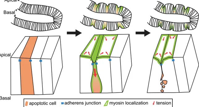
Figure 1 | Apoptosis and fold formation. As cells die through apoptosis, they shrink and
fragment, before being completely engulfed. Gettings et al.2 dissect how apoptosis causes
folding in the forming leg joints of fruit-fly larvae. They report that apoptotic cells in this epithelial sheet accumulate myosin along their apical–basal axis as they shrink. As the apoptotic cells pull into the tissue (red arrow, apoptotic cell), they remain attached to neighbouring cells via
adherens junctions; these neighbouring cells also accumulate myosin at their apical surfaces, causing elevated tension around the apoptotic cell (red arrows in non-apoptotic cells). As the dying cell is fragmented, neighbouring cells apically constrict and form a fold in the tissue (green cells).
Apical
Basal
adherens junction tension
apoptotic cell myosin localization
Basal Apical