HAL Id: hal-03194007
https://hal.archives-ouvertes.fr/hal-03194007
Submitted on 9 Apr 2021HAL is a multi-disciplinary open access
archive for the deposit and dissemination of sci-entific research documents, whether they are pub-lished or not. The documents may come from teaching and research institutions in France or abroad, or from public or private research centers.
L’archive ouverte pluridisciplinaire HAL, est destinée au dépôt et à la diffusion de documents scientifiques de niveau recherche, publiés ou non, émanant des établissements d’enseignement et de recherche français ou étrangers, des laboratoires publics ou privés.
Sterically Protected Molybdenum Trihydride Redox
Pairs: A Paramagnetic “Stretched” Dihydrogen
Complex?
Miguel Baya, Jennifer Houghton, Jean-Claude Daran, Rinaldo Poli, Louise
Male, Alberto Albinati, Matthias Gutman
To cite this version:
Miguel Baya, Jennifer Houghton, Jean-Claude Daran, Rinaldo Poli, Louise Male, et al.. Synthe-sis, Structure, and Electrochemical Properties of Sterically Protected Molybdenum Trihydride Redox Pairs: A Paramagnetic “Stretched” Dihydrogen Complex?. Chemistry - A European Journal, Wiley-VCH Verlag, 2007, 13 (19), pp.5347-5359. �10.1002/chem.200700293�. �hal-03194007�
Synthesis, structure and electrochemical properties of sterically protected
molybdenum trihydride redox pairs: A paramagnetic “stretched”
dihydrogen complex?
Miguel Baya,a Jennifer Houghton,a Jean-Claude Daran,a Rinaldo Poli,*a Louise Male,b Alberto Albinatib and Matthias Guttman,c
aLaboratoire de Chimie de Coordination, UPR CNRS 8241 liée par convention à l’Université
Paul Sabatier et à l’Institut National Polytechnique de Toulouse, 205 Route de Narbonne, 31077 Toulouse Cedex, France
bDepartment of Structural Chemistry (DCSSI), Università di Milano, Via G. Venezian 21,
20133 Milano, Italy.
cRutherford Appleton Laboratory, ISIS Facility, Chilton (Didcot) OX11 0QX U.K..
Proofs to:
Rinaldo Poli
Tel: +33-561333173 Fax: +33-561553003
Summary
Complexes Cp#Mo(PMe3)2H3 (Cp# = 1,2,4-C5H2tBu3, 2a; C5HiPr4, 2b) have been
synthesized from the corresponding compounds Cp#MoCl4 (1a, 1b) and fully characterized,
including by X-ray crystallography and by a neutron diffraction study for 2a. Protonation of
2a led to complex [(1,2,4-C5H2tBu3)Mo(PMe3)2H4]+ (3a) in THF and to
[(1,2,4-C5H2tBu3)Mo(PMe3)2(MeCN)H2]+ (4a) in MeCN. Complex 4b analogously derives from
protonation of 2b in MeCN, whereas the tetrahydride complex 3b is unstable. One-electron oxidation of 2a and 2b by [Cp2Fe]PF6 produces the EPR active 17-electron complexes [2a]+
and [2b]+. The former is thermally more stable than the latter and could be crystallographically characterized as the PF6- salt by X-ray diffraction, providing evidence for
the presence of a stretched dihydrogen ligand (H…H = 1.36(6) Å). Controlled thermal decomposition of [2a]+yielded the product of H2 elimination, the 15-electron monohydride
complex [(1,2,4-C5H2tBu3)Mo(PMe3)2H]PF6 (5a) which was characterized by X-ray
crystallography and by EPR spectroscopy at liquid He temperature. The compound establishes an equilibrium with the solvent adduct in THF. An electrochemical study by cyclic voltammetry provides further evidence for a rapid H2 elimination process from the
17-electron complexes. Contrary to the previously investigated [Cp*Mo(dppe)H3]+ system, the
decomposition of [2a]+ by H2 substitution with a solvent molecule appears to follow a
dissociative pathway in MeCN.
Keywords
Molybdenum, bulky cyclopentadienyl ligands, hydride ligands, paramagnetic hydride complexes, oxidatively induced reductive elimination, neutron diffraction
Introduction
Hydride complexes have paramount importance in light of their implication in a variety of catalytic processes and as models of a number of biological functions such as hydrogenase and nitrogenase.[1-6] Because of the strong covalent nature of the M-H bond and the absence of additional orbital interactions (i.e. of type), they are usually stable only in a closed-shell configuration. Open-shell versions are reactive, which is the very reason for their involvement as catalytic intermediates. An interesting subclass of open-shell hydride complexes are those with an odd-electron (mostly 17-electron) configuration, characterized by paramagnetism. These complexes have generally been accessed by one-electron oxidation of stable diamagnetic precursors. In most cases, they decompose by deprotonation,[7] disproportionation,[8] dihydrogen reductive elimination (for complexes containing at least two hydride ligands),[9] atom transfer,[10] and other pathways.[11] This multitude of available reaction pathways complicates their potential application, for instance in electrocatalysis. It is therefore useful to investigate in greater detail how the various pathways depend on the reaction conditions (e.g. solvent, available substrates) and molecular parameters (e.g. stereoelectronic properties of the ligand environment). For this purpose, it is necessary to develop more stable systems. We have learned from previous investigations[9, 12-16] that all decomposition pathways are disfavored by both a stronger electron donating and more sterically protecting ligand environment.
The oxidation of Cp*Mo(dppe)H3 (dppe = Ph2PCH2CH2PPh2) was studied in the
greatest detail.[9, 12, 16] It leads to the paramagnetic complex [Cp*Mo(dppe)H3]+, which is
stable at low temperatures and was characterized in situ by EPR spectroscopy. The detailed investigation of its decomposition at room temperature enabled us to quantify the relative rates of deprotonation (by the residual neutral precursor), disproportionation, and H2
elimination in various solvents.[9, 16] This was the first reported example where H
2 oxidatively
induced reductive elimination could be unambiguously demonstrated and distinguished from other decomposition pathways. Oxidation of a polyhydride complex {MHn} is expected to
favor its rearrangement to a nonclassical isomer, {MHn-2(H2)}+,[17, 18] but the multitude of
decomposition pathways, all possibly leading to H2 evolution,[19] have previously made the
identification of the H2 elimination pathway uncertain.[17, 20] For this specific trihydrido
molybdenum complex all three decomposition pathways were shown to occur via the nonclassical intermediate [Cp*Mo(dppe)H(H2)]+, although theoretical calculations and
circumstantial evidence indicates that the oxidized complex adopts a classical structure. Since the nonclassical tautomer is energetically less accessible for the related tungsten system, complex [Cp*W(dppe)H3]+ turned out to be sufficiently stable to be isolated and
crystallographically characterized.[9]
In this contribution, we report the synthesis and investigations into new molybdenum systems, isoelectronic with Cp*Mo(dppe)H3, that contain an even more strongly donating and
sterically encumbering coordination sphere. We used the two highly substituted cyclopentadienyl rings, C5HiPr4 and 1,2,4-C5H2tBu3, in place of Cp* and two PMe3 ligands in
place of bidentate dppe. Notable results of this investigation have been the isolation and structural characterization of the 17-electron oxidation product, [(1,2,4-C5H2tBu3)Mo(PMe3)2H3]+, and the observation of its subsequent H2 elimination process
leading to the 15-electron monohydride derivative, [(1,2,4-C5H2tBu3)Mo(PMe3)2H]+, which
was also structurally characterized. Some aspects of this investigation have been recently communicated.[21] While those preliminary results will be reported again here in fuller details, stronger emphasis will be placed on complementary investigations that have not previously been described.
Results and Discussion
(a) Synthesis and characterization of the diamagnetic trihydride complexes Cp#Mo(PMe3)2H3 (Cp# = 1,2,4-C5H2tBu3, 2a; C5HiPr4, 2b)
Adaptation of Schrock’s Cp*MoCl4 synthetic procedure[22, 23] to the bulkier Cp#
analogues (Cp# = 1,2,4-C5H2tBu3, a, and C5HiPr4, b) yielded the corresponding Cp#MoCl4
derivatives 1a and 1b in good yields, see Scheme 1. Subsequent reaction of these compounds with LiAlH4 in the presence of ≥ 2 equivalents of PMe3, followed by methanolysis and
crystallization from ether, yielded the corresponding trihydride derivatives, Cp#Mo(PMe3)2H3,
2a and 2b. It is interesting to compare these results with that previously reported for the
related Cp* system, which led to a mixture of Cp*Mo(PMe3)2H3 and Cp*Mo(PMe3)3H.[24]
The bulkier Cp# systems afford the trihydride derivatives 2 selectively and show no tendency to replace H2 in the presence of excess PMe3 under thermolytic conditions.
Na+Cp #-Mo(CO)6 + THF/ Cp#Mo(CO)3-Na+ MeI/THF Cp#Mo(CO)3(CH3) PhICl2/CH2Cl2 Cp#MoCl4 room T Cp# = 1,2,4-C5H2tBu3 C5HiPr4 1a 1b 1. LiAlH4/PMe3/THF Cp#Mo(PMe3)2H3 2a 2b 2. MeOH Scheme 1
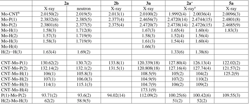
Both compounds gave single crystals suitable for a structural analysis. The crystal of 2a had sufficient quality to allow the location and refinement of the hydride positions from the X-ray data, as shown in the previous communication.[21] We have now completed the
structural investigation with a neutron diffraction experiment for 2a (the results of both refinements are compared in Table 1) and an X-ray diffraction experiment for 2b. The latter
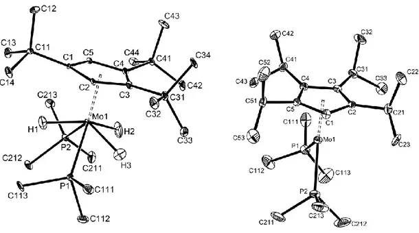
crystals had poorer quality and the hydride positions could not be located; the observable structural parameters are very close to those of 2a, see Table 1. A view of both geometries is shown in Figure 1. The molecular geometry is unusual for a half sandwich (ring)MoX3L2
compound, but parallels that previously reported for the related Cp*MoH3(dppe)
compound.[25, 26] This demonstrates that the unusual structure observed for Cp*MoH3(dppe)
is not enforced by the chelating nature of the dppe ligand. Another example for this structural type has recently been reported for the isoelectronic compound Cp*W(CO)2H2(SiH2Ph).[27]
As expected, the X-ray diffraction experiment yields shorter Mo-H distances than the neutron diffraction experiment for compound 2a. Only those afforded by neutron diffraction should be considered reliable. The parameters that do not involve hydrogen atom positions, on the other hand, are more precisely determined by the X-ray diffraction experiment.
The most interesting structural feature is the distance between atoms H2 and H3 [1.69(2) Å from the neutron structure]. This value is too long to envisage an interaction, but short in comparison to most nonbonded distances recorded by neutron diffraction for polyhydride complexes. This is evidence for a certain degree of “compression”.[28] Other relatively short nonbonded H∙∙∙H separations are 1.67(1) and 1.70(1) Å in [CpIr(PMe3)H3]+,[29] and values ranging from 1.637(4) to 1.668(4) Å between adjacent H
atoms in compound Os(PiPr2Ph)2H6,[30] whereas there are no shorter intramolecular H∙∙∙H
separations than 1.77 Å in compound Re(dppe)H7.[31] The longest H-H separations in
compounds that have been defined as “stretched H2 complexes” are 1.357(7) Å in
ReH7(P(C6H4-p-CH3)3)2[32] and 1.34(2) in [Os(H2)(en)2(O2CMe)]+PF6-,[33] all these values
resulting again from neutron diffraction experiments. Finally, complex OsH5(PMe2Ph)3+
exhibits an even longer separation (1.49(4) Å) and was described as falling in a “gray” region where there may or may not be a direct H/H attractive interaction.[34] The Cambridge Crystallographic Structural Database does not reveal H-H contacts between 1.0 and 1.7 Å for
Mo complexes, but a distance of ca. 1.18 Å has recently been estimated from T1 and JHD
measurements for complex [Mo(NPh)(PMe3)2(H2)(o-(Me3SiN)2C6H4)].[35] Interestingly, if
the H2 and H3 atoms are considered as defining a stretched H2 ligand, therefore occupying a
single coordination position in a MoII complex, then the coordination geometry would be described as a “four-legged piano stool”, which is indeed a quite common geometrical arrangement for MoII,[36] as exemplified by CpMo(PMe
2Ph)3Cl[37] and CpMo(dppe)(CO)H.[38]
<Table 1>
Figure 1. ORTEP view of compounds 2a (neutron diffraction, left) and 2b (right).
Hydrogen atoms, except those directly bonded to the Mo atom in compound 2a, are not shown for clarity.
The NMR properties of 2a and 2b confirm the presence of three hydride ligands. The single 31P{1H} resonance observed at room temperature is transformed into a binomial quartet in a 31P{sel. 1H} NMR experiment, showing that the P nuclei are coupled to three equivalent protons. In addition, the 1H spectrum shows a single, sharp triplet resonance. A rapid
exchange process between the inequivalent hydride positions must therefore be present. For the related Cp*Mo(dppe)H3 compound, the fluxional process could not be frozen out even at
the lowest attainable temperatures.[26] For compounds 2a and 2b, on the other hand, cooling results in decoalescence for the 1H NMR signal to yield two signals in a 1:2 ratio, consistent with the solid state structural investigation, see Figure 2. This shows quite clearly the effect of the bulky ring substituents on the dynamics of the hydride exchange process. For compound 2a, the lowest temperature (193 K) spectrum displayed well resolved triplets, due to coupling to the two equivalent P donor nuclei, with the unique hydride ligand being characterized by a greater JHP (64 Hz), relative to the two equivalent ones (44 Hz). No H-H
coupling between the inequivalent hydrides can be discerned. For compound 2b, on the other hand, the spectrum remained rather broad even at 193 K. A lineshape analysis yielded the activation parameters of the hydride scrambling process as H‡ = 9.0±0.7 (2a) and 8.5±0.3 (2b) kcal mol-1; S‡ = 17±3 (2a) and 21±1 (2b) e.u. The similar values of both activation parameters for the two compounds are in line with the similar structure of the two compounds. The longitudinal relaxation times (T1) of the various signals are also shown in
Figure 2. They confirm the classical nature of the compounds. Most notably, for compound
2a, the T1 value for the equivalent hydride ligands signal is not significantly shorter than that
-6 -5.5 -5 -4.5 (a) 193 K 213 K 233 K 253 K 273 K 710 ms 506 ms 359 ms 326 ms 303 ms 383 ms 372 ms -7 -6 -5 -4 -3 (b) 193 K 213 K 233 K 253 K 273 K 549 ms 378 ms 293 ms 319 ms 443 ms
Figure 2. Variable temperature 1H NMR spectrum of (a) compound 2a and (b) compound
2b in the hydride resonance region. The individual temperatures are shown on
each spectrum and the time values shown are the longitudinal relaxation times of the corresponding resonance.
The 31P NMR resonance of the two phosphine ligands remained sharp in the entire temperature range for compound 2a, in agreement with the chemical equivalence shown by the X-ray structure. For compound 2b, on the other hand, a decoalescence phenomenon was observed at low temperatures, yielding two singlet resonances with approximately equal intensities at 15.9 and 12.3 at 193 K (Figure shown in the Supporting Information). This behaviour can be rationalized by either the freezing out of a single Mo-(C5HiPr4) rotamer with
symmetry inequivalent PMe3 donors (but these ligands also need to be characterized by a
equilibrating rotamers having similar energy, perhaps differing by the relative conformation of the iPr groups in the C5HiPr4 ring. The latter phenomenon would not be expected to lead to
the existence of different rotamers for the 1,2,4-C5H2tBu3 substituted complex.
(b) Protonation studies
Compound 2a reacts with HBF4 at -80°C in THF to yield the tetrahydride complex
[(1,2,4-C5H2tBu3)MoH4(PMe3)2]+BF4-, 3a, see Scheme 2. This product is related to the
previously described [Cp*Mo(dppe)H4]+,[26] but shows a much greater thermal stability. It is
stable in THF solution at room temperature, whereas complex [Cp*Mo(dppe)H4]+
decomposes rapidly by loss of H2 via a presumed nonclassical [Cp*Mo(dppe)(H2)H2]+
intermediate. This stabilization effect is probably related to the greater donor power of the coordination sphere in 3a relative to [Cp*Mo(dppe)H4]+, with the corresponding stabilization
of the classical tetrahydrido structure with respect to the nonclassical tautomer. Compound
3a is diamagnetic and colorless, as expected for the d0 configuration of formally hexavalent molybdenum. It features, as expected, a triplet hydride signal in the 1H NMR spectrum (at -4.2 ppm) and a singlet (at 0.6 ppm) in the 31P{1H} NMR spectrum. Selective irradiation of
the PMe3 proton resonance yields a quintet 31P signal, in agreement with the presence of four
hydride ligands. The compound has also been structurally characterized by single crystal X-ray diffraction. Cp#Mo(PMe3)2H3 HBF4/THF [Cp#Mo(PMe3)2H4]+BF4 -HBF4/MeCN [Cp#Mo(PMe3)2(MeCN)H2]+BF4 -MeCN -H2 -H2 2a,b 3a,b 4a,b Scheme 2
The cation geometry is illustrated in Figure 3. The crystal quality allowed the location and refinement of all four hydride ligands. The geometry of the tetrahydride cation can be described as a highly distorted pentagonal bipyramid, when the bulky (1,2,4-C5H2tBu3) ligand
is considered to occupy a single coordination position at one of the vertices of the bipyramid. The second axial position is occupied by the hydride ligand H4, with the 5 equatorial ligands bent toward H4 and away from the cyclopentadienyl ligand (especially the two phosphine ligands, for steric reasons). The two PMe3 ligands occupy 1,3 positions in the
pseudo-pentagonal plane, with atom H1 bisecting the P1-Mo-P2 angle. The related complex [Cp*W(dppe)H4]+ was found to adopt an analogous coordination geometry, with the chelating
dppe ligand occupying one equatorial and one axial site.[26]
Figure 3. ORTEP view of the cation in compound 3a. Hydrogen atoms, except those
directly bonded to the Mo atom, are not shown for clarity.
The cationic tetrahydride complex 3a was also formed by protonation of 2a with HBF4
in MeCN, but further rapid evolution took place in this case. This reaction was only carried out on a spectroscopic scale in CD3CN and the resulting solution was monitored with time by
1H and 31P NMR spectroscopy. Within minutes at room temperature, resonances
corresponding to a second complex began to appear and the conversion was complete in 5 h. This product, [(1,2,4-C5H2tBu3)Mo(PMe3)2(MeCN)H2]+, 4a, derives from H2
elimination/MeCN coordination from 3a. It displays a singlet 31P{1H} resonance at 1.85 ppm, which converts into a triplet upon selective decoupling of the PMe3 ligand 1H resonance. At
room temperature a single broad hydride resonance is observed, however on cooling a CD3CN solution to -40o C, two triplet hydride resonances are resolved at -0.27 and -6.77 ppm
respectively. This suggests that the two PMe3 ligands occupy equivalent positions at all
temperatures, consistent with the structure shown in I.
Mo H Me3P PMe3 H MeCN + I Rn Rn = 1,2,4-tBu3, iPr4
The corresponding reaction of 2b, when carried out in THF at -80°C, led to the precipitation of a white solid, presumably corresponding to the [(C5HiPr4)Mo(PMe3)2H4]+
complex, 3b. However, this compound decomposes, even in the solid state, when warmed up to room temperature. Therefore, it could not be spectroscopically characterized. When the same protonation reaction was carried out in acetonitrile, the formation of complex [(C5HiPr4)Mo(PMe3)2(MeCN)H2]+, 4b, could be observed. As with 4a, the hydride ligands in
complex 4b are involved in a fluxional process at room temperature, appearing as a very broad resonance at -2.5 ppm. On cooling to 200 K in acetone-d6, two doublet of triplet
ligand couples to two equivalent phosphorus nuclei and to the other hydride ligand, in agreement with structure I. At this temperature, all four isopropyl groups on the cyclopentadienyl moiety also become inequivalent. In both complexes 4, a resonance integrating to 3 protons, assigned to the coordinated MeCN, can be observed at 2.17 (for 4a in CD3CN) and 2.71 (for 4b in acetone-d6). This behaviour is analogous to that of the related
Cp*Mo(dppe)H3 complex.[26] The greater coordinating ability of the acetonitrile solvent
induces a more facile H2 elimination by more efficiently trapping the resulting 16-electron
fragment.
(c) Oxidation studies: isolation and characterization
Preliminary electrochemical investigations indicated that both complexes 2a,b undergo an electrochemically reversible one-electron oxidation process and suggested that the oxidation product is relatively stable. The electrochemical behavior will be analysed in more detail later in section (e). The stoichiometric oxidation was accomplished by the use of Cp2Fe+PF6- in THF, see Scheme 3. The product for the 1,2,4-C5H2tBu3 system was
sufficiently stable to be isolated and crystallized. Its X-ray structure and its EPR spectrum (Figure 4) demonstrate its chemical identity as the PF6- salt of the one-electron oxidation
product, [2a]+, as discussed previously.[21]
Cp#Mo(PMe3)2H3 [Cp2Fe]+PF6 -[Cp#Mo(PMe3)2H3]+PF6 -Cp# = 1,2,4-C5H2tBu3 C5HiPr4 ([2a]+PF6-) ([2b]+PF6-) Scheme 3
50 G 50 G
10 G 10 G
(exp) (simul)
(exp) (simul) (simul)
[2a]+
[2b]+
LW = 2.07 G LW = 1.4 G
Figure 4. EPR spectra of complexes [2]+ in THF solution. Above: complex [2a]+ (T = 193). Below: complex [2b]+ (T = 183).
The most relevant metric parameters of the [2a]+ structure are compared with those of the parent compound 2a in Table 1. The overall geometry of the cation is essentially unchanged relative to that of the neutral precursor (see the previous communication for an ORTEP view).[21] There is no evidence of an interaction between the two ions, notably hydrogen bonding between the hydride ligands and the fluorine atoms of the anion. The Mo-CNT distance is slightly shorter, whereas the Mo-P distances is slightly longer, relative to the neutral precursor. The most notable change is a decrease of the H2…H3 contact from 1.69(2) Å (neutron diffraction) or 1.63(4) Å (X-ray diffraction) in the neutral complex to an average of 1.36(6) Å (from the X-ray data) in the cation. The difference is significant at the 2 level relative to the X-ray structure, at the 4 level relative to the neutron structure. Unfortunately, suitable crystals of compound [2a]+PF6-for a neutron diffraction analysis could not be grown.
Even when keeping the uncertainty into account, however, the H2…H3 separation in the cationic complex falls inside the range of compounds that have previously been described as “stretched” or “elongated” dihydrogen complexes, or alternatively as “compressed” dihydrides.[28, 39] Thus, it appears that the oxidation process has increased the interaction between the two hydride ligands H2 and H3.
Compound [2a]+PF
6- appears to be the first reported paramagnetic polyhydride complex
showing evidence for a stretched dihydrogen ligand (or compressed MH2 system). It is
interesting to compare this structure with that of the previously published isoelectronic [Cp*W(dppe)H3]+ complex. The two systems show a very different arrangement of the three
hydride ligands, the closest H∙∙∙H contact in the tungsten complex being 2.11 Å.[9] Although the H positions in this tungsten complex should again be considered with caution since this structure was also obtained from X-ray diffraction data, the relative arrangement of the heavy atoms that define the coordination sphere (the Cp ring, the metal, and the P donor atoms) is quite different in the two compounds. As we know, the W complex is stable and has no tendency to decompose, notably by H2 elimination.
The oxidation product obtained from 2b is rather short-lived at room temperature and could not be isolated. It was only characterized in situ by EPR spectroscopy, see Figure 4. Like [2a]+, it exhibits a rather broad spectrum at ambient temperature, which become sufficiently resolved at lower temperatures to allow the identification of the expected quartet of triplet feature, consistent with the presence of two phosphorus donor atoms and three hydride ligands, flanked by the 95Mo and 98Mo isotope satellites. This suggests, like for the
1,2,4-tBu3 analogue, that one-electron oxidation has afforded complex [2b]+.
The spectroscopic properties, however are peculiar in many respects. The simulation on the basis of any combination of spin ½ nuclei different than P2H3 (for instance, 2 P and 2 H)
failed to provide a spectrum sufficiently resembling the experimental one. However, the simulation for the P2H3 spin system was not nearly as satisfactory as for the [2a]+ homologue.
An unrestricted full parameter optimization resulted in too broad lateral features and a too sharp central one, compared to the experimental spectrum (Figure 4), for a broadening factor of 2.07 G. An artificial reduction of the line broadening parameter to 1.4 G allows a perfect match of the two lateral features of the triplet, but the central feature becomes too sharp
relative to the experiment. This may indicate a dynamic exchange process on the EPR time scale. Indeed, virtual triplets displaying artificially broadened central features are commonly found in the NMR spectra of diamagnetic compounds featuring suitable site exchange phenomena, such as the X signal for an ABX system where sites A and B are near the fast exchange limit.[40, 41] A rapid hydride scrambling process takes place for [2a]+ (symmetric EPR coupling pattern, inequivalent hydride positions by X-ray crystallography), as well as for the neutral precursors (vide supra) and for the related [Cp*Mo(dppe)H3]n+ (n = 0, 1)
complexes.[9] This exchange is possibly slower for the more encumbered (C5HiPr4)
derivative, causing the observed lineshape effect. It is interesting to note that the exchange rates are similar for the neutral precursors (slightly smaller for 2a), whereas the exchange process appears faster in 2a+ than in 2b+.
The next peculiar feature of the EPR spectrum of [2b]+ is a much lower value shown by aP and aH (namely aP = 6.2 G; aH = 1.7 G; aMo = 29.4 G) relative to both [2a]+ (aP = 36.2 G,
aH = 11.4 G and aMo = 30.8 G)[21] and [Cp*Mo(dppe)H3]+ (aP = 29.8 G; aH = 11.8 G).[9] The
cause of this phenomenon is not quite clear, but the different coupling values suggest that [2b]+ adopts a different geometry relative to that of [2a]+, possibly involving the complete
collapse of two hydrides to a dihydrogen ligand. It is easy to imagine how the greater bulk of the substituted cyclopentadienyl ring in [2b]+ might force the two H atoms closer together. This phenomenon may well be related to the slower hydride mutual exchange, as well as to our inability to isolate the compound. Related to this point, we recall that the tetrahydride protonation product is stable in the case of 3a but decomposes in the case of 3b, although the same system 4a,b is obtained in MeCN. Thus, we speculate that the extreme bulk of the C5HiPr4 ligand has the effect of pushing out an H2 ligand from both systems 3b and [2b]+.
(d) Oxidatively induced H2 reductive elimination
Although compound [2a]+PF6- is quite stable as a crystallized solid and in THF solution
at low temperatures, it slowly decomposed at T > 0°C, as indicated by a color change from orange to green. Well formed green crystals were obtained by slow crystallization from THF/pentane at -20°C. This product appeared thermally stable with no noticeable change over time in the solid state and in THF solution at room temperature. X-ray diffraction analysis revealed the identity of the compound as [(1,2,4-C5H2tBu3)MoH(PMe3)2]+PF6-, 5a.
A view of the structure is presented in our previous communication,[21] while selected bonding parameters are reported in Table 1. Therefore, the compound derives from its precursor [2a]+PF6- by H2 elimination, see Scheme 4.
[(1,2,4-C5H2tBu3)Mo(PMe3)2H3]+PF6
--H2
[(1,2,4-C5H2tBu3)Mo(PMe3)2H]+PF6 -THF, 0°C
Scheme 4
Although the quality of the data set allowed the identification of a single hydride ligand with a high level of confidence (see Experimental section), the question of the possible presence of additional hydride ligands in the structure of compound 5a has been considered carefully, since hydrogen atoms may be difficult to locate from X-ray diffraction data. Possibilities include the presence of one, two or three additional hydride ligands, giving a 16-electron dihydride, a 17-16-electron trihydride (a stereoisomer of the precursor [2a]+), and an 18-electron tetrahydride cation (i.e. complex 3a), as well as a dihydrogen ligand, yielding a nonclassical tautomer of [2a]+. The color of 5a relative to [2a]+PF6- excludes an isomeric
form of [2a]+ and also the tetrahydride formulation, although the bond distances and angles related to the heavy atoms in the cation of 5a, see Table 1, are not too different from those observed for compound 3a.
The 1H NMR spectrum of the isolated solid only revealed the resonances of the
tetrahydride complex 3a, indicating that this compound is a decomposition by-product (the solid was a mixture of well formed crystals and a powder). Complex 3a certainly arises from the transfer of a proton from acidic [2a]+ to residual 2a, similarly to what occurs for the Cp*Mo(dppe)H3 analogue.[16] A solid sample of the isolated compound showed bulk
paramagnetism, however a reliable value for the magnetic moment could not be obtained, given the impure nature of the sample. 1H NMR monitoring of the decomposition reaction also showed the formation of 3a, in addition to the formation of H2.
Positive identification of the green decomposition product as a 15-electron species comes from EPR spectroscopy. As detailed in the communication,[21] the solid sample shows features consistent with a spin quartet ground state at the liquid He temperature (Figure 5a): gx
and gy at 3.74 and 3.45 (±1/2 transition), plus a weak feature (gz for the forbidden ±3/2
transition) at 5.33. The g = 3.74 peak appears to display a fine structure, possibly due to coupling to the two equivalent P nuclei. The gz component of the ±1/2 transition is not visible
because it is overshadowed by stronger resonances in the g = 2 region (shown in Figure 5b). The resonance observed at g = 2.009 for the polycrystalline sample is very close to the position observed at higher temperature for the precursor complex [2a]+ and is therefore attributed to a residual amount of this material, which had co-crystallized with the H2
1000 1200 1400 1600 1800 2000 2200 X 10 gz(±3/2) gx,y(±1/2) H (Gauss) (a) 1000 1200 1400 1600 1800 2000 2200 X 10 gz(±3/2) X 10 gz(±3/2) gx,y(±1/2) H (Gauss) (a) 3000 3200 3400 3600 3800 g = 2.009 g = 1.922 H (Gauss) (b) 3000 3200 3400 3600 3800 g = 2.009 g = 1.922 H (Gauss) (b)
Figure 5. Liquid He EPR spectrum of compound 5a: (a) polycrystalline sample in the g =
4-6 region; (b) polycrystalline sample (solid line) and frozen THF glass (dashed line) in the g = 2 region.
Solutions of compound 5a in THF were EPR silent at room temperature and showed only a weak resonance at the liquid nitrogen temperature. On the other hand, they show an intense band at g = 1.922 at the liquid He temperature. The two resonances at g = 2.009 and 1.922 are visible for both polycrystalline and THF solution samples, but their relative intensity is opposite. The latter must belong to another S = ½ complex and we therefore assign it to the THF adduct, [(1,2,4-C5H2tBu3)Mo(PMe3)2(THF)H]+. Its presence for the
polycrystalline sample, which had been obtained by crystallization from THF (see Experimental section), represents evidence of solution equilibrium between the solvent-free,
15-electron, spin quartet monohydride complex and a spin doublet solvent adduct. The compound crystallizes preferentially in the solvent-free form, but the THF adduct also appears to exist in the solid state. Additional evidence for this equilibrium will be provided by the electrochemical analysis (vide infra).
As stated in the Introduction, we previously reported the first unambiguous oxidative induced reductive elimination of H2 from the one-electron oxidation of complex
Cp*Mo(dppe)H3. However, the elimination product could not be crystallized and was only
characterized in solution as the 17-electron solvent adduct [Cp*Mo(dppe)(solv)H]+ by EPR spectroscopy (solv = THF, CH2Cl2)[9] and by electrochemistry (solv = MeCN).[16] The steric
bulk of the PMe3 and 1,2,4-C5H2tBu3 ligands, in combination with the electron pairing
stabilization provided by the spin quartet state,[42, 43] accounts for the absence of solvent
coordination to complex 5a. The oxidatively induced reductive elimination of compounds containing two one-electron ligands {M(X)(Y), leading to the elimination of X-Y} has previously been demonstrated for dialkyl complexes {M(R)2} to give the alkane coupling
product R-R[18, 44-50] and for alkyl-hydride complexes {M(R)(H)} to give the corresponding alkane R-H,[18, 47] plus products originating from {M}+. To the best of our knowledge, the
15-electron {M}+ product was not isolated and characterized in any of those studies. For X = Y = H, as stated in the Introduction, oxidation often results in dihydrogen evolution but the multitude of decomposition pathways of the intermediate oxidized polyhydride complexes often obscure the clean identification of the oxidatively induced reductive elimination pathway. Therefore, the present investigation illustrates the first well defined example of an oxidatively induced reductive elimination of H2, through the full characterization of starting
and end product of the H2 elimination process.
Since the greater bulk of the C5HiPr4 ring should cause an even more favorable H2
lead to another 15-electron monohydride species, 5b, analogous to 5a. Upon warming to room temperature, orange solutions of [2b]+ change color to blue, but the transformation is accompanied by the development of new EPR signals indicative of other S = ½ species, which replaced the signal of the cationic trihydride complex. Thus, this decomposition is less well behaved than that of [2a]+, which led to an EPR silent solution (at room temperature). We cannot exclude the presence of species 5b in this solution, but attempts to crystallize one or more of the decomposition products from this solution were unsuccessful.
(e) Electrochemical studies
Both compounds 2 exhibit a reversible one-electron oxidation in both THF and MeCN at the usual scan rates. The measured E1/2 values for 2a and 2b are very similar (2a: -0.93 V
in MeCN, -0.89 V in THF; 2b: -0.95 V in MeCN, -0.88 in THF vs. the ferrocene standard). These potentials are slightly more negative than those measured for the related Cp*Mo(dppe)H3 compound (-0.85 V in MeCN and -0.73 V in THF)[9] in agreement with the
greater electron donating power of the coordination sphere. While the process is reversible in THF for scan rates as low as 10 mV s-1 for both compounds, the back reduction wave loses intensity relative to the oxidation wave at slow scan rates in MeCN. A figure is provided in the Supporting Information.
The cyclic voltammetry of compounds 2a and 2b has been investigated in MeCN and THF at variable scan rates and different potential ranges. The observed behavior is closely related to that of complex Cp*Mo(dppe)H3,[9, 16] a detailed study of which revealed three
simultaneous decomposition pathways for the one-electron oxidation product, [Cp*Mo(dppe)H3]+: deprotonation, disproportionation and H2 elimination. Each pathway
could be independently quantified (e.g. kdeprot = 2.8(2)∙102 s-1 M-1, kdisp = 3.98(9)∙103 s-1 M-1
thorough quantitative study. Rather, we focused only the voltammetric features that could provide additional information about the H2 elimination pathway.
All three decomposition pathways of [Cp*Mo(dppe)H3]+ have an associative character:
disproportionation is initiated by solvent coordination to the 17-electron [Cp*Mo(dppe)H(H2)]+ isomer; deprotonation needs the involvement of a molecule of neutral,
18-electron Cp*Mo(dppe)H3; and H2 elimination follows initial MeCN coordination.[16]
Indeed, the H2 elimination is much slower in THF (kelim = 2.2(2)∙10-5 s-1 M-1; measured
independently by decay of the EPR signal),[9] with no visible consequence on the cyclic voltammetric behavior in this solvent. Since compounds 2a and 2b have a more crowded coordination sphere than Cp*Mo(dppe)H3, the above three pathways should be slower.
However, whereas both disproportionation and deprotonation pathways demand a rate determining associative step, the H2 elimination may also occur dissociatively, in which case
the greater ligand steric pressure should accelerate it. We remind here that compound 5a is the stable product of H2 dissociation from [2a]+, although equilibrium amounts of the solvent
adduct are present in THF (see EPR characterization above). This suggests a dissociative H2
elimination process in THF but does not exclude an associative process in MeCN.
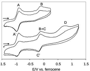
A two-scan cyclic voltammogram of 2a at a scan rate of 5 V s-1 is shown in Figure 6. When the scan is reversed at 0.7 V (vs. the reference Ag/AgCl electron, i.e. 0.27 V vs. ferrocene), only an additional oxidation peak B at -0.10 V (-0.05 V for 2b) is observed in the voltammogram, in addition to the reversible one-electron oxidation of 2a at A/A’. There is no return wave associated to this peak, indicating that the generated species decomposes rapidly. Note that the voltammogram does not change significantly in the second scan. When the potential sweep is switched at a higher potential, on the other hand, a third oxidation process
D, also irreversible, appears at ca. 0.8 V. Following the first potential sweep reversal, a new
C overlaps with peak B in the second scan. The behavior of compound 2b is very similar to
that of 2a. The C/C’ process shows a reduced reversibility for compound 2b. Voltammograms of 2a and 2b at variable scan rates are available in the Supporting Information.
-1.5 -1 -0.5 0 0.5 1 1.5 E/V vs. ferrocene A’ A B+C B D C’ -1.5 -1 -0.5 0 0.5 1 1.5 E/V vs. ferrocene A’ A B+C B D C’
Figure 6. Two-scan cyclic voltammograms of compound 2a in MeCN. Scan rate = 5000
mV s-1.
According to the previous study, the above observations are interpreted as follows, see Scheme 5. The 17-electron [M]H3+• complex produced at A yields (via the nonclassical
isomer [M]H(H2)+•) complex [M]H(MeCN)+•, which is responsible for the oxidation peak B.
The complete lack of reversibility for the latter is due to the immediate saturation by solvent coordination, to afford the 18-electron [M]H(MeCN)22+. On the other hand, subsequent
oxidation of [M]H3+• at D leads to the 16-electron [M]H32+, which is immediately followed by
proton transfer to the starting material [M]H3 with formation of [M]H4+ (redox inactive) and
[M]H2(MeCN)+. The latter is responsible for the reversible process C/C’. A deeper analysis
of these processes and complimentary investigations confirming their assignment were carried out during the previous study.[16] The two anodic waves of peaks B and C accidentally overlap for [M] = (1,2,4-C5H2tBu3)Mo(PMe3)2, whereas they differ only slightly for [M] =
[M]H(MeCN)+• and 18-electron [M]H2(MeCN)+ agree with a high covalent nature for the Mo-H bond. [M]H(H2)+. [M](S)H(H)2 +. +S [M]H3 [M]H3 +. +S [M]H3 2+ [M]H2S 2+ [M]H2S+ [M]H4 + [M]H3
Classical Non classical
Reductive elimination E°A - e -- e -E°D E°C - e -[M](S)H+. [M]H+. +S - e -E°B [M](S)H2+ - H2 dissociative associative Scheme 5
The essential point is that peak B is observed even at the highest scan speeds (up to 5 V s-1), whereas the same process for system Cp*Mo(dppe)H
3 became observable only at v < 0.6
V s-1 in neat MeCN. This illustrates that the H2 elimination process is fast for systems [2a]+
and [2b]+, faster than when [M] = Cp*Mo(dppe), thus strongly suggesting that it proceeds
dissociatively.
The voltammograms of 2a in THF, see Figure 7, show many similarities but also interesting differences with respect to the behavior of the same compound in MeCN and to that of compound Cp*Mo(dppe)H3 in THF. Following the oxidation at A, the irreversible
peak B and the second oxidation process D are observed like in MeCN. Transit over process
D does not generate a reversible C/C’ couple. Transit over process B, on the other hand,
generates a weak and broad reduction peak E at ca. -1.9 V, while peak A loses reversibility. A full rationalization of all these observations is not possible, as many of the species
implicated in Scheme 5 may be unstable and evolve to other unknown products when S = THF. Whereas the potentials of peaks A and D are close to the values observed in MeCN, peak B occurs at Ep,a ca. 0.25 V in THF (vs. -0.18 V in MeCN). The more positive potential
in THF may be related to a reduced donating power of THF relative to MeCN and/or to a more extensive solvent dissociation equilibrium established by the THF adduct with the 15-electron complex. Indeed, the presence of this equilibrium has been evidenced by the EPR study (see above). The most interesting feature, however, is the fact that compound 2a shows peak B also in THF, whereas compound Cp*Mo(dppe)H3 only shows it in MeCN.[16] This is
consistent with a dissociative mechanism for the H2 substitution in [2a]+, in which case the
rate should be essentially solvent independent. Indeed, peak B is observable in THF even at the fast scan rates (1 V s-1).
-2.3 -1.8 -1.3 -0.8 -0.3 0.2 0.7 1.2 E/V vs. ferrocene A B D B D A’ E F (a) (b) (c)
Figure 7. Cyclic voltammograms of compound 2a in THF with different switching
potentials. Scan rate = 200 mV s-1.
The behavior of 2b in THF shows similarities to that of 2a (see figure in the Supporting Information). Notably, a peak corresponding to process B is again present (Ep,a = 0.2 V vs.
ferrocene). The behavior at higher potential, however, is more complex and not fully understood. It is possible that only a fraction of [2b]+ undergoes H2 elimination, in
competition with other decomposition processes, in the THF solvent. This seems to be suggested also by the EPR monitoring of this decomposition (vide supra). It seems clear,
however, that the first decomposition mode (H2 elimination to afford a 15-electron derivative,
possibly in equilibrium with a 17-electron THF adduct) is also established by this system.
Conclusion
We have shown that the combination of greater ligand donor power and greater steric bulk in the coordination sphere of half sandwich MoIV trihydride complexes stabilizes the 17-electron products of one-17-electron oxidation. Complex [(1,2,4-C5H2tBu3)Mo(PMe3)2H3]+ is
sufficiently stable to be crystallized and structurally characterized. Its geometry is very close to that of the neutral precursor, except that the separation between two hydride ligands is shorter suggesting the presence of a stretched dihydrogen ligand (or compressed MH2
moiety). To the best of our knowledge, this is the first time that such evidence has been obtained for a paramagnetic polyhydride system. The steric control in this system, however, is rather subtle: on going from the 1,2,4-C5H2tBu system to the more encumbered C5HiPr4
system, the paramagnetic trihydride product becomes less stable and could not be isolated. The C5HiPr4 ligand might impose such steric pressure to the MoH3 system as to force a
stronger interaction between two hydride ligands and a more favorable expulsion of H2. This
also appears true for the tetrahydrido protonation product (3a vs. 3b). A peculiar difference between the ground state properties of [2a]+ and [2b]+ has been evidenced by EPR spectroscopy (Figure 4). The present investigation has also revealed a dissociative pathway for H2 substitution by a solvent molecule in the paramagnetic system. The oxidative behavior
of the half-sandwich Mo trihydride system is summarized in Scheme 6: H2 substitution by
MeCN on [Mo]H3+ is associative for [Mo] = Cp*Mo(dppe) and dissociative for
Cp#Mo(PMe3)2; an equilibrium has been established by EPR spectroscopy for the latter
no evidence was previously obtained for a 15-electron species in the Cp*Mo(dppe) case. The dissociative product has in fact been isolated and structurally characterized, providing the first well characterized example of an oxidatively induced reductive elimination of H2 from a
polyhydride compound. [M]H(H2) +. [M](S)H(H)2+. +S [M]H3 +.
Classical Non classical
[M](S)H+. [M]H+. +S - H2 dissociative associative isolated (5a) from THF observed by EPR (5a in THF) Scheme 6 Experimental Section
General procedures. All operations were carried out under an atmosphere of argon
using standard Schlenk line and glove box techniques. Solvents were dehydrated (CH2Cl2:
CaH2; THF, toluene, sodium benzophenone ketyl) and distilled under dinitrogen prior to use.
Mo(CO)6 and PMe3 (1 M solution in THF) were purchased from Aldrich and used as
received. Compounds 1,2,4-tri(tert-butyl)- and tetra(iso-propyl)-cyclopentadiene (as isomer mixtures) were prepared by literature methods[51, 52] and converted to their corresponding sodium salts by reaction with NaNH2.
(b) Measurements. NMR measurements were carried out on either a Bruker AC 200
or a Bruker AMX250 spectrometer and calibrated with the residual solvent resonances (1H) or with external 85% H3PO4 (31P). The lineshape analyses for the dynamic processes were
carried out by simulation with DNMR3, which is incorporated into the freely available SpinWorks program.[53] EPR spectra were measured on a Elexsys E500 BRUKER
spectrometer equipped with both a frequencymeter and gaussmeter. The spectrometer frequency was calibrated with diphenylpicrylhydrazyl (DPPH, g = 2.0037). EPR spectra simulations and fittings were carried out with the freely available WinSim program.[54] Cyclic voltammograms were recorded with an EG&G 362 potentiostat connected to a Macintosh computer through MacLab hardware/software. The electrochemical cell was fitted with an Ag-AgCl reference electrode, a platinum disk working electrode and a platinum wire counter-electrode. [Bu4N]PF6 (ca. 0.1 M) was used as supporting electrolyte. The ferrocene standard
had a potential of 0.43 V in MeCN and 0.62 V in THF under our experimental conditions.
Synthesis of (1,2,4-C5H2tBu3)Mo(CO)3CH3. A solution of Na(1,2,4-C5H2tBu3) (1.95
g, 8.3 mmol) in THF (20 mL) was transferred into a suspension of Mo(CO)6 (2.20 g, 8.3
mmol) in THF (15 mL). The mixture was heated to reflux for 15 h, during which time a colour change from pale yellow to dark red was observed. The mixture was then cooled and CH3I (1 mL,16 mmol) was added via syringe, causing an immediate colour change to bright
yellow. Subsequently, the mixture was heated to reflux for 2 h; following which it was cooled and the solvents evaporated. The residue was then extracted with pentane (100 mL) and the pentane solution evaporated to yield (1,2,4-C5H2tBu3)Mo(CO)3CH3 as a yellow solid.
Yield = 2.642 g; 74 %. IR (CH2Cl2): 2005, 1918 cm-1 (CO).
Synthesis of (C5HiPr4)Mo(CO)3CH3. A solution of Na(C5HiPr4) (2.82 g, 11.0 mmol)
in THF (25 mL) was transferred into a suspension of Mo(CO)6 (2.88 g, 10.9 mmol) in THF
(20 mL). The mixture was heated to reflux for 16 h, during which time a colour change from pale yellow to dark red was observed. The mixture was then cooled and CH3I (1.5 mL, 24
mmol) was added via syringe, causing an immediate colour change to bright yellow. Subsequently, the mixture was heated to reflux for 2 h; following which it was cooled and the solvents evaporated. The residue was then extracted with pentane and the pentane solution evaporated to yield (1,2,4-C5H2tBu3)Mo(CO)3CH3 as a yellow solid. Yield = 3.739 g; 80 %.
The compound was used directly for the synthesis of (C5HiPr4)MoCl4 (see below), without
characterisation.
Synthesis of (1,2,4-C5H2tBu3)MoCl4, 1a. A solution of PhICl2 (5 g, 38.2 mmol) in
dichloromethane (20 mL) was transferred slowly into a solution of (1,2,4-C5H2tBu3)Mo(CO)3CH3 (2.64 g, 6.2 mmol) in dichloromethane (15 mL). The mixture was
heated to reflux for 3 h during which time a colour change from brown to indigo was observed. Solvents were then concentrated to ca. 3 mL and the resulting suspension was filtered to give a purple solid, which was washed with portions of pentane (5 x 50 mL) and then dried under reduced pressure to give (1,2,4-C5H2tBu3)MoCl4, 1a, as a purple solid. Yield
= 2.32 g, 79 %. EPR: g = 1.992, aMo = 38.8 G.
Synthesis of (C5HiPr4)MoCl4, 1b. PhICl2 (6.87 g, 52.50 mmol) dissolved in
dichloromethane(20 mL) was added dropwise to a solution of (C5HiPr4)MoCO3CH3 (6.85 g,
15.98 mmol) in dichloromethane (20 mL). The solution was heated to reflux for 3 h, during which time a colour change from brown to indigo was observed. Solvents were then concentrated to ca. 4 mL and the resulting suspension was filtered. The solid was washed with portions of pentane (5 x 20 mL) and dried under reduced pressure to give (C5HiPr4)MoCl4, 1b, as an indigo solid. Yield = 5.27 g, 70 %.
Synthesis of (1,2,4-C5H2tBu3)Mo(PMe3)2H3, 2a. Compound 1a (1415 mg, 3.00 mmol) was dissolved in tetrahydrofuran (20 mL) and a solution of trimethylphosphine in tetrahydrofuran (1 M, 8 mL, 8 mmol) was added. The mixture was stirred for 30 min, and then a suspension of lithium tetrahydroaluminate (ca. 650 mg) in tetrahydrofuran (40 mL) was carefully added. Gas evolution was observed during the addition. The mixture was stirred for 5 h then methanol (ca. 6 mL) was added dropwise causing vigorous gas evolution. The resulting suspension was stirred for 1 h and vacuum-dried; the residue was then extracted with diethyl ether (ca. 150 mL) and filtered through Celite 545. The final solution was
vacuum-dried, and the residue washed three times with methanol (6, 4 and 4 mL) and dried in vacuo. The product 2a was obtained as a pale yellow solid. Yield: 727 mg (50 %). Anal. Calcd. for C23H50MoP2: C, 57.01; H, 10.40. Found: C, 56.48; H, 10.88. 1H NMR (C6D6): -5.20 (t, J =
51.0 Hz, 3 H, Mo-H), 1.37 (s, 9 H, tBu), 1.49 (br, 18 H, P(CH3)3), 1.58 (s, 18 H, 2 x tBu), 4.86
(s, 2 H, C5H2tBu3). 31P{1H} NMR (C6D6): 17.9 (s). A single crystal for the X-ray analysis
was obtained by slow diffusion of a MeOH layer into a pentane solution at 5ºC.
Synthesis of (C5HiPr4)Mo(PMe3)2H3, 2b. Compound 1b (890 mg, 1.89 mmol) was dissolved in tetrahydrofuran (20 mL), and a solution of trimethylphosphine in tetrahydrofuran (1 M, 5 mL, 5 mmol) was added. The mixture was stirred for 20 min and a suspension of lithium tetrahydroaluminate (ca. 500 mg) in tetrahydrofuran (40 mL) was carefully added. Gas evolution was observed during the addition. The mixture was stirred for 6 h, after which methanol (ca. 5 mL) was added drop by drop. Vigorous gas evolution was observed at this point. The resulting suspension was stirred for 1 h and then vacuum-dried. The residue was extracted with diethyl ether (ca. 100 mL) and filtered through Celite 545. The final solution was vacuum-dried and the residue washed with portions of methanol (5, 3 and 3 mL) and dried in vacuo. The product 2b was obtained as an orange-yellow solid. Yield: 449 mg (49 %). Anal. Calcd. for C23H50MoP2: C, 57.01; H, 10.40. Found: C, 56.91; H, 11.10. 1H NMR
(C6D6): -5.15 (t, J = 52.9 Hz, 3 H, Mo-H), 1.30 - 1.80 (42 H, CH(CH3)2, P(CH3)3), 2.79 (m,
2 H CH(CH3)2), 2.96 (m, 2 H CH(CH3)2), 4.72 (s, 1 H, C5HiPr4). 31P{1H} NMR (C6D6):
17.3 (s). A single crystal for the X-ray analysis was obtained by diffusion of a MeCN layer onto a THF solution at room temperature.
Synthesis of [(1,2,4-C5H2tBu3)Mo(PMe3)2H3]+PF6-, 2a+PF6-. A suspension of [Fe(5 -C5H5)2]PF6 (32 mg, 0.10 mmol) in tetrahydrofuran (5 mL) was added dropwise to a cold
solution (193 K) of compound 2a (53 mg, 0.11 mmol) in tetrahydrofuran (5 mL). The solution color immediately changed from pale yellow to dark blue and, within a few minutes,
to orange. The reaction mixture was slowly warmed up to 253 K and then concentrated to ca. 1 mL. Addition of cold pentane (253 K, 10 mL) afforded a brown precipitate that was decanted and further washed with cold pentane (253 K, 3x10 mL) and finally vacuum-dried. The product 2a+PF6- was obtained as a pale brown solid. Yield: 48 mg, 70 %. EPR (THF): g = 2.0185, aP = 36.2 G, aH = 11.4 G, aMo = 30.8 G. A single crystal for the X-ray analysis was
obtained by diffusion of a pentane layer onto a THF solution at -80°C.
Synthesis of [(1,2,4-C5H2tBu3)Mo(PMe3)2H4]+BF4-, 3a. A solution of (5 -C5H2tBu3)Mo(PMe3)2H3 (2a, 40 mg, 0.08 mmol) in diethyl ether (4 mL) was cooled to –80°C.
HBF4 (54 % solution in diethyl ether, 22μL, 0.16 mmol) was added via syringe. Within
minutes, a white precipitate formed. A further portion of diethyl ether (2 mL) was added. The solvent was then decanted and the solid washed with portions of diethyl ether (5 x 3 mL) and dried under reduced pressure to yield 3a as a white solid. Yield: 39 mg, 85 %. 31P{1H}
NMR (THF-d8): 0.4 (s). 1H NMR (THF-d8): -4.20 (t, J = 53.4 Hz, 3 H, Mo-H), 1.38 (s, 9
H, -tBu), 1.46 (s, 18 H, 2 x tBu), 1.78 (br, 18 H, P(CH3)3), 5.08 (s, 2 H, C5H2tBu3). A single
crystal for the X-ray analysis was obtained by diffusion of a diethyl ether layer onto a THF solution at room temperature.
Generation of [(1,2,4-C5H2tBu3)Mo(PMe3)2(MeCN)H2]+BF4-, 4a. [(1,2,4-C5H2tBu3
)-Mo(PMe3)2H4]+BF4-, (3a, 10 mg, 0.018 mmol) was measured into and NMR tube and
dissolved in CD3CN. 1H and 31P NMR spectra were recorded initially and again after 5 h. At
this time, the resonances corresponding to 3a were replaced by a new set of resonances, ascribed to [(1,2,4-C5H2tBu3)Mo(PMe3)2H2MeCN]+BF4-, 4a. 31P{1H} NMR (CD3CN): 1.85
(s). 31P{1H sel. decoupler at 1.61 ppm} 1.88 (t, J = 52.85 Hz) 1H NMR (CD3CN, 298 K):
1.07 (9H, s,C(CH3)3), 1.42, (18H, s, 2 x C(CH3)3), 1.61 (18H, d, J = 8.7 Hz, P(CH3)3), 2.17
(3H, s, CH3CN) 4.59 (m, 2H, C5H2tBu4). 1H NMR (CD3CN, 233 K): -6.77 (1H, t, JP-H =
C(CH3)3), 1.58 (18H, d, JP-H = 10 Hz, P(CH3)3 ), 2.40 (3H, s, CH3CN), 4.58 (2H, m,
C5H2tBu3).
Generation of [(C5HiPr4)Mo(PMe3)2H2(MeCN)]+BF4-, 4b. A solution of HBF4 (54 %
in diethyl ether, 7.4 μL, 0.05 mmol) was added to a solution of (5-C
5HiPr4)MoH3(PMe3)2 (26
mg, 0.05 mmol) in thf (1 mL) and MeCN (1 mL) at –80 oC. A color change from yellow to orange was observed immediately. Solvents were concentrated to 1 mL and diethyl ether (1 mL) was added to aid the precipitation of a yellow solid. The solution was filtered and the solid washed with portions of diethyl ether (5 x 2 mL) and dried under reduced pressure to give 4b as a yellow solid. 31P{1H} NMR (acetone-d6): 4.66 (s). 31P{1H sel. decoupler at
1.68 ppm} 4.68 (t, J = 54.7 Hz) 1H NMR (acetone-d6, 298 K): -2.5 (br, 2H, Mo-H), 1.18 (12H, d, J = 8.2 Hz, CH(CH3)2), 1.31, (12H, d, J = 8.6 Hz, CH(CH3)2), 1.69 (18H, d, P(CH3)3), 2.71 (7H, m, CH(CH3)2, CH3CN) 4.92 (s, 1H, C5HiPr4). 1H NMR (acetone-d6, 200 K): -5.27 (1H, td, JP-H = 32.5 Hz, JH-H = 10 Hz Mo-H), -0.13 (1H, ddd, JP-H = 47.5, 33.75, JH-H = 10 Hz, Mo-H), 1.07 (3H, d, J = 6.8 Hz, CH(CH3)2), 1.13 (3H, d, J = 6.8 Hz, CH(CH3)2), 1.18 – 1.23 (9H, 3 x d, J = 7.3, 6.9 Hz, CH(CH3)2), 1.29 (3H, d, J = 7.3 Hz, CH(CH3)2), 1.39 (6H, 2 x d, J = 6.5 Hz, CH(CH3)2), 1.65 (18H, 2 x d, JP-H = 8.0 Hz, P(CH3)3), 2.53 (1H, m, J = 6.8 Hz, CH(CH3)2,), 2.58 (1H, m, J = 7.3 Hz, CH(CH3)2), 2.71 (2H, 2 x CH(CH3)2, m, J = 6.5 Hz), 2.79 (3H, s, CH3CN), 5.02 (1H, d, J = 6 Hz, C5HiPr4).
Formation of [(1,2,4-C5H2tBu3)Mo(PMe3)2H]+PF6-, 5. A solution of compound
2a+PF
6- in THF was stored at -20°C for 2 days, after which time a mixture of green and dark orange-red crystals had formed. One of the green crystals was used for the X-ray analysis. For the spectroscopic properties, see Results and Discussion.
Single crystal X-ray and neutron diffraction studies. A single crystal of each
compound was mounted under inert perfluoropolyether at the tip of glass fibre and cooled in the cryostream of either an Oxford-Diffraction XCALIBUR CCD diffractometer for 2b, 4a
and 5a or a Stoe IPDS diffractometer for 2a, 2a+. Data were collected using the monochromatic MoK radiation (= 0.71073). The structures were solved by direct methods (SIR97)[55] and refined by least-squares procedures on F2 using SHELXL-97.[56] All H atoms attached to carbon were introduced in calculation in idealised positions and treated as riding models. In compound 2a+, there are two cations and anions in the asymmetric unit and all the tBu groups of one of the cations are disordered over two positions. In structure 2a, coordinates and Uiso for the hydrides, were fully refined whereas in 3a, the coordinates of the
hydrides were fully refined with an overall isotropic thermal parameter. In 2a+ and 5a, the coordinates of the hydrides were fully refined with Uiso= 1.2Ueq[Mo(2a+)] or
Uiso=1.5Ueq[Mo(5a)]. The disordered moieties were refined applying the restraints available
within SHELXL97.[56] Moreover, some residual electron density was difficult to model and therefore, the SQUEEZE function of PLATON[57] was used to eliminate the contribution of
the electron density in the solvent region from the intensity data, and the solvent-free model was employed for the final refinement. There are four cavities per unit cell and PLATON estimated that each cavity contains 32 electrons which could be attributed to a disordered THF molecules. The data collected for compound 2b were of very low quality and although the structural model is mainly correct, it was not possible to locate any hydride ligand. The drawing of the molecules was realised with the help of ORTEP32.[58] Crystal data and refinement parameters are shown in Table 2.
The neutron single-crystal diffraction study was performed using the time-of-flight Laue diffractometer SXD[59] installed at the ISIS pulsed spallation source. SXD uses the white beam Laue technique and a stationary crystal combined with eleven highly pixellated area detectors covering around half a sphere around the sample. Thus, large volumes in reciprocal space can be collected in a single shot. A suitable single crystal of the complex (2a) was fixed to an Aluminum pin with thin strips of adhesive Al tape and mounted on a He closed-cycle
refrigerator and cooled slowly to 20K. The space group P21/n was confirmed at 20K. No
significant change in the crystal mosaic or splitting of the peak was observed during cooling. Further crystallographic data and experimental details are given in Table 2 and in the Supporting Information. The unit cell dimensions were precisely calculated, at the end of the data collection, from the positions of 60 reflections per each detector orientation. Data were collected at nine different orientations at 20(1) K for ca. 24 hour per orientation. The range of wavelengths used for the data collection was 0.37 < λ < 8.8 Å, even though the bulk of the diffraction information is obtained from the wavelength range 0.5 < λ < 7.0 Å. Data reduction and a Gaussian absorption correction were performed using the standard SXD procedure implemented in the SXD2001 software[60] resulting in a total of 8686 reflections of which 3203 were unique. The starting structural model for the refinement was based on the atomic co-ordinates for the non hydrogen atoms taken from the X-ray structural determination. The structure was refined by full matrix least squares, minimising the function [w(Fo2 -
(1/k)Fc2)2] and using all independent data. During the refinement, the difference-Fourier maps
clearly showed all H atoms of the ligands and the three hydrides. The final structure model included co-ordinates and anisotropic displacement parameters for all atoms. Upon convergence the final Fourier difference map showed no significant features. The coherent scattering amplitudes used were those tabulated by Rauch and Waschkowski.[61] All calculations were carried out by using the PC version of the programs WINGX,[62]
SHELX-97[56] and ORTEP.[58]
Crystallographic data (excluding structure factors) have been deposited with the Cambridge Crystallographic Data Centre as supplementary publication no. CCDC 631893 - 631898. Copies of the data can be obtained free of charge on application to the Director, CCDC, 12 Union Road, Cambridge CB2 1EZ, UK (Fax: (+44) 1223-336-033; E-mail: [email protected]).
Acknowledgements
We thank the European Commission through the HYDROCHEM program (contract HPRN-CT-2002-00176) for support of this work. MB thanks the Spanish Ministerio de Educación y Ciencia for a post-doctoral fellowship.
Supporting Information available
Figures showing the structure of 2a from the neutron diffraction experiment, the variable temperature 31P{1H} experiment for compound 2b, and a variety of cyclic voltammograms for


![Figure 4. EPR spectra of complexes [2] + in THF solution. Above: complex [2a] + (T = 193)](https://thumb-eu.123doks.com/thumbv2/123doknet/13662980.429763/15.892.111.465.102.344/figure-epr-spectra-complexes-thf-solution-complex-t.webp)