HAL Id: pasteur-01371190
https://hal-pasteur.archives-ouvertes.fr/pasteur-01371190
Submitted on 24 Sep 2016HAL is a multi-disciplinary open access archive for the deposit and dissemination of sci-entific research documents, whether they are pub-lished or not. The documents may come from teaching and research institutions in France or abroad, or from public or private research centers.
L’archive ouverte pluridisciplinaire HAL, est destinée au dépôt et à la diffusion de documents scientifiques de niveau recherche, publiés ou non, émanant des établissements d’enseignement et de recherche français ou étrangers, des laboratoires publics ou privés.
Distributed under a Creative Commons Attribution| 4.0 International License
The skin is a significant but overlooked anatomical
reservoir for vector-borne African trypanosomes
Paul Capewell, Christelle Cren-Travaillé, Francesco Marchesi, Pamela
Johnston, Taylor-Anne Gorman, Estefania Calvo-Alvarez, Aline Crouzols,
Grégory Jouvion, Vincent Jammoneau, William Weir, et al.
To cite this version:
Paul Capewell, Christelle Cren-Travaillé, Francesco Marchesi, Pamela Johnston, Taylor-Anne Gor-man, et al.. The skin is a significant but overlooked anatomical reservoir for vector-borne African trypanosomes. eLife, eLife Sciences Publication, 2016, �10.7554/eLife.17716�. �pasteur-01371190�
The skin is a significant but overlooked anatomical reservoir for vector-borne African
1
trypanosomes
2 3
Paul Capewell1*, Christelle Cren-Travaillé2*, Francesco Marchesi3, Pamela Johnston3, Caroline
4
Clucas1, Robert A Benson4, Taylor-Anne Gorman1,4, Estefania Calvo-Alvarez2, Aline Crouzols2,
5
Grégory Jouvion5, Vincent Jammoneau6, William Weir1, M Lynn Stevenson3, Kerry O’Neill1, Anneli
6
Cooper1, Nono-raymond Kuispond Swar7, Bruno Bucheton6, Dieudonné Mumba Ngoyi8, Paul
7
Garside4, Brice Rotureau2** and Annette MacLeod1**
8 9
1Wellcome Trust Centre for Molecular Parasitology, College of Medical, Veterinary and Life
10
Sciences, Henry Wellcome Building for Comparative Medical Sciences, Garscube Estate, Glasgow,
11
United Kingdom, G61 1QH
12
2Trypanosome Transmission Group. Trypanosome Cell Biology Unit, INSERM U1201 & Department
13
of Parasites and Insect Vectors, Institut Pasteur, Paris, France
14
3Veterinary Diagnostic Services, Veterinary School, University of Glasgow, Garscube Estate,
15
Glasgow, United Kingdom, G61 1QH
16
4Institute of Infection, Immunology and Inflammation, College of Medical, Veterinary and Life
17
Sciences, Glasgow Biomedical Research Centre, University of Glasgow, Glasgow, United Kingdom,
18
G12 8TA
19
5Human Histopathology and Animal Models Unit, Institut Pasteur, Paris, France
20
6Institut de Recherche pour le Développement, Unité Mixte de Recherche IRD-CIRAD 177, Campus
21
International de Baillarguet, Montpellier, France
22
7University of Kinshasa, Kinshasa, Democratic Republic of the Congo
23
8Department of Parasitology, National Institute of Biomedical Research (INRB), Kinshasa,
24
Democratic Republic of the Congo
25 26
* Joint first authors
27
** Joint last authors
28
The authors have no competing financial interests.
29
Keywords
30
Skin, blood, reservoir, transmission, infection, Human African Trypanosomiasis, trypanosomes,
31
Trypanosoma brucei, tsetse fly, vector-borne, evolution 32
Abstract
34
The role of mammalian skin in harbouring and transmitting arthropod-borne protozoan parasites has
35
been overlooked for decades as these pathogens have been regarded primarily as blood-dwelling
36
organisms. Intriguingly, infections with low or undetected blood parasites are common, particularly
37
in the case of Human African Trypanosomiasis caused by Trypanosoma brucei gambiense. We
38
hypothesise, therefore, the skin represents an anatomic reservoir of infection. Here we definitively
39
show that substantial quantities of trypanosomes exist within the skin following experimental
40
infection, which can be transmitted to the tsetse vector, even in the absence of detectable
41
parasitaemia. Importantly, we demonstrate the presence of extravascular parasites in human skin
42
biopsies from undiagnosed individuals. The identification of this novel reservoir requires a
re-43
evaluation of current diagnostic methods and control policies. More broadly, our results indicate that
44
transmission is a key evolutionary force driving parasite extravasation that could further result in
45
tissue invasion-dependent pathology.
46 47
Introduction
48
Understanding the process of parasite transmission is essential for the design of rational control
49
measures to break the disease cycle and requires the identification of all reservoirs of infection. In a
50
number of vector-borne diseases, it is becoming evident that asymptomatic individuals, be they
51
humans or animals, can represent a significant proportion of the infected population and therefore
52
an important reservoir of disease that requires targeting by control measures1-4. The recent
53
identification in West Africa of asymptomatic individuals with human trypanosomiasis (long-term
54
seropositives) but undetected parasitaemia, raises the question of what role these individuals play
55
in disease transmission3,5-7. Therapy is currently only directed towards microscopy-positive
56
individuals and thus a proportion of the infected population remain untreated.
57
There is convincing evidence that seropositive individuals with low or undetected parasitaemia
58
contain transmissible trypanosomes. Xenodiagnosis experiments, in which tsetse flies are fed on
59
microscopy-negative infected humans8 or, more recently, experimentally-infected pigs9, have shown
60
that these apparently aparasitaemic hosts contain the parasite since the tsetse flies became
61
infected. It is uncertain where the trypanosomes reside in the host but, given the telmophagus
62
(slash and suck) feeding habit of the tsetse fly, they could be skin-dwelling parasites ingested with
63
the blood meal. Our findings suggest that parasites may be sufficiently abundant in the skin to allow
64
transmission and therefore the skin may represent an anatomical reservoir of infection.
65
Detection of trypanosomes in the skin is not well documented, although there are descriptions of
66
cutaneous symptoms associated with African trypanosomiasis and distinct ‘trypanid’ skin lesions10.
67
Imaging data from mouse models of infection suggest that trypanosomes sequester to major organs
68
such as the spleen, liver and brain11,12 and recent evidence has demonstrated trypanosomes in
69
extravascular adipose tissue13. These adipose-associated trypanosomes appear to be a new
life-70
cycle stage with a distinct transcriptional profile and, while tsetse bite-site associated transmission
71
has been suggested14, and a historical study made a passing observation of localised deposition of
72
trypanosomes in the skin matrix15, the broader role of skin-dwelling trypanosomes in transmission
73
remains unclear. In this paper we report the investigation of a possible anatomical reservoir in the
74
skin of the mammalian host. We provide conclusive evidence of T.b. brucei, (a causative agent of
75
animal trypanosomiasis) and the human-infective trypanosome, T.b. gambiense, invading the
76
extravascular tissue of the skin (including but not restricted to the adipose tissue) and undergoing
77
onward transmission despite undetected vascular parasitaemia. We also provide evidence of
78
localisation of trypanosomes within the skin of undiagnosed humans. The presence of a significant
79
transmissible population of T. brucei in this anatomical compartment is likely to impact future control
80
and elimination strategies for both animal and human trypanosomiases.
81 82
Results
83
In order to investigate the potential for extravascular skin invasion by T. brucei, BALB/c mice were
84
inoculated via IP injection with the low virulence STIB247 strain of T. brucei and skin sections were
85
assessed over a 36-day time-course. The presence and relative quantities of extravascular
86
parasites were evaluated by semi-quantitative scoring of the histological samples (Figure 1-source
87
data 1) and compared to blood parasitaemia (Figure 1-source data 2). Extravascular parasites were
88
first observed in the skin 12 days post-infection and remained throughout the experiment. Skin
89
parasite numbers fluctuated to a lesser extent than blood parasitaemia and the apparent periodicity
90
in the skin may be due to one particularly high data point on day 24 (Figure 1). Parasites were found
91
in the dermis, subcutaneous adipose tissue (Figure 2) and in fascia beneath the panniculus
92
carnosus muscle. We did not detect any particular clustering around dermal adipocytes. The
93
presence of parasites in the skin was not associated with major inflammation (Supplementary File
94
1). To confirm that skin invasion by this parasite was not strain or sub-species specific, the more
95
virulent TREU927 strain of T. brucei and the human-infective T.b. gambiense strain, PA, were used
96
to infect mice. Extravascular skin invasion of the dermis, subcutaneous adipose tissue and fascial
97
planes (Figure 2-figure supplement 1) was evident with associated mild to moderate inflammation
98
(Supplementary File 2), in some instances the degree of skin invasion in the T.b. gambiense
99
infected mice was far greater than T.b. brucei, perhaps suggesting a greater propensity for
100
sequestration in this sub-species.
101
To confirm that the extravascular distribution of parasites was not an artefact of the route of
102
inoculation, infections by natural vector transmission were carried out using a bioluminescent T.b.
103
brucei strain, AnTat1.1E AMLuc/tdTomato. Mice were infected by a single infective bite of an
104
individual G.m. morsitans. After 4 to 11 days and up to the end of the experiment, parasites were
105
observed in the skin with a dynamic distribution (Figure 3A) and a variable density (Figure 3B).
106
Parasites were first detected in the blood between 5 and 19 days after natural transmission and
107
parasitaemia remained lower than 107 parasites/ml. Observed bioluminescence directly reflects the
108
total number of living parasites in the entire organism, including blood and viscera, but the intensity
109
of the signal decreases with tissue depth. Therefore, at the end of each experiment, mice were
110
sacrificed and their organs were checked for bioluminescence. The presence of extravascular
111
parasites in cutaneous and subcutaneous tissues was first demonstrated by bioluminescence
112
imaging in entire dissected skins (Figure 3C) and was not necessarily localised to the bite site. In
113
addition to the skin, only the spleen, some lymph nodes and adipose tissue were observed to be
114
positive for bioluminescence in several individuals (Figure 3-figure supplement 1). This suggests
115
that the observed bioluminescence is likely to originate predominantly, but not solely, from parasites
116
in the skin. In addition, only mild inflammation was observed after 29 days (Figure 3-figure
117
supplement 2).
To confirm that trypanosomes in the skin are a viable population, fluorescent parasites were
119
monitored by two intravital imaging methods following IP injection and natural transmission (Figure
120
4). First, IP-injected fluorescent T. brucei STIB247 were imaged in vivo using 2-photon microscopy
121
(Figure 4A). Extravascular trypanosomes observed in the dermal layer of dorsal skin were highly
122
motile, consistent with viability (Video 1). Second, naturally-transmitted fluorescent T. brucei
123
AnTat1.1E AMLuc/tdTomato were imaged in vivo using spinning-disk confocal microscopy in the
124
C57BL/6J-Flk1-EGFP mouse line that has green fluorescent endothelial cells in the lymphatic and
125
blood vessels16 (Figure 4B). Extravascular trypanosomes were observed in the dermal layer of the
126
ear and were highly motile (Videos 2, 3 and 4).
127
Differentiation of dividing trypanosomes to non-dividing stumpy forms is essential for transmission to
128
the tsetse. Using the relative transcript abundance of the stumpy marker Protein Associated with
129
Differentiation 1 (PAD1)17 to an endogenous control, Zinc Finger Protein 3 (ZFP3)18, we estimated
130
that approximately 20% of skin-dwelling parasites were stumpy (Supplementary File 3). To directly
131
determine the proportion of stumpy forms in skin sections, histological staining for PAD1 was also
132
performed (Table 1-source data 4). PAD1-positive cells were observed in variable proportions (from
133
8 to 80%) in all bioluminescent skin samples examined after IP injection (Supplementary File 4).
134
Following natural transmission, up to 38% of parasites detected using VSG surface markers (Figure
135
3-figure supplement 3A-B) also expressed PAD1 (Figure 3-figure supplement 3C-D) (n=441 cells
136
from 8 skin sections). In all skin sections, stumpy parasites were homogenously distributed in the
137
dermis and subcutaneous adipose tissues.
138
We next assessed the ability of skin-dwelling parasites to infect tsetse flies. Teneral flies (immature
139
flies that have not yet taken a blood meal) were fed on different regions of skin from mice infected
140
with AnTat1.1E AMLuc/TY1/tdTomato with differing levels of bioluminescence across the skin
141
(Table 1-source data 1 and Table 1-source data 2). This was repeated in 20 mice with differing
142
levels of parasitaemia. Flies were dissected and checked for the presence of fluorescent
143
trypanosomes after two days (Table 1). In mice with undetectable or low parasitaemia (<5x10⁴
144
parasites/ml), no parasites were found in flies fed on non-bioluminescent regions. Conversely, a
145
median of 36% (±23%, n=70) of flies that fed on bioluminescent regions of low parasitaemic mice
146
became infected (Table 1 and Table 1-source data 3). This demonstrates that skin parasites, from
147
mice without visible parasitaemia, can contribute to tsetse infection, possibly reflecting human
148
asymptomatic infections. When parasites were detected in the blood and skin, tsetse infection rates
149
increased up to 100% (median of 61% ±22%, n=120) (Table 1 and Table 1-source data 3).
150
Parasites taken-up from the skin were able to further develop through the life-cycle to early procyclic
151
forms in the fly as evidenced by GPEET-procyclin marker on the parasites surface (n=721 cells from
152
16 flies) (Table 1-source data 4 and Supplementary File 4).
153
To demonstrate that skin invasion by trypanosomes can occur in humans as well as the murine
154
model, slides of historical skin biopsies, taken as part of a diagnostic screening programme for
Onchocerca microfilaria in the trypanosomiasis-endemic Democratic Republic of the Congo, were 156
examined for trypanosomes. At the time of sampling, the incidence of trypanosomiasis was 1.5-2%
157
and we hypothesised that some individuals may have harboured undiagnosed infections.
Re-158
examination of 1,121 skin biopsies by microscopy revealed six individuals with trypanosomes in
159
their skin (Figure 5). These individuals had not previously been diagnosed with human
160
trypanosomiasis by clinical signs or the presence of blood parasites.
161 162
Discussion
163
We have shown that there exists a significant yet overlooked population of live, motile,
164
extravascular T. brucei in the dermis and subcutis of animal models infected by artificial routes or by
165
vector transmission. It is likely that once injected the parasites disseminate via the lymph and blood
166
to the skin where they are ingested during tsetse fly pool-feeding and readily initiate cyclical
167
development in the vector. Given the relative volume of the skin organ compared to the vasculature,
168
it is possible, depending on the density of skin invasion, that more parasites exist in the skin than in
169
the blood. The skin, therefore, represents an unappreciated reservoir of infection. The extravasation
170
of trypanosomes was described previously in major organs such as liver, spleen11,12 and visceral
171
adipose tissue13, but the importance of these parasites in transmission was not investigated. Here
172
we show that these skin-dwelling trypanosomes contribute to transmission and could explain the
173
maintenance of disease foci, despite active screening and treatment. Skin invasion for enhanced
174
transmission is likely a powerful evolutionary force driving extravasation, suggesting that the
175
generalised tissue penetration underlying pathogenesis (i.e. splenomegaly, hepatomegaly, CNS
176
invasion) is a secondary epiphenomenon. A skin reservoir also presents a novel target for
177
diagnostics (e.g. skin biopsies), allowing the prevalence of infection to be accurately determined
178
and the identification of any previously undetected animal reservoirs of human disease.
179
The skin as an anatomical reservoir of parasites is a recurring theme in arthropod-borne human
180
diseases such as Leishmania19,20 and Onchocerca21,22. Here we present evidence of trypanosomes
181
in the skin of hitherto undiagnosed individuals. This anatomical reservoir may serve to explain how
182
HAT foci re-emerge and persist despite low numbers of reported cases even in the absence of an
183
animal reservoir23-25.
184
HAT was once widespread across much of sub-Saharan Africa but concerted control efforts brought
185
it close to elimination during the 1960s26,27. However, disease foci persisted, with a resurgence in
186
the number of reported cases to over 300,000 in the 1990s26-28. Currently, HAT is approaching
187
elimination in many areas27,29. Understanding how HAT evaded elimination in the latter half of the
188
20th century and how it continues to persist is vital to efforts to eliminate the disease. For example,
189
our results indicate that it may be necessary to develop novel therapeutics capable of accessing
190
extravascular compartments. The current policy in most endemic countries is not to treat
191
serologically positive individuals unless they demonstrate active infection, due to the long duration
and high toxicity of treatment and the low predictive value of the serological tests. We suggest that
193
this policy should be reconsidered in light of our compelling evidence that they represent a carrier
194
population which may maintain HAT foci and explain previously thwarted efforts to eliminate this
195
major pathogen.
196
Acknowledgments
197
We acknowledge P. Solano, D. Engman, K. Matthews, M. Taylor, M. Carrington, R. Amino, E.
198
Myburgh and K. Gull for providing various material, cell lines, antibodies and plasmids and to A. Tait
199
for critically reviewing this manuscript.
200
References
201
1. Lindblade, K. A., Steinhardt, L., Samuels, A., Kachur, S. P. & Slutsker, L. The silent threat:
202
asymptomatic parasitemia and malaria transmission. Expert Review of Anti-infective Therapy
203
11, 623–639 (2014).
204
2. Fakhar, M. et al. Asymptomatic human carriers of Leishmania infantum: possible reservoirs
205
for Mediterranean visceral leishmaniasis in southern Iran. Annals of Tropical Medicine &
206
Parasitology 102, 577–583 (2013). 207
3. Koffi, M. et al. Aparasitemic serological suspects in T. b. gambiense human African
208
trypanosomiasis: A potential human reservoir of parasites? Acta Tropica 98, 183–188 (2006).
209
4. Berthier, D. et al. Tolerance to Trypanosomatids: A Threat, or a Key for Disease Elimination?
210
Trends in Parasitology 32, 157–168 (2016). 211
5. Jamonneau, V. et al. Untreated human infections by Trypanosoma brucei gambiense are not
212
100% fatal. PLoS Neglected Tropical Diseases 6, e1691 (2012).
213
6. Bucheton, B., MacLeod, A. & Jamonneau, V. Human host determinants influencing the
214
outcome of Trypanosoma brucei gambiense infections. Parasite Immunol. 33, 438–447
215
(2011).
216
7. Kanmogne, G. D., Asonganyi, T. & Gibson, W. C. Detection of T. brucei gambiense, in
217
serologically positive but aparasitaemic sleeping-sickness suspects in Cameroon, by PCR.
218
Ann Trop Med Parasitol 90, 475–483 (1996). 219
8. Frezil, J. L. [Application of xenodiagnosis in the detection of T. gambiense trypanosomiasis in
220
immunologically suspect patients]. Bull Soc Pathol Exot Filiales 64, 871–878 (1971).
221
9. Wombou Toukam, C. M., Solano, P., Bengaly, Z., Jamonneau, V. & Bucheton, B.
222
Experimental evaluation of xenodiagnosis to detect trypanosomes at low parasitaemia levels
223
in infected hosts. Parasite 18, 295–302 (2011).
224
10. McGovern, T. W. et al. Cutaneous manifestations of African trypanosomiasis. Arch Dermatol
225
131, 1178–1182 (1995).
226
11. Blum, J. A., Zellweger, M. J., Burri, C. & Hatz, C. Cardiac involvement in African and
227
American trypanosomiasis. Lancet Infect Dis 8, 631–641 (2008).
228
12. Kennedy, P. G. E. Human African trypanosomiasis of the CNS: current issues and
229
challenges. Journal of Clinical Investigation 113, 496–504 (2004).
230
13. Trindade, S. et al. Trypanosoma brucei parasites occupy and functionally adapt to the
231
adipose tissue in mice. Cell Host & Microbe (2016).
232
14. Caljon, G. et al. The Dermis as a Delivery Site of Trypanosoma brucei for Tsetse Flies. PLoS
233
Pathogens 12, e1005744 (2016). 234
15. Goodwin, L. G. Pathological effects of Trypanosoma brucei on small blood vessels in rabbit
235
ear-chambers. Trans. R. Soc. Trop. Med. Hyg. 65, 82–88 (1971).
236
16. Ema, M., Takahashi, S. & Rossant, J. Deletion of the selection cassette, but not cis-acting
237
elements, in targeted Flk1-lacZ allele reveals Flk1 expression in multipotent mesodermal
progenitors. Blood 107, 111–117 (2006).
239
17. Dean, S., Marchetti, R., Kirk, K. & Matthews, K. R. A surface transporter family conveys the
240
trypanosome differentiation signal. Nature 459, 213–217 (2009).
241
18. Walrad, P., Paterou, A., Acosta-Serrano, A. & Matthews, K. R. Differential Trypanosome
242
Surface Coat Regulation by a CCCH Protein That CoAssociates with procyclin mRNA cis
-243
Elements. PLoS Pathogens 5, e1000317 (2009).
244
19. Sacks, D. in Leishmania After the Genome (2008).
245
20. Schlein, Y. Leishmania and sandflies: interactions in the life cycle and transmission.
246
Parasitol. Today (Regul. Ed.) (1993). doi:10.1016/0169-4758(93)90070-V 247
21. Onchocerciasis. Symptomatology, pathology, diagnosis. (1974).
248
22. Dalmat, H. T. in Onchocerciasis 425 (1955).
249
23. Kagbadouno, M. S. et al. Epidemiology of sleeping sickness in Boffa (Guinea): where are the
250
trypanosomes? PLoS Neglected Tropical Diseases 6, e1949 (2012).
251
24. Cordon-Obras, C. et al. Trypanosoma brucei gambiense in domestic livestock of Kogo and
252
Mbini foci (Equatorial Guinea). Tropical Medicine and International Health 14, 535–541
253
(2009).
254
25. Balyeidhusa, A. S. P., Kironde, F. A. S. & Enyaru, J. C. K. Apparent lack of a domestic animal
255
reservoir in Gambiense sleeping sickness in northwest Uganda. Veterinary Parasitology 187,
256
157–167 (2012).
257
26. World Health Organization. Dept. of Epidemic, Alert, P.Response. WHO report on global
258
surveillance of epidemic-prone infectious diseases. (2000).
259
27. World Health Organization. Control and surveillance of human African trypanosomiasis.
260
World Health Organ Tech Rep Ser 1–237 (2013). 261
28. Steverding, D. The history of African trypanosomiasis. Parasites Vectors 1, 3 (2008).
262
29. Simarro, P. P. et al. Estimating and Mapping the Population at Risk of Sleeping Sickness.
263
PLoS Neglected Tropical Diseases 6, e1859 (2012). 264
30. Geigy, R., Kauffmann, M. & Jenni, L. Wild mammals as reservoirs for Rhodesian sleeping
265
sickness in the Serengeti, 1970-71. Trans. R. Soc. Trop. Med. Hyg. 67, 284–286 (1973).
266
31. Goedbloed, E. et al. Serological studies of trypanosomiasis in East Africa. II. Comparisons of
267
antigenic types of Trypanosoma brucei subgroup organisms isolated from wild tsetse flies.
268
Ann Trop Med Parasitol 67, 31–43 (1973).
269
32. Tait, A., Babiker, E. A. & Le Ray, D. Enzyme variation in Trypanosoma brucei ssp. I Evidence
270
for the sub-speciation of Trypanosoma brucei gambiense. Parasitology 89, 311–326 (1984).
271
33. Myburgh, E. et al. In vivo imaging of trypanosome-brain interactions and development of a
272
rapid screening test for drugs against CNS stage trypanosomiasis. PLoS Neglected Tropical
273
Diseases 7, e2384 (2013). 274
34. Le Ray, D., Barry, J. D., Easton, C. & Vickerman, K. First tsetse fly transmission of the
275
‘AnTat’ serodeme of Trypanosoma brucei. Ann Soc Belg Med Trop (1977).
276
35. Xong, H. V. et al. A VSG expression site-associated gene confers resistance to human serum
277
in Trypanosoma rhodesiense. Cell 95, 839–846 (1998).
278
36. Fragoso, C. M. & Roditi, I. Highly efficient stable transformation of bloodstream forms of
279
Trypanosoma brucei. Mol Biochem Parasitol 153, 220–223 (2007).
280
37. Branchini, B. R., Southworth, T. L., Khattak, N. F., Michelini, E. & Roda, A. Red- and
green-281
emitting firefly luciferase mutants for bioluminescent reporter applications. Anal. Biochem.
282
345, 140–148 (2005).
283
38. Bastin, P., Bagherzadeh, Z., Matthews, K. R. & Gull, K. A novel epitope tag system to study
284
protein targeting and organelle biogenesis in Trypanosoma brucei. Mol Biochem Parasitol 77,
285
235–239 (1996).
286
39. Magez, S. & Caljon, G. Mouse models for pathogenic African trypanosomes: unravelling the
287
immunology of host-parasite-vector interactions. Parasite Immunol. 33, 423–429 (2011).
288
40. Lumsden, W. H. Quantitative methods in the study of trypanosomes and their applications:
289
With special reference to diagnosis. Bull. World Health Organ. 28, 745–752 (1963).
290
41. Rotureau, B., Subota, I., Buisson, J. & Bastin, P. A new asymmetric division contributes to
291
the continuous production of infective trypanosomes in the tsetse fly. Development 139,
292
1842–1850 (2012).
42. MacGregor, P., Savill, N. J., Hall, D. & Matthews, K. R. Transmission Stages Dominate
294
Trypanosome Within-Host Dynamics during Chronic Infections. Cell Host & Microbe 9, 310–
295
318 (2011).
296
43. Zamze, S. E., Ferguson, M. A., Collins, R., Dwek, R. A. & Rademacher, T. W.
297
Characterization of the cross-reacting determinant (CRD) of the glycosyl-phosphatidylinositol
298
membrane anchor of Trypanosoma brucei variant surface glycoprotein. Eur. J. Biochem. 176,
299
527–534 (1988).
300
44. Kohl, L., Sherwin, T. & Gull, K. Assembly of the paraflagellar rod and the flagellum
301
attachment zone complex during the Trypanosoma brucei cell cycle. J. Eukaryot. Microbiol.
302
46, 105–109 (1999).
303
45. Makenga Bof, J. C. et al. Onchocerciasis control in the Democratic Republic of Congo (DRC):
304
challenges in a post‐war environment. Tropical Medicine and International Health 20, 48–62
305 (2015). 306 307 308 Methods 309
Trypanosome strains and cultures
310
All strains used in this study are pleomorphic. STIB247 is a low virulence T.b. brucei strain that
311
induces a chronic infection and was isolated from a hartebeest in the Serengeti in 197130. TREU927
312
is the genome reference strain for T. brucei and is more virulent than STIB247. This strain was
313
isolated from a tsetse fly in Kenya in 1969/197031. PA is a human-infective group 1 T.b. gambiense
314
strain isolated from a patient in the Democratic Republic of the Congo in 197532. mCherry STIB247
315
was created by transfection of STIB247 with pHD1034-mCherry33. This strain constitutively
316
expresses fluorescent mCherry from the ribosomal RNA promoter and its expression is stable over
317
repeated passage.
318
The AnTat 1.1E clone of T.b. brucei was derived from a strain originally isolated from a bushbuck in
319
Uganda in 196634. Bloodstream forms were cultivated in HMI11 medium supplemented with 10%
320
foetal calf serum at 37°C in 5% CO2. This strain was genetically engineered to produce two strains
321
continuously expressing the red-shifted luciferase (PpyRE9H) and the tdTomato red fluorescent
322
protein either individually or in combination.
323
For the first AnTat 1.1E strain, the pTbAMLuc plasmid (M. Taylor, London School of Hygiene and
324
Tropical Medicine, UK) was used for continuous cytosolic expression of the red-shifted luciferase
325
PpyRE9H. The tdTomato coding sequence was cloned with HindIII and BamHI into the pTSARib
326
vector35, generating the final pTSARib-tdTomato construct. The two plasmids were linearised with
327
KpnI and SphI restriction enzymes, respectively, and used to transform procyclic parasites with an 328
Amaxa Nucleofector (Lonza)36. After 24h, transfected cells were selected by the addition of
329
blasticidin or puromycin (10μg/ml). After one week, the population was examined (i) for red
330
fluorescence by fluorescence microscopy and (ii) for both red fluorescence and bioluminescence by
331
using a fluorimeter Infinite® 200 (Tecan). Cells were sub-cloned by limiting dilution, and clone
332
selection was performed after 15 days by measuring both bioluminescence in a microplate reader
Infinite® 200 (Tecan) and fluorescence with a Muse® cell Analyzer (Merck-Millipore). This strain,
334
named AnTat1.1E AMLuc/tdTomato, was used for natural transmission experiments.
335
The second AnTat 1.1E strain expressing the 3.1 Kb chimeric multiplex reporter protein
336
PpyRE9H::TY1::tdTomato was named AnTat 1.1E AMLuc/TY1/tdTomato. This cytoplasmic reporter
337
is composed of three distinct markers: the red-shifted luciferase (PpyRE9H) is fused to the
338
tdTomato red fluorescent protein by a TY1 tag. Briefly, the 1.68 Kb optimised version of the North
339
American firefly Photinus pyralis luciferase37 was fused with a 10-bp sequence known as TY1-tag38
340
and cloned into the pTSARib plasmid35 by using XhoI and HindIII restriction enzymes to obtain the
341
pTSARib-PpyRE9H-TY1 plasmid. Finally, the 1.4 Kb coding region of the tdTomato fluorescent
342
protein was inserted downstream using HindIII and BamHI. The resulting 8.9 Kb vector, containing a
343
blasticidin S resistance cassette, was linearised with SphI to integrate the rDNA promoter locus.
344
Bloodstream parasites were transformed with an Amaxa Nucleofector (Lonza)36, sub-cloned by
345
limiting dilution, and clone selection was performed by measuring both bioluminescence in a
346
microplate reader Infinite® 200 (Tecan) and fluorescence with a Muse® cell Analyzer
(Merck-347
Millipore). The selected AnTat 1.1E AMLuc/TY1/tdTomato sub-clone was comparable to the
348
parental wild-type strain in terms of growth rate (Table source data 1A), pleomorphism (Table
1-349
source data 4C), tsetse infectivity and virulence in mice (Table 1-source data 2). In order to verify
350
the reliability of the bioluminescent marker as well as to define the bioluminescence detection
351
threshold of the AnTat 1.1E AMLuc/TY1/tdTomato selected sub-clone, a parasite density /
352
bioluminescence intensity analysis was performed in 96-micro-well plates with an IVIS® Spectrum
353
imager (Perkin Elmer). Parasite density and bioluminescence intensity were correlated when
354
bioluminescence levels were higher than 104 p/s/cm²/sr, corresponding to about 103 parasites,
355
allowing estimation of the parasite density from in vivo imaging over this threshold (Table 1-source
356
data 1B). This correlation was verified by quantification in a microplate reader Infinite® 200 (Tecan)
357
at the very beginning of the first in vivo experiment as well as the end of the last one, demonstrating
358
the stability of the triple reporter expression in the AnTat 1.1E AMLuc/TY1/tdTomato selected
sub-359
clone over time, especially after at least one full in vivo parasite cycle in the tsetse fly and the
360
mammalian host (Table 1-source data 1C). This strain was used for xenodiagnosis experiments and
361
quantification of parasite densities.
362 363
Mouse strains
364
BALB/c and C57BL/6J mice were used as models for chronic disease39. In addition, to allow for
365
further 3D intravital imaging of the lymphatic and blood systems, C57BL/6J-Flk1-EGFP mice
366
expressing a GFP tagged Kdr (Flk1) gene encoding the vascular endothelial growth factor receptor
367
2 (VEGFR-2) were used16.
Ethical statements
369
This study was conducted under Home Office and SAPO regulations in the UK and in strict
370
accordance with the recommendations from the Guide for the Care and Use of Laboratory Animals
371
of the European Union (European Directive 2010/63/UE) and the French Government. The protocol
372
was approved by the “Comité d’éthique en expérimentation animale de l’Institut Pasteur” CETEA 89
373
(Permit number: 2012-0043 and 2016-0017) and undertaken in compliance with Institut Pasteur
374
Biosafety Committee (protocol CHSCT 12.131).
375 376
Skin invasion time-course
377
A total of 36 eight-week old BALB/c mice (Harlan, UK) were inoculated by intra-peritoneal (IP)
378
injection with 104 parasites of strain STIB 247. Parasitaemia was assayed on each subsequent day
379
using phase microscopy40. Twenty-four uninfected animals served as controls. Every three days for
380
36 days, three infected animals and two uninfected animals were culled and 2cm2 skin samples
381
removed from the dorsum. Skin samples were fixed in 10% neutral buffered formalin prior to
382
histological analysis.
383 384
Natural infections using tsetse flies
385
Tsetse flies (Glossina morsitans morsitans) were maintained, infected and dissected at the Institut
386
Pasteur as described previously41. Flies were infected with AnTat1.1E AMLuc/tdTomato parasites.
387
Positive flies were selected first by screening the abdominal fluorescence (midgut infection) 15 days
388
after the infective meal, and then by a salivation test (mature salivary gland infection) after one
389
month. Single flies with salivary gland infections were used to infect the abdomen of mice
390
anaesthetised by IP injection of ketamine (Imalgene 1000 at 125mg/kg) and xylazine (Rompun 2%
391
at 12.5mg/kg) and feeding was confirmed by visual observation of the fly abdomen full of blood.
392
Control mice were either not bitten or bitten by uninfected flies. The presence and density of
393
parasites in the blood was determined daily by automated fluorescent cell counting with a Muse
394
cytometer (Merck-Milllipore, detection limit 5.102 parasites/ml) or by direct examination under a
395
phase microscope with standardised one-use haemocytometers (Hycor Kova, detection limit 104
396
parasites/ml), according to the manufacturer’s recommendations.
397 398
Infection for xenodiagnosis
399
A total of 20 seven-week-old male C57BL/6J Rj mice (Janvier, France) were IP injected with 105
400
AnTat 1.1E AMLuc/TY1/tdTomato bloodstream forms. Parasitaemia was assayed daily by
401
automated fluorescent cell counting with a Muse cytometer (Merck-Millipore, detection limit 5.102
402
parasites/ml) according to the manufacturer’s recommendations.
PAD1/ZFP3 relative expression
404
Three BALB/c were infected with T.b. brucei strain TREU927 and culled at day 11. The mice were
405
perfused and a 2cm2 region of skin removed from the flank. Skin sections were lysed using a
406
Qiagen Tissuelyzer LT and RNA extracted using a Qiagen RNAeasy kit following the manufacturer’s
407
instructions. 100ng of RNA from each sample was reverse-transcribed using an Invitrogen
408
Superscript III RT kit. qPCR was performed on each sample using 5µl of cDNA using a protocol and
409
primers validated previously42 on an Agilent Technologies Stratagene Mx3005P qPCR machine. 410
The ratio of PAD1 to ZFP3, and hence the proportion of cells transcribing the PAD1 gene, was
411
estimated using the Agilent Technologies MXPro software.
412
Xenodiagnosis
413
Mice were first anaesthetised by IP injection of ketamine (Imalgene 1000 at 125 mg / kg) and
414
xylazine (Rompun 2% at 12.5 mg/kg). Batches of 10 teneral male tsetse flies (from 8 to 24 h
post-415
eclosion) were then placed in 50 ml Falcon tubes closed with a piece of net through which they
416
were allowed to feed directly on mouse skin regions of interest for 10 minutes. The selection of the
417
skin regions for fly feeding was based on mice bioluminescence profiles and parasitaemia. Unfed
418
flies were discarded and fed flies were maintained as previously described. Anaesthetised mice
419
were finally sacrificed by cervical dislocation and their skin was dissected for controlling
420
bioluminescence with an IVIS® Spectrum imager (Perkin Elmer). All the flies were dissected and
421
checked for the presence of trypanosomes either 2 or 14 days after their meal on mouse skin by
422
two entomologists blinded to group assignment and experimental procedures. Dissections were
423
performed as previously described41, entire midguts were scrutinised by fluorescence microscopy to
424
detect and count living red fluorescent parasites, and positive midguts were further treated for IFA.
425
A total of 420 flies were used in 3 independent xenodiagnosis experiments.
426 427
In vitro bioluminescence imaging
428
To perform the parasite density / bioluminescence intensity assay with AnTat 1.1E
429
AMLuc/TY1/tdTomato bloodstream forms, parasites were counted, centrifuged and resuspended in
430
fresh HMI11 medium at 10.106 cells/ml. Then, 100μl (or 106 parasites) of this suspension were
431
transferred into black clear-bottom 96-well plates and serial 2-fold dilutions were performed in
432
triplicate adjusting the final volume to 200μl of HMI11 with 300 μg/ml of beetle luciferin (Promega,
433
France). Luciferase activity was quantified after 10 minutes of incubation with a microplate reader
434
Infinite® 200 (Tecan), following the instructions of the Promega Luciferase Assay System. After
435
background removal, results were analysed as mean ±SD of three independent experiments.
In vivo bioluminescence imaging
437
Infection with bioluminescent parasites was monitored daily by detecting the bioluminescence in
438
whole animals with the IVIS® Spectrum imager (Perkin Elmer). The equipment consists of a cooled
439
charge-coupled camera mounted on a light-tight chamber with a nose cone delivery device to keep
440
the mice anaesthetised during image acquisition with 1.5% isofluorane. D-luciferin potassium salt
441
(Promega) stock solution was prepared in phosphate buffered saline (PBS) at 33.33 mg/ml,
filter-442
sterilised and stored in a -20°C freezer. To produce bioluminescence, mice were inoculated IP with
443
150 μl of D-luciferin stock solution (250mg/kg). After 10 minutes of incubation to allow substrate
444
dissemination, all mice were anaesthetised in an oxygen-rich induction chamber with 2%
445
isofluorane, and images were acquired by using automatic exposure (30 seconds to 5 minutes)
446
depending on signal intensity. Images were analysed with Living Image software version 4.3.1
447
(Perkin Elmer). Data were expressed in average radiance (p/s/cm²/sr) corresponding to the total flux
448
of bioluminescent signal according to the selected area (total body of the mouse here). The
449
background noise was removed by subtracting the bioluminescent signal of the control mouse from
450
the infected ones for each acquisition.
451
2-photon microscopy
452
Intravital multi-photon microscopy studies were carried out using a Zeiss LSM7 MP system
453
equipped with a tuneable titanium:sapphire solid-state two-photon excitation source (4W,
454
Chameleon Ultra II, Coherent Laser Group) coupled to an Optical Parametric Oscillator (Chameleon
455
Compact OPO; Coherent). Movies were acquired for 10 to 15 minutes with an X:Y pixel resolution
456
of 512 x 512 in 2μm Z increments producing up to 40μm stacks. 3D tracking was performed using
457
Volocity 6.1.1 (Perkin Elmer, Cambridge, UK). Values representing the mean velocity, displacement
458
and meandering index were calculated for each object. Mice were anaesthetised IP using
459
medetomidine (Domitor 0.5mg/kg) and ketamine (50mg/kg) and placed on a heated stage.
460
Following removal of hair with a depilatory cream, dorsal skin was imaged. An intravenous injection
461
of non-targeted quantum dots (Qdot705) (Life Technologies, UK) prior to imaging allowed
462
visualisation of blood vessels.
463
Spinning-disk confocal microscopy
464
AnTat1.1E AMLuc/tdTomato parasites were monitored in the ear of Kdr (Flk1) C57BL/6J Rj mice by
465
spinning-disk confocal microscopy as described previously41. Briefly, mice were first anaesthetised
466
by IP injection of ketamine (Imalgene 1000 at 125mg/kg) and xylazine (Rompun 2% at 12.5mg/kg).
467
Mice were wrapped in a heating blanket and placed on an aluminium platform with a central round
468
opening of 21mm in diameter. A coverslip was taped on the central hole and the mouse was
469
positioned so that the ear was lying on this oiled coverslip. Imaging was performed using an
470
UltraView ERS spinning-disk confocal system (Perkin Elmer) with a x40 oil objective (1.3 numerical
471
aperture). Movies were acquired by an EM-CCD camera (Hamamatsu) controlled by the Volocity
software (Perkin Elmer) with an exposure time of 500ms for a total of 30 to 120s. Images were
473
analysed using ImageJ 1.48v and its plugin Bio-formats importer (NIH).
474
Histological and immunohistochemical evaluation of the skin
475
Paraformaldehyde-fixed skin samples were trimmed and processed into paraffin blocks. Sections
476
were stained with Haematoxylin and Eosin (HE). Additional serial sections were processed for
477
immunohistochemical staining using a polyclonal rabbit antibody raised against the invariant surface
478
glycoprotein 65 (IGS65) (M. Carrington, Cambridge, UK) using a Dako Autostainer Link 48 (Dako,
479
Denmark) and were subsequently counterstained with Gill’s Haematoxylin.
480
Histopathological assessment of inflammation in the skin
481
The extent of cutaneous inflammatory cell infiltration was assessed in haematoxylin and eosin
482
stained sections with a semi-quantitative scoring system applied by two pathologists blinded to
483
group assignment and experimental procedures. The extent of mixed inflammatory cell infiltration in
484
the dermis and/or subcutis was assessed on a 0 to 3 grading scale (0 = no inflammation or only few
485
scattered leukocytes; 1 = low numbers of inflammatory cells; 2 = moderate numbers of inflammatory
486
cells; 3 = large numbers of inflammatory cells). Ten high-power fields were scored for each skin
487
sample. An inflammation score calculated as the average of the scores in the 10 high-power fields
488
was determined for each sample.
489
Semi-quantitative evaluation of the parasite burden in skin sections
490
Parasite burden was assessed in skin sections stained with anti-IGS65 antibody by two pathologists
491
blinded to group assignment and experimental procedures. Presence of parasites defined as
492
intravascular (parasites within the lumen of dermal or subcutaneous small to medium-sized vessels)
493
and extravascular (parasites located outside blood vessels, scattered in the connective tissue of the
494
dermis or in the subcutis) was evaluated in 5 randomly selected high-power fields at x40
495
magnification with a 0 to 3 semi-quantitative grading scale (0 = no parasites detectable; 1 = low
496
numbers of parasites (< 20); 2 = moderate numbers of parasites (20 < 50); 3 = large numbers of
497
parasites (> 50). An average parasite burden score was calculated for each sample.
498
Immunofluorescence analysis
499
Cells were treated for immunofluorescence after paraformaldehyde or methanol fixation as
500
described previously17. Parasites were stained with one or two of the following antibodies: (i) the
501
anti-CRD polyclonal rabbit antibody (1:300) to label the cross-reactive determinant of the
502
glycosylphosphatidylinositol anchors of proteins, predominantly the variant surface glycoproteins43,
503
(ii) the anti-PAD1 polyclonal rabbit antibody (1:100) targeting the carboxylate-transporter Proteins
504
Associated with Differentiation 1 (PAD1) (Keith Matthews, Edinburgh, UK)17, (iii) the anti-GPEET
505
mouse IgG3 monoclonal antibody (1:500) targeting the T. brucei GPEET-rich procyclin (Acris
506
Antibodies GmbH, San Diego, USA), (iv) the L8C4 mouse IgG1 monoclonal antibody labelling an
epitope of the PFR2 protein44. Specific antibodies with minimal cross-reactions with mice and
508
coupled to AlexaFluor 488, Cy3 or Cy5 (Jackson ImmunoResearch, USA) were used as secondary
509
antibodies. DNA was stained with 4,6-diamidino-2-phenylindole (DAPI). IFA image acquisition was
510
carried out on a Leica 4000B microscope with a x100 objective lens using a Hamamatsu
ORCA-511
03G camera controlled by Micro-manager and images were normalised and analysed with ImageJ
512
1.49v (NIH).
513
Histopathology of Historical Human Skin Samples
514
Historical human skin samples were collected from 1991 to 1995 as part of The National
515
Onchocerciasis Task Force (NOTF)45. Of this collection, 1,121 paraffin embedded skin samples
516
were cut into 2.5 micron sections and stained with Giemsa (Sigma-Aldrich). Slides were screened
517
for the presence of parasites by two pathologists independently and representative images taken at
518
x100 magnification.
520
Figure 1. Parasite densities in the blood and in the extravascular tissue of the skin over a
521
time-course
522
The blood parasitaemia of T.b. brucei strain STIB247 (red) and the semi-quantitative score of
523
extravascular parasites in the skin (blue) are shown over a 36-day time-course following infection in
524
Balb/C mice. Blood parasitaemia was measured using phase microscopy using methodology
525
outlined in 40. Skin parasite burden is an average of five high-power fields scored by histological
526
analysis (0 = no parasites detectable; 1 = low numbers of parasites; 2 = moderate numbers of
527
parasites; 3 = large numbers of parasites). Standard error shown (n=3).
528 529
530
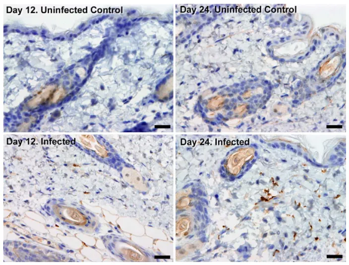
Figure 2. Extravascular localisation of trypanosomes during an infection
531
Histological sections of dorsal skin from uninfected and infected Balb/C mice stained with
532
trypanosome-specific anti-ISG65 antibody (brown), counterstained with Gill’s Haematoxylin stain
533
(blue) at 12 days and 24 days post-inoculation with T.b. brucei strain STIB247. Parasites are visible
534
in extravascular locations of the skin including the deep dermis and subcutaneous adipose tissue
535
from day 12. The scale bar represents 20µm.
536 537
538
Figure 3. Dynamics of parasite distribution in the extravascular tissue of the skin and in the
539
blood during a representative course of infection following natural transmission
540
A total of seven mice were infected by the single infective bite of an individual G.m. morsitans on
541
the belly with the T.b. brucei AnTat1.1E AMLuc/tdTomato strain. Panels A and C depict
542
representative patterns. (A) Examples of bioluminescence profiles of 3 mice (+ bitten by an infected
543
fly, - bitten by an uninfected fly and 0 not bitten) 6, 12, 20 and 26 days after the bite are shown. (B)
544
Ventral (blue) and dorsal (green) bioluminescence (BL) intensities (in p/s/cm2/sr on the left Y-axis)
545
and parasitaemia (in parasites/ml in red on the right Y-axis) were measured daily for 29 days and
546
plotted as mean ±SD (n=7 mice). (C) The entire skins of mice (+) and (-) were dissected for
547
bioluminescence imaging 29 days after the bite. For the mouse (+), Figure 3–figure supplement 1
548
shows the bioluminescence profile of dissected organs, Figure 3–figure supplement 2 presents the
549
skin inflammation, and Figure 3–figure supplement 3 shows labelled parasites in skin sections.
551
Figure 4. Extravascular localisation of trypanosomes during an infection visualised using
552
multi-photon microscopy (A) and spinning-disk confocal microscopy (B)
553
(A) Still-image extracted from video (Video 1) of multi-photon live imaging of dorsal skin during a
554
trypanosome infection. Intravenous non-targeted quantum dots (white) highlight blood vessels. T.b.
555
brucei STIB 247 parasites transfected with mCherry to aid visualisation (red) are clearly visible and 556
motile outside the vasculature and within the extravascular skin matrix (green). (B) Still-image
557
extracted from (Video 3) of spinning-disk confocal live imaging of the ear of an Kdr (Flk1) C57BL/6J
558
Rj mouse during a trypanosome infection. T.b. brucei AnTat1.1E AMLuc/tdTomato parasites
559
expressing tdTomato (red) are moving in the extravascular region surrounding a vessel of the
560
dermis (green).
561 562
563
564
Figure 5. Extravascular localisation of trypanosomes in previously unidentified human cases
565
of trypanosomiasis
566
Histological sections of skin collected from previously unidentified cases of human trypanosomiasis
567
from the Democratic Republic of Congo, showing the presence of extravascular parasites in
568
biopsies from three individuals (A, B and C). Skin biopsies were collected as part of a national
569
onchocerciasis screening programme that took place in the same geographic region as an active
570
trypanosomiasis focus. Slides were stained with Giemsa and examined under oil immersion at 100x
571
magnification. In addition to visible slender forms (black arrows) in the extravascular tissue of the
572
skin, a clearly identifiable stumpy transmission form with typical morphology and an unattached
573
undulating membrane is also present in the skin of one individual (red arrow in A). The scale bar
574
represents 5µm.
575 576
Fly batches Parasites in blood (per ml)
Parasites in skin (per cm2)
Dissected flies Fly infection rates (%) C1 0 0 32 0% C 0 0 8 0% A4B < 104 < 103 16 0% B1B 2.2x10⁴ < 103 13 0% A4A < 104 6.6x10⁵ 17 35% A2A 1.1x10⁴ 3.8x10⁶ 7 86% B1A 2.2x10⁴ 4.6x10⁷ 16 31% 3B 4.4x10⁴ 2.6x10⁴ 14 36% 3A 4.4x10⁴ 2.6x10⁴ 16 38% 1B 1.8x10⁵ 8.0x10³ 12 67% 1A 1.8x10⁵ 8.0x10³ 14 79% 4B 2.2x10⁵ 1.2x10⁴ 17 53% 4A 2.2x10⁵ 1.2x10⁴ 18 56% 2B 1.6x10⁶ 8.0x10³ 14 36% 2A 1.6x10⁶ 3.2x10⁴ 18 39% B4B 4.3x10⁶ 6.7x10⁷ 10 80% B4A 4.3x10⁶ 6.7x10⁷ 17 100% 577
Table 1. Skin parasites are ingested during tsetse pool-feeding
578
Mice were IP infected with T.b. brucei AnTat1.1E AMLuc/TY1/tdTomato and the parasitaemia and
579
bioluminescence were monitored daily until the day of xenodiagnosis. The number of parasites in
580
the blood was determined using a haemocytometer or a flux cytometer. The number of parasites in
581
the skin was estimated from the measured bioluminescence intensity by using a standard curve
582
(Table 1-source data 1 and Table 1-source data 2). Batches of teneral flies were fed on different
583
skin regions of mice infected with differing levels of bioluminescence across the skin and with
584
differing levels of parasitaemia (Table 1-source data 2 and Table 1-source data 3). Fly batches A4A,
585
A2A, B1A, 3B and 3A were used to assess tsetse transmission in hosts with low numbers of blood
586
parasites but high numbers skin parasites, while fly batches 1B, 1A, 4B, 4A, 2B, 2A, B2B and B4A
587
were used to investigate the impact of high numbers of parasites in both the skin and blood. Flies
588
were dissected and their midguts checked for the presence of fluorescent trypanosomes after two
589
days to determine the proportion of infected flies (Table 1-source data 4A-B). For some of these
590
experiments, results of an in-depth quantification of parasite stages by IFA is provided in
591
Supplementary file 4. Stumpy forms were observed only in the blood of mice with parasitaemia
592
values highlighted in light grey. Bioluminescence was detected in the skin of mice with values
593
highlighted in dark grey.
Figure Supplements
595 596
Figure 2-figure supplement 1. Skin invasion by T.b. brucei strain TREU927 and T.b.
597
gambiense strain PA
598
Histological sections of dorsal skin from a mouse infected with T.b. brucei strain TREU927 at 20x
599
magnification and two mice infected with T.b. gambiense strain PA at 40x magnification 10-days
600
post-inoculation. Trypanosome-specific anti-ISG65 antibody reveals the presence of extravascular
601
parasites (brown) and the slides were counterstained with Gill’s Haematoxylin stain (blue) to reveal
602
host skin structure.
603 604
Figure 3-figure supplement 1. Bioluminescence mostly originates from parasites in the skin
605
Mouse (+) was sacrificed and dissected for bioluminescence imaging 29 days after the infective
606
bite. Fig 3C shows the bioluminescence profile of its entire skin and dissected organs are shown
607
here.
608 609
Figure 3-figure supplement 2. Mild inflammation of skin tissues one month after an infection
610
by natural transmission
611
After 29 days, the most bioluminescent skin region of mouse (+) was dissected, fixed in
612
paraformaldehyde, embedded in paraffin and stained with HE. Multifocal inflammatory infiltrates
613
containing neutrophils were located in the dermis and subcutaneous tissue and associated with
614
oedema. Inflammatory foci were generally centred on blood vessels (arrows).
615 616
Figure 3-figure supplement 3. Extravascular parasites in the skin express both VSGs and
617
PAD1 surface markers
618
After 29 days, the most bioluminescent skin region of mouse (+) was dissected, fixed in
619
paraformaldehyde, embedded in paraffin and treated for IFA with the anti-CRD antibody that
620
predominately labels parasites expressing VSGs (A-B), or the anti-PAD1 antibody specific to
621
transmission form “stumpy” cells (C-D).
Source Data
623 624
Table 1-source data 1. Characterisation of the AnTat 1.1E AMLuc/TY1/tdTomato sub-clone
625
(A) The in vitro growth of the selected AnTat1.1E AMLuc/TY1/tdTomato sub-clone (red) was similar
626
to that of the parental wild-type strain (blue). Bloodstream forms were cultured in HMI11, counted
627
daily in a Muse cytometer (Merck-Millipore) and diluted after 4 days. (B) A parasite density /
628
bioluminescence intensity analysis was performed by measuring the bioluminescence in successive
629
2-fold dilutions in 96-micro-well plates with an IVIS® Spectrum imager (Perkin Elmer). When plotted
630
as mean ±SD (n=3), parasite densities and bioluminescence intensities were correlated when the
631
bioluminescence levels were higher than 104 p/s/cm²/sr, corresponding to about 103 parasites,
632
allowing estimation of the parasite density from in vivo imaging over this threshold. This standard
633
curve was used to estimate the number of parasites in the skin from measured values of
634
bioluminescence. (C) This correlation was verified by quantification in a microplate reader Infinite®
635
200 (Tecan) at the very beginning of the first in vivo experiment as well as the end of the last one
636
(mean ±SD, n=3).
637 638
Table 1-source data 2. Parasite densities in extravascular tissue of the skin and in the blood
639
of mice used for differential xenodiagnosis
640
Mice were injected IP with AnTat1.1E AMLuc/TY1/tdTomato and monitored daily for
641
bioluminescence and parasitaemia. (A) Bioluminescence profile of four mice (- uninfected control
642
and (1-3) three infected mice) four days after infection. (B) The entire skins of the uninfected control
643
mouse (-) and mouse 3 were dissected for bioluminescence imaging four days after infection. (C)
644
Parasite densities in the blood and in the skin (calculated from the mean dorsal bioluminescence
645
intensity measurement and from the standard curve in Figure 4-figure supplement 3B, in
646
parasites/cm2 in blue) were calculated daily over one week and plotted as mean
± SD (n=13 mice).
647
Table 1-source data 3. Skin parasites are sufficient to initiate a tsetse infection
648
Schematics summarising the principal results from the xenodiagnosis experiment. In a mouse with
649
no detected transmissible parasites in the blood (absence of stumpy forms by IFA and absence of
650
infection of flies fed on a non-bioluminescent region of the skin), flies can ingest transmissible
651
parasites from the bioluminescent region of the skin (left panel). When a mouse presents
652
transmissible forms in the blood, fly infection rates increase with the concomitant ingestion of
653
parasites from the skin (right panel). Values correspond to those obtained for mouse A4 and B4.
654 655
Table 1-source data 4. Parasite stage determination by labelling of specific surface markers
656
Parasites recovered from infected tsetse midguts (A-B) or included in bloodsmears (C) were fixed in
657
methanol for 5 seconds and stained either with the anti-GPEET antibody detecting early procyclic
forms (red in A-B) and the L8C4 antibody labelling the flagellum PFR (green in A-B), or with the
659
anti-PAD1 antibody detecting intermediate and stumpy forms (green in C), respectively.
660 661
Figure 1-source data 1. Semi-quantitative evaluation of the parasite burden in skin sections
662
(STIB247)
663
Every three days for 36 days of a STIB247 T.b. brucei infection, five mice were culled (three
664
infected, two control) and skin sections stained with parasite-specific anti-IGS65 antibody. Parasite
665
burden was assessed by two pathologists blinded to group assignment and experimental
666
procedures. Presence of parasites defined as intravascular (parasites within the lumen of dermal or
667
subcutaneous small to medium-sized vessels) and extravascular (parasites located outside blood
668
vessels, scattered in the connective tissue of the dermis or in the subcutis) was evaluated in 5
high-669
power fields at x40 magnification with a 0 to 3 semi-quantitative grading scale (0 = no parasites
670
detectable; 1 = low numbers of parasites; 2 = moderate numbers of parasites; 3 = large numbers of
671
parasites).
672 673
Figure 1-source data 2. Daily parasitaemia during STIB247 infection in Balb/C mice
674
The daily parasitaemia during a 36-day STIB247 T.b. brucei infection was estimated using phase
675
microscopy and methodology outlined in 40.
676 677