HAL Id: hal-02933756
https://hal.archives-ouvertes.fr/hal-02933756
Submitted on 30 Sep 2020
HAL is a multi-disciplinary open access
archive for the deposit and dissemination of
sci-entific research documents, whether they are
pub-lished or not. The documents may come from
teaching and research institutions in France or
abroad, or from public or private research centers.
L’archive ouverte pluridisciplinaire HAL, est
destinée au dépôt et à la diffusion de documents
scientifiques de niveau recherche, publiés ou non,
émanant des établissements d’enseignement et de
recherche français ou étrangers, des laboratoires
publics ou privés.
Distributed under a Creative Commons Attribution - NonCommercial| 4.0 International
License
variations in the mantle
B. Moine, N. Bolfan-Casanova, I. Radu, D. Ionov, G. Costin, A. Korsakov, A.
Golovin, O. Oleinikov, E. Deloule, J. Cottin
To cite this version:
B. Moine, N. Bolfan-Casanova, I. Radu, D. Ionov, G. Costin, et al.. Molecular hydrogen in minerals as
a clue to interpret ∂D variations in the mantle. Nature Communications, Nature Publishing Group,
2020, 11 (1), �10.1038/s41467-020-17442-8�. �hal-02933756�
ARTICLE
Molecular hydrogen in minerals as a clue to
interpret
∂D variations in the mantle
B. N. Moine
1
✉
, N. Bolfan-Casanova
2
, I. B. Radu
1,3
, D. A. Ionov
4
, G. Costin
5
, A. V. Korsakov
6
,
A. V. Golovin
6,7
, O. B. Oleinikov
8
, E. Deloule
9
& J. Y. Cottin
1
Trace amounts of water dissolved in minerals affect density, viscosity and melting behaviour
of the Earth’s mantle and play an important role in global tectonics, magmatism and volatile
cycle. Water concentrations and the ratios of hydrogen isotopes in the mantle give insight
into these processes, as well as into the origin of terrestrial water. Here we show the
presence of molecular H
2in minerals (omphacites) from eclogites from the Kaapvaal and
Siberian cratons. These omphacites contain both high amounts of H
2(70 to 460 wt. ppm)
and OH. Furthermore, their
∂D values increase with dehydration, suggesting a positive H
isotope fractionation factor between minerals and H
2–bearing fluid, contrary to what is
expected in case of isotopic exchange between minerals and H
2O-
fluids. The possibility of
incorporation of large quantities of H as H
2in nominally anhydrous minerals implies that the
storage capacity of H in the mantle may have been underestimated, and sheds new light on H
isotope variations in mantle magmas and minerals.
https://doi.org/10.1038/s41467-020-17442-8
OPEN
1Université de Lyon, UJM-Saint-Etienne, UCA, IRD, CNRS, Laboratoire Magmas et Volcans, UMR6524, Saint-Etienne, France.2Laboratoire Magmas et Volcans, Université Clermont-Auvergne, CNRS UMR 6524, Clermont-Ferrand IRD R 163, France.3Department of Geological Sciences, University of Cape Town, Rondebosch, Cape Town 7701, South Africa.4Géosciences Montpellier, Université de Montpellier, Montpellier 34095, France.5Department of Earth, Environmental and Planetary Sciences, Rice University, Houston, TX 77005, USA.6Sobolev Institute of Geology and Mineralogy, Siberian Branch Russian Academy of Sciences (SB RAS), Koptyuga 3, Novosibirsk 630090, Russia.7Novosibirsk State University, Pirogova 2, Novosibirsk 630090, Russia.8Diamond and Precious Metal Geology Institute, SB RAS, Yakutsk 677007, Russia.9CRPG, UMR7358, CNRS, Université de Lorraine, Vandoeuvre-lès-Nancy, France. ✉email:bertrand.moine@univ-st-etienne.fr
123456789
H
ydrogen (or water in its oxidised form) plays a key role in
the evolution, dynamics and habitability of the Earth. Even
in minor amounts, it decreases the mechanical strength
and melting temperatures of rocks and minerals, properties that
govern volcanism and mantle convection. Hydrogen was
incor-porated into the Earth’s interior during its accretion
1–3and then
evolved through degassing by volcanism and recycling by
sub-duction. It is an ubiquitous trace component of nominally
anhy-drous minerals (NAMs) in the upper mantle, estimated to
amount, as water, to 0.5–1 times the mass of the oceans
4,5, with
cosmochemical arguments leading to an estimate of up to seven
oceanic masses in the initial Bulk Silicate Earth
6. So far, hydrogen
was thought to exist in the mantle in the form of hydroxyl (OH)
with storage capacity depending on depth
7,8. An equivalent to a
few hundred ppm by weight of H
2O was measured in peridotite
xenoliths (mantle fragments brought up by volcanic eruptions)
9,10and up to 1.2 wt% H
2O in a ringwoodite inclusion in an ultradeep
diamond from the transition zone
11. On the other hand, the
studies of eclogite xenoliths from Slave, West African and
Zim-babwe cratons indicate that oxygen fugacity,
ƒO
2, ranges from
ΔlogƒO
2−2 to −4.5
12relative to the Fayalite–Magnetite–Quartz
buffer
13,14. At such fugacity, a substantial amount of H should be
present in a reduced form
15. Reducing conditions were
demon-strated experimentally to greatly decrease the solubility of OH in
olivine
16and it was recently discovered that H
2
could also be
dissolved in NAMs, while it had remained undetectable due to its
low infra-red extinction coefficient
17.
Here we report H concentration, speciation and isotope ratios
for omphacite (sodic clinopyroxene) from 12 eclogite xenoliths
from the Kaapvaal (Roberts Victor Mine) and Siberian
(Obnaz-hennaya kimberlite) cratons. These eclogites are bi-mineralic
(omphacite, garnet) and corundum-bearing rocks (Fig.
1
) with
estimated equilibrium conditions of 2.1−3.4 GPa, 805−1110 °C,
and 2.8–4.1 GPa, 923−1140 °C, respectively (Supplementary
Table 1). They are believed to represent subducted oceanic crust
preserved in the cratonic root for >2 billion years and unaffected
by host kimberlite or older melts
18. The samples were analysed
for their hydrogen abundances (expressed as water) and isotopic
composition by Thermal Conversion-Elemental Analyser coupled
with continuous
flow mass spectrometer (TC/EA-IRMS
19,20).
Hydrogen was also measured in the omphacite by SIMS
(Sec-ondary Ion Mass Spectrometry) and FTIR (Fourier Transform
Infra-red Spectroscopy). The hydrogen abundances obtained by
TC/EA-IRMS and SIMS (that detect all forms of hydrogen) are
consistently higher than those obtained by FTIR (that only
detects OH and H
2O efficiently), see Table
1
. We propose that the
Omph Omph Omph Gt 2 mm Cor 1mm
a
b
GtFig. 1 Thin section images of eclogite samples. a bimineralic eclogite from Roberts Victor Mine (South Africa) andb corundum-bearing eclogite from
Obnazhennaya kimberlite (Siberia).
Table
1
Water
content
and
hydrogen
isotope
composition
of
omphacites.
Sample n ∂ D ‰ vSMOW ±‰ H2 O (ppm) TC/EA-MS 1 S D n H2 Otot (ppm) FTIR 3000-3800 cm − 1 ±30% H2 O (ppm) SIMS 1 S D Abs norm 5200 cm − 1 H2 Omol (ppm) FTIR 5200 cm − 1 Abs Int norm 4000 –4300 cm − 1 H2 (ppm) 1 S D Obn108 12 − 107 11 2750 100 7 1177 353 0.0536 178 4.62 175 41 Obn110 12 − 120 11 4850 260 6 1249 375 4850 340 0.0417 138 6.02 400 51 Obn111 9 − 116 4 4650 540 9 1413 424 0.0425 141 9.42 360 76 Obn112 9 − 126 5 5065 340 12 929 279 0.0520 172 14.1 460 49 RV179 1 − 92 720 120 9 120 36 67 4 RV203 3 − 92 2 1315 130 RV233 5 − 99 5 1770 200 9 354 106 157 25 RV360 5 − 107 2 2400 300 9 123 37 253 34 RV377 4 − 93 3 1040 10 RV469 3 − 99 2 1467 65 RV488 3 − 91 2 1230 280 9 173 52 117 32 RV513 4 − 89 3 1256 256 9 255 77 111 30 Water content and H isotopic composi tion were measured using a Ther mal con version/Elemental Analyser coupled with Isotope-Ratio Mass spectromete r (TC/EA-I RMS) , Fourier Tra nsform Infra Red (FTIR) and Seconda ry Ion Mass Spectrometry (SIMS ) analyses. FTIR concent rations were calculated using prev iously published absorption coef fi cient 34 . Molecula r water con tent was estimated based on the speci fi c IR absorption peak (5200 cm − 1) and the absorption coef fi cient of 27 . Molecular H2 was calculated by di fference between total H2 O content (TC/EA-IRMS ) and FTIR integrated in tensity in the range of 3000 –380 0 c m − 1, acco unting for both OH and potentia l H2 Omolecular . Abs. norm absorba nce correspon ding to 5200 cm − 1peak height, 1S D standa rd devi ation, n number of anal yses“missing” hydrogen undetected by FTIR is stored in minerals as
molecular H
2, which implies that current estimates for total
hydrogen in the mantle may be too low. Our results also show
that H
2concentrations are correlated with H isotope
composi-tions, providing new insights into H-isotope systematics in
mantle rocks and minerals.
Results and discussion
H contents. The water concentrations (Table
1
) in the omphacite
measured by TC/EA-IRMS range from 720 to 5065 wt ppm,
consistent with data for orogenic (crustal) eclogites (1200−6000
ppm
21–23), and are generally much higher than the maximum of
600 ppm H
2O reported previously for mantle pyroxenes
10,24,25.
Thus, eclogites could represent a significant reservoir of water in
the cratonic lithosphere, despite their relatively low average
abundance (2%)
22,23, as well as generally in the convecting
man-tle
26. FTIR spectra in the OH region (2800−3800 cm
−1; Fig.
2
)
systematically indicate much lower water contents (100−1500
ppm H
2O) than those obtained by TC/EA-IRMS or SIMS
(Table
1
). The most important difference between the two
methods is that TC/EA-IRMS records the totality of H atoms
whereas the FTIR value only relates to structurally bound OH and
molecular water
27. This suggests that some hydrogen is stored in
the omphacite not only as OH but in a different form.
Dis-crepancies between bulk and spectroscopic methods have already
been observed for omphacites in orogenic eclogites and magmatic
clinopyroxene
21,28and tentatively explained by the presence of
nano-bubbles
29,30of molecular water in the minerals. Although
previous work did not identify molecular water spectroscopically,
except maybe through the band at 3400 cm
−131, we observed it
in some of the analysed grains, albeit with a very noisy signal. Its
contribution is very low, 140−290 ppm, within the error of TC/
EA-IRMS and FTIR measurements (Table
1
), estimated based on
the 5200 cm
−1specific band
32using published absorbance
coef-ficients
27. Molecular water, therefore, cannot account for the
measured H excess. It thus looks like most of the total water in
minerals measured in the 2800−3800 cm
−1range is present in
the form of hydroxyl (OH). The SIMS H
2O measurements for
sample Obn110 are consistent with TC/EA-IRMS data (4850
ppm, Table
1
). Since SIMS is a microbeam technique (that
measures all H atoms independently of their speciation), the fact
that the water content it provides agrees with the bulk water
measurements by TC-EA/IRMS indicates that the amount of H in
occasional micro-inclusions and/or fractures is not significant.
H speciation. We also detected a very small and pleochroic peak
in the infra-red spectra near 4100 cm
−1for the most water-rich
omphacites from corundum-bearing Obnazhennaya eclogites that
contain 2750−5065 wt ppm water based on the TC/EA-IRMS
method (Fig.
2
). This peak was previously proposed to
corre-spond to molecular H
217. Molecular H
2does not normally
respond to infra-red excitation due to its symmetry. However, if
H
2is dissolved in an ionic environment, the weak forces cause the
appearance of a dipole interacting with infra-red radiation
33. The
centroid of this peak is reduced by ~50 cm
−1compared with that
of molecular H
2vapour determined by Raman spectroscopy
17.
We calculated the amount of molecular H
2(70−460 wt ppm;
Table
1
) by difference between the FTIR and TC/EA IRMS data,
and found H/OH molar ratios > 3.
It is further possible to estimate the IR absorption coefficient
for H
2from the FTIR absorbance areas and H
2contents
determined above using the Beer-Lambert law (see
“Methods”
and Supplementary Fig. 1). The fact that the concentration of
molecular H
2calculated as the difference between bulk water
obtained from TC-EA-IRMS and OH obtained from FTIR
correlates positively with the integrated absorbance indicates that
the assignment of the 4100 cm
−1peak to H
2is correct.
Furthermore, the average linear absorption coefficient of H
2that
we calculate (Table
1
) is 1500 times lower than for OH (0.13 ± 0.3
l mol(H
2)
−1cm
−1or ~44 ± 10 l mol(H
2)
−1cm
−2for H
2vs.
65,000 l mol
−1cm
−2for OH
34). This is much lower than those
previously proposed (46.4 l mol(H
2)
−1cm
−1or 1650 l mol(H
2)
−1cm
−2)
17for NAMs (orthopyroxene) annealed under reducing
conditions, but in agreement with previous measurements on
silica (0.26 l mol(H
2)
−1cm
−1)
33. This further highlights the very
low infra-red activity of H
2and therefore the very low
detectability of molecular H
2by spectroscopic methods. This
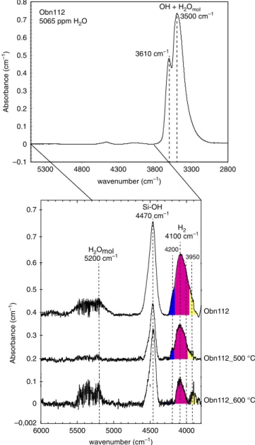
H2 4100 cm–1 –0.1 0 0.1 0.2 0.3 0.4 0.5 0.6 0.7 0.8 2800 3300 3800 4300 4800 5300 OH + H2Omol Absorbance (cm –1 ) wavenumber (cm–1) H2Omol 5200 cm–1 Si-OH 4470 cm–1 Obn112 5065 ppm H2O –0,002 0 0.1 0.2 0.3 0.4 0.5 4000 4500 5000 5500 6000 0.6 4200 wavenumber (cm–1) Absorbance (cm –1 ) 3950 0.7 0.7 Obn112_600 °C Obn112_500 °C Obn112 3500 cm–1 3610 cm–1
Fig. 2 Unpolarised FTIR spectra of omphacite (Obn112 sample). In the Fourier Transform Infra Red (FTIR) spectra, the 2800–5500 cm−1range show main peaks in the 3000–3800 cm−1range corresponding to OH and molecular H2O (stretching). Secondary peaks in the 3800–6000 cm−1
interval correspond to molecular H2(4100 cm−1), Si-OH (4470 cm−1), and
molecular water (5200 cm−1). A comparison between unheated and heated samples (500 and 600 °C) shows decreasing intensity of the peak at 4100 cm−1with increasing temperature. The widening of the peak with increasing H2content is due to overlapping with two smaller peaks at 4200
and 3950 cm−1. Note that the low intensity of the band at 5200 cm−1 related to molecular water indicates negligiblefluid inclusion contributions.
strongly suggests that the amounts of H determined in previously
studied mantle xenoliths have been greatly underestimated.
H isotopic composition. We examine the effects of hydrogen
speciation in minerals on H isotope fractionation in the deep
water cycle. The
∂D values in the omphacites from this study
correlate well with H concentrations expressed either as H
2O or
as H
2(Fig.
3
a, b). The well-defined trend indicates an increase of
the
∂D values in the omphacites with decreasing water content.
To confirm the speciation of H, Obnazhennaya samples were
heated under vacuum (10
−3mbar) at 400 °C, with sample
Obn112 incrementally heated at 250, 400, 500, and 600 °C. After
each heating step, that lasted 20 min, the samples were
re-analysed with FTIR and TC-EA/IRMS. These stepwise
experi-ments show that (1) the bulk water content and the isotopic
composition of the samples are little affected by heating up to
400 °C (see Supplementary Table 2). Only two samples (Obn110
and Obn108) suffered up to 15% water loss during heating up to
400 °C. This means that if any inclusions were present, the
amount of water stored in them must be negligible or within 15%
because otherwise, like in
fluid inclusion studies, or for garnets
containing water-inclusions, the speciation and concentration of
bulk water would be affected
31,35. If we consider only the samples
annealed at the highest temperature and consider them as the
most free of the contribution from inclusions then we get an
absorptivity coefficient of 30 l mol(H
2)
−1cm
−2, instead of the
average of 44 l mol(H
2)
−1cm
−2determined on the samples
before heating at high temperature. (2) Incremental heating of
sample Obn112 up to 600 °C yields an increase in the
∂D values
of residual H along with a decrease in OH and H
2concentrations
in omphacite (Fig.
4
), in agreement with the general trend
described/recorded by the samples. One way to explain this
negative correlation between the concentration of total water and
isotopic composition is by loss during heating of a component
with more negative
∂D values, such as H
2or a mixture of H
2and
OH
36. Indeed, the global partitioning of H
2
and OH between
mineral and
fluid would enrich the fluid in
1H and the residual
solid in
2D
36. Previous reports of such negative correlation on
eclogites from Dabie Sulu have been interpreted by the loss of
isotopically light molecular water due to kinetic fractionation of
H-D during dehydration in the course of exhumation
37. Given
that the ascent of kimberlites is very fast, we propose instead that
the H-D isotopic fractionation is controlled by the presence of H
2.
Structural H
2is indeed observed in omphacite, and the linear
relationship between the absorbance of the 4100 cm
−1band and
the calculated H
2content indicates that its quantification is robust
(see Supplementary Fig. 1). Molecular water present in
nano-inclusions seems to be negligible in these samples. However, if
present in large quantities, nano-inclusions could also be
filled
with H
2given that the conditions of equilibration of the present
eclogites are very close to the conditions where H
2and H
2O are
miscible within the mantle
38.
As shown in Fig.
2
, the absorbance of the 4100 cm
−1band
decreases with increasing temperature. This decrease is decoupled
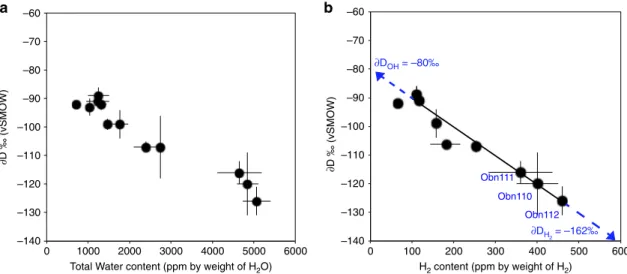
–140 –130 –120 –110 –100 –90 –80 –70 –60 0 100 200 300 400 500 600 H2 content (ppm by weight of H2) ∂DOH = –80‰ ∂DH2 = –162‰ ∂D ‰ (vSMOW) Obn110 Obn112 Obn111
b
Total Water content (ppm by weight of H2O)
∂D ‰ (vSMOW)
a
–140 –130 –120 –110 –100 –90 –80 –70 –60 0 1000 2000 3000 4000 5000 6000Fig. 3 Hydrogen isotope composition of omphacites versus H content. a∂D (relative to Vienna Standard Mean Ocean Water – V-SMOW) versus total water content determined by Thermal conversion/Elemental Analyser coupled with Isotope-Ratio Mass spectrometer (TC/EA-IRMS, andb∂D (relative to V-SMOW) versus calculated H2content (wt ppm). Error bars correspond to 1 SD.
–140 –130 –120 –110 –100 –90 –80 –70 –60 –50 0 1000 2000 3000 4000 5000 6000
Total water (ppm by weight of H2O)
∂D ‰ (vsmow) 600 °C 500 °C 400 °C 250 °C Obn112 Dehydrogenation (H2) Dehydration (OH) Fluid inclusions (H2O)
Fig. 4∂D versus H2O for heated and unheated omphacite Obn112. Total
water content (determined by Thermal conversion/Elemental Analyser coupled with Isotope-Ratio Mass spectrometer - TC/EA-IRMS) and∂D (relative to Vienna Standard Mean Ocean Water– V-SMOW) show a robust linear correlation. The loss of H with increasing temperature implies higher∂D values supporting the loss of a component with much more negative∂D (thus H2rather than OH or H2O), as also observed in the
infra-red spectra where the integrated absorbance of the H2band decreases
from that of the band at ~4500 cm
−1(assigned unambiguously to
a Si-OH vibration) implying that these two bands cannot be
attributed to the same species, i.e. the 4100 cm
−1band is not the
result of some combination of OH mode (see Supplementary
Figs. 2, 3). Indeed, if these two bands were due to the same species
the ratio of their intensities would stay constant, which is not the
case: it varies depending on temperature (see Supplementary
Fig. 3). While the integrated absorbance of the band at 4500 cm
−1decreases, between 100 and 400 °C, that of the 4100 cm
−1band
increases (Supplementary Fig. 2). This translates into H
2being
produced while OH is being consumed. Thus, we interpret the
contrasting behaviour shown in Supplementary Figs. 2, 3 as a
transition from OH to H
2during the heating stage at low
temperatures following the oxidation-dehydrogenation shown in
reaction 1, similar to what is inferred for natural or experimental
samples
16,39,40:
H
2O
þ 2 FeO <¼> H
2þ Fe
2O
3ð1Þ
Such behaviour also indicates that the kinetics of H
2and H
+diffusion are close in this temperature range and cross over at
higher temperatures in agreement with previous
measure-ments
41–44(Fig.
5
).
Reaction 1 is probably responsible for the stabilisation of H
2in
NAMs linked to a change in iron valence in ferro-magnesian
silicates, via reduction of water by ferrous iron. Ferric iron
solubility increases in clinopyroxene and garnet with increasing
pressure
15. Thus, we can expect that eclogitic clinopyroxenes
containing 7000−16000 wt ppm Fe, with Fe
3+/∑Fe estimated at
20−30% due to a high jadeite component [Na
+(Al
3+Fe
3+)
Si
4+2O
2−6]
45, can easily incorporate the H
2concentrations
measured in this study via reaction 1. Such a reaction is common
for dehydration metamorphism in subduction zones
39. In the
absence of available oxygen, another reaction producing H
2has
been proposed for the formation of diamond in the cratonic
mantle:
CH
4¼ C
diamondþ 2 H
2:
ð2Þ
However, the preservation within the mantle of high amounts
of H (>2000 ppm), considered highly mobile, at high temperatures
and for a long time, needs to be explained. Diffusivity of H
2in
minerals is currently unknown but existing data for silicate glasses
indicate that H
2diffusion is not very fast, no faster than for H
+at
mantle conditions. For example, the diffusivity of H
2in silica
glass
43,44is 9.3 × 10
−16m
2s
−1at 23 °C and 2.4 × 10
−12m
2s
−1at
250 °C. Using the activation energy of 44 kJ mol
−1provided in
these studies we calculate a diffusivity of H
2of 1.5 × 10
−10m
2s
−1at 600 °C or 1.0 × 10
−9m
2s
−1at 1000 °C (Fig.
5
). Such values are
very similar to those determined for the rate of the oxidation
reaction
46as well as for OH diffusivity in olivine
41or diopside
42.
The validity of these estimates ultimately hinges on the knowledge
of diffusion mechanisms for molecular H
2in minerals (vacancy vs.
interstitial diffusion, polaron or Franck-Turbull mechanisms),
which are currently unknown. Considering that H
2diffusivity
experiments in silicate glass have shown a strong dependence
(three orders of magnitude)
46(Fig.
5
) on H
2
partial pressure, the
above diffusivity (hence H loss) estimates are likely to be
exaggerated. Such diffusivity also implies that equilibrium is
geologically fast at the mineral grain scale at moderate to high
temperatures, yet high H concentrations could be maintained on
the scale of oceanic crust fragments hundreds of metres or
kilometres in size. If such eclogitic blocks are preserved for several
Ga in the mantle, they will develop hydrogen zoning in terms of
abundance and isotope ratios, reflecting their progressive
re-equilibration with the ambient mantle.
The surface water cycle fractionates hydrogen isotopes,
creating a wide range of isotopically distinct reservoirs, such as
Greenland ice caps standard precipitation [∂D = −190‰],
sea-water [VSMOW
∂D ~ 0‰] and rainwater [∂D = 0−130‰]). The
deep water cycle may fractionate hydrogen isotopes as well, but
the processes involved are different. Upper mantle (MORB)
magmas typically have uniform
∂D values of −60 ± 5‰
47,
whereas oceanic island magmas, thought to come from the lower
mantle, may have much lower
∂D down to −218‰
48. The
∂D
values for omphacites in this study range from
−89 to −126‰;
they are similar to those reported for orogenic eclogites from the
ultra-high pressure (UHP) Sulu terrane (−82 to −128‰
18), and
lower than those for MORB-like sources (−60‰ ± 5)
47. Both in
our study and previous reports of analyses of UHP rocks by TC/
EA-IRMS, the isotopic composition of H is observed to increase
with decreasing bulk H of omphacite. This contradicts previous
inferences that
∂D decreases during subduction, hence with
dehydration, typically from modern oceanic crustal segments
with
∂D of −35 ± 15‰
49–51, to orogenic or cratonic eclogites
(remnants of ancient subducted oceanic crust), with
∂D of −82 to
−128‰
18. Experimentally determined mineral-water H isotope
fractionation factors are generally negative
52,53implying that the
hydrogen remaining in the slab becomes increasingly depleted in
deuterium which preferentially partitions into expelled
fluids.
Subduction-related dehydration thus causes a decrease in
∂D in
slab materials with depth
54, as modelled in Fig.
6
(blue trend).
The overall mineral-fluid H isotope fractionation factor is difficult
to estimate because it varies greatly depending on temperature,
pressure and mineral species during subduction-related slab
metamorphism. Nevertheless, known H-D isotope fractionation
between water and common hydrous minerals (serpentine,
amphibole, chlorite, epidote, zoisite, brucite, and clays) range
from
−10 and −77‰ in the 100−800 °C temperature range, with
decreasing fractionation at higher temperatures. However, is
dehydration the actual process taking place in the present
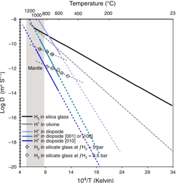
–20 –18 –16 –14 –12 –10 –8 4 9 14 19 24 29 34 Log D (m 2 S –1 ) 104/T (Kelvin) H2in silica glass H+ in olivineH2 in silicate glass at ƒH2 = 5 bar
H2 in silicate glass at ƒH2 = 0.5 bar
23 200 400 600 800 1000 1200 Mantle Temperature (°C) H+ in diopside H+ in diopside [001] or [100] H+ in diopside [010]
Fig. 5 Experimental diffusivity of H in silicate materials. Diffusivity of hydrogen in silica glass43,44, effective diffusivity in olivine41, diffusivity in
diopside as a function of crystallographic orientation42, and the effect of H
2
partial pressure on diffusivity in glass46. The greyfield corresponds to the
temperature range of the lithospheric mantle (dashed lines correspond to low temperature extrapolation of experimental data).
samples? In this work, we propose that dehydrogenation plays an
important role in the isotopic compositions observed (see Fig.
6
,
green trend).
Here we model the change in the D/H ratio of altered oceanic
crust (starting at 2 wt% H
2O,
∂D = −35‰
55,56) during subduction
as it dehydrates. By using a Rayleigh fractionation process and a
calculated fractionation factor
54(α
Mineral-H2O
= 0.9277), it is
possible to account for the highest water abundances and the
highly negative
∂D values, observed here for the cratonic
omphacites ([H
2O]
= 5065 ppm, ∂D = −126‰) (Table
1
).
How-ever, such a low fractionation factor cannot explain why the
∂D
values increase upon dehydration from 5000 to 700 ppm
water (Fig.
3
a). The enrichment of a mineral in
2D concomitant
with dehydration can only be explained if the mineral-fluid
fractionation factor is positive, which is the case for H partitioning
between minerals and H
236. In such a case, the de-volatilisation is
accompanied by the release of a H
2fluid enriched in
1H instead of a
H
2O
fluid enriched in
2D, which leads to less negative
∂D values of
residual hydrogen in the mineral. Since the isotope fractionation
factor for molecular H
2is positive and high
57, extraction of H
2results in a positive mineral-(OH-H
2) fractionation factor at high
temperatures such that
∂D values of the eclogite become less
negative (Figs.
6
,
7
). Nonetheless, these isotopic compositions
cannot be explained by H
2release alone, which would induce a
much faster isotopic evolution. The most likely scenario is a
combination of dehydration and dehydrogenation processes (Figs.
6
,
7
) by diffusion during the very long residence time in the mantle of
these eclogitic units as H
2and H
+have similar diffusivities at
mantle temperature (Fig.
5
).
The studied samples show a very good linear correlation between
the calculated molecular H
2content and
∂D values of total H (R² of
0.93, n
= 12, p < 0.001; Fig.
3
b). This correlation between H
2concentration and isotopic composition indicates that the
∂D in
omphacites reflects a mixture of two components, OH and
molecular H
2, each having distinct isotopic compositions. From
the proportions of molecular H
2and the respective isotopic ratios of
each sample, it is possible to determine the isotopic compositions of
each end-member:
∂D
MineralOH= −80‰ and ∂D
MineralH2=
−162‰. We can then calculate the intra-mineral fractionation
factor between OH and H
2for the Obnazhennaya omphacites
as follows:
α
MineralOH-MineralH2= 1.098 (10
3lnα
MineralOH-MineralH2=
+93‰). Because hydrogen isotope fractionation between the
structurally bound OH (MineralOH) and structurally bound H
2(MineralH
2) is >20‰, the fractionation factor was calculated as
follows:
α
A-B= (1000 + ∂D
A)/(1000
+ ∂D
B)
53. The intra-mineral
fractionation between OH and H
2calculated here is equivalent to
the fractionation between molecular water and molecular hydrogen
(10
3lnα
H2O-H2
: ~+100‰) at very high temperature (≥1100 °C
57).
This suggests that high temperature isotopic equilibrium was
reached and preserved in our samples. The diffusive loss of H
during the transport of mantle xenoliths close to the surface by
kimberlitic magma is low because the magma ascent is very fast
58.
Since molecular H
2is most likely to be the dominant form of H
in the reduced deep mantle (refs.
3,17), it follows that isotopic
fractionation of H in the mantle should be controlled by
equilibria involving H
2-bearing minerals rather than H
2O- or
OH-bearing minerals. This must be taken into account when
interpreting the H isotopic distribution in the mantle and models
involving deep mantle volatile loss
59. Similar to
findings of this
study, clinopyroxene (augite) megacrysts from alkaline basalts at
Nushan
28yield different H
2
O contents by FTIR and
TC/EA-IRMS (or manometrically), which correlate negatively with
measured
∂D (see Supplementary Table 1 and Supplementary
Fig. 4). Assuming that the measured concentration difference is
due to molecular H
2, the calculated 10
3lnα
MineralOH-MineralH2is
estimated to be
+111‰, which is realistic at magmatic
temperatures. In addition, the presence of structurally bound
H
2could explain the large difference in
∂D values of coexisting
richterites (−132‰ vSMOW) and phlogopites (−65‰ vSMOW)
in MARID xenolith suites from South African kimberlites, which
was previously interpreted as a result of fractional crystallisation
or re-equilibration during ascent
60. Also, a recent discovery of
highly negative
∂D in deep magmas trapped in melt inclusions
(e.g.
48) could be due to different H
2
O
tot-H
2proportions and
isotopic fractionation controlled by
ƒO
261or H
2loss by diffusion,
rather than a primary composition as previously believed. An
–150 –130 –110 –90 –70 –50 –30 –10 0.00 0.0005 0.0010 0.0015 0.0020 1/H2O (ppm–1) ∂D (‰ vs vSMOW)Altered Oceanic crust 2 wt% H2O, ∂D = –35‰
Rayleigh fractionation trend Dehydration
103lnαMin-H20 = –75‰
Rayleigh fractionation trend dehydration-dehydrogenation 103lnα MinOH-H2O = –5‰ 103lnαMinOH-H2 = +93‰ 103lnα Min-H~+25‰ Mantle box
Fig. 6 Model for evolution of H content and∂D in mantle omphacites. Hydrogen isotope composition (∂D relative to Vienna Standard Mean Ocean Water—V-SMOW) versus 1/H2O (water content determined by Thermal Conversion /Elemental Analyser coupled with Isotope Ratio Mass Spectrometer
—TC/EA-IRMS). The blue trend models the compositional evolution of subducting oceanic crust dominated by dehydration. The magenta trend models the compositional evolution of oceanic material, during or post-subduction, dominated by dehydrogenation and dehydration. The model is based on Rayleigh fractionation. The blue box corresponds to the typical isotopic composition of oceanic crust altered by seawater55,56and the green box to the“normal”
accurate determination of H speciation in mantle samples,
allowing the quantification of hydrogen in molecular and
hydroxyl forms, is therefore a prerequisite for any isotopic
measurement and interpretation.
In this study we provide three main pieces of evidence for H
2in minerals: (i) the discrepancy between hydrogen contents from
mass spectrometry and FTIR, (ii) the presence of an absorption
band in the infra-red spectra at 4100 cm
−1, which scales with the
H
2content, (iii) the isotopic data indicating a preferred
partitioning of
1H into the
fluid during H loss. Still, further
experimental work is needed to constrain the speciation and
mobility of H
2in mantle minerals and test the model
presented here.
Methods
Water content and stable H isotopes determination
“On-line” procedure. Samples were analysed using a continuous flow elemental analyser (TC/EA) operating on-line with mass spectrometer19,20,31. The system used is a ThermoFisher HTFlash IRMS©working on-line with DeltaV+©mass
spectrometer monitored by ConflowIV©diluter hosted at the Magmas and
Vol-canoes laboratory from Université Jean Monnet, Saint-Etienne. The DeltaV+©
used an electrostaticfilter to prevent isobaric interferences between the helium carrier gas and the generated mass 3 of hydrogen. The elemental analyses used the pyrolysis line consisting of a glassy carbon tubefilled with glassy carbon grains placed inside an alumina ceramic tube heated at 1450 °C andflushed by helium (100 ml min−1). All hydrogenous gasses were reduced by glassy carbon, H2was
separated from other gas species (CO) in a chromatographic column heated at 90 °C and transferred to mass spectrometer. ConflowIV©diluter monitored the
flux and the two injections of H2reference gas manufactured by Air Liquid
company. The duration of a complete analysis was 300 s. Aliquots of minerals weighting between 0.3 and 25 mg, depending on H2O content, were analysed.
Samples were crushed tofine grains as suggested by31,62to prevent incomplete extraction and fractionation of H and D. All samples were preheated at 100 °C for 24 h to eliminate adsorption water on sample surfaces.
To estimate the impact of tinyfluid inclusions on water content and the role of molecular H2on∂D, four samples experimented heating. 400 mg of pure
handpicked omphacite with grain size ranging between 500 and 1000 μm were put in a 6 mm tube of fused quartz and connected to a vacuum preparation line (10−3 mbar). Each sample was held under vacuum and heated with a heat gun by steps of 20 min at 250, 400, 500, and 600 °C. Between each step, an aliquot is taken for FTIR and TC/EA-IRMS analyses.
The relation between hydrogen contents and peak area detected by mass spectrometer was calibrated with benzoic acid (4.952 wt% H), and water concentrations were determined by mass H2peak area, the uncertainty is estimated
to be ±0.05 wt%.
We have also investigated/addressed linearity issues by loading 36 aliquots of Biotite NBS30 of different weights (0.318–3.423 mg) and thus obtaining different peak sizes on mass 2 (amplitude) in the range of 741 mV to 9396 mV. At the beginning of each analytical session we applied the H3+correction factor at
different pressures of the reference gas to correct for different peak heights.
D/H measurements were calibrated against NBS30 biotite and IAEA CH7 polyethylene and previously measured amphibole and mica on VSMOW-GISP isotope scale63by modified off-line method of64(see below). These have been chosen based on their extremely different∂D values (Amphibole AJE 282: ∂D = −130 ± 0.5‰ and Mica AJE361 ∂D = −40 ± 3‰ vs VSMOW) spanning over a range of 90‰ and overlapping with the range of unknown samples. A mean ∂D value for the NBS30 biotite standard of−65.4 ± 1‰ and water content of 3.67 ± 0.12 wt% (n= 36) were obtained during the course of this study.
“Off-line” procedure. A suite of amphibole (richterite) and mica (phlogopite) from the South African MARID suite wasfirst analysed using an “off-line” vacuum extraction line. 30–80 mg of pure hand-picked mica and amphibole with grain size ranging between 100 and 200 μm were put in a 6 mm tube of fused quartz and connected to a vacuum preparation line (10−9mbar). Each sample was held at 150–200 °C for 1 h under vacuum to desorb atmospheric water, and heated gra-dually with a butane-oxygen torch to release all hydrogenous gas to reach the melting point of quartz tube (1700–1800 °C). On the line, a CuO grain furnace constantly held at 575 °C allowed to transform all hydrogenous gas to H2O that was
subsequently collected at liquid nitrogen temperature in a 10 mm pyrex coldfinger. The trap temperature was increased to−90 °C with a mixture of ethanol-liquid nitrogen to allow the non-condensable gases to be pumped away. Then the col-lected H2O was reduced to H2with U metal at 800 °C64. H2was trapped into a
coconut charcoal coldfinger at liquid nitrogen temperature and expanded at room temperature in a calibrated volume connected to a capacitance gauge allowing to measure the“total water” content of minerals. On the same line, water standards (IAEA VSMOW, GISP and lab standards) were converted to H2with the same
procedure. The D/H ratios were determined using an Elementar Isoprime dual-inlet mass spectrometer at the Magmas and Volcanoes laboratory, Jean Monnet University, Saint-Etienne. The results are expressed in the∂-notation as permil relative to VSMOW. A mean∂D value for the IAEA NBS30 biotite standard of −65.7 ± 0.3‰ and water content of 3.68 ± 0.1 wt% (n = 4) were obtained during the course of this study.
FTIR. Ten grains of each sample were doubly polished withfinal thicknesses of 150–450 μm depending on the grains. The OH content has been determined on a Bruker Vertex 70 FTIR (Fourier transform infra-red spectroscope) coupled with a Hyperion microscope equipped with ×15 objective and condenser at LMV. Beam size in the analyses varied from 30 to 50 μm. The spectra were collected through a CaF2plate with a resolution of 2 cm−1and with up to 300 scans. After the
appli-cation of a linear baseline with anchor points outside the OH stretching region, the absorbance was integrated from 3000 to 3800 cm−1and the absorbance coefficient for omphacite was applied34. The calculation of the water concentration was performed using the Beer-Lambert law: A= ε·C·t, where A is the absorbance, ε is the absorptivity, C the concentration and t the thickness (in cm). Quantification was based on the average of ~10 unpolarised measurements performed on ran-domly oriented grains within the doubly polished thin sections. The absolute absorbance of the crystal is then equal to three times the unpolarised value as demonstrated by65. Absorbances of molecular H
2and H2O followed the same
procedure and were integrated respectively from 4000 to 4300 cm−1and around 5200 cm−1. The concentration of molecular water was calculated using previously published absorptivity28. The absorptivity of H
2was calculated using the
Beer-Lambert law and the concentrations calculated in Table1from the difference H isotope cycle trough subduction zones and in the mantle
Dehydrogenation
Mantle ∂D = –60‰ 100–400ppm H2O
less than 75% of slab water ∂D = –10 to –34‰ Dehydrated and hydrogenated slab 25% of slab “water” ∂D = –125‰ seawater ∂D = 0‰ Dehydration Subducting slab ∂D = –35‰ 20000 ppm H2O Craton Graphite Diamond Spinel Garnet 5000 ppm H 2O 2500 ppm <1000 ppm H 2O Dehydrated and dehydrogenated slab 20 to 10% of slab “water” ∂D = –90 to –75‰
Fig. 7 H recycling and isotope evolution in subduction zone. The sketch shows how the recycled crust gets enriched in1H during subduction due to dehydration and subsequently gets enriched in D due to H2-OH equilibration and diffusion within the upper mantle.
between the total water content measured by TC-EA-IRMS and the water content measured by FTIR in the OH+H2O frequency region (see Supplementary Fig. 1).
SIMS. In situ water contents were measured on polished sections, gold coated, with the Cameca IMS1280 HR ion microprobe at CRPG-CNRS, Nancy. A 13 kV, 5 nA O- primary beam was focused onto the sample to a diameter of 20 μm. The secondary beam mass resolution was set at 1600, with an energy window of 35 eV and no energyfiltering. Secondary ions of H+and D+were measured by peak switching for 10 min by ion counting. Under these analytical conditions, counting rates on H+ varied between 1 × 10⁵and 5 × 10⁵counts per second and statistical
precision ranged from 0.5 to 3%. Samples were carefully degassed before intro-duction in the analytical chamber. The samples were doubly polished thin sections that were glued on a glass plate and gold coated. They were introduced in the vacuum chamber of the SIMS the night prior to analysis at 2 μPa (2 × 10⁻⁹atm) associated with a liquid N2cold trap. A presputering of 3 min with a 20 μm raster
was used to clean the sample surface before measurement, and a raster of 5 μm and an electronic gate of 90% was used for the analysis. The background level was lower than 10 ppm of water. The water content of samples was calculated by comparing the measured hydrogen secondary ion intensity relative to the primary ion beam intensity of samples with that of pyroxene of known composition66measured during the same session as reference material. The estimated precision on the calculated water content was about 15% (1 sigma). All hydrogen signal is converted into water content, without considering its initial form.
2-Pressure–temperature estimates. Temperatures were calculated with a pressure-dependent garnet-clinopyroxene Fe–Mg geothermometer67for 1, 3, and 7 GPa. Pressures were calculated by projecting the temperature estimates to local conductive model geotherms68corresponding to a surface heatflow of 39 mW m−2
for Roberts Victor69and of 45 mW m−2for Obnazhennaya (estimated at 40 to >50
mW m−2)70.
Data availability
All data in this study are presented in Table1and available in Supplementary Tables 1 and 2.
Received: 4 September 2019; Accepted: 26 June 2020;
References
1. Clesi, V. et al. Effect of H2O on metal–silicate partitioning of Ni, Co, V, Cr,
Mn and Fe: implications for the oxidation state of the Earth and Mars. Geochim. Cosmochim. Acta 192, 97–121,https://doi.org/10.1016/j. gca.2016.07.029(2016).
2. Füri, E., Deloule, E., Gurenko, A. & Marty, B. New evidence for chondritic lunar water from combined D/H and noble gas analyses of single Apollo 17 volcanic glasses. Icarus 229, 109–120,https://doi.org/10.1016/j.
icarus.2013.10.029(2014).
3. Hirschmann, M. M., Withers, A. C., Ardia, P. & Foley, N. T. Solubility of molecular hydrogen in silicate melts and consequences for volatile evolution of terrestrial planets. Earth Planet. Sci. Lett. 345-348, 38–48,https://doi.org/ 10.1016/j.epsl.2012.06.031(2012).
4. Peslier, A. H., Schönbächler, M., Busemann, H. & Karato, S.-I. Water in the Earth’s interior: distribution and origin. Space Sci. Rev. 210, 1–68 (2017). 5. Jacobsen, S. D. Effect of water on the equation of state of nominally anhydrous
minerals. Rev. Mineral. Geochem. 62, 321–342,https://doi.org/10.2138/ rmg.2006.62.14(2006).
6. Marty, B. The origins and concentrations of water, carbon, nitrogen and noble gases on Earth. Earth Planet. Sci. Lett. 313-314, 56–66,https://doi.org/ 10.1016/j.epsl.2011.10.040(2012).
7. Férot, A. & Bolfan-Casanova, N. Water storage capacity in olivine and pyroxene to 14 GPa: implications for the water content of the Earth’s upper mantle and nature of seismic discontinuities. Earth Planet. Sci. Lett. 349-350, 218–230,https://doi.org/10.1016/j.epsl.2012.06.022(2012).
8. Smyth, J. R. Hydrogen in high pressure silicate and oxide mineral structures. Rev. Mineral. Geochem. 62, 85–115,https://doi.org/10.2138/rmg.2006.62.5
(2006).
9. Bell, D. R. & Rossman, G. R. Water in Earth’s mantle: the role of nominally anhydrous minerals. Science 255, 1391 (1992).
10. Demouchy, S. & Bolfan-Casanova, N. Distribution and transport of hydrogen in the lithospheric mantle: a review. Lithos 240-243, 402–425,https://doi.org/ 10.1016/j.lithos.2015.11.012(2016).
11. Pearson, D. G. et al. Hydrous mantle transition zone indicated by ringwoodite included within diamond. Nature 507, 221,https://doi.org/10.1038/ nature13080(2014).
12. Aulbach, S. et al. Evidence for a dominantly reducing Archaean ambient mantle from two redox proxies, and low oxygen fugacity of deeply subducted oceanic crust. Sci. Rep. 9, 20190,https://doi.org/10.1038/s41598-019-55743-1
(2019).
13. Goncharov, A. G., Ionov, D. A., Doucet, L. S. & Pokhilenko, L. N. Thermal state, oxygen fugacity and COHfluid speciation in cratonic lithospheric mantle: new data on peridotite xenoliths from the Udachnaya kimberlite, Siberia. Earth Planet. Sci. Lett. 357-358, 99–110,https://doi.org/10.1016/j. epsl.2012.09.016(2012).
14. Woodland, A. B. & Koch, M. Variation in oxygen fugacity with depth in the upper mantle beneath the Kaapvaal craton, Southern Africa. Earth Planet. Sci. Lett. 214, 295–310,https://doi.org/10.1016/S0012-821X(03)00379-0
(2003).
15. McCammon, C. A. & Frost, D. J. The effect of oxygen fugacity on the olivine to wadsleyite transformation: implications for remote sensing of mantle redox state at the 410 km seismic discontinuity. Am. Mineral. 94, 872–882,https:// doi.org/10.2138/am.2009.3094(2009).
16. Yang, X. Effect of oxygen fugacity on OH dissolution in olivine under peridotite-saturated conditions: an experimental study at 1.5–7 GPa and 1100–1300 °C. Geochim. Cosmochim. Acta 173, 319–336,https://doi.org/ 10.1016/j.gca.2015.11.007(2016).
17. Yang, X., Keppler, H. & Li, Y. Molecular hydrogen in mantle minerals. Geochem. Perspect. Lett., 160–168,https://doi.org/10.7185/geochemlet.1616
(2016).
18. Radu, I. B., Harris, C., Moine, B. N., Costin, G. & Cottin, J. Y. Subduction relics in the subcontinental lithospheric mantle, evidence from∂18O variations in eclogite xenoliths from the Kaapvaal craton. Contrib. Miner. Petrol. 174,https://doi.org/10.1007/s00410-019-1552-z(2019).
19. Gong, B., Zheng, Y.-F. & Chen, R.-X. An online method combining a thermal conversion elemental analyzer with isotope ratio mass spectrometry for the determination of hydrogen isotope composition and water concentration in geological samples. Rapid Commun. Mass Spectrom. 21, 1386–1392,https:// doi.org/10.1002/rcm.2973(2007).
20. Sharp, Z. D., Atudorei, V. & Durakiewicz, T. A rapid method for
determination of hydrogen and oxygen isotope ratios from water and hydrous minerals. Chem. Geol. 178, 197–210,https://doi.org/10.1016/S0009-2541(01) 00262-5(2001).
21. Chen, R.-X., Zheng, Y.-F. & Gong, B. Mineral hydrogen isotopes and water contents in ultrahigh-pressure metabasite and metagranite: constraints on fluid flow during continental subduction-zone metamorphism. Chem. Geol. 281, 103–124,https://doi.org/10.1016/j.chemgeo.2010.12.002(2011). 22. Katayama, I., Nakashima, S. & Yurimoto, H. Water content in natural eclogite
and implication for water transport into the deep upper mantle. Lithos 86, 245–259,https://doi.org/10.1016/j.lithos.2005.06.006(2006).
23. Smyth, J. R., Bell, D. R. & Rossman, G. R. Incorporation of hydroxyl in upper-mantle clinopyroxenes. Nature 351, 732–735,https://doi.org/10.1038/ 351732a0(1991).
24. Doucet, L. S. et al. High water contents in the Siberian cratonic mantle linked to metasomatism: an FTIR study of Udachnaya peridotite xenoliths. Geochim. Cosmochim. Acta 137, 159–187,https://doi.org/10.1016/j.gca.2014.04.011
(2014).
25. Peslier, A. H., Schönbächler, M., Busemann, H. & Karato, S.-I. Water in the Earth’s interior: distribution and origin. Space Sci. Rev. 212, 743–810,https:// doi.org/10.1007/s11214-017-0387-z(2017).
26. Allègre, C. J. & Turcotte, D. L. Implications of a two-component marble-cake mantle. Nature 323, 123,https://doi.org/10.1038/323123a0(1986). 27. Newman, S., Stolper, E. M. & Epstein, S. Measurement of water in rhyolitic
glasses; calibration of an infrared spectroscopic technique. Am. Mineral. 71, 1527–1541 (1986).
28. Kovács, I. et al. Water concentrations and hydrogen isotope compositions of alkaline basalt-hosted clinopyroxene megacrysts and amphibole clinopyroxe-nites: the role of structural hydroxyl groups and molecular water. Contrib. Miner. Petrol. 171,https://doi.org/10.1007/s00410-016-1241-0(2016). 29. Su, W., You, Z., Cong, B., Ye, K. & Zhong, Z. Cluster of water molecules in
garnet from ultrahigh-pressure eclogite. Geology 30, 611–614,https://doi.org/ 10.1130/0091-7613(2002)030<0611:cowmig>2.0.co;2(2002).
30. Xu, Z., Zheng, Y.-F., Zhao, Z.-F. & Gong, B. The hydrous properties of subcontinental lithospheric mantle: constraints from water content and hydrogen isotope composition of phenocrysts from Cenozoic continental basalt in North China. Geochim. Cosmochim. Acta 143, 285–302,https://doi. org/10.1016/j.gca.2013.12.025(2014).
31. Gong, B., Zheng, Y.-F. & Chen, R.-X. TC/EA-MS online determination of hydrogen isotope composition and water concentration in eclogitic garnet. Phys. Chem. Miner. 34, 687–698,https://doi.org/10.1007/s00269-007-0184-4
(2007).
32. Bartholomew, R. F., Butler, B. L., Hoover, H. L. & Wu, C. K. Infrared spectra of a water-containing glass. J. Am. Ceram. Soc. 63, 481–485,https://doi.org/ 10.1111/j.1151-2916.1980.tb10748.x(1980).
33. Shelby, J. E. Protonic species in vitreous silica. J. Non-Cryst. Solids 179, 138–147 (1994).
34. Koch-Müller, M., Abs-Wurmbach, I., Rhede, D., Kahlenberg, V. & Matsyuk, S. Dehydration experiments on natural omphacites: qualitative and quantitative characterization by various spectroscopic methods. Phys. Chem. Miner. 34, 663–678,https://doi.org/10.1007/s00269-007-0181-7(2007).
35. Ishiyama, D., Shinoda, K., Shimizu, T., Matsubaya, O. & Aikawa, N. Structural states and isotopic compositions of water in hydrothermal quartz, Koryu Deposit, Japan. Econ. Geol. 94, 1347–1351,https://doi.org/10.2113/ gsecongeo.94.8.1347(1999).
36. Vennemann, T. W. & O’Neil, J. R. Hydrogen isotope exchange reactions between hydrous minerals and molecular hydrogen: I. a new approach for the determination of hydrogen isotope fractionation at moderate temperatures. Geochim. Cosmochim. Acta 60, 2437–2451,https://doi.org/10.1016/0016-7037 (96)00103-2(1996).
37. Sheng, Y.-M. & Gong, B. Hydrous species in eclogitic omphacite: implication for metamorphic dehydration during exhumation. J. Asian Earth Sci. 145, 123–129,https://doi.org/10.1016/j.jseaes.2016.12.020(2017).
38. Bali, E., Audétat, A. & Keppler, H. Water and hydrogen are immiscible in Earth’s mantle. Nature 495, 220–222,https://doi.org/10.1038/nature11908
(2013).
39. Peslier, A. H., Luhr, J. F. & Post, J. Low water contents in pyroxenes from spinel-peridotites of the oxidized, sub-arc mantle wedge. Earth Planet. Sci. Lett. 201, 69–86,https://doi.org/10.1016/S0012-821X(02)00663-5(2002). 40. Skogby, H. O. H. incorporation in synthetic clinopyroxene. Am. Mineral. 79,
240–249 (1994).
41. Demouchy, S. Diffusion of hydrogen in olivine grain boundaries and implications for the survival of water-rich zones in the Earth’s mantle. Earth Planet. Sci. Lett. 295, 305–313,https://doi.org/10.1016/j.epsl.2010.04.019
(2010).
42. Hercule, S. & Ingrin, J. Hydrogen in diopside; diffusion, kinetics of extraction-incorporation, and solubility. Am. Mineral. 84, 1577–1587,https://doi.org/ 10.2138/am-1999-1011(1999).
43. Shang, L., Chou, I. M., Lu, W., Burruss, R. C. & Zhang, Y. Determination of diffusion coefficients of hydrogen in fused silica between 296 and 523K by Raman spectroscopy and application of fused silica capillaries in studying redox reactions. Geochim. Cosmochim. Acta 73, 5435–5443,https://doi.org/ 10.1016/j.gca.2009.06.001(2009).
44. Lee, R. W. Diffusion of hydrogen in natural and synthetic fused quartz. J. Chem. Phys. 38, 448–455,https://doi.org/10.1063/1.1733679(1963). 45. Schmid, R. et al. Micro-XANES determination of ferric iron and its
application in thermobarometry. Lithos 70, 381–392,https://doi.org/10.1016/ S0024-4937(03)00107-5(2003).
46. Gaillard, F., Schmidt, B., Mackwell, S. & McCammon, C. Rate of hydrogen–iron redox exchange in silicate melts and glasses. Geochim. Cosmochim. Acta 67, 2427–2441,https://doi.org/10.1016/S0016-7037(02) 01407-2(2003).
47. Clog, M., Aubaud, C., Cartigny, P. & Dosso, L. The hydrogen isotopic composition and water content of southern Pacific MORB: a reassessment of the D/H ratio of the depleted mantle reservoir. Earth Planet. Sci. Lett. 381, 156–165,https://doi.org/10.1016/j.epsl.2013.08.043(2013).
48. Hallis, L. J. et al. Evidence for primordial water in Earth’s deep mantle. Science 350, 795 (2015).
49. Satake, H. & Matsuda, J. i. Strontium and hydrogen isotope geochemistry of fresh and metabasalt dredged from the Mid-Atlantic Ridge. Contrib. Mineral. Petrol. 70, 153–157,https://doi.org/10.1007/BF00374444(1979).
50. Sheppard, S. M. F. & Epstein, S. D/H and 18O/16O ratios of minerals of possible mantle or lower crustal origin. Earth Planet. Sci. Lett. 9, 232–239 (1970).
51. Wenner, D. B. & Taylor, H. P. D/H and O18/O16 studies of serpentinization of ultramaflc rocks. Geochim. Cosmochim. Acta 38, 1255–1286,https://doi. org/10.1016/0016-7037(74)90120-3(1974).
52. Graham, C. M., Harmon, R. S. & Sheppard, S. M. F. Experimental hydrogen isotope studies: hydrogen isotope exchange between amphibole and water. Am. Miner. 69, 128–138 (1984).
53. Suzuoki, T. & Epstein, S. Hydrogen isotope fractionation between OH-bearing minerals and water. Geochim. Cosmochim. Acta 40, 1229–1240,https://doi. org/10.1016/0016-7037(76)90158-7(1976).
54. Shaw, A. M., Hauri, E. H., Fischer, T. P., Hilton, D. R. & Kelley, K. A. Hydrogen isotopes in Mariana arc melt inclusions: implications for subduction dehydration and the deep-Earth water cycle. Earth Planet. Sci. Lett. 275, 138–145,https://doi.org/10.1016/j.epsl.2008.08.015(2008). 55. Ito, E., Harris, D. M. & Anderson, A. T. J. Alteration of oceanic crust and
geologic cycling of chlorine and water. Geochim. Cosmochim. Acta 47, 1613–1624 (1983).
56. Peacock, S. M. Fluid processes in subduction zones. Science 248, 329–337 (1990).
57. Richet, P., Bottinga, Y. & Javoy, M. A review of hydrogen, carbon, nitrogen, oxygen, sulphur, and chlorine stable isotope fractionation among gaseous molecules. Annu. Rev. Earth Planet. Sci. 5, 65–110,https://doi.org/10.1146/ annurev.ea.05.050177.000433(1977).
58. Peslier, A. H., Woodland, A. B. & Wolff, J. A. Fast kimberlite ascent rates estimated from hydrogen diffusion profiles in xenolithic mantle olivines from southern Africa. Geochim. Cosmochim. Acta 72, 2711–2722,https://doi.org/ 10.1016/j.gca.2008.03.019(2008).
59. Sharp, Z. D., McCubbin, F. M. & Shearer, C. K. A hydrogen-based oxidation mechanism relevant to planetary formation. Earth Planet. Sci. Lett. 380, 88–97,https://doi.org/10.1016/j.epsl.2013.08.015(2013).
60. Kuroda, Y., Suzuoki, T., Matsuo, S. & Aoki, K. I. D/H ratios of the coexisting phlogopite and richterite from mica nodules and a peridotite in South African kimberlites. Contrib. Mineral. Petrol. 52, 315–318,https://doi.org/10.1007/ BF00401460(1975).
61. Mysen, B. Hydrogen isotope fractionation and redox-controlled solution mechanisms in silicate-COH melt+fluid systems. J. Geophys. Res.: Solid Earth 120, 7440–7459,https://doi.org/10.1002/2015JB011954(2015).
62. Qi, H., Coplen, T. B., Olack, G. A. & Vennemann, T. W. Caution on the use of NBS 30 biotite for hydrogen-isotope measurements with on-line high-temperature conversion systems. Rapid Commun. Mass Spectrom. 28, 1987–1994,https://doi.org/10.1002/rcm.6983(2014).
63. Coplen, T. B. Reporting of stable hydrogen, carbon, and oxygen isotopic abundances. Geothermics 24, 707–712,https://doi.org/10.1016/0375-6505(95) 00024-0(1995).
64. Bigeleisen, J., Perlman, M. L. & Prosser, H. C. Conversion of hydrogenic materials to hydrogen for isotopic analysis. Anal. Chem. 24, 1356–1357 (1952).
65. Kovács, I. N. et al. Quantitative absorbance spectroscopy with unpolarized light: Part II. Experimental evaluation and development of a protocol for quantitative analysis of mineral IR spectra. Am. Mineral. 93, 765–778,https:// doi.org/10.2138/am.2008.2656(2008).
66. Xia, Q.-K., Dallai, L. & Deloule, E. Oxygen and hydrogen isotope
heterogeneity of clinopyroxene megacrysts from Nushan Volcano, SE China. Chem. Geol. 209, 137–151,https://doi.org/10.1016/j.chemgeo.2004.04.028
(2004).
67. Ravna, K. The garnet–clinopyroxene Fe2+–Mg geothermometer: an updated
calibration. J. Metamorph. Geol. 18, 211–219, https://doi.org/10.1046/j.1525-1314.2000.00247.x(2000).
68. Pollack, H. N. & Chapman, D. S. Mantle heatflow. Earth Planet. Sci. Lett. 34, 174–184,https://doi.org/10.1016/0012-821X(77)90002-4(1977).
69. Griffin, W., O’Reilly, S. Y., Natapov, L. M. & Ryan, C. G. The evolution of lithospheric mantle beneath the Kalahari Craton and its margins. Lithos 71, 215–241,https://doi.org/10.1016/j.lithos.2003.07.006(2003).
70. Ionov, D. A., Doucet, L. S., Xu, Y., Golovin, A. V. & Oleinikov, O. B. Reworking of Archean mantle in the NE Siberian craton by carbonatite and silicate melt metasomatism: evidence from a carbonate-bearing, dunite-to-websterite xenolith suite from the Obnazhennaya kimberlite. Geochim. Cosmochim. Acta 224, 132–153,https://doi.org/10.1016/j.gca.2017.12.028
(2018).
Acknowledgements
This research was supported by the Laboratory of Excellence ClerVolc, Jean Monnet University-Saint-Etienne and CPER AURA. AK, AG, and OO were supported by Russian Federation state assignment projects of IGM SB RAS and of DPMGI SB RAS. This research wasfinanced by the French Government Laboratory of Excellence Initiative n° ANR-10-LABX-0006 and by the TelluS programme of CNRS/INSU. This is Laboratory of Excellence ClerVolc contribution n°415. The authors thank Prof. Chris Harris for discussions and his improvements on the English writing.
Author contributions
B.N.M. performed the H2Ototand∂D measurements (TC/EA-IRMS and
off-line/Dual-inlet method). I.B.R., B.N.M., and N.B-C. performed FTIR measurements, and E.D. performed SIMS measurements. I.B.R. calculated pressure–temperature equilibration conditions. G.C., D.I., A.G., A.K., and O.O. provided the samples. B.N.M., N.B-C. I.B.R., D.I., J-Y.C, A.K., A.G., and E.D contributed ideas, models, plots, and participated in writing of the manuscript. B.N.M. took the lead in preparing the manuscript with input from the other authors.
Competing interests
The authors declare that they have no known competingfinancial interests or personal relationships that could have appeared to influence the work reported in this paper
Additional information
Supplementary information is available for this paper at https://doi.org/10.1038/s41467-020-17442-8.
Correspondence and requests for materials should be addressed to B.N.M.
Peer review information Nature Communications thanks Hans Keppler, Xiaozhi Yang and the other, anonymous, reviewer(s) for their contribution to the peer review of this work. Peer reviewer reports are available.
Reprints and permission information is available athttp://www.nature.com/reprints
Publisher’s note Springer Nature remains neutral with regard to jurisdictional claims in published maps and institutional affiliations.
Open Access This article is licensed under a Creative Commons Attribution 4.0 International License, which permits use, sharing, adaptation, distribution and reproduction in any medium or format, as long as you give appropriate credit to the original author(s) and the source, provide a link to the Creative Commons license, and indicate if changes were made. The images or other third party material in this article are included in the article’s Creative Commons license, unless indicated otherwise in a credit line to the material. If material is not included in the article’s Creative Commons license and your intended use is not permitted by statutory regulation or exceeds the permitted use, you will need to obtain permission directly from the copyright holder. To view a copy of this license, visithttp://creativecommons.org/ licenses/by/4.0/.