RESEARCH OUTPUTS / RÉSULTATS DE RECHERCHE
Author(s) - Auteur(s) :
Publication date - Date de publication :
Permanent link - Permalien :
Rights / License - Licence de droit d’auteur :
Bibliothèque Universitaire Moretus Plantin
Institutional Repository - Research Portal
Dépôt Institutionnel - Portail de la Recherche
researchportal.unamur.be
University of Namur
Long-term kinetics of anti-SARS-CoV-2 antibodies in a cohort of 197 hospitalized and
non-hospitalized COVID-19 patients
Favresse, Julien; Elsen, Marc; Eucher, Christine; Laffineur, Kim; Van Eeckhoudt, Sandrine;
Nicolas, Jean-Baptiste; Gillot, Constant; Dogné, Jean-Michel; Douxfils, Jonathan
Published in:
Clinical Chemistry and Laboratory Medicine DOI:
10.1515/cclm-2020-1736
Publication date: 2020
Document Version
Publisher's PDF, also known as Version of record
Link to publication
Citation for pulished version (HARVARD):
Favresse, J, Elsen, M, Eucher, C, Laffineur, K, Van Eeckhoudt, S, Nicolas, J-B, Gillot, C, Dogné, J-M & Douxfils, J 2020, 'Long-term kinetics of anti-SARS-CoV-2 antibodies in a cohort of 197 hospitalized and non-hospitalized COVID-19 patients', Clinical Chemistry and Laboratory Medicine. https://doi.org/10.1515/cclm-2020-1736
General rights
Copyright and moral rights for the publications made accessible in the public portal are retained by the authors and/or other copyright owners and it is a condition of accessing publications that users recognise and abide by the legal requirements associated with these rights. • Users may download and print one copy of any publication from the public portal for the purpose of private study or research. • You may not further distribute the material or use it for any profit-making activity or commercial gain
• You may freely distribute the URL identifying the publication in the public portal ?
Take down policy
If you believe that this document breaches copyright please contact us providing details, and we will remove access to the work immediately and investigate your claim.
Letter to the Editor
Julien Favresse, Marc Elsen, Christine Eucher, Kim Laf
fineur, Sandrine Van Eeckhoudt,
Jean-Baptiste Nicolas, Constant Gillot, Jean-Michel Dogné and Jonathan Doux
fils*
Long-term kinetics of anti-SARS-CoV-2 antibodies
in a cohort of 197 hospitalized and
non-hospitalized COVID-19 patients
https://doi.org/10.1515/cclm-2020-1736
Received November 20, 2020; accepted December 2, 2020; published online December 15, 2020
Keywords: antibody; COVID-19; kinetics; long-term; SARS-CoV-2.
To the Editor,
The quite recent emergence of the SARS-CoV-2 pandemic precludes long-term investigations of the immunologic response towards this new pathogen. Depending on the pathogen, serological persistence has been shown to last for months to years, as for SARS-CoV or other human corona-viruses (HCoV) [1]. Antibody responses to SARS-CoV-2 can be detected in most infected individuals 14 days after the symptom onset [2–4]. Recent reports are inconsistent regarding the persistence of antibodies directed against SARS-CoV-2 [5, 6]. These differences may be explained by multiple reasons but are more probably related to method-ological issues than real different immunogenic effects. The aim of this study was to evaluate the long-term kinetics of anti-SARS-CoV-2 antibodies in a population of RT-PCR confirmed positive SARS-CoV-2 subjects and to describe the kinetics of antibodies in hospitalized patients compared to
the one of non-hospitalized patients, including asymptom-atic individuals.
A total of 197 patients with a confirmed SARS-CoV-2 RT-PCR were retrospectively included from March 21 to October 27, 2020. Demographic of patient participants are present in Supplementary Table 1. A total of 314 serum samples was analyzed for the detection of anti-SARS-CoV-2 antibodies. The World Health Organization (WHO) clinical progression scale was use to categorize patients according to disease severity (score 1 = asymptomatic, non-hospitalized; score 2–3 = mild disease, non-hospitalized; score >3 = mod-erate-severe disease, hospitalized) [7]. Information of symptom onset was gathered in clinicalfiles of patients and/ or by contacting the medical practitioners. Blood samples were collected into serum-gel or in lithium-heparin plasma tubes (BD Vacutainer® tubes, Becton Dickinson, New Jer-sey, USA) according to standardized operating procedures. Samples were centrifuged for 10 min at 1,885 × g (ACU Modular® Pre-Analytics, Roche Diagnostics®). The Elecsys anti-SARS-CoV-2 nucleocapsid (NCP) electro-chemiluminescent immunoassay (ECLIA) (Cobas e801, Roche Diagnostics®, Basel, Switzerland) for the in vitro qualitative detection of total antibodies (IgG, IgM and IgA) to SARS-CoV-2 was used. The test result is given as a cut-off index (COI). According to the manufacturer, a result <1.0 is considered negative while a result≥1.0 is considered posi-tive. An optimized cut-off of 0.165 was also considered based on our previous validation [8] which has been confirmed by a recent study performed by the National SARS-CoV-2 Serology Assay Evaluation Group (i.e. 0.128) [9]. The speci-ficity of the test was excellent in several independent eval-uations (99.8–100%) [8–11]. The RT-PCR for SARS-CoV-2 determination in respiratory samples (nasopharyngeal swab samples) was performed on the LightCycler® 480 Instru-ment II (Roche Diagnostics®) using the LightMix® Modular SARS-CoV E-gene set.
Samples were subdivided according to the following cat-egories since symptom onset, 0–1 week: 44 sera; 1–2 weeks: 30 sera; 2–4 weeks: 60 sera; 4–6 weeks: 18 sera; 6–11 weeks days:
*Corresponding author: Jonathan Douxfils, Department of Pharmacy, University of Namur 5000 Namur, Belgium; and Qualiblood sa, Namur, Belgium, Phone:+32 81 72 43 91, E-mail: jonathan.douxfils@unamur.be. https://orcid.org/0000-0002-7644-5298
Julien Favresse, Department of Laboratory Medicine, Clinique St-Luc Bouge, Namur, Belgium; and Department of Pharmacy, Namur Research Institute for LIfe Sciences, University of Namur, Namur, Belgium Marc Elsen, Christine Eucher and Kim Laffineur, Department of Laboratory Medicine, Clinique St-Luc Bouge, Namur, Belgium Sandrine Van Eeckhoudt and Jean-Baptiste Nicolas, Department of Internal Medicine, Clinique St-Luc Bouge, Namur, Belgium Constant Gillot and Jean-Michel Dogné, Department of Pharmacy, Namur Research Institute for LIfe Sciences, University of Namur, Namur, Belgium
47 sera; 11–17 weeks: 57 sera; 17–26 weeks: 34 sera and 26–32 weeks: 24 sera. The antibody kinetics was determined separately for hospitalized and non-hospitalized patients. In asymptomatic patients, the day of RT-PCR positivity was used instead of the day of symptom onset. A kinetics for patients that had at least 3 blood sampling with a last collection time at more than 7 weeks since symptom onset was also evaluated separately (10 non-hospitalized and 11 hospitalized patients). Descriptive statistics were used to analyze the data. The mean COI results (and 95% CI) were plotted against the different timeframes. Sensitivity was defined as the pro-portion of correctly identified COVID-19 positive patients initially positive by RT-PCR SARS-CoV-2 determination. Smoothing splines with four knots were used to estimate the time kinetics curve in hospitalized (WHO score >3) and non-hospitalized patients (WHO score 2–3). Dunn’s multi-ple comparisons test was used to assess potential differ-ences between sampling timings. Data analysis was performed using GraphPad Prism® software (version 9.0.0, California, USA). p-value<0.05 was used as a sig-nificance level. The study fulfilled the Ethical principles of the Declaration of Helsinki.
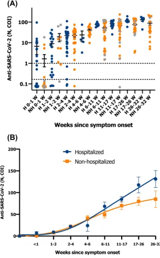
In symptomatic patients, a gradual increase in anti-body titers up to the last timepoint was observed (Figure 1). We confirm that sampling before 2 weeks does not permit to identify previous or ongoing infection due to insufficient sensitivity. However, within the first week, the positivity trend was higher in hospitalized patients (i.e. 50%) compared to non-hospitalized patients (i.e. 20%), an observation already made by Long et al. [12] and by Gillot et al. [13]. From 4 to 6 weeks, excellent sensitivities were observed (Table 1, Figure 1). Individual results for hospitalized patients were largely above the manufacturer’s cut-off. In non-hospitalized patients, one asymptomatic subject did not developed antibodies against the NCP (Figure 1A).
A trend towards higher antibody titers in hospitalized patients was also observed from weeks 6 to 11. The difference was higher if considering weeks 17 to 32 (Figure 1B). Other studies also reported higher levels of antibodies in patients with more severe disease [2, 14–16]. Of the 21 patients for which at least three independent blood drawn were avail-able for a minimal follow-up period of 7 weeks, a decrease in antibody titer was observed for 4 non-hospitalized patients out 10 (40%). In hospitalized patient, the titer gradually increased to reach a plateau without any decrease (n=11; 100%) (Supplementary Figure 1). Nevertheless, the association was not found to be significant in our study (p>0.05).
Importantly, the antibody kinetics may vary ac-cording to the type of assay considered. Recent studies
are in line with the current results and also found a sustained antibody response against the NCP antigen using the Roche total antibody assay, on a lower follow-up period (i.e. 3–4 months) [15, 17]. A sustained antibody response against the receptor-binding domain (RBD) antigen, as assessed by the Wantai and the Siemens total antibody assays, was also observed up to 4 months [15, 17]. A decrease in anti-RDB IgG and anti-spike IgG levels was similarly observed over a period of up to 5 months in recent reports [6, 14, 15, 18, 19]. A significant decrease in sensitivity was also found using the Abbott assay (NCP IgG), in studies with up to 4 months of follow-up [15, 17].
(A)
(B)
Figure 1: Anti-SARS-CoV-2 titers and long-term kinetics. (A) Anti-SARS-CoV-2 titers (mean COI and 95% CI) from symptom onset in hospitalized (blue points) and non-hospitalized
(orange points) COVID-19 patients (timeframe in weeks). Grey points correspond to asymptomatic patients that had a positive RT-PCR. (B) Long-term kinetics of anti-SARS-CoV-2 in hospitalized (blue points) and non-hospitalized (orange points) COVID-19 patients (timeframe in weeks). Smoothing splines with four knots were used to estimate the time kinetics curve (mean standard± error of the mean). Asymptomatic patients were excluded from the analysis.
The sustained antibody response as measured with total antibody assays (NCP and RBD) compared to IgG assays may be due to the additional response of non-IgG antibody iso-types. The reasons for the differences in assay performance over time for assays targeting the same antigen remain however unclear [17]. Whether the antibodies measured with commercial assays has a neutralizing capacity is paramount to indicate the potential level of protective immunity against SARS-CoV-2 infection. Antibody titers generated with avail-able assays correlated differently with neutralizing antibody titers [17, 20–22]. The Roche assay was the weaker predictor of neutralizing capacity (r=0.56, p=0.0001) compared to the Abbott assay (NCP IgG) (r=0.69, p<0.0001), Siemens assay (RDB total antibodies) (r=0.74, p<0.001), and the S1/S2-based DiaSorin assay (S1/S2 IgG ) (r=0.84, p<0.0001) [17]. Jahrs-dörfer et al. and Padoan et al. confirm that the weaker cor-relation was observed using the Roche assay [11, 22], and McAndrews et al. found that 86% of individuals positive for RBD-directed antibodies exhibited neutralizing capacity, whereas only 76% of positive NCP-directed antibodies exhibited neutralizing capacity [23]. The fact that anti-NCP assays have the lowest neutralizing capacity could be ex-pected, as neutralizing antibodies are directed to the spike protein responsible for enabling cell entry. Indeed, a strong correlation between levels of RBD or spike anti-bodies and neutralizing capacity has been found in inde-pendent evaluations [5, 6, 14, 19, 24]. Neutralizing capacity remained robust from 1 to 5 months in several studies [14, 19, 20], although modest declines at 3–5 months were observed by Wajnberg et al. and Isho et al. [5, 18]. Other studies how-ever observed a significant decrease of 2 to 4-fold, in neutralizing capacity up to 3 months [16, 17, 25–27].
It is important to keep in mind that some patients may develop anti-spike or anti-RBD antibodies but may not have detectable neutralizing antibodies. These are only correla-tion studies which are not related to direct measures of neutralizing capacity [17]. The fact that neutralizing anti-bodies constitute a major protective mechanism against SARS-CoV-2 infection deserves that further investigation are done in this area to assess to long-term inhibition capacity of SARS-CoV-2 antibodies [5, 9, 17]. The contribution of B cells and T cells to immunity to SARS-CoV-2 should also be more explored and it seems important to remind that previous exposure to SARS-CoV-2 might not guarantee total immu-nity in all cases since reinfection with SARS-CoV-2 have been described [28, 29].
In conclusion, we found a gradual increase in anti-NCP total antibody titers for up to 32 weeks since symptom onset. Even if some non-hospitalized patients showed a slight tendency towards a decrease of their antibody titer, this study found that detection rates were similar in
Tab le : Anti-SARS-CoV - titers (mean COI and % CI) from sym ptom onset in hospitalized and non-hospitalized COV ID- patie nts. Time inter vals (we eks since symptom onse t) – – – – – – – – Non -hospitalized WHO score – (+ ) n (+ ) (+ ) (+ ) (+ ) (+ ) (+ ) (+ ) (+ ) Mean ( % CI) . (− . to . ) . ( to . ) . ( . to . ) . ( . to . ) . ( . to . ) . ( . to . ) . ( . to . ) . ( . to . ) Sensit ivity (%) . ( . – . ) . ( . – . ) . ( . – . ) ( . – ) ( . – ) . ( . – . ) ( . – ) ( . – ) Hospitalized WHO score > n Mean ( % CI) . (− . to . ) . ( . to . ) . ( . to . ) . (− . to . ) . ( . to . ) . ( . to . ) . ( . to . ) . ( . to . ) Sensit ivity (%) ( % CI) . ( . – . ) . ( . – . ) . ( . – . ) ( . – ) ( . – ) ( . – ) ( . – ) ( . – ) Num bers bet ween brackets corr espond to asymp tomatic patients (WHO score ). The cut-off use d to calc ulate sensitivity w a s . .
hospitalized or non-hospitalized patients after one week from symptom onset and last at least 7.5 months. Although the majority of asymptomatic patients (95%) developed a sustained antibody response, one patient did not devel-oped antibodies 11 weeks after the RT-PCR positivity sup-porting the claim that caution is advised when interpreting anti-SARS-CoV-2 antibodies in asymptomatic subjects. Research funding: None declared.
Author contributions: All authors have accepted responsibility for the entire content of this manuscript and approved its submission.
Competing interests: Authors state no conflict of interest. Informed consent: Informed consent was obtained from all individuals included in this study.
Ethical approval: Research involving human subjects complied with all relevant national regulations, institutional policies and is in accordance with the tenets of the Helsinki Declaration (as revised in 2013), and has been approved by the authors’ Institutional Review Board or equivalent committee.
References
1. Huang AT, Garcia-Carreras B, Hitchings MDT, Yang B, Katzelnick LC, Rattigan SM, et al. A systematic review of antibody mediated immunity to coronaviruses: kinetics, correlates of protection, and association with severity. Nat Commun 2020;11:4704.
2. Bohn MK, Loh TP, Wang CB, Mueller R, Koch D, Sethi S, et al. IFCC interim guidelines on serological testing of antibodies against SARS-CoV-2. Clin Chem Lab Med 2020;58:2001–8.
3. Favresse J, Eucher C, Elsen M, Laffineur K, Dogne JM, Douxfils J. Response of anti-SARS-CoV-2 total antibodies to nucleocapsid antigen in COVID-19 patients: a longitudinal study. Clin Chem Lab Med 2020;58:e193-6.
4. Favresse J, Cadrobbi J, Eucher C, Elsen M, Laffineur K, Dogne JM, et al. Clinical performances of three fully automated anti-SARS-CoV-2 immunoassays targeting the nucleocapsid or spike proteins. J Med Virol 2020. https://doi.org/10.1002/jmv.26669. 5. Wajnberg A, Amanat F, Firpo A, Altman DR, Bailey MJ, Mansour M,
et al. Robust neutralizing antibodies to SARS-CoV-2 infection persist for months. Science 2020;370:1227–30.
6. Ibarrondo FJ, Fulcher JA, Goodman-Meza D, Elliott J, Hofmann C, Hausner MA, et al. Rapid decay of anti-SARS-CoV-2 antibodies in persons with mild COVID-19. N Engl J Med 2020; 383:1085–7.
7. Characterisation WHOWGotC, Management of C-i. A minimal common outcome measure set for COVID-19 clinical research. Lancet Infect Dis 2020;20:e192–7.
8. Favresse J, Eucher C, Elsen M, Tre-Hardy M, Dogne JM, Douxfils J. Clinical performance of the elecsys electrochemiluminescent immunoassay for the detection of SARS-CoV-2 total antibodies. Clin Chem 2020;66:1104–6.
9. National S-C-SAEG. Performance characteristics offive immunoassays for SARS-CoV-2: a head-to-head benchmark comparison. Lancet Infect Dis 2020;20:1390–400.
10. Egger M, Bundschuh C, Wiesinger K, Gabriel C, Clodi M, Mueller T, et al. Comparison of the Elecsys(R) Anti-SARS-CoV-2
immunoassay with the EDI enzyme linked immunosorbent assays for the detection of SARS-CoV-2 antibodies in human plasma. Clin Chim Acta 2020;509:18–21.
11. Jahrsdorfer B, Kroschel J, Ludwig C, Corman VM, Schwarz T, Korper S, et al. Independent side-by-side validation and comparison of four serological platforms for SARS-CoV-2 antibody testing. J Infect Dis 2020. https://doi.org/10.1093/ infdis/jiaa656.
12. Long QX, Liu BZ, Deng HJ, Wu GC, Deng K, Chen YK, et al. Antibody responses to SARS-CoV-2 in patients with COVID-19. Nat Med 2020;26:845–8.
13. Gillot C, Douxfils J, Cadrobbi J, Laffineur K, Dogne JM, Elsen M, et al. An original ELISA-based Multiplex method for the simultaneous detection of 5 SARS-CoV-2 IgG antibodies directed against different antigens. J Clin Med 2020;9. https://doi.org/10. 3390/jcm9113752.
14. Figueiredo-Campos P, Blankenhaus B, Mota C, Gomes A, Serrano M, Ariotti S, et al. Seroprevalence of anti-SARS-CoV-2 antibodies in COVID-19 patients and healthy volunteers up to six months post disease onset. Eur J Immunol 2020. https://doi.org/ 10.1002/eji.202048970.
15. Gudbjartsson DF, Norddahl GL, Melsted P, Gunnarsdottir K, Holm H, Eythorsson E, et al. Humoral immune response to SARS-CoV-2 in Iceland. N Engl J Med 2020;383:1724–34.
16. Seow J, Graham C, Merrick B, Acors S, Pickering S, Steel KJA, et al. Longitudinal observation and decline of neutralizing antibody responses in the three months following SARS-CoV-2 infection in humans. Nat Microbiol 2020;5:1598–607.
17. Muecksch F, Wise H, Batchelor B, Squires M, Semple E, Richardson C, et al. Longitudinal analysis of clinical serology assay performance and neutralising antibody levels in COVID19 convalescents. medRxiv 2020. https://doi.org/10.1101/2020.08. 05.20169128.
18. Isho B, Abe KT, Zuo M, Jamal AJ, Rathod B, Wang JH, et al. Persistence of serum and saliva antibody responses to SARS-CoV-2 spike antigens in COVID-19 patients. Sci Immunol 2020;5. https://doi.org/10.1126/sciimmunol.abe5511.
19. Iyer AS, Jones FK, Nodoushani A, Kelly M, Becker M, Slater D, et al. Persistence and decay of human antibody responses to the receptor binding domain of SARS-CoV-2 spike protein in COVID-19 patients. Sci Immunol 2020;5:eabe0367.
20. Brigger D, Horn MP, Pennington LF, Powell AE, Siegrist D, Weber B, et al. Accuracy of serological testing for SARS-CoV-2 antibodies: first results of a large mixed-method evaluation study. Allergy 2020. https://doi.org/10.1111/all.14608.
21. Kohmer N, Westhaus S, Ruhl C, Ciesek S, Rabenau HF. Brief clinical evaluation of six high-throughput SARS-CoV-2 IgG antibody assays. J Clin Virol 2020;129:104480.
22. Padoan A, Bonfante F, Pagliari M, Bortolami A, Negrini D, Zuin S, et al. Analytical and clinical performances offive immunoassays for the detection of SARS-CoV-2 antibodies in comparison with neutralization activity. EBioMedicine 2020;62:103101. 23. McAndrews KM, Dowlatshahi DP, Dai J, Becker LM, Hensel J,
Snowden LM, et al. Heterogeneous antibodies against SARS-CoV-2 spike receptor binding domain and nucleocapsid with
implications for COVID-19 immunity. JCI Insight 2020;5. https://doi.org/10.1172/jci.insight.142386.
24. Premkumar L, Segovia-Chumbez B, Jadi R, Martinez DR, Raut R, Markmann A, et al. The receptor binding domain of the viral spike protein is an immunodominant and highly specific target of antibodies in SARS-CoV-2 patients. Sci Immunol 2020;5. https://doi.org/10.1126/sciimmunol.abc8413.
25. Crawford KHD, Dingens AS, Eguia R, Wolf CR, Wilcox N, Logue JK, et al. Dynamics of neutralizing antibody titers in the months after SARS-CoV-2 infection. J Infect Dis 2020. https://doi.org/10.1093/ infdis/jiaa618.
26. Prevost J, Gasser R, Beaudoin-Bussieres G, Richard J, Duerr R, Laumaea A, et al. Cross-sectional evaluation of humoral responses against SARS-CoV-2 spike. Cell Rep Med 2020;1: 100126.
27. Wang K, Long QX, Deng HJ, Hu J, Gao QZ, Zhang GJ, et al. Longitudinal dynamics of the neutralizing antibody response to SARS-CoV-2 infection. Clin Infect Dis 2020. https://doi.org/10.1093/cid/ciaa1143. 28. Tillett RL, Sevinsky JR, Hartley PD, Kerwin H, Crawford N,
Gorzalski A, et al. Genomic evidence for reinfection with SARS-CoV-2: a case study. Lancet Infect Dis 2020. https://doi.org/10.1016/s1473-3099(20)30764-7.
29. To KK, Hung IF, Ip JD, Chu AW, Chan WM, Tam AR, et al. COVID-19 re-infection by a phylogenetically distinct SARS-coronavirus-2 strain confirmed by whole genome sequencing. Clin Infect Dis 2020. https://doi.org/10.1093/cid/ciaa1275.
Supplementary Material: The online version of this article offers supplementary material (https://doi.org/10.1515/cclm-2020-1736).