RESEARCH OUTPUTS / RÉSULTATS DE RECHERCHE
Author(s) - Auteur(s) :
Publication date - Date de publication :
Permanent link - Permalien :
Rights / License - Licence de droit d’auteur :
Bibliothèque Universitaire Moretus Plantin
Institutional Repository - Research Portal
Dépôt Institutionnel - Portail de la Recherche
researchportal.unamur.be
University of Namur
Formation of carbon nitride compounds during successive implantations in copper
Thome, Tristan; Colaux, Julien; Louette, Pierre; Terwagne, Guy
Published in:
Journal of Electron Spectroscopy and Related Phenomena
Publication date:
2006
Document Version
Early version, also known as pre-print
Link to publication
Citation for pulished version (HARVARD):
Thome, T, Colaux, J, Louette, P & Terwagne, G 2006, 'Formation of carbon nitride compounds during
successive implantations in copper', Journal of Electron Spectroscopy and Related Phenomena, vol. 151, no. 1, pp. 19-23. <http://www.sciencedirect.com/>
General rights
Copyright and moral rights for the publications made accessible in the public portal are retained by the authors and/or other copyright owners and it is a condition of accessing publications that users recognise and abide by the legal requirements associated with these rights. • Users may download and print one copy of any publication from the public portal for the purpose of private study or research. • You may not further distribute the material or use it for any profit-making activity or commercial gain
• You may freely distribute the URL identifying the publication in the public portal ?
Take down policy
If you believe that this document breaches copyright please contact us providing details, and we will remove access to the work immediately and investigate your claim.
Formation of carbon nitride compounds during
successive implantations in copper
T. Thom´e
a,∗, J.L. Colaux
a, P. Louette
b, G. Terwagne
aaLaboratoire d’Analyses par R´eactions Nucl´eaires, Facult´es Universitaires Notre-Dame de la Paix, 61 rue de Bruxelles, B-5000 Namur, Belgium bLaboratoire Interdisciplinaire de Spectroscopie Electronique, Facult´es Universitaires Notre-Dame de la Paix, 61 rue de Bruxelles, B-5000 Namur, Belgium
Received 15 April 2005; received in revised form 3 October 2005; accepted 7 October 2005 Available online 21 November 2005
Abstract
Copper substrates are successively implanted with carbon and nitrogen (13C+ and14N+) at high fluences (5× 10¹⁷ and 1× 10¹⁷at. cm−2,
respectively) in order to synthesize specific carbon nitride compounds. The concentration as well as the depth distribution of carbon13C and
nitrogen14N are determined using non resonant nuclear reactions induced by a 1.05 MeV deuteron beam. The use of (d,p) and (d,␣) reactions
allows us to profile both13C and14N elements with a single and relatively rapid measurement and a quite good resolution. The bonded states
of carbon and nitrogen are studied as a function of depth by X-ray photoelectron spectroscopy (XPS). The curve fitting of the C 1s and N 1s photopeaks shows that carbon and nitrogen atoms exist in different chemical states depending on the analysis depth, which correspond to specific kinds of chemical bonds. At least two characteristic C–N bonds are detected indicating that different carbon nitride compounds have been formed during the implantations.
© 2005 Elsevier B.V. All rights reserved.
Keywords: Carbon; Nitrogen; XPS; NRA; Implantation
1. Introduction
In recent years, the synthesis of crystalline carbon nitrides has been extensively investigated as they are expected to show remarkable physical properties such as wide band gap and high hardness and wear resistance. Indeed, some calculations pre-dicted that␣- and -C3N4would be harder than diamond[1].
A wide variety of elaboration techniques have been used such as reactive sputtering, chemical vapour deposition, pyrolysis of organic materials, laser deposition and ion implantations[2–7]. So far, whatever the technique employed, mixed phase layers are quite often obtained and it remains very difficult to achieve fully crystalline phase formation. Some studies have also shown that fullerene-like structures can be formed from nitrogen-poor CNxphase[8]. For instance, carbon nitride systems close to the
C11N4stoichiometry, that present interesting mechanical
prop-erties as high compliance, have been found to be stable [9]. Although ion implantation may be one solution to synthesize these types of new compounds due to the possibility to control
∗Corresponding author. Tel.: +32 81 72 54 77.
E-mail address: [email protected] (T. Thom´e).
precisely the modifications on surface properties of materials, successive carbon and nitrogen implantations in metals have not yet succeeded in that way[10]. Nevertheless, even amorphous carbon nitride layers have suitable physical properties for the use in many tribological applications, for instance as protective coatings.
The aim of our study is to form at least homogeneous, repro-ducible and well characterized carbon nitride compounds by means of implantation. We performed successive implantations of 13C and 14N into copper at 200◦C using a 2 MV Tande-tron accelerator. Then, we determined the concentration depth distributions of carbon 13C and nitrogen14N using non reso-nant nuclear reactions (NRA) induced by a 1.05 MeV deuteron beam. To obtain information on the nature of the chemical bonds created between atoms in the carbon–nitride layer formed, we carried out XPS measurements.
2. Experimental
2.1. Materials and substrate implantation
The samples are polished polycrystalline copper substrates. Successive carbon13C+and nitrogen14N+implantations were
0368-2048/$ – see front matter © 2005 Elsevier B.V. All rights reserved. doi:10.1016/j.elspec.2005.10.001
20 T. Thom´e et al. / Journal of Electron Spectroscopy and Related Phenomena 151 (2006) 19–23
carried out using the 2 MV ALTAIS Tandetron accelerator (Acc´el´erateur Lin´eaire Tandetron pour 1’Analyse et 1’Implan-tation des Solides). During both implan1’Implan-tations, the sample was maintained at 200◦C using a specific sample heater and the vacuum pressure did not exceed 10−5Pa. The implantation of carbon was performed in first place. It was implanted at 400 keV with a current density of about 6A cm−2 and the final dose was 5× 10¹⁷at. cm−2over a 5 mm× 5 mm area. The nitrogen implantation was performed at 450 keV with a current density of 4A cm−2and the final dose was 1017at. cm−2(on the same area). The energies of nitrogen and carbon ions were chosen to obtain identical implantation projected ranges close to 450 nm in copper, according to SRIM code calculations[11].
2.2. Characterization
The concentration as well as the distribution of carbon13C and nitrogen14N were determined using non resonant nuclear reactions induced by a 1.05 MeV deuteron beam before and after the14N implantation. NRA experiments were performed using the same facilities as for the implantations (ALTAIS Tandetron accelerator). We considered the nuclear reactions14N(d,p0)15N, 14N(d,p
12)15N,14N(d,p3)15N,14N(d,␣1)12C, and13C(d,p0)14C
to depth profile14N and13C, and12C(d,p0)13C to determine the
quantity of12C carbon contamination brought during implanta-tion. Two silicon surface barrier detectors were placed at 150◦ and 165◦relative to the incident beam to measure respectively NRA and RBS signals. A 12m mylar absorber foil was set in front of the NRA detector at 150◦in order to stop backscat-tered ions and to measure␣ and proton energies. Since the cross sections of the nuclear reactions on14N and13C are very low, we used a large solid angle (25.7 msr) for this detector to min-imize the acquisition time (about 20 min). The RBS detector was collimated (0.18 msr) to allow the detection of backscat-tered deuterons without any absorber foil. The ␣ and protons emitted from nuclear reactions at low energy were also detected with a better resolution than with the NRA detector. The RBS detector was used as a monitor to determine the concentrations of nitrogen and carbon atoms implanted in the copper sample from the quantity of incident deuterons detected. The distribu-tion homogeneity of carbon and nitrogen was controlled carrying out several measurements on the implanted area of the sample. XPS measurements were performed to study the composi-tion and the nature of the carbon–nitride layer formed in the implanted copper sample. XPS spectra were recorded with a SSX 100 Spectrometer system (Surface Science Instrument) equipped with a hemispherical electron analyser. All reported spectra were recorded at a 35◦ take-off angle relative to the substrate using monochromatized Al K␣ radiation as excitation source (1486.6 eV). Nominal resolution was measured as full width at half maximum of 1.0 and 1.7 eV for core-levels and survey spectra, respectively. The argon ion gun used for sputter-ing was equipped with a special regulatsputter-ing system which enabled automated operation during long time depth profile procedures. The depth profiling conditions were the followings: raster size of about 2 mm× 2 mm, Ar+ion energy of 4 keV and sputter rates around 3 nm min−1. Depth calibration was done measuring the
crater depth generated by argon sputtering on the copper sam-ple. The chemical composition was obtained from the areas of the detected XPS peaks in the C 1s, N 1s, O 1s and Cu 2p3/2
regions, performing Shirley background subtraction and tak-ing into account sensitivity factors for each constituent. The analyzed core-level lines were referenced with respect to the component C 1s binding energy. The peaks were analysed using mixed Gaussian–Lorentzian curves (90% of Gaussian charac-ter). Binding state information was determined from chemical shifts observed on the binding energy scale after the curve fitting of XPS peaks.
3. Results and discussion
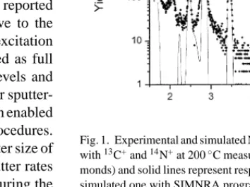
We first present NRA results from carbon and nitrogen nuclear reactions with the 1.05 MeV deuteron beam. Then, the formation of characteristic carbon nitrogen bonds in the implanted layer is discussed on the basis of XPS measurements. Fig. 1shows the experimental NRA spectrum measured at 150◦ after carbon and nitrogen implantations. A very intense peak is observed just below 3 MeV, which can be attributed to12C surface contamination. Around 6 MeV, the (d,p) peak corresponds to the implanted13C ions. All the other peaks are assigned to (d,p) and (d,␣) reactions with14N element. From this spectrum, we determined the total concentrations and the depth distributions of12C,13C and14N elements using the SIMNRA program[12]in which specific nuclear reaction cross sections are considered and the copper sample is sliced into layers con-taining all the elements concerned. The simulated SIMNRA spectrum is represented inFig. 1and the different concentration depth distributions of12C,13C and14N calculated are shown in Fig. 2.
The13C depth distribution determined before the14N implan-tation is slightly narrower than the final one but both areas are identical (distributions not shown here)[13], which means that a small carbon diffusion process occurs during the nitrogen implantation. Although13C and14N concentrations correspond
Fig. 1. Experimental and simulated NRA spectra of the copper sample implanted with13C+and14N+at 200◦C measured at 150◦(NRA detector). Symbols (dia-monds) and solid lines represent respectively the experimental spectrum and the simulated one with SIMNRA program.
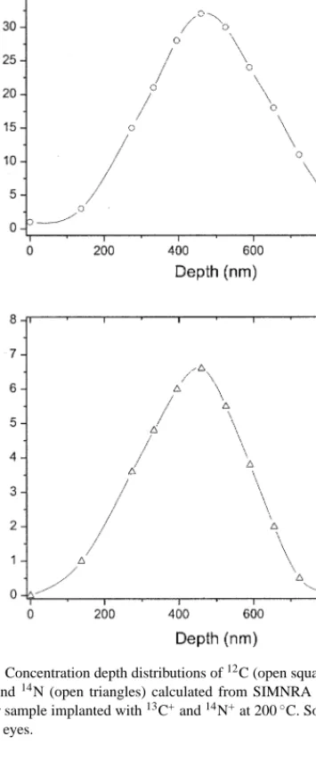
Fig. 2. Concentration depth distributions of12C (open squares),13C (open
cir-cles) and14N (open triangles) calculated from SIMNRA simulations for the
copper sample implanted with13C+and14N+at 200◦C. Solid lines are guides
for the eyes.
well to the implanted ones expected, a large amount of12C is also detected in the sample (nearly 5× 10¹⁷at. cm−2).
A surface contamination layer mainly composed of12C is clearly observed (about 30 nm wide) while almost no 12C is
Table 1
Experimental and calculated projected ranges (Rexpand RSRIM) and full widths at
half maximum (FWHMexpand FWHMSRIM) of the concentration distributions
in the copper sample implanted with13C+and14N+at respectively 400 and
450 keV
Element Rexp(nm) RSRIM(nm) FWHMexp(nm) FWHMSRIM(nm)
13C 480 450 350 130
14N 460 430 300 130
detected around the 13C and 14N implantation mean depth (Fig. 2). This contamination may be explained by some car-bon build-up phenomena during implantations as no carcar-bon is detected on the virgin sample surface. Gaussian like dis-tribution curves are observed for 13C and 14N. Although the experimental distribution maxima (close to 470 nm in the sam-ple) correspond well with the projected ranges calculated with SRIM program, the widths of the curves are larger than SRIM calculated ones (Table 1). That can be explained by a diffusion process of nitrogen and carbon during the implantations. The relative concentrations of13C and14N at the profile maximum correspond to our implanted dose estimations (N/C ratio of about 0.2).
Then, XPS measurements were carried out to know whether specific carbon nitrogen bonding is created in the implanted copper sample.
Survey scan XPS spectra indicate the presence of carbon, nitrogen and oxygen in the copper implanted sample. The oxy-gen contamination is not studied in this work because it is mainly localized in the near surface. The concentrations of nitrogen and carbon can be deduced from the areas of the different XPS peaks. The carbon contamination layer detected by NRA is confirmed by XPS results. The maximum concentration range (around 450 nm) also corresponds approximately to the one determined by NRA. Nevertheless, the atomic concentration ratio of nitro-gen to carbon (N/C) is less important than the one calculated from NRA experiments (by a factor 1.5). That can be explained by some contribution of the surface contamination carbon to the determination of the carbon concentration value at 450 nm.
Core level photoelectron spectra of C and N 1s, acquired at the surface and at a depth where the concentrations of nitrogen and carbon are maximum, are presented inFigs. 3 and 4. No clear peak components can be observed in the different spectra in C 1s region but the peaks broaden with the analysis depth. One main peak component, which is centred around 285 eV, can be resolved at the surface. It corresponds to C–C bonds in an amorphous carbon contamination layer. A tail in the high bind-ing energies indicates the presence of low quantities of C–N and C–O bonds (a few %) due to the higher electronegativity of nitrogen and oxygen. When, the analysis goes further in depth, the peak broadening is accompanied with an increase of the tail component intensity. To achieve curve fitting of the C 1s peak at the 450 nm depth, four Gaussian components (C0, C1,
C2, C3) have been used, that we attributed to different bonding
states of carbon atoms. The C0component (around 284.8 eV) is
attributed to carbon atoms bonded to carbon neighbours, as in graphite or amorphous carbon. No distinction between sp2and
22 T. Thom´e et al. / Journal of Electron Spectroscopy and Related Phenomena 151 (2006) 19–23
Fig. 3. C 1s XPS spectra at the surface (a) and at a 450 nm depth (b) for the copper sample implanted with13C+ and14N+ at 200◦C. Experimental data are represented by open squares. Solid lines are the results of the curve fitting Gaussian components in dotted lines. Meaning of Cinotations is discussed in
the text.
sp3has been made for this first component whatever the analy-sis depth. C1(286.2 eV) and C2(287.8 eV) peak components are
related to nitrogen presence. As suggested in different reports [14,15], we ascribed C1 to sp2 C–N bonds (trigonal carbon
configuration) and C2to sp3C–N bonds (tetrahedral carbon
con-figuration). The final peak component with the highest binding energy (289.9 eV) may be assigned to C–O bonds. The position and the intensity of this last peak are rather difficult to obtain pre-cisely as they depend very much on the background subtraction procedure. Thus, the presence of C–O bonds is not discussed here.
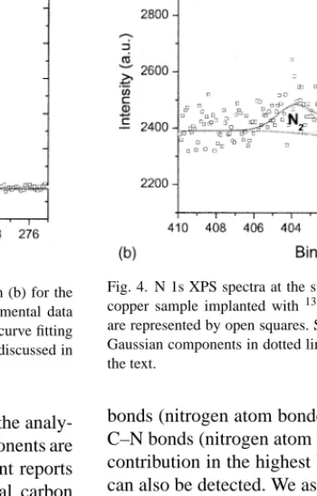
As shown inFig. 4, the N 1s core level photoelectron spectra exhibit more resolved peak components even if their intensities are lower due to a smaller implanted concentration. No curve fitting and decomposition were performed on the first spectrum because of poor statistics due to very low nitrogen concentration detected at the surface. To perform curve fitting of the N 1s peak at the 450 nm depth, three Gaussian components (N0, N1, N2)
have been used. We attributed the N0(398.7 eV) and N1(around
400.7 eV) dominant peak components to respectively, sp3C–N
Fig. 4. N 1s XPS spectra at the surface (a) and at a 450 nm depth (b) for the copper sample implanted with13C+and14N+at 200◦C. Experimental data
are represented by open squares. Solid lines are the results of the curve fitting Gaussian components in dotted lines. Meaning of Ninotations is discussed in
the text.
bonds (nitrogen atom bonded to tetrahedral carbon) and to sp2 C–N bonds (nitrogen atom bonded to trigonal carbon). A small contribution in the highest binding energies (around 403.7 eV) can also be detected. We assigned it to N–O bonds and we will not discuss it further. This peak component identification for C 1s and N 1s core level photoelectron spectra is in good agreement with recent reports in literature[15,16].
These results show that several carbon nitride compounds are created during the successive implantations. At least two kinds of carbon nitride regions, that are characterized by dif-ferent hybridisations of C–N bonds (sp2 and sp3), mixed in a dominant amorphous carbon matrix and surrounded by copper, may be envisioned.
4. Conclusion
This study showed that carbon nitride formation was possible by performing successive carbon and nitrogen implantations at 200◦C. The atomic depth distributions of nitrogen and carbon were determined by NRA. The depth profile peaks corresponded
well to the calculated ion projected ranges (around 470 nm for both profiles), which enabled us to maximize the interactions of nitrogen and carbon at this depth. XPS measurements clearly indicated that characteristic types of chemical bonds between carbon and nitrogen atoms were created at this depth and that they might correspond to sp3 and sp2 carbon hybridisations. These results suggest that at least two different carbon nitride regions exist and that they are embedded in a dominant amor-phous carbon matrix.
Acknowledgments
T. Thom´e was granted a CERUNA post-doctoral position by the University of Namur (FUNDP).
References
[1] A. Lui, M. Cohen, Science 247 (1989) 688.
[2] K.M. Yu, M.L. Cohen, B.E. Haller, W.L. Hansen, A.Y. Liu, J.C. Wu, Phys. Rev. B 49 (1994) 5034.
[3] L. Maya, D.R. Cole, E.W. Hagaman, J. Am. Ceram. Soc. 74 (1991) 1686.
[4] C. Niu, Y.Z. Lu, C.M. Lieber, Science 261 (1993) 334.
[5] E. Gyorgy, V. Nelea, I. Mihailescu, A. Perrone, H. Pelletier, A. Cornet, S. Ganatsios, J. Werckmann, Thin Solid Films 388 (2001) 93. [6] A. Hoffman, I. Gouzman, R. Brener, Appl. Phys. Lett. 64 (1994) 845. [7] E. Romanowsky, O. Bespalova, A. Borisov, N. Goryaga, V. Kulikauskas,
V. Sukharev, V. Satekin, Nucl. Instr. Meth. B 139 (1998) 355. [8] N. Hellgren, M.P. Johansson, E. Broitman, L. Hultman, J.-E. Sundgren,
Phys. Rev. B 59 (1999) 5162.
[9] H. Sj¨ostr¨om, L. Hultman, J.-E. Sundgren, S. Hainsworth, T. Page, G.S. Theunissen, J. Vac. Sci. Technol. A 14 (1996) 56.
[10] R. Sanchez, J.A. Garcia, A. Medrano, M. Rico, R. Martinez, R. Rodriguez, C. Fernandez-Ramos, A. Fernandez, Surf. Coat. Technol. 158–159 (2002) 630.
[11] J.F. Ziegler, J.P. Biersack, U. Littmark, The Stopping and Range of Ions in Solids, Pergamon Press, New York, 1967.
[12] M. Mayer, SIMNRA, a Simulation Program for the Analysis of NRA, RBS and ERDA, in: J.L. Duggan, I.L. Morgan (Eds.), Proceedings of the 15th International Conference Appl. Accelerators in Research and Industry, AIP Conf. Proc. 475 (1999) 541.
[13] J. Colaux, G. Terwagne, Nucl. Instr. Meth. B 240 (2005) 429. [14] B. Angleraud, N. Mubumbila, P.Y. Tessier, V. Ferandez, G. Turban,
Diamond Rel. Mater. 10 (2001) 1142.
[15] N. Hellgren, J. Guo, Y. Luo, C. Sathe, A. Agui, S. Kashtanov, J. Nord-gren, H. ANord-gren, J.E. SundNord-gren, Thin Solid Films 471 (2005) 19. [16] S.E. Rodil, S. Muhl, Diamond Rel. Mater. 13 (2004) 1521.