Author(s) - Auteur(s) :
Publication date - Date de publication :
Permanent link - Permalien :
Rights / License - Licence de droit d’auteur :
Bibliothèque Universitaire Moretus Plantin
Subchronic exposure to titanium dioxide nanoparticles modifies cardiac structure and
performance in spontaneously hypertensive rats
Rossi, Stefano; Savi, Monia; Mazzola, Marta; Pinelli, Silvana; Alinovi, Rossella; Gennaccaro,
Laura; Pagliaro, Alessandra; Meraviglia, Viviana; Galetti, Maricla; Lozano-Garcia, Omar;
Rossini, Alessandra; Frati, Caterina; Falco, Angela; Quaini, Federico; Bocchi, Leonardo; Stilli,
Donatella; Lucas, Stéphane; Goldoni, Matteo; Macchi, Emilio; Mutti, Antonio; Miragoli,
Michele
Published in:Particle and Fibre Toxicology DOI:
10.1186/s12989-019-0311-7
Publication date: 2019
Document Version
Publisher's PDF, also known as Version of record
Link to publication
Citation for pulished version (HARVARD):
Rossi, S, Savi, M, Mazzola, M, Pinelli, S, Alinovi, R, Gennaccaro, L, Pagliaro, A, Meraviglia, V, Galetti, M, Lozano-Garcia, O, Rossini, A, Frati, C, Falco, A, Quaini, F, Bocchi, L, Stilli, D, Lucas, S, Goldoni, M, Macchi, E, Mutti, A & Miragoli, M 2019, 'Subchronic exposure to titanium dioxide nanoparticles modifies cardiac structure and performance in spontaneously hypertensive rats', Particle and Fibre Toxicology, vol. 16, no. 1, 25, pp. 25. https://doi.org/10.1186/s12989-019-0311-7
General rights
Copyright and moral rights for the publications made accessible in the public portal are retained by the authors and/or other copyright owners and it is a condition of accessing publications that users recognise and abide by the legal requirements associated with these rights. • Users may download and print one copy of any publication from the public portal for the purpose of private study or research. • You may not further distribute the material or use it for any profit-making activity or commercial gain
• You may freely distribute the URL identifying the publication in the public portal ?
Take down policy
If you believe that this document breaches copyright please contact us providing details, and we will remove access to the work immediately and investigate your claim.
R E S E A R C H
Open Access
Subchronic exposure to titanium dioxide
nanoparticles modifies cardiac structure
and performance in spontaneously
hypertensive rats
Stefano Rossi
1,2†, Monia Savi
3†, Marta Mazzola
1,8, Silvana Pinelli
1,2, Rossella Alinovi
1,2, Laura Gennaccaro
4,5,10,
Alessandra Pagliaro
4,5, Viviana Meraviglia
4,5, Maricla Galetti
1,2, Omar Lozano-Garcia
6,9, Alessandra Rossini
4,5,
Caterina Frati
1, Angela Falco
1, Federico Quaini
1, Leonardo Bocchi
3, Donatella Stilli
3, Stéphane Lucas
6,
Matteo Goldoni
1,2, Emilio Macchi
2,3, Antonio Mutti
1,2,7and Michele Miragoli
1,2,8*Abstract
Background: Non-communicable diseases, intended as the results of a combination of inherited, environmental and biological factors, kill 40 million people each year, equivalent to roughly 70% of all premature deaths globally. The possibility that manufactured nanoparticles (NPs) may affect cardiac performance, has led to recognize NPs-exposure not only as a major Public Health concern, but also as an occupational hazard. In volunteers, NPs-exposure is problematic to quantify. We recently found that inhaled titanium dioxide NPs, one of the most produced engineered nanomaterials, acutely increased cardiac excitability and promoted arrhythmogenesis in normotensive rats by a direct interaction with cardiac cells. We hypothesized that such scenario can be exacerbated by latent cardiovascular disorders such as hypertension.
Results: We monitored cardiac electromechanical performance in spontaneously hypertensive rats (SHRs) exposed to titanium dioxide NPs for 6 weeks using a combination of cardiac functional measurements associated with toxicological, immunological, physical and genetic assays.
Longitudinal radio-telemetry ECG recordings and multiple-lead epicardial potential mapping revealed that atrial activation times significantly increased as well as proneness to arrhythmia. At the third week of nanoparticles administration, the lung and cardiac tissue encountered a maladaptive irreversible structural remodelling starting with increased pro-inflammatory cytokines levels and lipid peroxidation, resulting in upregulation of the main pro-fibrotic cardiac genes. At the end of the exposure, the majority of spontaneous arrhythmic events terminated, while cardiac hemodynamic deteriorated and a significant accumulation of fibrotic tissue occurred as compared to control untreated SHRs. Titanium dioxide nanoparticles were quantified in the heart tissue although without definite accumulation as revealed by particle-induced X-ray emission and ultrastructural analysis.
Conclusions: The co-morbidity of hypertension and inhaled nanoparticles induces irreversible hemodynamic impairment associated with cardiac structural damage potentially leading to heart failure. The time-dependence of exposure indicates a non-return point that needs to be taken into account in hypertensive subjects daily exposed to nanoparticles.
Keywords: Titanium dioxide nanoparticles, Nanotoxicology, Cardiac electrophysiology, Cardiac fibrosis, Arrhythmias
© The Author(s). 2019 Open Access This article is distributed under the terms of the Creative Commons Attribution 4.0 International License (http://creativecommons.org/licenses/by/4.0/), which permits unrestricted use, distribution, and reproduction in any medium, provided you give appropriate credit to the original author(s) and the source, provide a link to the Creative Commons license, and indicate if changes were made. The Creative Commons Public Domain Dedication waiver (http://creativecommons.org/publicdomain/zero/1.0/) applies to the data made available in this article, unless otherwise stated.
* Correspondence:[email protected]
†Stefano Rossi and Monia Savi contributed equally to this work. 1
Department of Medicine and Surgery, University of Parma, Via Gramsci, n° 14, 43126 Parma, Italy
2CERT, Center of Excellence for Toxicological Research, INAIL, ex-ISPESL,
University of Parma, Parma, Italy
have been raised and need to be addressed [2]. Manufac-tured nanoparticles have become a new component of the air we breathe [3]. Since the heart and the lungs are intim-ately linked, it is difficult to identify the specific susceptibil-ity of either organ to the effects of nanoparticles (NPs) [4]. It is possible to better define the impact of particle matter (PM) on individual organ impairment or disease [5, 6] through the use of animal models. Recently, in order to fur-ther clarify the relationships between fine particles and car-diopulmonary diseases, the spontaneously hypertensive rat (SHR) has been widely used as a model of cardiovascular hypertension for researches on toxicology and pathogenesis of cardiovascular adverse effects of exposure to PM [7]. Limited experimental evidence from cardiopulmonary-compromised animal models has shown increased mortal-ity from chronic exposure to simulated particle/gaseous urban air pollution in SHR [8]. Chronic systemic hyperten-sion and associated cardiomyopathy may increase the sus-ceptibility to PM-induced morbidity. In fact, in control SHR the ECG showed a depression in ST-segment area which was not present in Wistar Kyoto (WKY) rats and a further depression in ST-segment area in SHR during the first and second days of PM exposure. This enhanced de-pression was less evident following 3 days of exposure, per-haps suggesting adaptive changes [9].
The first study to report long-term cardiovascular toxicity of an inhaled engineered nanomaterial, clearly demon-strated that long-term exposure to inhaled nickel NPs can induce oxidative stress and inflammation, not only in the lung but also in the cardiovascular system [10]. Recent data showed that long-term silica inhalation in rat, at realistic concentrations, induced very low systemic toxicity and neg-ligible pulmonary fibrogenicity [11].
Titanium dioxide (TiO2) is one of the most widely
pro-duced engineered nanomaterials. As consumption grows the chance of population exposure to fine or ultrafine TiO2
increases. In 2007 the National Institute for Occupational Safety and Health (NIOSH) estimated 68.000 workers in U.S. directly in contact with TiO2pigments, with an
esti-mated production of TiO2of 1.45 million metric tons / year
(https://www.cdc.gov/niosh/docs/2011-160/pdfs/2011-160. pdf) and the demand of TiO2 increases exponentially
worldwide. Compared to the high risk of heart disease by PM exposure, the TiO2effects are still partially known [12].
fine TiO2 and 0.3 mg/m3 for ultrafine TiO2 were
recom-mended, as time-weighted average concentrations for up to 10 h per day during a 40-h work/week [15]. Subjects with underlying health issues such as asthma and hypertension may be at increased risk of TiO2-NPs toxicity [16]. A
sig-nificant association between inhalable TiO2-NPs exposure
and declines in pulmonary function has been demon-strated, as well as increase in blood pressure, especially in small airways functions and systolic blood pressure in healthy workers at the end of shift [17]. Exposure of rats, by intra-tracheal instillation, to a well-dispersed suspension of ultra-fine TiO2-NPs caused dose-dependent pulmonary
damage and inflammation, which persisted 42 days post-exposure [2]. Another work demonstrated that exposures to nano sized TiO2 had no significant long-term adverse
pulmonary effects, even if acute levels of inflammation and cytotoxicity were observed for TiO2-NPs, at the highest
dose of 5 mg/kg, 1 day post-exposure [18]. We recently showed that a single acute intra-tracheal instillation of TiO2-NPs in normotensive animals, induces cardiac
elec-trophysiological alterations especially in the repolarization phase, in both isolated cardiomyocytes and in vivo rat heart, giving rise to a pro-arrhythmic substrate [19] due to the dir-ect formation of nano pores across cell membrane [20]. It also has been demonstrated that, when SHR and WKY rats were exposed to the same dose of fine particles including metallic elements such as Fe, Mg, Zn, Pb and Cu, the lung injury in SHR was greater than in WKY rats, due to an in-crease in cytokines [21], and rats with cardiopulmonary dis-eases were more susceptible than healthy rats [22].
The aim of the present study was to investigate the ef-fects of subchronical exposure to TiO2-NPs on cardiac
structure and electromechanical performance in SHRs, being aware that TiO2-NPs can rapidly translocate from
the lung into the bloodstream and finally to the heart [19]. Here, we demonstrated a pathological cardiac remodeling associated with an increment of spontaneous arrhythmic events mainly including sinus pauses and atria-ventricular blocks (AV blocks), changes in conduction velocity, and worsened hemodynamic performance. Interestingly, our approach identified a time-point characterized by cardiac and pulmonary inflammatory response, nanotoxicity and up-regulation of genes deputed to control collagen depos-ition in the heart, ultimately resulting in an irreversible
increment of diffuse cardiac fibrosis compared to the un-treated SHRs.
Results
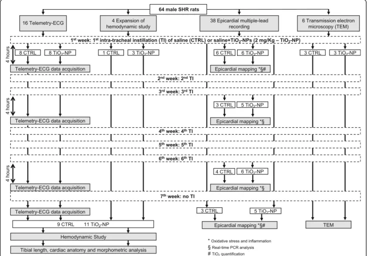
The experimental design and measurements performed in the various animal groups are summarized in Fig.1.
Telemetry-ECG study
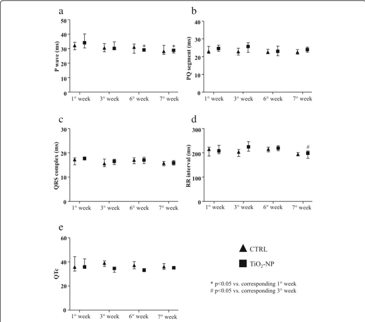
Electrocardiographic wave and interval durations showed only time-dependent effects as illustrated in Fig.2. Atrial activation (P wave duration) in NPs exposed SHRs was re-duced at the 6th week and the end of the longitudinal protocol (7th week) compared to the first week (Fig.2a). We did not observe significant differences for PQ seg-ment, QRS complex and QTc (Fig.2b, c and e). RR inter-val was significantly reduced only at the end of the experimental protocol in the treated animals (Fig.2d).
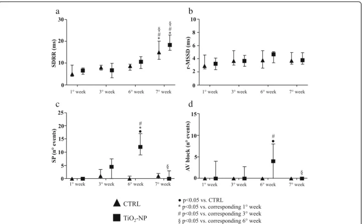
The heart rate variability, evaluated as standard devi-ation of RR interval (SDRR), increased only at final stage in both groups (Fig.3a), while no differences were seen in
the root mean square of successive RR interval square dif-ferences (r-MSSD) value (Fig.3b). ECG-telemetry analysis revealed different types of arrhythmic events (Add-itional file 1: Figure S1). Arrhythmia quantification dem-onstrated that sinus pauses and AV blocks significantly increased in TiO2-NPs group in comparison with control
(CTRL) group only after the 6th instillation (nine and sixteen-fold increase, respectively; Fig.3c and d). This in-crease disappeared when TiO2treatment was suspended.
Instead, supraventricular and ventricular arrhythmias were similar in the two experimental groups during the entire protocol (data not shown).
Hemodynamic study
The heart rate measured under anesthesia was similar in both groups as well as systolic and diastolic arterial blood pressure, and left ventricular systolic pressure (LVSP) whose values were coherent with the degree of hyperten-sion expected in 19-week old SHR (Table 1). Compared with CTRL animals, TiO2-NP rats exhibited a global
Fig. 1 Study population and experimental protocol. Flowchart describing the number of animals subjected to the different experimental procedures before and after intra-tracheal instillation of saline solution (CTRL group) or saline solution added with TiO2, at a final concentration of 2 mg/kg (TiO2-NP
group). * timepoints for oxidative stress and inflammation; § timepoints for real-time PCR analysis; # timepoints for PIXE. For more details see“Outline of the experimental protocols” section
deterioration of hemodynamic performance, as indicated by the significant increase in the left ventricular (LV) end-diastolic pressure (+ 28%, LVEDP, Table 1), the decrement in the maximal rate of LV pressure rise (− 15%, +dP/dtmax,
Table 1) and decline (− 15%, −dP/dtmax, Table1), and the
marked prolongation of isovolumic contraction time (+ 10%, IVCT, Table1).
Epicardial mapping study
In-vivo electrophysiological parameters were evaluated by means of epicardial multiple leads recording. Cardiac excit-ability remained unchanged (Fig. 4b-d) during the entire protocol while refractoriness decreased at the 6th week in
the CTRL group and then completely recovered (7th week) to the values measured at the beginning of the experimen-tal protocols (Fig. 4a). As compared to CTRL, the treat-ment induced a significant decrease of ventricular conduction velocity as well as anisotropy ratio by about 25% after first instillation (Fig.4e and g). Afterwards the dif-ferences between the two groups disappeared. A time-dependent increment of longitudinal conduction velocity (CVl) was observed in the TiO2-NP group at the 6th week.
After 1 week of recovery (7th week) CVl and transverse conduction velocity (CVt) decreased in both groups (Fig.4e and f). Arrhythmia inducibility in TiO2-NP rats displayed
an increasing significant trend till 6th week reducing its
c
d
e
Fig. 2 Electrocardiographic waveform and interval durations. Basic electrophysiological parameters evaluated in CTRL (triangles) and TiO2-NPs treated
(square) animals. a P wave duration (ms). b PQ segment duration (ms). c QRS complex duration (ms). d RR interval duration (ms). e QTc duration (ms). Kruskal-Wallis (post hoc analyses: Dunn’s multiple comparison) was performed and statistical significance was set at p < 0.05. * vs corresponding 1° week; # vs corresponding 3° week. Data are represented as median and interquartile range (IQR)
value at the end of experimental protocol after 1 week of suspended treatment (data not shown).
Tibial length, cardiac anatomy and morphometric analysis
Body weight (BW) at sacrifice was similar between the two groups, while tibial length and left ventricular
weight (LVW) were significantly reduced in TiO2-NP
rats (− 7% for both parameters; Table 2) showing a reduced growth. LV wall thickness, chamber diameter, chamber length, and LVW/BW were not different be-tween the two groups. Conversely, LV mass was sig-nificantly decreased (− 7%) and LV chamber volume markedly increased (+ 30%), causing a reduction in LV mass/chamber volume (− 23%; p = 0.07), in TiO2
-NP rats in comparison with CTRL (Table 2). In addition, Masson’s trichrome staining demonstrated that TiO2-NP rats displayed an increased volume
fraction of total fibrosis in the LV myocardium (Fig. 5) mostly due to a significant increase in diffuse fibrosis (+ 64%; Table 2).
Titanium dioxide quantification
We demonstrated that at the final stage (7th week), the lung level of TiO2 increased by about 280% as
com-pared with the value measured after the first intra-tracheal instillation (mean values: 1st week 1093 ± 571 ppm; 7th week 4232 ± 1403 ppm, p < 0.05) while TiO2
-NPS were practically absent in trachea (mean values:
1st week 900 ± 230 ppm; 7th absent). Conversely, heart
a
b
c
d
Fig. 3 Heart rate variability indexes and arrhythmia evaluation. Heart rate variability parameters and spontaneous arrhythmic events in CTRL (triangles) and TiO2-NP (square) groups. a SDRR duration (ms). b r-MSSD duration (ms). c Sinus pauses (SP, number of events). d Atrio-ventricular blocks (AV block, number
of events). Kruskal-Wallis (post hoc analyses: Dunn’s multiple comparison’s) was performed and statistical significance was set at p < 0.05. ● vs CTRL; * vs corresponding 1° week; # vs corresponding 3° week; § vs corresponding 6° week. Data are represented as median and IQR
Table 1 Hemodynamic study
CTRL (n = 9) TiO2-NP (n = 11)
Heart rate (beats/min) 188 ± 5 190 ± 5 Systolic arterial BP (mmHg) 197.5 ± 2.12 198.16 ± 7.08 Diastolic arterial BP (mmHg) 122.4 ± 4.36 123.0 ± 4.92 LVSP (mmHg) 195.9 ± 2.90 195.4 ± 5.96 LVEDP (mmHg) 6.51 ± 0.55 8.32 ± 0.64* +dP/dtmax(mmHg/s) 9639.6 ± 86.37 8190.1 ± 264.5* -dP/dtmax(mmHg/s) − 7790.6 ± 117.7 − 6608.2 ± 91.28* IVCT (s) 0.020 ± 0.0002 0.022 ± 0.0003*
Values are means ± standard error of the mean (SEM)
Hemodynamic measurements were performed at 7th week, before sacrifice. Unpaired Student’s t-test
BP blood pressure, LVSP left ventricular systolic pressure, LVEDP left ventricular end-diastolic pressure, +dP/dtmaxmaximal rate of left ventricular (LV) pressure
rise,−dP/dtmaxmaximal rate of LV pressure decline, IVCT isovolumic
contraction time *p < 0.05 vs CTRL
tissue TiO2 level remained high, although without a
definite accumulation (mean values: 1st week 156 ± 37 ppm; 7th week 116 ± 16 ppm, n.s.)
Titanium dioxide detection
We found the presence of TiO2-NPs in both, the lungs
(Fig. 6) and the hearts (Fig.7). In particular, several NPs
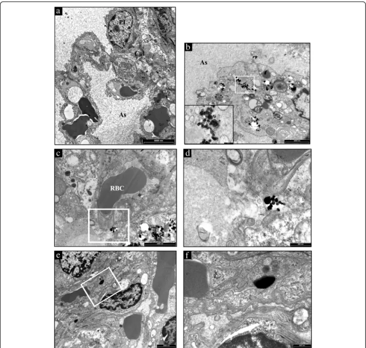
aggregates were found free in the alveolar space (Add-itional file 2: Figure S2), in the interstitial space sur-rounding alveolar capillary (Fig.6c and d) and inside the cells (Fig.6e and f ) as in alveolar cells displaying macro-phagic/autophagic features (Fig. 6b). Morphologic evi-dence provided by Transmission Electron Microscopy (TEM) suggested that in the heart NPs left the capillary lumen localizing in the interstitial perivascular space
c
d
e
g
f
Fig. 4 Cardiac refractoriness, excitability, anisotropic conduction velocities and arrhythmia induction. Electrophysiological parameters and the index of arrhythmia inducibility in CTRL (triangles) and TiO2-NPs treated (squares) animals. a Effective refractory period (ERP, ms). b Rheobase (μA). c Chronaxie (ms).
d Threshold intensity for a 1 ms duration impulse (μA). e ventricular conduction velocities along fiber (CVl, m/s). f ventricular conduction velocities across fiber (CVt, m/s). g ventricular conduction velocities ratio (CVl/CVt). Kruskal-Wallis (post hoc analyses: Dunn’s multiple comparison) was performed and statistical significance was set at p < 0.05.● vs CTRL; * vs corresponding 1° week; # vs corresponding 3° week; § vs corresponding 6° week. Data are represented as median and interquartile range (IQR)
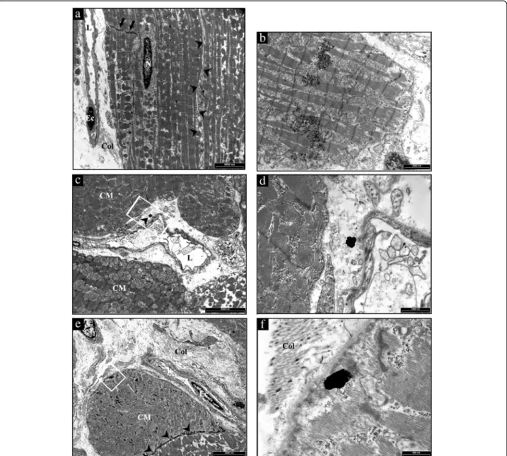
(Fig. 7b-d), penetrated the sarcolemma and reached the myoplasm by establishing intimate contact with myofi-brils and mitochondria (Fig.7e and f ).
Oxidative stress and inflammation
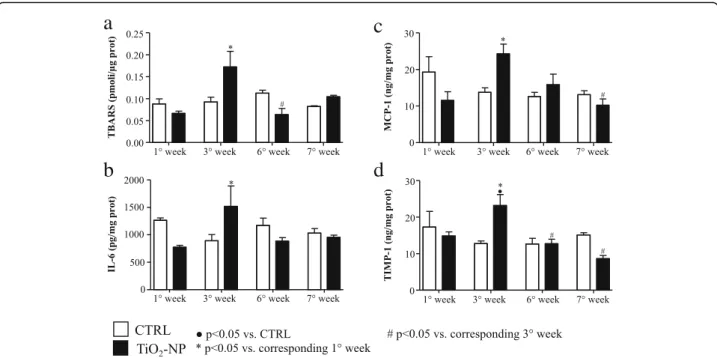
Lipid peroxidation analysis in cardiac and lung tissues dem-onstrated that at the 3rd week the Thiobarbituric acid re-active substances (TBARS) level significantly increased in TiO2-NP rats compared to the 1st week of treatment,
dis-playing the maximum value (Fig.8a, Additional file3: Fig-ure S3). Similarly, significant alterations were observed in cardiac tissue at the 3rd week for Interleukin-6 (IL-6) and Monocyte Chemoattractant Protein-1 (MCP-1) (Fig. 8b and c). To note, tissue inhibitor metalloproteinase 1 (TIMP-1) levels increased as well at the 3rd week (Fig.8d). The treatment induced approx. 50% increment of all cyto-kines at the 3rd week although the difference became statis-tically significant only for TIMP-1.
Gene expression analysis
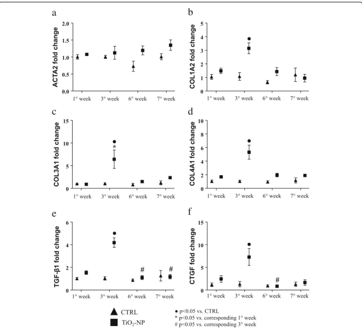
The expression of different genes related to myocardial fi-brosis was assessed in the rat hearts, revealing that at the 3rd week TiO2-NPs instillation the expression of different
collagen isoform genes (namely COL1A2, COL3A1, COL4A1) were definitively upregulated in treated animals
Table 2 Tibial length, cardiac anatomy and morphometric analysis CTRL (n = 9) TiO2-NP (n = 11) Tibial length (mm) 38.2 ± 0.95 35.7 ± 0.25 * BW (g) 359.3 ± 6.47 356.1 ± 5.70 LVW (mg) 1200 ± 25.50 1119.13 ± 27.91 * LVW/BW (mg/g) 3.39 ± 0.10 3.16 ± 0.11 LV mass (mm3) 1132.1 ± 24.06 1055.8 ± 26.33 * LV wall thickness (mm) 2.28 ± 0.09 2.26 ± 0.06 LV chamber diameter (mm) 7.43 ± 0.40 7.68 ± 0.25 LV chamber length (mm) 15.44 ± 0.50 15.11 ± 0.42 LV chamber volume (mm3) 361.3 ± 38.16 469.3 ± 31.29 * LV mass/chamber volume 3.11 ± 0.33 2.39 ± 0.16 Total fibrosis (%) 7.64 ± 1.11 15.44 ± 3.09 * Diffuse Fibrosis (%) 4.24 ± 0.82 11.69 ± 3.18 * Perivascular Fibrosis (%) 3.59 ± 0.49 4.69 ± 0.45
Values are means ± SEM Unpaired Student’s t-test
BW body weight measured before sacrifice, LV left ventricular, LVW left ventricular weight
*p < 0.05 vs CTRL
a
b
c
d
Fig. 5 Cardiac tissue fibrosis evaluation. Masson’s trichrome staining sections were analyzed by optical microscopy to evaluate perivascular and interstitial fibrosis in the LV myocardium (greenish). Representative images of heart sections from control (a and b) and TiO2-NP treated animals (c and d). a CTRL LV
myocardium; black rectangle area is shown at higher magnification in panel (b). c TiO2-NPs treated LV myocardium; black rectangle area is shown at
compared to CTRL (Fig. 9b-d). Interestingly, at the same time point, also TGF-β1 (transforming growth factor beta-1) and CTGF (connective tissue growth factor) showed sig-nificantly increased expression (Fig.9e and f).
Discussion
Inhaled NPs are on the current concern for their effect on the cardiovascular system after translocation from
the air-blood barrier [23]. Evidences are provided for in-haled NPs accumulation in the sites of vascular lesions [24], lymph nodes, liver, kidneys [25] and the heart [4].
Ambient (indoor) air pollution, due to the rapid urbanization worldwide, is known to be an important trigger of cardiovascular diseases especially for the people/workers that exposed continuously to NPs emit-ting sources with poor ventilation [26]. Following our Fig. 6 Ultrastructural features of the alveolar lung parenchyma from a representative untreated CTRL and TiO2-NPs treated SHR. a a type I pneumocyte
(PI) and three capillaries (*) are present in the alveolar septum separating air spaces (As) in CTRL lung. b several TiO2-NPs are present in the cytoplasm
of a large alveolar macrophage showing, in addition to vacuoles and microvescicles, onionskin-like multimembrane ultrastructures suggestive of autophagy. The area inscribed by white rectangle is shown at higher magnification in the inset. c an aggregate of electrondense TiO2-NPs is located
nearby an endothelial cell lining the lumen of a capillary recognizable by the red blood cell (RBC). The white rectangle inscribes an area shown at higher magnification in d to document that TiO2-NPs are not yet internalized. e internalization of TiO2-NPs in a capillary endothelial cell that is better appreciable
recent findings that showed inhaled TiO2 NPs in the
heart [19], recent literature have shown that inhaled ti-tanium dioxide NPs activate a plethora of cardiovascular effects, i.e., activation of complement cascade in the heart [27], induction of cytotoxicity in cardiomyoblast [28], microvascular and mitochondrial dysfunction in progeny of female SD rats exposed to TiO2 [29],
induction of myocarditis [30] and depression diastolic function in response to adrenergic stimuli [31].
In this study, we observed two concomitant responses in terms of both electrical remodeling (reversible) and mechanical and structural remodeling (irreversible) in SHRs subjected to 2 mg/kg TiO2 weekly repeated
tra-cheal instillation compared with saline treated animals. Fig. 7 TEM analysis of the heart from a representative untreated CTRL and TiO2-NP treated SHR. a low magnification image of a CTRL heart to illustrate
the sarcolemma (arrowheads) lining the surface and gap junctions (arrows) delimiting cardiomyocytes filled of mitochondria and myofibrils. On the left, collagen bundles (Col) are present in the interstitial space between an endothelial cell (Ec) lining a capillary lumen (L) and cardiomyocytes. N: cardiomyocyte nucleus. b aggregates of TiO2-NPs within the cardiomyocyte cytoplasm showing effaced myofibrils and swollen mitochondria. c the white
rectangle inscribes an area shown at higher magnification in (d) in which the arrowhead points to NP located in the interstitial space between the vessel wall inscribing a lumen (L) and cardiomyocytes (CM). e low magnification image of a treated SHR myocardium to illustrate widening of the interstitial space by abundant fibrotic deposition (Col) surrounding cardiomyocytes (CM) and an endothelial cell (Ec) lining a capillary. Arrowheads point to gap junctions delimiting two CMs. The white rectangle inscribes an area shown at higher magnification in f to document the internalization of TiO2-NP located
Electrical remodeling
The electrical remodeling is related to a direct interaction between TiO2-NPs and cardiac cell membrane. The
elec-trical remodelling and the arrhythmic events are related to a direct interaction between TiO2-NPs and cardiac cell
membrane via ionic leakage as previously described [19]. The almost complete recovery of electrical functional properties, can be explain by the recruitment and presence of myofibroblasts that can concur in entrapping TiO2-NPs
in the interstitial regions and thus reduced the amount of NPs passing the cardiomyocytes cell membrane (cf. Fig-ure7d). Because of that, the subsequent depolarization is now minimal but enough for enhancing excitability. The latter is the base of the biophysical phenomena of super-normal (increment) conduction velocity as described by us in fibrotic engineered in-vitro cardiac tissue [32] and in normotensive animals subjected to a single instillation of TiO2-NPs [19].
Noteworthy, in treated SHRs, only the number of hypo-kinetic arrhythmic events, including SP events (correlating with altered P wave duration) and AV blocks, as well as inducible arrhythmic events significantly increased at the 6th week and completely terminated at the end of the treatment. This scenario is presumably due to cardiac tis-sue damage associated with a direct acute effect of NPs, further confirmed here by ultrastructural analysis. Import-antly, the number of arrhythmic events in the treated animals (sinus pauses and AV blocks) was significantly
reduced approaching the values observed in CTRL (saline, positive control).
Conversely, neither the NPs treatment nor the sub-chronic exposure influenced QRS complex. Neverthe-less, RR variability increased in both groups at the end of treatment, suggesting a time-dependency effect of this parameters independently of the treatment.
Mechanical and structural irreversible remodeling
The irreversible mechanical and structural remodeling is related to the subchronic exposure to TiO2-NPs that
causes in SHRs a series of maladaptive cardiac responses mainly characterized by inflammation, upregulation of genes promoting collagen deposition and fibrosis, ultim-ately leading to alterations in hemodynamic performance.
Electrocardiographic measurements in freely moving animals demonstrated a time-dependency for P wave duration (6th and 7th weeks) and RR interval (7th weeks). We are unable to interpret this finding and fur-ther specific atrial electrophysiological and morpho-logical measurements are required.
Epicardial multiple lead recording demonstrated a de-crease in the CVl only after the first NPs instillation, possibly due to a massive depolarization of the resting potential created by the NPs entry in the cardiac tissue [20]. It is known that SHR cardiomyocytes have a reduc-tion in IK1 current responsible for diastolic membrane
potential depolarization [33], and thus modulating Fig. 8 Inflammation and toxicological markers in the heart tissue. Lipid peroxidation products and different inflammatory and toxicological markers evaluated in CTRL (white bars) and TiO2-NPs treated (black bars) animals. a TBARS evaluation in the heart tissue. b IL-6 evaluation in the heart tissue r. c
MCP-1 evaluation in the heart tissue. d TIMP-1 evaluation in the heart tissue. Two-way ANOVA (post hoc analyses: Bonferroni test) was performed and statistical significance was set at p < 0.05.● vs CTRL; * vs corresponding 1° week; # vs corresponding 3° week. Data are represented as mean ± SEM
cardiac excitability. Moreover, the treated animals de-noted a major time-dependency because at the 6th week, we observed a significant increment in CVl and CVt in the treated group vs. CTRL, at the first and third week. Such increment disappeared at the 7th week after the “washout” period. The increment at the 6th week can be explained if we take into account two factors: i) the CVs are calculated only for portions of the epicardial multiple lead arrays that denoted absence of conduction blocks and ii) a role exerted by the cardiac myofibroblasts that are populating the fibrotic tissue.
Repeated exposure to TiO2-NPs doubled the total
amount of cardiac fibrosis and triplicated the diffuse
fibrosis known to fasten the progression towards heart failure [34]. These findings are in agreement with the sig-nificant upregulation of genes involved in (myo)fibroblast recruitment and collagen protein expression: TGF-β1 and COL1A2 (threefold increment), COL3A1 and CTGF (six-fold increment), COL4A1 (five(six-fold increment). Those genes were upregulated exactly at the third week of instil-lation, when also the inflammatory response initiated in the heart. Indeed, the third week of exposure is the critical time-point, when we observed the increment in oxidative stress markers and cytokines in cardiac tissue in agree-ment with Gurguiera et al. [35] showing that heart oxida-tive stress significantly correlated with inhaled titanium
a
b
c
d
e
f
Fig. 9 Molecular analysis displaying different gene expression linked to fibrosis deposition. Graph of different gene expression in CRTL (triangles) and TiO2
-NPs treated (square) animals. a ACTA2 gene expression; b COL1A2 gene expression; c COL3A1 gene expression; d COL4A1 gene expression; e TGF-β1 gene expression; f CTGF gene expression. Kruskal-Wallis (post hoc analyses: Dunn’s multiple comparison) was performed and statistical significance was set at p < 0.05.● vs CTRL; * vs corresponding 1° week; # vs corresponding 3° week. Data are represented as median and IQR
diac characteristics, including chamber dilation and re-duction in LV mass, as well as worsening of cardiac hemodynamic performance.
Conclusions
The comorbidity of hypertension and inhaled nanoparti-cles has been largely studied in terms of hazard at epi-demiological and meta-analysis levels. We provide here, for the first time, a mechanistic evidence that this co-morbidity induces irreversible hemodynamic impairment associated with cardiac structural damage, while electro-physiological alterations are mostly reversible. Translat-ing this animal-based data to human hypertension is questionable due to species-related differences, different adaptation to the pathology and treatment, and the pro-cedure we adopted for tracheal administration. Never-theless, we provided here experimental evidence that the most produced engineered nanomaterial, considered to be inert, activates functional and structural remodelling of the hypertensive heart, potentially fastening the pro-gression towards cardiac failure. Specifically, our results indicate that prolonged TiO2-NP exposure may reach a
point of no-return (third week of treatment), in terms of myocardial tissue damage and reduces cardiac mechan-ical efficiency. This needs to be considered for the vul-nerable subjects/workers daily exposed to nanomaterials.
Limitations
Invasive hemodynamic and morphological measure-ments have been acquired only at the end of the treat-ment. On the other hand, it should be considered that the upregulation of genes promoting fibrotic damage and inflammation occurred at the third week and this finding prompted us to focus on the consequent later morphofunctional irreversible changes.
In this study, we limited our investigation to the ven-tricular cardiac fibrosis as one of the leading causes to-wards heart failure, in both NP-treated and untreated SHR. However, similar investigations need to be addressed for a plethora of hypertension-related diseases including atrial dysfunction, vascular endothelial dysfunction and pulmon-ary responses and clearance, known to be primpulmon-ary barriers for the inhalator route of nanoparticles. Lastly, we included
The bedding of the cages consisted of wood shavings with food and water available ad libitum. All surgery procedures and experiments, when specified, were per-formed under anesthesia which consisted of a mixture of ketamine chloride (40 mg/kg ip; Imalgene, Merial, Mi-lano, Italy) and medetomidine hydrochloride (0.15 mg/ kg ip; Domitor, Pfizer Italia s.r.l., Latina, Italy). The period of study was selected by taking into consideration the frailty of SHR animals and their suffering due to the hypertensive condition. This study was carried out in ac-cordance with the recommendations in the Guide for the Care and Use of Laboratory Animals of the National Institute of Health (Bethesda, MD, USA, revised 1996). The protocol was approved by the Veterinary Animal Care and Use Committee of the University of Parma (Permit: n. PMS 53/2009) and conforms to the National Ethical Guidelines of the Italian Ministry of Health. All effort was made to minimize suffering. We noticed 15% of spontaneous animal loss during the repeated anesthesia procedures from the 3rd week onwards be-cause of the SHR hyperreactivity [38]. We did not ob-serve animal loss for the surgical procedure.
Particle suspension
TiO2-NPs (Titanium (IV) oxide, code 677469,
Sigma-Aldrich, Milan, Italy) were previously characterized by us [19, 39]. Nanoparticles were suspended in physio-logical saline solution (10 mg/mL, stock solution). Im-mediately before the experiments, the suspension was vortexed and immersed in a sonication bath (Branson Ultrasonics, Danbury, CT, USA) for 5 min at 37 °C in order to minimize particle aggregation.
Intra-tracheal instillation
Although inhalation studies are considered to be the gold standard, data from intra-tracheal instillation studies can be used for hazard assessment [16]. The instillation process was extensively described in our previous work [19]. Briefly, after anesthesia, a 16-gauge catheter was gently inserted into the trachea of rats in order to deliver 20μL/100 g BW of saline solution or saline solution plus 2 mg/kg TiO2by
means of a laboratory bench P200 pipette (Gilson, Dun-stable, UK). We adopted repeated weekly doses of 2 mg/kg by taking into consideration the NIOSH recommendation
(https://www.cdc.gov/niosh/docs/2011-160/pdfs/2011-160. pdf) of 0.3 mg/m3of TiO2NPs exposure as time-weighted
average concentration for 10 h per day during 40-h work week and the species-specificity [19]. With such a concen-tration we are further beneath the maximal exposure limit for both human [40] and rats [41–43]. Furthermore, an empty sterile syringe (1 mL) was used to gradually inflate the lung with air twice prior to the connection of the can-nula port to a ventilator (Rodent ventilator UB 7025, Ugo Basile, Comerio, Italy) set to 85 cycle/min. Finally, admin-istration of 0.15 mg/kg atipamezole hydrochloride (Antise-dan, Pfizer, Milan, Italy) has been performed to wake up the animal. This protocol was repeated consecutively once a week for 6 weeks. A six-week period was selected be-cause the SHR at this stage of age had an average systolic blood pressure of 200 mmHg, and we choose 7 weeks as longitudinal study in order to avoid physical suffering due to high systolic blood pressure and aged related cardiac implication [44].
Outline of the experimental protocols
Telemetry-ECG, hemodynamic, cardiac anatomy and morphometry studies
The experimental design and measurements performed in the two experimental groups are summarized in Fig.1. Six-teen rats were chronically instrumented with a telemetry-ECG system to enable the evaluation of arrhythmia vulnerability and the duration of the different ECG waves and intervals, in conscious freely moving animals. One week later, the animals started the experimental instillation protocol which consisted in a weekly intra-tracheal instilla-tion of saline soluinstilla-tion (CTRL group,n = 8) or saline solu-tion added with TiO2at a final concentration of 2 mg/kg
(TiO2-NP group, n = 8), as described above. Four hours
later, a 30-min telemetry-ECG recording was performed while the experimental animal was left alone and undis-turbed in its cage. ECG recordings were also performed at the 3rd week, 6th week, and at the end of the experimental protocol (7th week) 1 week after the last intra-tracheal in-stillation (Fig.1). Afterward, hemodynamic data were inva-sively collected in all animals and in additional 3 TiO2-NP
and 1 CTRL rats for a total of 11 TiO2-NP and 9 CTRL.
Fi-nally, at sacrifice, the hearts were perfusion fixed for ana-tomical and structural studies.
Epicardial multiple-lead recording, PIXE, and molecular studies
In 38 rats belonging to CTRL (n = 16) and TiO2-NP
(n = 22) groups, electrophysiological investigation was performed by epicardial multiple-lead recording at the same time points specified above for telemetry-ECG re-cordings. At death, for each time point, fresh prepara-tions of the excised hearts and lungs were used for molecular analyses (real time PCR: n = 3 CTRL, n = 5
TiO2-NP), immunological assay (n = 3 CTRL and n = 4
TiO2-NP) while for TiO2-NP quantification (PIXE, see
specific section below for details) the analysis has been performed at the 1st week (n = 3 TiO2-NP) and at the
end of the treatment, 7th week (n = 2 TiO2-NP).
Transmission electron microscopy study
In 3 CTRL and 3 TiO2-NP rats, heart and lung samples
were analyzed by TEM in order to document the presence of NPs within these tissues at the end of the experimental protocol (7th week), 1 week after the 6th intra-tracheal in-stillation, given that we have already documented the presence of TiO2-NPs in both tissues 4 hours after
tra-cheal instillation [15].
Animal instrumentation for telemetry-ECG recording
After anesthesia, animals were chronically instrumented with a miniaturized transmitter for telemetry-ECG record-ing (model TA11CTA-F40, Data Sciences International, St. Paul, MN, USA). The details of the surgical procedure have been already published by us [45]. Briefly, a ventral celiotomy was performed and the body of the transmitter (25x15x8mm) was placed in the abdominal cavity. One re-cording lead was fixed to the dorsal aspect of the xiphoid process, close to the apex of the heart. The other lead was subcutaneously tunneled on the thorax toward the upper insertion of the sternohyoid muscle. Here, the wire was formed into a U shape, pushed under the muscle and then along the trachea into the anterior mediastinum, to get the tip of the recording lead close to the right atrium. Fi-nally, the muscle and skin layers were separately sutured. After surgery, all animals were given Antisedan 0.15 mg/ kg im (Pfizer), flunixin 5 mg/kg im (Finadyne, Schering-Plough s.p.a, Milan, Italy), gentamicin sulphate 10 mg/kg im (Aagent, Fatro, Milan, Italy), and kept warm with infra-red lamp radiation. Antibiotic therapy was continued in the following 3 days while the animals were individually housed. Rats were allowed to recover for 7 days before the onset of the instillation protocol and ECG acquisition.
Telemetry-ECG data acquisition and processing
ECG signals were collected by a receiver (model CTR85-SA, Data Sciences International, St. Paul, MN, USA) placed under the housing cage, monitored on an oscillo-scope and simultaneously routed to a personal computer, via an analog-to-digital conversion board (12 bits, 1 kHz sampling rate), for a permanent storage. The data were analyzed by using a software package developed in our laboratory, in order to measure:
i. RR interval duration, the time interval between two consecutive R wave peaks;
ii. P wave duration (atrial depolarization), between the onset and offset of P wave;
variability included: the SDRR and r-MSSD, which quantify the state of the balance between sympa-thetic and parasympasympa-thetic activities on the heart and the influence on heart rate of the parasympa-thetic branch respectively [46];
viii.Number and type of arrhythmic events. In detail, we recorded both, supraventricular
(Supraventricular Extrasystole and Sinus
Arrhythmia, SVA; SP) and ventricular (AV block; Ventricular Extrasystole, VE) events.
For the evaluation of ECG wave and interval duration, 30 measures for each animal were performed.
Hemodynamic study
After anesthesia, the right carotid was cannulated with a microtip pressure transducer catheter (Millar SPC-320; Millar Instruments, Houston, TX, U.S.A.) connected to a recording system (Power Laboratory ML 845/4 channels; 2Biological Instruments, Besozzo, Italy), and systolic and diastolic arterial blood pressures were determined. The catheter was then advanced into the left ventricle to meas-ure (software package CHART B4.2): LVSP; LVEDP; +dP/ dtmaxand -dP/dtmax, taken as indexes of ventricular
mech-anical efficiency; IVCT.
Tibial length, cardiac anatomy and morphometric analysis
At the end of the hemodynamic procedure, the heart was arrested in diastole by injecting 5 mL of cadmium chloride solution (100 mmol/L, iv), and excised. The right ventricle and the left ventricle inclusive of the septum were separ-ately weighed and fixed in 10% buffered formalin solution. In all animals, the length of the tibia was also determined. The volume of the LV myocardium was computed by div-iding LV weight by the specific gravity of the tissue (1.06 g/mL). LV chamber length was measured from the apex to the aortic valve. The equatorial transverse section of the left ventricle (1 mm thick), cut perpendicularly to the major axis, allowed the morphometric computation of the LV equatorial diameter and wall thickness (Image Pro-plus, Media Cybernetics, Bethesda, MD, USA, version 7.0). The LV chamber volume was then calculated according to the Dodge equation, which equalizes the ventricular cavity to an ellipsoid [47]. The section was finally embedded in
aid of a grid defining a tissue area of 0.160mm2and con-taining 42 sampling points, each covering an area of 0.0038mm2. To define the volume fraction of fibrosis, the number of points overlying myocardial fibrosis was counted and expressed as percentage of the total number of points explored.
Epicardial multiple-lead recording
In rats under anesthesia and artificial respiration, the heart was exposed through a longitudinal sternotomy and sus-pended in a pericardial cradle. Body temperature was maintained with infrared lamp radiation. An 8 × 8 row electrode matrix with 1-mm inter-nodal resolution was fabricated from surgical cotton gauze. The electrode array was positioned in order to cover part of the anterior sur-face of the right and left ventricles [49], and the following measurements were carried out: refractoriness (by means of S1-S2 protocol) and inducible ectopic activity (n = 8 for each animal), ventricular excitability (Rheobase and Chronaxie,n = 5 for each animal and Threshold intensity for a 1 ms duration impulse, n = 13) and conduction vel-ocities (CVl and CVt, n = 10 for each animal, as well as their ratio), as previously described [19]. Moreover, arrhythmia inducibility was evaluated as percentage of electrodes showing at least one ventricular extrasystole during effective refractory period (ERP) protocol.
Titanium dioxide quantification by PIXE
The trachea, lungs, and heart were prepared into pellets following the previously described procedure [50]. Briefly, biological samples were dried in a 37 °C oven for 24 h, then they were freeze dried and chromium nitride (Cr2N) powder (Goodfellow SARL, Lille, France) was
added at 8.5 ± 1.5 wt.%. Biological samples and Cr2N
powder were ball milled to produce a homogeneous powder mixture which was hard pressed into a 2 cm diameter, 1-2 mm thick pellet. We added Cr2N in order
to avoid charge accumulation in the sample during ion beam irradiation and have an internal beam monitor for quantitative measurements. In addition, Cr2N does not
interfere with the measurement of other elements, be-cause it is neither present in the biological matter nor as an impurity in TiO2.
PIXE measurements were performed with the University of Namur ALTAÏS accelerator (Accélérateur Linéaire Tan-detron pour l’Analyse et l’Implantation des Solides). PIXE is an ion beam technique, whose physical principles can be found extensively explained elsewhere [51], providing pre-cise quantification of chemical elements in toxicological assessments [52]. PIXE and Rutherford Back-scattering (RBS) measurements were done simultaneously with an in-cident ion beam of 2.5 MeV protons with an intensity of 1–1.5 nA. With respect to the beam direction, the sample was tilted at 45°, a Canberra LEGe (Low Energy Germa-nium) detector was located at 90° for PIXE measurements (variable solid angle up to 35 msr), and a Canberra PIPS (Passivated Implanted Planar Silicon) detector was posi-tioned at 145° for RBS measurements (solid angle of 23.8 msr). An aluminum collimator (1 mm aperture, 0.2 mm thick) was used in front of the PIXE detector to equalize the ratio of low-energy and high-energy X-rays. The samples were mounted on a rotating device providing a total analyzed area of 140.5 mm2. RBS was used to derive the number of incident ions, and to quantify carbon, oxy-gen and nitrooxy-gen contents for matrix (lung tissue) correc-tion of X-ray absorpcorrec-tion effects. A BCR-126 lead glass standard was used to calibrate both detectors. The PIXE setup allowed for a detection limit of Ti in biological matrices of 10 wt.ppm [52].
Data were analyzed with the GUPIXWIN software [53]. The concentration of TiO2-NPs in tissues was calculated
from the Ti X-ray emission, then converted to TiO2
assum-ing a 1:2 ratio of Ti:O.
Oxidative stress and inflammation
Tissue samples were washed with PBS and included in cryovials prior to freezing at− 80 °C. Frozen tissue samples (lungs, and heart) were homogenized and sonicated in PBS supplemented with protease inhibitor cocktail (Sigma-Al-drich, Milan, Italy). Insoluble debris was pelleted and super-natants were analyzed.
Lipid peroxidation was evaluated by TBARS method, using Malondialdehyde as a standard for the calibration curve [39]. The fluorescence intensity was measured by Cary Eclipse fluorescence spectrophotometer (Varian/Agi-lent, Santa Clara, CA, USA) (excitation 515 nm, emission 545 nm). Four independent experiments were carried out and TBARS values were normalized to protein concentra-tions. Commercially available enzyme-linked immunosorb-ent assays (ELISA, Cloud-Clone Corp., Katy, TX, USA) were used to evaluate biomarkers of tissue inflammation (IL-6 and MCP-1), and tissue remodeling (TIMP-1). All data were related to protein concentration.
Real-time PCR analysis
Frozen heart samples were kept in dry ice and cut into small pieces of about 50–70 mg using a scalpel. TRIzol
reagent (1 ml) (Invitrogen, Thermos Fisher Scientific) was then added to each sample, and the tissue was homoge-nized using the TeSeE PRECESS 24 (BioRad) performing three agitation cycles at 5100 rpm for 10s at 4 °C. Homog-enized tissues were centrifuged at 12000×g for 1 min, in order to remove particulate debris. The RNA was subse-quently extracted from the supernatants using the kit Direct-zol RNA MiniPrep (Zymo research), following manufacturer’s instructions.
RNA concentration and quality was assessed by Nano-Drop 1000 spectrophotometer (Thermo fisher Scientific): only samples with a value of A260/280 > 1.8 and A260/ 230 > 1.8 were used for subsequent gene expression ana-lysis. RNA integrity was evaluated with Experion electro-phoresis system using the Experion™RNA StdSens Analysis Kit (Bio-Rad).
Total RNA (1μg) was reversely-transcribed using the SuperScript VILO cDNA synthesis Kit (Thermo Fisher Scientific) according to manufacturer’s instructions.
The amplification of the cDNA was then performed using All-in-One SYBR® Green qPCR Mix (GeneCo-poeia) on a CFx96 Real-Time System C1000 Thermal Cycler (BioRad). The list of primers used for cDNA amplification is reported in Additional file4: Table S1.
Raw expression intensities of genes of interest were nor-malized to the Ct value of Glyceraldehyde 3-phosphate dehydrogenase (GAPDH). Relative quantitation was per-formed using the ΔΔCt method in comparison with the mean expression of the same genes in CTRL animals. Fold changes in gene expression were calculated as 2(ΔΔCt)[54].
Titanium dioxide detection in heart and lung tissues by TEM
Heart and lung samples from 3 CTRL and 3 TiO2-NP
groups at the 7th week of the experimental protocol, 1 week after the last intra-tracheal instillation, were ana-lyzed by TEM in order to document the presence of NPs within these tissues. Samples were fixed in Kar-novsky solution (4% formaldehyde, 5% glutaraldehyde) for 3 h at room temperature. After washing several times with 0.1 M phosphate buffer, pH 7.2, the tissues were post-fixed in 1% osmium tetroxide (OsO4) for 90 min at
room temperature and dehydrated by increasing concen-tration of alcohol. Then, samples were washed with propylene oxide and embedded in epoxy resin. Sections of 0.5μm thickness were stained with methylene blue and safranin to morphologically select the field of inter-est. Subsequently, ultrathin sections of 60-80 nm thick-ness were collected on a 300-mesh copper grid and, after staining with uranyl acetate and lead citrate, were qualitatively examined under a transmission electron microscope (EM 208S model, Philips, Amsterdam, The Netherlands).
Kruskal-Wallis (post hoc analyses: Dunn’s multiple com-parison). Prism 5.0 software (GraphPad Software) was used to assess the normality of the data and for statistical calculation. The details on the specific test used for each experiment are reported in the figure legends. Statistical significance was set atp < 0.05.
Additional files
Additional file 1: Figure S1. Spontaneous arrhythmic events. Different examples of arrhythmic events. Both, supraventricular (Supraventricular Extrasystole and Sinus Arrhythmia, SVA; Sinus Pause, SP) and ventricular (Atrio-ventricular block, AV block; Ventricular Extrasystole, VE) events were recorded. (PDF 324 kb)
Additional file 2: Figure S2. TEM analysis of the alveolar lung parenchyma from a SHR rat seven weeks after intratracheal instillation of TiO2-NPs. Individual or microaggregates of small electrondense NPs are present in the air space (As) as scattered within the alveolar septum in which a type II pneumocyte (PII) is recognized. NPs are also apparent in endothelial cells lining a capillary (*) and the lumen of a larger venule containing red blood cells (RBC) polymorphonuclear (PMN) neutrophils, lymphocytes (Lym) and platelets (arrow). Magnification 1800X. Scale Bar: 10μm. (PDF 2011 kb)
Additional file 3: Figure S3. TBARS measurement in lungs evaluated in CTRL (white bars) and TiO2-NPs treated (black bars) animals. Two-way ANOVA (post hoc analyses: Bonferroni test) was performed and statistical significance was set at p<0.05.● vs CTRL; * vs corresponding 1° week; # vs corresponding 3° week. Data are represented as mean ± SEM. (PDF 60 kb)
Additional file 4: Table S1. List of primer sequences 5′-3′ used for real-time PCR analysis. (PDF 118 kb)
Abbreviations
+dP/dtmax:Maximum rate of ventricular pressure rise; AV block: Atrio-ventricular
block; BW: Body weight; Cr2N: Chromium nitride; CTGF: Connective tissue growth
factor; CTRL: Control; CVl: Longitudinal conduction velocity; CVt: Transversal conduction velocity; -dP/dtmax: Maximum rate of ventricular pressure reduction;
ERP: Effective refractory period; GAPDH: Glyceraldehyde 3-phosphate dehydrogenase; IL-6: Interleukin-6; IVCT: Isovolumic contraction time; LEGe: Low energy germanium; LV: Left ventricular; LVEDP: Left ventricular end diastolic pressure; LVSP: Left ventricular systolic pressure; LVW: Left ventricular weight; MCP-1: Monocyte Chemoattractant Protein-1; NIOSH: National institute for occupational health; NPs: Nanoparticles; OsO4: Osmium tetroxide; PIPS: Passivated
implant planar silicon; PIXE: Particle-Induced X-ray emission; PM: Particulate matter; RBS: Rutherford back scattering; r-MSSD: Root mean square of successive RR interval square differences; SDRR: Standard deviation of average RR interval; SHRS: Pontaneously hypertensive rat; SP: Sinus pause; SVA: Supraventricular Extrasystole and Sinus Arrhythmia; TBARS: Thiobarbituric acid reactive substances; TEM: Transmission Electron Microscopy; TGF-β1: Transforming growth factor beta-1; TIMP-1: Tissue inhibitor metalloproteinase beta-1; TiO2: Titanium dioxide;
VE: Ventricular Extrasystole; WKY: Wistar Kyoto
LG, AP, VM, AR performed real-time PCR analysis. SR, MS, MGaletti, MGoldoni take care of statistical evaluation and graphical representation. CF, AF, FQ de-signed and performed TEM experiments and ultrastructural analysis; MMira-goli, DS, EM, AM conceptual and technical advises; MMiraMMira-goli, SR and MS conceptual design, experimental design, data analysis, preparation of the manuscript. All authors critically read and approved the final manuscript. Funding
Italian Ministry of Health young research grant GR-2009-1530528 to Mi.Mi, A.M. FP7- Quality Nano Research and Infrastructure Project FUNDP-TAF-225, FP7 to Mi.Mi., L.G., and to O.L.G.
Italian Ministry of Health, Ricerca Finalizzata grant NanOI-LuCaS RF-2009-1472550 to A.M.
Department of Innovation, Research and Universities of the Autonomous Province of Bolzano-South Tyrol (Italy) to L.G., A.P., V.M, A.R.
Fondo Guido Erluison per la Ricerca Clinica, Dipartimento di Medicina e Chirurgia postdoctorate salary to S.R.
CSEIA 2018 Open-Up - Outgoing Publications grant for scientific publication to S.R. Ethics approval and consent to participate
The protocol was approved by the Veterinary Animal Care and Use Committee of the University of Parma (Permit: n. PMS 53/2009) and conforms to the National Ethical Guidelines of the Italian Ministry of Health.
Consent for publication Not Applicable. Competing interests
The authors declare that they have no competing interests. Author details
1Department of Medicine and Surgery, University of Parma, Via Gramsci, n°
14, 43126 Parma, Italy.2CERT, Center of Excellence for Toxicological Research, INAIL, ex-ISPESL, University of Parma, Parma, Italy.3Department of Chemistry,
Life Sciences and Environmental Sustainability, University of Parma, Parma, Italy.4Institute for Biomedicine, Eurac Research, Bolzano, Italy.5Affiliated
Institute of the University of Lübeck, Lübeck, Germany.6Namur Nanosafety Centre (NNC), Namur Research Institute for Life Sciences (NARILIS), Research Centre for the Physics of Matter and Radiation (PMR), University of Namur, B-5000 Namur, Belgium.7Azienda Ospedaliera-Universitaria, Unità di Medicina
del lavoro e Tossicologia industriale, Parma, Italy.8Humanitas Clinical and Research Center, Rozzano, Milan, Italy.9Present address: Cátedra de
Cardiología y Medicina Vascular, Escuela de Medicina y Ciencias de la Salud Tecnologico de Monterrey, Monterrey, Mexico.10Present address:
Department of Biomedical and Neuromotor Sciences, University of Bologna, 40126 Bologna, Italy.
Received: 3 December 2018 Accepted: 6 June 2019
References
1. Harries AD, Kumar AM, Satyanarayana S, Lin Y, Takarinda KC, Tweya H, et al. Communicable and non-communicable diseases: connections, synergies and benefits of integrating care. Public Health Action. 2015;5(3):156–7.https://doi. org/10.5588/pha.15.0030 http://www.ncbi.nlm.nih.gov/pubmed/26393110. 2. Sager TM, Kommineni C, Castranova V. Pulmonary response to intratracheal
area. Part Fibre Toxicol. 2008;5:17.https://doi.org/10.1186/1743-8977-5-17 http://www.ncbi.nlm.nih.gov/pubmed/19046442.
3. Grassian VH, O'Shaughnessy PT, Adamcakova-Dodd A, Pettibone JM, Thorne PS. Inhalation exposure study of titanium dioxide nanoparticles with a primary particle size of 2 to 5 nm. Environ Health Perspect. 2007;115(3):397– 402.https://doi.org/10.1289/ehp.9469 http://www.ncbi.nlm.nih.gov/ pubmed/17431489.
4. Miragoli M, Ceriotti P, Iafisco M, Vacchiano M, Salvarani N, Alogna A, et al. Inhalation of peptide-loaded nanoparticles improves heart failure. Sci Transl Med. 2018;10(424).https://doi.org/10.1126/scitranslmed.aan6205 https:// www.ncbi.nlm.nih.gov/pubmed/29343624.
5. Paulin L, Hansel N. Particulate air pollution and impaired lung function. F1000Res. 2016;5.https://doi.org/10.12688/f1000research.7108.1 http://www. ncbi.nlm.nih.gov/pubmed/26962445.
6. Pietroiusti A, Bergamaschi E, Campagna M, Campagnolo L, De Palma G, Iavicoli S, et al. The unrecognized occupational relevance of the interaction between engineered nanomaterials and the gastro-intestinal tract: a consensus paper from a multidisciplinary working group. Part Fibre Toxicol. 2017;14(1):47.https://doi.org/10.1186/s12989-017-0226-0 http://www.ncbi. nlm.nih.gov/pubmed/29178961.
7. Kodavanti UP, Schladweiler MC, Ledbetter AD, Watkinson WP, Campen MJ, Winsett DW, et al. The spontaneously hypertensive rat as a model of human cardiovascular disease: evidence of exacerbated cardiopulmonary injury and oxidative stress from inhaled emission particulate matter. Toxicol Appl Pharmacol. 2000;164(3):250–63.https://doi.org/10.1006/taap.2000.8899 http://www.ncbi.nlm.nih.gov/pubmed/10799335.
8. Cassee FR, Heroux ME, Gerlofs-Nijland ME, Kelly FJ. Particulate matter beyond mass: recent health evidence on the role of fractions, chemical constituents and sources of emission. Inhal Toxicol. 2013;25(14):802–12. https://doi.org/10.3109/08958378.2013.850127 http://www.ncbi.nlm.nih.gov/ pubmed/24304307.
9. Kodavanti UP, Moyer CF, Ledbetter AD, Schladweiler MC, Costa DL, Hauser R, et al. Inhaled environmental combustion particles cause myocardial injury in the Wistar Kyoto rat. Toxicol Sci. 2003;71(2):237–45http://www.ncbi.nlm. nih.gov/pubmed/12563109.
10. Kang GS, Gillespie PA, Gunnison A, Moreira AL, Tchou-Wong KM, Chen LC. Long-term inhalation exposure to nickel nanoparticles exacerbated atherosclerosis in a susceptible mouse model. Environ Health Perspect. 2011;119(2):176–81.https://doi.org/10.1289/ehp.1002508 http://www.ncbi. nlm.nih.gov/pubmed/20864429.
11. Sutunkova MP, Solovyeva SN, Katsnelson BA, Gurvich VB, Privalova LI, Minigalieva IA, et al. A paradoxical response of the rat organism to long-term inhalation of silica-containing submicron (predominantly nanoscale) particles of a collected industrial aerosol at realistic exposure levels. Toxicology. 2017;384:59–68.https://doi.org/10.1016/j.tox.2017.04.010 http:// www.ncbi.nlm.nih.gov/pubmed/28450064.
12. Sha B, Gao W, Wang S, Li W, Liang X, Xu F, et al. Nano-titanium dioxide induced cardiac injury in rat under oxidative stress. Food Chem Toxicol. 2013;58:280–8.https://doi.org/10.1016/j.fct.2013.04.050 http://www.ncbi.nlm. nih.gov/pubmed/23665316.
13. Chockalingam A, Campbell NR, Fodor JG. Worldwide epidemic of hypertension. Can J Cardiol. 2006;22(7):553–5http://www.ncbi.nlm.nih.gov/ pubmed/16755308.
14. Mills KT, Bundy JD, Kelly TN, Reed JE, Kearney PM, Reynolds K, et al. Global disparities of hypertension prevalence and control: a systematic analysis of population-based studies from 90 countries. Circulation. 2016;134(6):441–50. https://doi.org/10.1161/CIRCULATIONAHA.115.018912 http://www.ncbi.nlm. nih.gov/pubmed/27502908.
15. National Institute for Occupational Safety and Health. NIOSH current intelligence bulletin 63: occupational exposure to titanium dioxide. 2011. https://www.cdc.gov/niosh/docs/2011-160/pdfs/2011-160.pdf. 16. Shi H, Magaye R, Castranova V, Zhao J. Titanium dioxide nanoparticles: a
review of current toxicological data. Part Fibre Toxicol. 2013;10:15.https://doi. org/10.1186/1743-8977-10-15 http://www.ncbi.nlm.nih.gov/pubmed/23587290. 17. Zhen S, Qian Q, Jia G, Zhang J, Chen C, Wei Y. A panel study for
cardiopulmonary effects produced by occupational exposure to inhalable titanium dioxide. J Occup Environ Med. 2012;54(11):1389–94.https://doi.org/ 10.1097/JOM.0b013e3182611a49 http://www.ncbi.nlm.nih.gov/pubmed/ 23059552.
18. Warheit DB, Webb TR, Reed KL, Frerichs S, Sayes CM. Pulmonary toxicity study in rats with three forms of ultrafine-TiO2 particles: differential
responses related to surface properties. Toxicology. 2007;230(1):90–104. https://doi.org/10.1016/j.tox.2006.11.002 http://www.ncbi.nlm.nih.gov/ pubmed/17196727.
19. Savi M, Rossi S, Bocchi L, Gennaccaro L, Cacciani F, Perotti A, et al. Titanium dioxide nanoparticles promote arrhythmias via a direct interaction with rat cardiac tissue. Part Fibre Toxicol. 2014;11:63. https://doi.org/10.1186/s12989-014-0063-3 http://www.ncbi.nlm.nih.gov/pubmed/25487314.
20. Miragoli M, Novak P, Ruenraroengsak P, Shevchuk AI, Korchev YE, Lab MJ, et al. Functional interaction between charged nanoparticles and cardiac tissue: a new paradigm for cardiac arrhythmia? Nanomedicine (Lond). 2013; 8(5):725–37.https://doi.org/10.2217/nnm.12.125 http://www.ncbi.nlm.nih. gov/pubmed/23140503.
21. Cao Q, Zhang S, Dong C, Song W. Pulmonary responses to fine particles: differences between the spontaneously hypertensive rats and wistar Kyoto rats. Toxicol Lett. 2007;171(3):126–37.https://doi.org/10.1016/j.toxlet.2007.05. 007 http://www.ncbi.nlm.nih.gov/pubmed/17606336.
22. Kodavanti UP, Mebane R, Ledbetter A, Krantz T, McGee J, Jackson MC, et al. Variable pulmonary responses from exposure to concentrated ambient air particles in a rat model of bronchitis. Toxicol Sci. 2000;54(2):441–51http:// www.ncbi.nlm.nih.gov/pubmed/10774827.
23. Bazyar J, Pourvakhshoori N, Khankeh H, Farrokhi M, Delshad V, Rajabi E. A comprehensive evaluation of the association between ambient air pollution and adverse health outcomes of major organ systems: a systematic review with a worldwide approach. Environ Sci Pollut Res Int. 2019;26(13):12648–61. https://doi.org/10.1007/s11356-019-04874-z https://www.ncbi.nlm.nih.gov/ pubmed/30903465.
24. Miller MR, Raftis JB, Langrish JP, McLean SG, Samutrtai P, Connell SP, et al. Inhaled nanoparticles accumulate at sites of vascular disease. ACS Nano. 2017;11(5):4542–52.https://doi.org/10.1021/acsnano.6b08551 https://www. ncbi.nlm.nih.gov/pubmed/28443337.
25. Takenaka S, Karg E, Roth C, Schulz H, Ziesenis A, Heinzmann U, et al. Pulmonary and systemic distribution of inhaled ultrafine silver particles in rats. Environ Health Perspect. 2001;109(Suppl 4):547–51.https://doi.org/10. 1289/ehp.01109s4547 https://www.ncbi.nlm.nih.gov/pubmed/11544161. 26. Farmer SA, Nelin TD, Falvo MJ, Wold LE. Ambient and household air
pollution: complex triggers of disease. Am J Physiol Heart Circ Physiol. 2014; 307(4):H467–76.https://doi.org/10.1152/ajpheart.00235.2014 https://www. ncbi.nlm.nih.gov/pubmed/24929855.
27. Husain M, Wu D, Saber AT, Decan N, Jacobsen NR, Williams A, et al. Intratracheally instilled titanium dioxide nanoparticles translocate to heart and liver and activate complement cascade in the heart of C57BL/6 mice. Nanotoxicology. 2015;9(8):1013–22.https://doi.org/10.3109/17435390.2014. 996192 https://www.ncbi.nlm.nih.gov/pubmed/25993494.
28. Huerta-Garcia E, Zepeda-Quiroz I, Sanchez-Barrera H, Colin-Val Z, Alfaro-Moreno E, Ramos-Godinez MDP, et al. Internalization of titanium dioxide nanoparticles is cytotoxic for H9c2 rat Cardiomyoblasts. Molecules. 2018; 23(8).https://doi.org/10.3390/molecules23081955 https://www.ncbi.nlm.nih. gov/pubmed/30082584.
29. Stapleton PA, Nichols CE, Yi J, McBride CR, Minarchick VC, Shepherd DL, et al. Microvascular and mitochondrial dysfunction in the female F1 generation after gestational TiO2 nanoparticle exposure. Nanotoxicology. 2015;9(8):941–51.https://doi.org/10.3109/17435390.2014.984251 https:// www.ncbi.nlm.nih.gov/pubmed/25475392.
30. Hong F, Wang L, Yu X, Zhou Y, Hong J, Sheng L. Toxicological effect of TiO2 nanoparticle-induced myocarditis in mice. Nanoscale Res Lett. 2015;10(1): 1029.https://doi.org/10.1186/s11671-015-1029-6 https://www.ncbi.nlm.nih. gov/pubmed/26269254.
31. Kan H, Wu Z, Lin YC, Chen TH, Cumpston JL, Kashon ML, et al. The role of nodose ganglia in the regulation of cardiovascular function following pulmonary exposure to ultrafine titanium dioxide. Nanotoxicology. 2014; 8(4):447–54.https://doi.org/10.3109/17435390.2013.796536 https://www. ncbi.nlm.nih.gov/pubmed/23593933.
32. Miragoli M, Gaudesius G, Rohr S. Electrotonic modulation of cardiac impulse conduction by myofibroblasts. Circ Res. 2006;98(6):801–10. https://doi.org/10.1161/01.RES.0000214537.44195.a3 http://www.ncbi.nlm. nih.gov/pubmed/16484613.
33. Sridhar A, Dech SJ, Lacombe VA, Elton TS, McCune SA, Altschuld RA, et al. Abnormal diastolic currents in ventricular myocytes from spontaneous hypertensive heart failure rats. Am J Physiol Heart Circ Physiol. 2006;291(5): H2192–8.https://doi.org/10.1152/ajpheart.01146.2005 http://www.ncbi.nlm. nih.gov/pubmed/16766638.
doi.org/10.1161/HYPERTENSIONAHA.117.09045 http://www.ncbi.nlm.nih.gov/ pubmed/28373589.
37. Fan D, Takawale A, Lee J, Kassiri Z. Cardiac fibroblasts, fibrosis and extracellular matrix remodeling in heart disease. Fibrogenesis Tissue Repair. 2012;5(1):15.https://doi.org/10.1186/1755-1536-5-15 http://www.ncbi.nlm. nih.gov/pubmed/22943504.
38. Schaefer CF, Brackett DJ, Gunn CG, Wilson MF. Behavioral hyperreactivity in the spontaneously hypertensive rat compared to its normotensive progenitor. Pavlov J Biol Sci. 1978;13(4):211–6https://www.ncbi.nlm.nih.gov/ pubmed/748844.
39. Alinovi R, Goldoni M, Pinelli S, Campanini M, Aliatis I, Bersani D, et al. Oxidative and pro-inflammatory effects of cobalt and titanium oxide nanoparticles on aortic and venous endothelial cells. Toxicol In Vitro. 2015; 29(3):426–37.https://doi.org/10.1016/j.tiv.2014.12.007 http://www.ncbi.nlm. nih.gov/pubmed/25526690.
40. Kreyling WG, Holzwarth U, Haberl N, Kozempel J, Wenk A, Hirn S, et al. Quantitative biokinetics of titanium dioxide nanoparticles after intratracheal instillation in rats: part 3. Nanotoxicology. 2017;11(4):454–64.https://doi.org/ 10.1080/17435390.2017.1306894 https://www.ncbi.nlm.nih.gov/pubmed/ 28290735.
41. Suker DK, Jasim FA. Liver histopathological alteration after repeated intra-tracheal instillation of titanium dioxide in male rats. Gastroenterol Hepatol Bed Bench. 2018;11(2):159–68https://www.ncbi.nlm.nih.gov/pubmed/ 29910858.
42. Oyabu T, Myojo T, Lee BW, Okada T, Izumi H, Yoshiura Y, et al. Biopersistence of NiO and TiO(2) nanoparticles following Intratracheal instillation and inhalation. Int J Mol Sci. 2017;18(12).https://doi.org/10.3390/ ijms18122757 https://www.ncbi.nlm.nih.gov/pubmed/29257061. 43. Okada T, Ogami A, Lee BW, Kadoya C, Oyabu T, Myojo T. Pulmonary
responses in rat lungs after intratracheal instillation of 4 crystal forms of titanium dioxide nanoparticles. J Occup Health. 2016;58(6):602–11.https:// doi.org/10.1539/joh.16-0094-OA https://www.ncbi.nlm.nih.gov/pubmed/ 27725490.
44. Rossi S, Fortunati I, Carnevali L, Baruffi S, Mastorci F, Trombini M, et al. The effect of aging on the specialized conducting system: a telemetry ECG study in rats over a 6 month period. PLoS One. 2014;9(11):e112697.https:// doi.org/10.1371/journal.pone.0112697 http://www.ncbi.nlm.nih.gov/ pubmed/25398004.
45. Sgoifo A, Stilli D, Medici D, Gallo P, Aimi B, Musso E. Electrode positioning for reliable telemetry ECG recordings during social stress in unrestrained rats. Physiol Behav. 1996;60(6):1397–401http://www.ncbi.nlm.nih.gov/ pubmed/8946481.
46. Heart rate variability: standards of measurement, physiological interpretation and clinical use. Task Force of the European Society of Cardiology and the North American Society of Pacing and Electrophysiology. Circulation. 1996; 93(5):1043–65http://www.ncbi.nlm.nih.gov/pubmed/8598068.
47. Dodge HT, Baxley WA. Left ventricular volume and mass and their significance in heart disease. Am J Cardiol. 1969;23(4):528–37http://www. ncbi.nlm.nih.gov/pubmed/5781880.
48. Stilli D, Bocchi L, Berni R, Zaniboni M, Cacciani F, Chaponnier C, et al. Correlation of alpha-skeletal actin expression, ventricular fibrosis and heart function with the degree of pressure overload cardiac hypertrophy in rats. Exp Physiol. 2006;91(3):571–80.https://doi.org/10.1113/expphysiol.2005. 032607 http://www.ncbi.nlm.nih.gov/pubmed/16452123.
49. Rossi S, Baruffi S, Bertuzzi A, Miragoli M, Corradi D, Maestri R, et al. Ventricular activation is impaired in aged rat hearts. Am J Physiol Heart Circ Physiol. 2008;295(6):H2336–47.https://doi.org/10.1152/ajpheart.00517.2008 http://www.ncbi.nlm.nih.gov/pubmed/18849337.
54. Livak KJ, Schmittgen TD. Analysis of relative gene expression data using real-time quantitative PCR and the 2(−Delta Delta C(T)) method. Methods. 2001;25(4):402–8.https://doi.org/10.1006/meth.2001.1262 http://www.ncbi. nlm.nih.gov/pubmed/11846609.
Publisher’s Note
Springer Nature remains neutral with regard to jurisdictional claims in published maps and institutional affiliations.