HAL Id: hal-00972078
https://hal.archives-ouvertes.fr/hal-00972078
Submitted on 4 Jul 2017
HAL is a multi-disciplinary open access
archive for the deposit and dissemination of
sci-entific research documents, whether they are
pub-lished or not. The documents may come from
teaching and research institutions in France or
abroad, or from public or private research centers.
L’archive ouverte pluridisciplinaire HAL, est
destinée au dépôt et à la diffusion de documents
scientifiques de niveau recherche, publiés ou non,
émanant des établissements d’enseignement et de
recherche français ou étrangers, des laboratoires
publics ou privés.
S1PR5 is pivotal for the homeostasis of patrolling
monocytes.
Emilie Debien, Katia Mayol, Vincent Biajoux, Cécile Daussy, Mercedes
Gomez de Aguero, Morgan Taillardet, Nicolas Dagany, Lilia Brinza, Thomas
Henry, Bertrand Dubois, et al.
To cite this version:
Emilie Debien, Katia Mayol, Vincent Biajoux, Cécile Daussy, Mercedes Gomez de Aguero, et al..
S1PR5 is pivotal for the homeostasis of patrolling monocytes.. European Journal of Immunology,
Wiley-VCH Verlag, 2013, 43 (6), pp.1667-75. �10.1002/eji.201343312�. �hal-00972078�
S1PR5 is pivotal for the homeostasis of patrolling
monocytes
Emilie Debien
∗1,2,3, Katia Mayol
∗1,2,3, Vincent Biajoux
4,5,
C´ecile Daussy
1,2,3, Mercedes Gomez De Aguero
1,2,3, Morgan Taillardet
1,2,3,
Nicolas Dagany
1,2,3, Lilia Brinza
1,2,3, Thomas Henry
1,2,3,
Bertrand Dubois
1,2,3, Dominique Kaiserlian
1,2,3, Jacqueline Marvel
1,2,3,
Karl Balabanian
4,5and Thierry Walzer
1,2,31Universit´e de Lyon, France 2INSERM U1111, Lyon, France
3Universit´e Lyon1, UMS3444 / US8, Lyon, France
4Univ. Paris-Sud, Laboratoire “Cytokines, Chimiokines et Immunopathologie”, UMR_S996,
Clamart, France
5INSERM, Laboratory of Excellence in Research on Medication and Innovative Therapeutics
(LERMIT), Clamart, France
Patrolling Ly6C− monocytes are blood-circulating cells that play a role in
inflamma-tion and in the defense against pathogens. Here, we show that similar to natural killer (NK) cells, patrolling monocytes express high levels of S1PR5, a G-coupled receptor for
sphingosine-1 phosphate. We found that S1pr5−/−mice lack peripheral Ly6C−monocytes
but have a normal number of these cells in the bone marrow (BM). Various lines of
evi-dence exclude a direct contribution of S1PR5 in the survival of Ly6C−monocytes at the
periphery. Rather, our data support a role for S1PR5 in the egress of Ly6C− monocytes
from the BM. In particular, we observed a reduced frequency of patrolling monocytes in BM sinusoids of S1PR5 KO mice. Unexpectedly, S1P was not a chemoattractant for patrolling monocytes and had no significant effect on their viability in vitro. Moreover,
the disruption of S1P gradients in vivo did not alter Ly6C−monocyte trafficking and
viabil-ity. These data suggest that S1PR5 regulates the trafficking of monocytes via a mechanism independent of S1P gradients.
Keywords: Bone marrow r Patrolling monocytes r Sphingosine-1 phosphate r Survival r
Trafficking
Introduction
Blood monocytes are bone marrow (BM) derived phagocytic cells that play an important role in innate immunity against different classes of pathogens [1]. Human and mouse monocytes have been subdivided into at least two subsets on the basis of expression of
CD14 and CD16 (human) and Ly6C (mouse) and several func-tional, migratory [2] and transcriptomic [3–5] parameters.
Mouse Ly6C+ monocytes are classical inflammatory
mono-cytes, equivalent to human CD14+CD16−monocytes, as recently
confirmed by gene profiling experiments [4, 5]. They are rapidly recruited to inflamed tissues in response to CC chemokine
Receptor 2 (CCR2) [6] or CCR6 [7] ligands. During infection by various pathogens (intracellular bacteria, parasites, or viruses), they differentiate into TNF/iNOS producing dendritic cells
(Tip-DCs) that produce large amounts of TNF-α, reactive oxygen
species, and nitric oxide [8]. They can also differentiate into other
types of DCs in other inflammatory contexts [9]. Moreover, Ly6C+
monocytes are involved in atherosclerosis and can also differenti-ate into macrophages or myeloid suppressor cells [2].
The role of Ly6C− monocytes remains more elusive. Ly6C−
monocytes express high levels of CX3CR1, which allows them to
patrol healthy tissues through long-range crawling on the surface of blood endothelium at the luminal side [10], in response to
membrane-anchored endothelial CX3CL1 [11]. This interaction is
also required for their survival [11]. They express low levels of CCR2 and migrate less efficiently to inflamed tissues than inflam-matory monocytes [12]. They have been proposed to be precursors of resident macrophage populations [13]. Moreover, their human
equivalent, the CD16+CD14dimmonocytes respond to virus
infec-tion through TLR7 and TLR8 (where TLR is Toll-like receptor) and
produce TNF-α, IL-1β, and CC chemokine Ligand 3 (CCL3) [4]. A
recent article also reported that Ly6C−monocytes were uniquely
equipped with high levels of Fcγ receptors involved in
antibody-dependent cell cytotoxicity such as FcγR1 and FcγR4 [14]. Finally,
they could also have a role in tissue repair and angiogenesis [13]. Monocytes are produced in the BM from macrophage-DC pre-cursor [13]. Upon development, monocytes reach the blood
circu-lation via BM sinusoids. Egress of Ly6C+monocytes from BM has
been shown to be dependent on CCR2. This egress is weak under steady-state conditions but increases massively upon inflammation induced by bacterial infection [6]. During infections, low concen-trations of TLR ligands in the bloodstream drive CCR2-dependent emigration of monocytes from the BM. BM mesenchymal stem cells and CXC chemokine ligand 12 abundant reticular cells rapidly express CCL2 in response to TLR ligands or bacterial infection and
induce monocyte egress into the blood [15]. How Ly6C−
mono-cytes reach the peripheral blood is however still unknown.
Here, we report that Ly6C−monocytes expressed high levels of
sphingosine-1 phosphate receptor 5 (S1PR5), previously involved
in BM egress of natural killer (NK) cells [16]. S1pr5−/−mice lack
peripheral Ly6C−monocytes. Our data support a role for S1PR5
together with CCR2 in their egress from the BM. Modulation of extracellular S1P levels did not affect monocyte trafficking to the blood while it reduced T-cell egress from lymphoid organs, show-ing that S1P receptors regulate the traffickshow-ing of monocytes and lymphocytes using different mechanisms.
Results
S1PR5 is required for the presence of Ly6C− monocytes in the periphery
We measured using quantitative RT-PCR the expression of all S1PR in different lymphocyte and monocyte populations sorted by flow cytometry from the BM. S1PR5 showed the highest
expres-Ly6C+
Ly6C-
T cells
NK cells
0.001 0.01 0.1 1 10
S1PR1 S1PR2 S1PR3 S1PR4 S1PR5
ND ND ND ND NDRelative to GAPDH
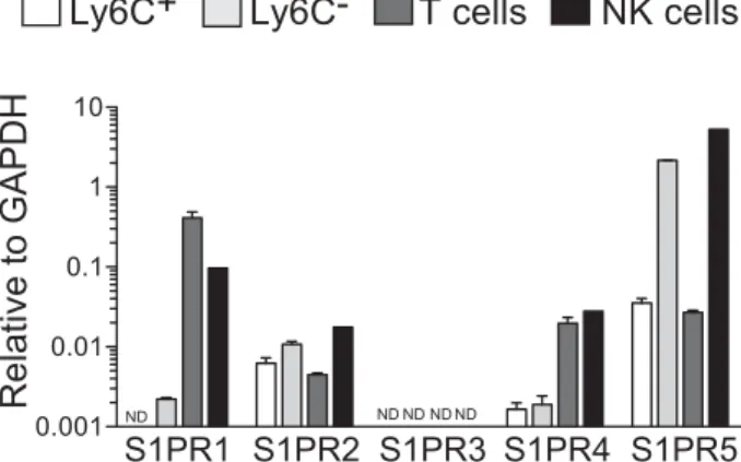
Figure 1. Expression of S1P receptors in different immune cell types.
Ly6C+and Ly6C−monocytes, NK cells, and T cells were sorted by flow cytometry from the BM and their mRNA expression of S1P receptors was measured using semi quantitative RT-PCR, relative to GAPDH. Results are representative of two independent FACS sorting experiments and show the mean+ SD of triplicate PCRs for each S1P receptor.
sion in monocyte subsets. S1PR5 was expressed 30 times higher
in Ly6C− monocytes than in Ly6C+ monocytes (Fig. 1). A
sim-ilar difference in S1PR5 expression between monocyte subsets has been measured using microarrays by the Immgen consor-tium (http://www.immgen.org/databrowser/index.html) [17].
S1PR5 levels in Ly6C− monocytes were comparable to those
in NK cells (Fig. 1), in which S1PR5 plays a role in BM egress [16].
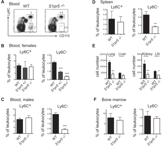
To investigate the function of S1PR5 in monocytes, we first compared the percentage of monocyte subsets in the blood of
wild-type (WT) and S1pr5−/− mice [18] by flow cytometry.
Results in Figure 2A–C showed a significant reduction of Ly6C−
monocytes in the blood of S1pr5−/− mice. This reduction was
observed both in S1pr5−/−female (Fig. 2A and B) and male mice
(Fig. 2C). S1pr5+/−heterozygous mice also showed a mild
phe-notype (Fig. 2B). A strong reduction in the frequency of Ly6C−
monocytes was also observed in the spleen, which is known to be an important reservoir for this subset [19] (Fig. 2D), in the lymph nodes and in non-lymphoid organs such as the lung, liver, and
kidney (Fig. 2E). By contrast, the percentage of Ly6C−monocytes
appeared normal in the BM of S1pr5−/−mice (Fig. 2F). Moreover,
the percentage of Ly6C+monocytes was normal in all lymphoid
organs of S1pr5−/−mice tested (Fig. 2, all panels).
To test if the role of S1PR5 in monocytes was cell-intrinsic, we generated mixed BM chimeras by reconstituting lethally irradiated
mice with equal amounts of BM from WT (CD45.1+) and S1pr5−/−
(CD45.2+) mice. Six weeks after reconstitution, we measured
CD45.1 and CD45.2 expression in different immune subsets in
the blood and BM, and calculated the corresponding S1pr5−/−to
WT ratio for each subset. As previously reported [20], for mature NK (mNK) cells, this ratio was very high in the BM and very low in the blood (Fig. 3, left panel), reflecting the important role of
S1PR5 in NK cell exit from the BM. For Ly6C+monocytes, the
S1pr5−/−to WT ratio was nearly 1 in both blood and BM (Fig. 3, right panel), confirming the absence of a role of S1PR5 in this
A
B
D
F
0 5 10 0.0 0.5 1.0 1.5 Ly6C+ Ly6C- Blood, females Ly6C+ Ly6C- Bone marrow 0.0 0.1 0.2 0.3 0.4 0.5 Ly6C+ Spleen 0.0 0.2 0.4 0.6 Ly6C- Ly 6 C CD115 2 3 0.5 3.5 WT S1pr5 -/-Blood WT S1pr5 +/-S1pr5 -/-0 1 2 3 4 WT S1pr5 +/- S1pr5-/-0.0 1.0 2.0 3.0C
Ly6C+ Ly6C- Blood, males 0 5000 10000 15000 20000E
0 200 400 600 800 WT S1pr5 -/-0 1 2 3 4 WT S1pr5 -/-0.0 0.5 1.0 1.5 2.0 2.5 WT S1pr5 -/-WT S1pr5 -/-WT S1pr5 -/-WT S1pr5 -/-WT S1pr5 -/-WT S1pr5 -/-WT S1pr5 -/-WT S1pr5-/-Lung Liver Kidney LN
*** *** *** ** ** ** * % of leukocytes % of leukocytes % of leukocytes
% of leukocytes % of leukocytes % of leukocytes
cell number
% of leukocytes
% of leukocytes
cell number
Figure 2. S1pr5−/−mice lack peripheral Ly6C−monocytes. (A–F) Flow cytometric analyses of the percentage or absolute number of Ly6C+and Ly6C−monocytes in (A–C) the blood, (D) the spleen, (E) lung, liver, kidney, lymph nodes, and (F) the BM of WT and S1pr5−/−mice, as indicated. (A) A representative flow cytometric analysis in the blood of WT and S1pr5−/−mice is shown. (C–F) Data are shown as the mean+ SD percentage or absolute number of monocyte subsets among total leukocytes in groups of 5 (spleen) to 20 (blood and BM) age- and sex-matched mice, and are pooled from three experiments performed. *p< 0.05, **p < 0.01, ***p < 0.001, t-test.
subset. By contrast, for Ly6C− monocytes, the S1pr5−/− to WT
ratio was near 0.5 in the BM and 0.1 in the blood (Fig. 3, left panel). These data suggest that S1PR5 is important both for the
development of Ly6C−monocytes and for their trafficking or their
survival at the periphery.
S1PR5 and CCR2 but not CX3CR1 are essential for localization of Ly6C−monocytes to BM sinusoids
The paucity of patrolling monocytes in the periphery of S1pr5−/−
mice could be explained by a role of this receptor either in their egress from the BM or in their survival at the periphery. To try and discriminate between both hypotheses, we performed a series
of experiments using Cx3Cr1gfp/gfpand Ccr2−/− mice as controls.
Indeed, CX3CR1 has been shown to regulate peripheral survival of
patrolling monocytes but is devoid of chemotactic activity involved in BM egress. Reciprocally, CCR2 is essential for monocyte egress from the BM but is not involved in their survival. The distribution
of Ly6C−monocytes in Cx3cr1gfp/gfpand Ccr2−/−mice is in fact very
similar to that of S1pr5−/− mice, with a near normal frequency
0.0 0.5 1.0 1.5 0.0 0.5 1.0 3.5 4.0 4.5
BM
Blood
S1pr5 KO / WT
ratio
Ly6C+ Ly6C-
mNK
*** *** *** ***Figure 3. The role of S1PR5 in Ly6C−monocytes is cell-intrinsic. Mixed BM chimeric mice were generated by reconstituting irradiated CD45.1 × CD45.2 mice with a 1:1 mixture of WT CD45.1 BM cells and S1pr5−/− CD45.2 BM cells as indicated. Eight weeks after reconstitution, the per-centage of CD45.2 positive cells in the indicated subsets was measured by flow cytometry in the blood and BM. Results are shown as the mean+ SD of four mice in each group and are expressed as the ratio between S1pr5−/−and WT cells for each subset, as indicated. This exper-iment is representative of four independent experexper-iments performed. ***p< 0.001, t-test.
Blood Bone marrow
A
BM sinusoids 0.0 0.5 1.0 1.5 0 1 2 3 ** ** ** *** ** * 0 5 10 Blood BM 0.0 0.5 1.0 1.5 2.0Transfer: Cx3cr1 KO:WT Transfer: S1pr5 KO:WT
0.0 0.5 1.0 1.5 2.0 2.5 KO / WT ratio 16 h after transfer S1pr5 -/-WT Ccr2 Cx3cr1 -/-WT Ccr2 Cx3cr1 -/-WT Ccr2 Cx3cr1
-/-C
B
0 20 40 60 80 100 Before transferD
Blood BM ** * S1pr5 -/-WT S1pr5 -/-WT 0 10 20 30 40 50E
F
Bcl2 ( ) vs isotype ( ) WT Cx3cr1-/- S1pr5-/-WT S1pr5-/-control M-CSF S1pr5-/-control M-CSF % of leukocytes Percent CD45+ % viable cells % of leukocytes % viable cells Blood BM 16 h after transfer Before transferFigure 4. S1PR5 promotes BM egress of Ly6C−monocytes and not survival. (A) Flow cytometric analyses of the percentage of Ly6C−monocytes in the blood and the BM of WT, Cx3cr1gfp/gfpand Ccr2−/−mice. Data are shown as the mean+ SD of 4–10 mice in each group. (B) In some experiments,
the frequency of sinusoidal cells (CD45+) within total BM Ly6C−monocytes was measured. Data are shown as mean+ SD of five mice in each group. (A, B) Data shown are pooled from three experiments performed. (C) Adoptive transfer of BM cells from Cx3cr1gfp/gfpmice and CD45.1 WT
mice or S1pr5−/−and CD45.1 WT mice (1:1 ratio) into CD45.1× CD45.2 recipient mice. Sixteen hours after transfer the number of Ly6C−monocytes from each donor was measured in the BM and blood of recipient mice by flow cytometry. Results are expressed as a ratio between KO and CD45.1 WT Ly6C−monocytes, for each combination, before and after transfer in the BM and blood. Each symbol represents an individual recipient mouse. Data are pooled from three independent experiments. (D) Freshly isolated blood and BM cells from WT S1pr5−/−chimeric mice were stained with Annexin V and 7-AAD and antibodies to detect monocyte cells. The frequency of viable Ly6C−monocytes was determined by flow cytometry. Data are shown as the mean+ SD percentage of viable Annexin V−7-AAD−cells among total Ly6C−monocytes of each donor mouse in the BM and blood. Results are representative of three independent experiments with at least three mice in each group. (E) Ly6C−monocytes were sorted from BM cell suspensions by flow cytometry and cultured for 24 h in medium alone or supplemented with M-CSF as indicated. Data are shown as the mean± SD viability of cells as assessed by Annexin V / 7-AAD staining and are pooled from three independent experiments. (F) Flow cytometric measurement of intracellular Bcl2 expression in blood Ly6C−monocytes from different mouse strains as indicated. Data are representative of
n= 3 mice in each group. *p < 0.05, **p < 0.01, ***p < 0.001, t-test.
in the BM and a low frequency of these cells at the periphery (Fig. 4A).
First, we compared the frequency of sinusoidal Ly6C−
mono-cytes in the BM of WT, Cx3Cr1gfp/gfp, Ccr2−/−, and S1pr5−/−
mice using in vivo labeling of sinusoidal cells. This measurement has been shown to be proportional to the BM exit rate. Indeed,
newly developed BM leukocytes transit from the BM parenchyma through the endothelium and into the BM sinusoids where they are transiently retained until their release into the blood circu-lation. Results presented in Fig. 4B showed that the percentage
of sinusoidal Ly6C−monocytes was significantly decreased in the
By contrast, the percentage of sinusoidal Ly6C− monocytes was
significantly increased in the BM of Cx3Cr1gfp/gfpmice compared to
the BM of WT mice. These results support a role for S1PR5 in the
migration of Ly6C−monocytes from the parenchyma to the
sinu-soidal compartment of the BM, a process essential for exit from
the BM. This process could be negatively regulated by CX3CR1,
perhaps as a result of adhesive properties of CX3CR1.
Second, we compared the fate of monocytes of different geno-types adoptively transferred into recipient mice. We performed intravenous injection of a 1:1 mixture of WT (CD45.1) and
S1pr5−/− or Cx3cr1gfp/gfp (CD45.2) BM cells into recipient WT
(CD45.1× CD45.2) mice. Sixteen hours after transfer, we
mea-sured the frequency of donor monocyte subsets in the blood and the BM of recipient mice. We calculated the ratio between WT and KO donors for each subset before transfer and 16 h after
transfer in the blood and the BM. Cx3cr1gfp/gfpLy6C−monocytes
were barely detectable in both BM and blood of recipient mice,
confirming the important role of CX3CR1 in the survival of Ly6C−
monocytes (Fig. 4C, left panel). By contrast, transferred S1pr5−/−
Ly6C− monocytes were almost absent from the blood but were
represented at similar frequency as WT Ly6C−monocytes in the
BM of recipient mice (Fig. 4C, right panel). These data support a
role for S1PR5 in the egress of Ly6C−monocytes rather than in
their survival.
Third, we compared the ex vivo viability of WT and S1pr5−/−
Ly6C−monocytes in the blood and BM of WT S1pr5−/−chimeric
mice using AnnexinV/7-AAD staining. In both compartments, the
viability of S1pr5−/− Ly6C− monocytes was slightly lower than
that of WT Ly6C− monocytes (Fig. 4D). Moreover, irrespective
of the mouse genotype, the viability of Ly6C− monocytes was
lower in the BM than in the blood. We also assessed viability
of WT and S1pr5−/−Ly6C−monocytes sorted by flow cytometry
and cultured in the presence or absence of M-CSF. After 24 h,
the viability of WT and S1pr5−/− Ly6C−monocytes was similar
in both culture conditions (Fig. 4E). Finally, we measured the expression of Bcl2, an important anti-apoptotic molecule that has
been shown to be down regulated in Cx3cr1gfp/gfpLy6C−monocytes
and to regulate their survival. The expression of Bcl2 was similar in
Ly6C−monocytes from WT and S1pr5−/−mice but it was reduced
in Cx3cr1gfp/gfpLy6C−monocytes (Fig. 4F), as previously reported
[21].
Altogether, this series of in vivo experiments demonstrate that
CX3CR1 and S1PR5 regulate homeostasis of the peripheral pool
of Ly6C−monocytes via different mechanisms. While CX3CR1 is
clearly involved in their survival, S1PR5 is rather implicated in their egress from the BM although it may also contribute indirectly in their survival.
Modulation of S1P levels does not alter the trafficking and survival of Ly6C−monocytes
Finally, we investigated the role of S1P in the physiology of Ly6C−
monocytes using in vitro and in vivo experiments.
A
NK cells Ly6C+
Ly6C-1.0 1.5 2.0 2.5
B
NK cells Ly6C+
Ly6C-0 5 10 15 100 120 140 160 180 200
E
Cell number (per ml of blood x 10
3)
0 500 1000 1500
Ly6C+ Ly6C- T cells
0 500 1000 1500 2000 Day 3 Day 10 *** ***
F
D
BSA S1P 0 10 20 30 H2O DOP 0 20 40 60 80 100 H2O DOP BM BloodC
WT S1PR5 KO 0 2 4 6 8 10 medium CCL2Migration index Migration index
Migration index % viable cells
% viable cells medium CCL2 medium S1P 1 nM S1P 10 nM S1P 100 nM H2O DOP
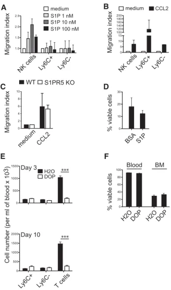
Figure 5. S1P gradient is not required for homeostasis of Ly6C− mono-cytes. (A and B) Transwell migration assay of purified Ly6C+and Ly6C− monocytes or NK cells (A) in response to different concentrations of S1P or (B) in response to CCL2 (50 ng/mL) as indicated. Values are reported as the mean ratio+SD between the number of cells that migrated during the 2 h incubation with S1P or CCL2 and the number of cells migrating in the control (no chemokine) condition. Results are representative of three independent experiments. (C) Transwell migration of BM Ly6C− monocytes from WT and S1pr5−/−mice in response to CCL2 gradients, as indicated. (D) Ly6C−monocytes were sorted from BM cell suspen-sions by flow cytometry and cultured for 24 h in medium without FCS supplemented with BSA, in the presence or absence of S1P, as indi-cated. Data are shown as mean+ SD of six samples pooled from three independent experiments. (E and F) C57BL/6 mice were treated with DOP given in the drinking water for 3 or 10 days. (E) The percentage of Ly6C+and Ly6C−monocytes and the percentage of CD3+T cells was measured on days 3 (top) and 10 (bottom) by flow cytometry. (F) The viability of Ly6C−-monocytes was measured after 3 days of treatment in the BM and in the blood. Results are shown as the mean+ SD of six mice in each group and are pooled from three experiments performed. ***p< 0.001, t-test.
In vitro, we measured responsiveness of monocytes to S1P gra-dients in chemotaxis chambers. No consistent migration of either population of monocytes was observed (Fig. 5A), whereas both monocyte populations migrated in response to CCL2 gradients (Fig. 5B). In the same experiments, NK cells migrated in response
to both S1P and CCL2 gradients (Fig. 5A and B), as previously
reported [16]. WT and S1pr5−/− Ly6C− monocytes migrated
equally to CCL2 gradients, excluding a possible cross talk between
CCR2 and S1PR5 (Fig. 5C). We also cultured Ly6C−monocytes
with S1P at concentrations similar to those observed in vivo. The addition of S1P at any concentration did not change monocyte viability in vitro (Fig. 5D and data not shown).
Next, we treated mice with the sphingosine lyase inhibitor deoxypyridoxine (DOP), which has been shown to dramati-cally increase S1P levels in tissues and disrupt S1P gradients in vivo [22]. Upon treatment with DOP, peripheral T-cell numbers dropped, as previously reported [22]. However, DOP had no effect
on the trafficking or the number of Ly6C−monocytes (Fig. 5E) and
NK cells [22] even after prolonged (10 days) treatment (Fig. 5E).
The ex vivo viability of blood and BM Ly6C−monocytes was not
modified either (Fig. 5F).
Altogether, these results suggest that S1P and S1P gradients are not involved in monocyte survival and unexpectedly not in their trafficking.
Discussion
In this article, we report for the first time a high expression of S1PR5 in patrolling monocytes and the paucity of these cells in
the peripheral compartment of S1pr5−/−mice. The following body
of evidences supports a role for S1PR5 in BM egress of patrolling monocytes: (i) We previously showed that S1PR5 was involved in NK-cell egress from the BM to blood [20, 23]. Moreover, several other members of the family of S1P receptors (S1PR1, S1PR3) are clearly involved in egress of different leukocyte subsets from
cen-tral and peripheral lymphoid organs [24]. (ii) Ly6C−monocytes
are reduced in BM sinusoids of S1pr5−/−mice, whereas they are
preserved, or even slightly increased in Cx3cr1gfp/gfpmice, which
only exhibit impaired survival of Ly6C−monocytes at the
periph-ery. (iii) The phenotype of S1pr5−/−mice is very similar to that
of Ccr2−/−mice in which monocyte egress from the BM has been
shown to be clearly impaired [15]. In particular, the number of
Ly6C−monocytes was normal in the BM of S1pr5−/−and Ccr2−/−
mice but reduced in the blood circulation and in BM sinusoids.
(iv) Upon adoptive transfer into recipient mice, S1pr5−/−Ly6C−
monocytes recirculate to the BM and rapidly disappear from the
blood circulation. For comparison, Cx3cr1gfp/gfpLy6C−monocytes
do not survive either in the BM or in the blood after transfer. An intriguing observation is the absence of accumulation of
S1pr5−/− Ly6C− monocytes in the BM of S1pr5−/−mice or WT
S1pr5−/− BM chimeric mice. A similar phenomenon (i.e. lack of
accumulation of Ly6C−monocytes) was also observed in Ccr2−/−
mice and WT Ccr2−/−BM chimeric mice. This suggests that the
trafficking machinery of Ly6C−monocytes regulates somehow the
developmental fitness of these cells and that an impairment of this machinery results in an impaired survival. As a matter of fact, we
found that the ex vivo viability of Ly6C− monocytes in the BM
was very low, confirming previous findings [25]. It is therefore possible that an impairment of their trafficking by means of CCR2
or S1PR5 deletion could further decrease the viability of these fragile cells.
In vivo modulation of S1P levels by pharmacological means did
not alter homeostasis of Ly6C−monocytes (this report), while they
dramatically reduced the number of T cells in circulation. These results show that S1P receptors operate through different modes of action in monocytes and in T cells. Several hypotheses could
explain this paradox. First, the role of S1PR5 in Ly6C−monocytes
could be S1P-independent. Other physiological ligands for this receptor have not yet been described but specific S1PR5 analogs binding with high affinity to this receptor have been synthesized [26], and may therefore exist in vivo. Second, it has been reported that S1PR5 could act as a constitutively active receptor [27] like other G-protein-coupled receptors [28]. S1PR5 was in fact shown to decrease adenylyl cyclase and ERK activity in several cell lines in the absence of S1P, inducing cell rounding and detachment without promoting apoptosis [27]. This effect could contribute or even induce cell migration by preventing strong attachment to the stromal substrate of the BM. In this scenario, S1PR5 would not be a chemotactic receptor in monocytes, which would explain why we could not detect migration of these cells in response to S1P gradients in vitro. An alternative possibility could be that the form of S1P physiologically active in monocytes is different from the one we use in vitro. In fact, S1P can be found under differ-ent forms in vivo that could have differdiffer-ential activities on leuko-cyte subsets. Further studies are required to test these points. It remains also to be determined whether S1PR5 acts differently in
monocytes and NK cells. Indeed, S1pr5−/−mice lack both
periph-eral NK cells and Ly6C− monocytes but only NK cells
accumu-late in the BM of these mice and migrate in vitro in response to S1P.
Altogether, our findings shed light on the long-sought
mech-anisms of exit of Ly6C−monocytes from the BM [12, 29]. They
also show fundamental differences between both monocyte
sub-sets, as the exit of Ly6C+monocytes from the BM relies only on
pro-inflammatory signals mediated by CCR2 [6] whereas that of
Ly6C−monocytes is primarily mediated by S1PR5. Furthermore,
S1pr5−/−mice constitute an interesting model to study the role of
Ly6C−monocytes in immunity, a point that remains unclear.
Materials and methods
Mice and reagentsWT C57BL/6 mice were purchased from Charles River
Laborato-ries (L’Arbresle, France). S1pr5−/−mice [18], Ccr2−/−[30], and
Cx3cr1gfp/gfp mice [31] have been previously described. In some
experiments, we also used C57BL/6 CD45.1 mice or C57BL/6
CD45.1 × CD45.2 mice that were bred in our animal house.
Female mice 8–24 week-old were used unless specified. DOP (Sigma, St. Louis, MO, USA) was provided in the drinking water
and mice housing were approved by the local Ethics Committee and carried out according to the French and European laws.
Generation of BM chimera
C57BL/6 CD45.1× CD45.2 mice were irradiated twice at 450 rad
within a 4-h interval. Four hours after the last irradiation, they received an intravenous injection of a 1:1 mixture of BM cells
from WT CD45.1 and S1pr5−/−CD45.2 mice. BM chimeras were
analyzed 6–12 weeks after reconstitution.
In vivo labeling of sinusoidal lymphocytes
This technique was previously described [32]. Briefly, mice were
injected intravenously with 1μg anti-CD45 Mab (30F11) coupled
to phycoerythrin (PE) or PE-cyanin-5 (BD Biosciences, San Jose, USA). Mice were sacrificed 2 min after antibody injection. BM was then collected and analyzed by flow cytometry.
Adoptive transfers
BM cells from WT CD45.1 and S1pr5−/−or Cx3cr1gfp/gfp(CD45.2)
mice were prepared and mixed at a 1:1 ratio before intravenous
injection (1× 10⁷cells of each genotype in PBS) into anesthetized
CD45.1× CD45.2 C57BL/6 mice. Sixteen hours later, mice were
sacrificed, blood and bone marrow was collected and the percent-age of monocyte subsets of each donor mice was measured by flow cytometry after staining for CD45.1 and CD45.2 expression.
Cell viability
Cell viability was measured in ex vivo isolated cell suspensions using Annexin V and 7-AAD staining (BD Biosciences) and flow cytometry.
Antibodies and flow cytometry
BM, spleen, lung, lymph node, kidney, and blood cells were iso-lated and stained as previously described [33]. Cell counts were determined using an accuri C6 flow cytometer (BD Accuri
Cytome-ters, Ann Arbor, MI, USA). Monocytes were identified as CD115+
in the blood or as CD11b+CD11clowNK1.1−CD19−Ly6G− in the
BM and spleen. The following Mabs from eBioscience (San Diego, CA, USA) or BD Biosciences (Becton Dickinson, San Jose, USA) were used: anti-CD115 (AFS98), anti-Ly6C (HK1.4), anti-Ly6G (1A8), anti-CD19 (ebio1D3), anti-CD3 (145–2C11), anti-NK1.1 (PK136), anti NKp46 (29A1.4), anti-CD11b (M1/70), anti-CD45.1 (A20), anti CD45.2 (104), and relevant isotype controls. Bcl2 expression was measured using a commercial kit (BD Biosciences) according to the manufacturer’s instructions. Flow cytometry was
carried out on a FACS Canto, a FACS Canto II or a FACS LSR II (Becton Dickinson).
Chemotaxis assays
For S1P migration assays, monocytes were purified from BM cells using a negative selection procedure. Briefly, freshly isolated BM cells were stained with purified antibodies against CD4, CD8, CD19, and Ly6G (eBioscience). Cells were then incubated with magnetic beads covered with goat anti-rat IgG (Qiagen, Hilden, Germany) before magnetic separation. After washing steps, cells were then suspended in RPMI1640 supplemented with 4 mg/mL fatty acid-free bovine albumin (Sigma). The same medium was
used to prepare S1P (Sigma) at 10−8 M or CCL2 at 50 ng/mL
(R&D systems). Cell migration was measured in Transwell
cham-bers (Costar, Cambridge, MA, USA) with 5-μm pore-width
poly-carbonate filters. After 2 h, transmigrated cells were stained for CD3, NK1.1, CD27 and CD11b or CD11b, Ly6G and Ly6C and counted by flow cytometry as described previously [16].
Quantitative RT-PCR
Lymphocyte or monocyte subsets stained with the appropriate antibodies were sorted using a FACS Aria cell sorter (Becton Dick-inson). RNA was extracted with the RNeasy micro kit (Qiagen), which includes treatment with DNase I. We used Superscript II reverse transcriptase (Invitrogen, Carlsbad, CA, USA) to gener-ate cDNA for RT-PCR. PCR was carried out with a SybrGreen-based kit (FastStart Universal SYBR Green Master, Roche, Basel, Switzerland) on a StepOne plus instrument (Applied biosystems, Carlsbad, CA, USA). Primers were designed using the oligop-erfect software (Invitrogen, Carlsbad, CA, USA). The following primers were used: S1pr1 (F: AAATGCCCCAACGGAGACTCTG, R: TTGCTGCGGCTAAATTCCATGC), S1pr2 (F: CCCAACTCCGGGA-CATAGA, R: ACAGCCAGTGGTTGGTTTTG), S1pr3 (F: TCAGTG-GTTCATCATGCTGG, R: CAGGTCTTCCTTGACCTTCG), S1pr4 (F: AAGACCAGCCGTGTGTATGG, R: TCAGCACGGTGTTGAGTAGC),
S1pr5 (F: GCCTGGTGCCTACTGCTACAG, R:
CCTCCGTCGCTG-GCTATTTCC), Gapdh (F: GCATGGCCTTCCGTGTTC, R: TGTCAT-CATACTTGGCAGGTTTCT). S1PR expression level in the different cell subsets was normalized to GAPDH expression levels.
In vitro culture of Ly6C−monocytes
Ly6C−monocytes were sorted by flow cytometry using a FACS Aria
cell sorter (Becton Dickinson). They were cultured in flat bottom 96 well plates (25000/condition) in duplicates. For cultures with M-CSF, the culture medium was supplemented with 10% FCS in the presence or absence of 5% of an M-CSF-containing cell culture supernatant. In some experiments, cells were resuspended in medium supplemented with 4 mg/mL fatty acid-free bovine albumin (Sigma). The same medium was used to prepare S1P
(Sigma), which was added or not to the cultures at a concentration
of 10−6M.
Statistical analyses
Statistical analyses were performed using two-tailed t-tests or nonparametric tests when appropriate. These tests were run on the Prism software (GraphPad, La Jolla, CA, USA). Levels of
significance are expressed as p-values (*p< 0.05, **p < 0.01,
***p< 0.001).
Acknowledgments: Authors thank the Plateau de Biologie
Exp´erimentale de la Souris, and the flow cytometry facility of
the SFR Biosciences Gerland. We also thank Andrew Calver (Glax-oSmithKline) for providing S1PR5 KO mice and Steffen Jung for
the CX3CR1gfp/gfpmice. The T. W. lab is supported by the FINOVI
foundation, Agence Nationale de la Recherche (ANR JC sphinks), European Research council (ERC-Stg 281025), Institut National
de la Sant´e et de la Recherche M´edicale (INSERM).
References
1 Serbina, N. V., Jia, T., Hohl, T. M. and Pamer, E. G., Monocyte-mediated defense against microbial pathogens. Annu. Rev. Immunol. 2008. 26: 421–452.
2 Auffray, C., Sieweke, M. H. and Geissmann, F., Blood monocytes: devel-opment, heterogeneity, and relationship with dendritic cells. Annu. Rev. Immunol. 2009. 27: 669–692.
3 Ancuta, P., Liu, K.-Y., Misra, V., Wacleche, V. S., Gosselin, A., Zhou, X. and Gabuzda, D., Transcriptional profiling reveals developmental rela-tionship and distinct biological functions of CD16+and CD16- monocyte subsets. BMC Genomics. 2009. 10: 403–422.
4 Cros, J., Cagnard, N., Woollard, K., Patey, N., Zhang, S.-Y., Senechal, B., Puel, A. et al., Human CD14dim monocytes patrol and sense nucleic acids and viruses via TLR7 and TLR8 receptors. Immunity 2010. 33: 375–386. 5 Ingersoll, M. A., Spanbroek, R., Lottaz, C., Gautier, E. L., Frankenberger,
M., Hoffmann, R., Lang, R. et al., Comparison of gene expression profiles between human and mouse monocyte subsets. Blood 2010. 115: e10–e19. 6 Serbina, N. V. and Pamer, E. G., Monocyte emigration from bone mar-row during bacterial infection requires signals mediated by chemokine receptor CCR2. Nat. Immunol. 2006. 7: 311–317.
7 Le Borgne, M., Etchart, N., Goubier, A., Lira, S. A., Sirard, J. C., van Rooijen, N., Caux, C. et al., Dendritic cells rapidly recruited into epithelial tissues via CCR6/CCL20 are responsible for CD8+ T cell crosspriming in vivo. Immunity 2006. 24: 191–201.
8 Serbina, N. V., Salazar-Mather, T. P., Biron, C. A., Kuziel, W. A. and Pamer, E. G., TNF/iNOS-producing dendritic cells mediate innate immune defense against bacterial infection. Immunity 2003. 19: 59–70. 9 Tacke, F. and Randolph, G. J., Migratory fate and differentiation of blood
monocyte subsets. Immunobiology. 2006. 211: 609–618.
10 Auffray, C., Fogg, D., Garfa, M., Elain, G., Join-Lambert, O., Kayal, S., Sarnacki, S. et al., Monitoring of blood vessels and tissues by a population of monocytes with patrolling behavior. Science 2007. 317: 666–670. 11 Kim, K.-W., Vallon-Eberhard, A., Zigmond, E., Farache, J., Shezen, E.,
Shakhar, G., Ludwig, A. et al., In vivo structure/function and expression analysis of the CX3C chemokine fractalkine. Blood 2011. 118: e156–e167. 12 Shi, C. and Pamer, E. G., Monocyte recruitment during infection and
inflammation. Nat. Rev. Immunol. 2011. 11: 762–774.
13 Geissmann, F., Manz, M. G., Jung, S., Sieweke, M. H., Merad, M. and Ley, K., Development of monocytes, macrophages, and dendritic cells. Science 2010. 327: 656–661.
14 Biburger, M., Aschermann, S., Schwab, I., Lux, A., Albert, H., Danzer, H., Woigk, M. et al., Monocyte subsets responsible for immunoglob-ulin G-dependent effector functions in vivo. Immunity 2011. 35: 932–944.
15 Shi, C., Jia, T., Mendez-Ferrer, S., Hohl, T. M., Serbina, N. V., Lipuma, L., Leiner, I. et al., Bone marrow mesenchymal stem and progenitor cells induce monocyte emigration in response to circulating toll-like receptor ligands. Immunity 2011. 34: 590–601.
16 Mayol, K., Biajoux, V., Marvel, J., Balabanian, K. and Walzer, T., Sequen-tial desensitization of CXCR4 and S1P5 controls natural killer cell traf-ficking. Blood 2011. 118: 4863–4871.
17 Heng, T. S. P., Painter, M. W. and Immunological Genome Project Consor-tium, The Immunological Genome Project: networks of gene expression in immune cells. Nat. Immunol. 2008. 9: 1091–1094.
18 Jaillard, C., Harrison, S., Stankoff, B., Aigrot, M. S., Calver, A. R., Duddy, G., Walsh, F. S. et al., Edg8/S1P5: an oligodendroglial receptor with dual function on process retraction and cell survival. J. Neurosci. 2005. 25: 1459–1469.
19 Swirski, F. K., Nahrendorf, M., Etzrodt, M., Wildgruber, M., Cortez-Retamozo, V., Panizzi, P., Figueiredo, J.-L. et al., Identification of splenic reservoir monocytes and their deployment to inflammatory sites. Science 2009. 325: 612–626.
20 Walzer, T., Chiossone, L., Chaix, J., Calver, A., Carozzo, C., Garrigue-Antar, L., Jacques, Y. B. et al., Natural killer cell trafficking in vivo requires a dedicated sphingosine 1-phosphate receptor. Nat. Immunol. 2007. 8: 1337–1344.
21 Jakubzick, C., Tacke, F., Ginhoux, F., Wagers, A. J., van Rooijen, N., Mack, M., Merad, M. and Randolph, G. J., Blood monocyte subsets differentially give rise to CD103+ and CD103- pulmonary dendritic cell populations. J. Immunol. 2008. 180: 3019–3027.
22 Schwab, S. R., Pereira, J. P., Matloubian, M., Xu, Y., Huang, Y. and Cys-ter, J. G., Lymphocyte sequestration through S1P lyase inhibition and disruption of S1P gradients. Science 2005. 309: 1735–1739.
23 Jenne, C. N., Enders, A., Rivera, R., Watson, S. R., Bankovich, A. J., Pereira, J. P., Xu, Y. et al., T-bet-dependent S1P5 expression in NK cells pro-motes egress from lymph nodes and bone marrow. J. Exp. Med. 2009. 206: 2469–2481.
24 Cyster, J. G. and Schwab, S. R., Sphingosine-1-Phosphate and lymphocyte egress from lymphoid organs. Annu. Rev. Immunol. 2012. 30: 69–94. 25 Hanna, R. N., Carlin, L. M., Hubbeling, H. G., Nackiewicz, D., Green, A. M.,
Punt, J. A., Geissmann, F. et al., The transcription factor NR4A1 (Nur77) controls bone marrow differentiation and the survival of Ly6C- mono-cytes. Nat. Immunol. 2011. 12: 778–785.
26 Mattes, H., Dev, K. K., Bouhelal, R., Barske, C., Gasparini, F., Guerini, D., Mir, A. K. et al., Design and synthesis of selective and potent orally active S1P5 agonists. Chem. Med. Chem. 2010. 5: 1693–1696.
27 Niedernberg, A., Blaukat, A., Sch ¨oneberg, T. and Kostenis, E., Regulated and constitutive activation of specific signalling pathways by the human S1P5 receptor. Br. J. Pharmacol. 2003. 138: 481–493.
28 Rosenbaum, D. M., Rasmussen, S. G. F. and Kobilka, B. K., The struc-ture and function of G-protein-coupled receptors. Nastruc-ture 2009. 459: 356–363.
29 Ingersoll, M. A., Platt, A. M., Potteaux, S. and Randolph, G. J., Monocyte trafficking in acute and chronic inflammation. Trends Immunol. 2011. 32: 470–477.
30 Boring, L., Gosling, J., Chensue, S. W., Kunkel, S. L., Farese, Jr, R. V., Broxmeyer, H. E. and Charo, I. F., Impaired monocyte migration and reduced type 1 (Th1) cytokine responses in C-C chemokine receptor 2 knockout mice. J. Clin. Invest. 1997. 100: 2552–2561.
31 Jung, S., Aliberti, J., Graemmel, P., Sunshine, M. J., Kreutzberg, G. W., Sher, A. and Littman, D. R., Analysis of fractalkine receptor CX(3)CR1 function by targeted deletion and green fluorescent protein reporter gene insertion. Mol. Cell Biol. 2000. 20: 4106–4114.
32 Pereira, J. P., An, J., Xu, Y., Huang, Y. and Cyster, J. G., Cannabinoid receptor 2 mediates the retention of immature B cells in bone marrow sinusoids. Nat. Immunol. 2009. 10: 403–411.
33 Walzer, T., Bl ´ery, M., Chaix, J., Fuseri, N., Chasson, L., Robbins, S. H., Jaeger, S. et al., Identification, activation, and selective in vivo abla-tion of mouse NK cells via NKp46. Proc. Natl. Acad. Sci. U S A. 2007. 104: 3384–3389.
Abbreviations:DOP: deoxypyridoxine· S1P: sphingosine-1 phosphate ·
S1PR5: sphingosine-1 phosphate receptor 5
Full correspondence: Dr. Thierry Walzer, INSERM U1111, 21 avenue Tony Garnier, Lyon, France 69007
Fax:+33-(0) 43728 2341