Publisher’s version / Version de l'éditeur:
AJP : Regulatory, Integrative and Comparative Physiology, 295, 2, pp.
R611-R623, 2008-08-01
READ THESE TERMS AND CONDITIONS CAREFULLY BEFORE USING THIS WEBSITE. https://nrc-publications.canada.ca/eng/copyright
Vous avez des questions? Nous pouvons vous aider. Pour communiquer directement avec un auteur, consultez la première page de la revue dans laquelle son article a été publié afin de trouver ses coordonnées. Si vous n’arrivez pas à les repérer, communiquez avec nous à PublicationsArchive-ArchivesPublications@nrc-cnrc.gc.ca.
Questions? Contact the NRC Publications Archive team at
PublicationsArchive-ArchivesPublications@nrc-cnrc.gc.ca. If you wish to email the authors directly, please see the first page of the publication for their contact information.
NRC Publications Archive
Archives des publications du CNRC
This publication could be one of several versions: author’s original, accepted manuscript or the publisher’s version. / La version de cette publication peut être l’une des suivantes : la version prépublication de l’auteur, la version acceptée du manuscrit ou la version de l’éditeur.
For the publisher’s version, please access the DOI link below./ Pour consulter la version de l’éditeur, utilisez le lien DOI ci-dessous.
https://doi.org/10.1152/ajpregu.00917.2007
Access and use of this website and the material on it are subject to the Terms and Conditions set forth at
Mild endotoxemia, NF-κB translocation, and cytokine increase during
exertional heat stress in trained and untrained individuals
Selkirk, G. A.; McLellan, T. M.; Wright, H. E.; Rhind, S. G.
https://publications-cnrc.canada.ca/fra/droits
L’accès à ce site Web et l’utilisation de son contenu sont assujettis aux conditions présentées dans le site
LISEZ CES CONDITIONS ATTENTIVEMENT AVANT D’UTILISER CE SITE WEB.
NRC Publications Record / Notice d'Archives des publications de CNRC:
https://nrc-publications.canada.ca/eng/view/object/?id=b0eac582-8202-4b41-94b3-64ccbd36f3b2 https://publications-cnrc.canada.ca/fra/voir/objet/?id=b0eac582-8202-4b41-94b3-64ccbd36f3b2Mild endotoxemia, NF-B translocation, and cytokine increase during
exertional heat stress in trained and untrained individuals
G. A. Selkirk,2T. M. McLellan,1,2H. E. Wright,2and S. G. Rhind1,2
1Defence Research and Development Canada-Toronto, Toronto; and2Kinesiology and Health Science Graduate Programme, York University, Toronto, Ontario, Canada
Submitted 21 December 2007; accepted in final form 17 June 2008
Selkirk GA, McLellan TM, Wright HE, Rhind SG.Mild endo-toxemia, NF-B translocation, and cytokine increase during exer-tional heat stress in trained and untrained individuals. Am J Physiol
Regul Integr Comp Physiol 295: R611–R623, 2008. First published June 18, 2008; doi:10.1152/ajpregu.00917.2007.—This study exam-ined endotoxin-mediated cytokinemia during exertional heat stress (EHS). Subjects were divided into trained [TR; n ⫽ 12, peak aerobic power (V˙O2peak) ⫽ 70 ⫾ 2 ml 䡠 kg lean body mass⫺1䡠 min⫺1] and untrained (UT; n ⫽ 11, V˙O2peak ⫽ 50 ⫾ 1 ml 䡠 kg lean body
mass⫺1
䡠 min⫺1) groups before walking at 4.5 km/h with 2% elevation in a climatic chamber (40°C, 30% relative humidity) wearing protec-tive clothing until exhaustion (Exh). Venous blood samples at baseline and 0.5°C rectal temperature increments (38.0, 38.5, 39.0, 39.5, and 40.0°C/Exh) were analyzed for endotoxin, lipopolysaccharide binding protein, circulating cytokines, and intranuclear NF-B translocation. Baseline and Exh samples were also stimulated with LPS (100 ng/ml) and cultured in vitro in a 37°C water bath for 30 min. Phenotypic determination of natural killer cell frequency was also determined. Enhanced blood (104 ⫾ 6 vs. 84 ⫾ 3 ml/kg) and plasma volumes (64 ⫾ 4 vs. 51 ⫾ 2 ml/kg) were observed in TR compared with UT subjects. EHS produced an increased concentration of circulating endotoxin in both TR (8 ⫾ 2 pg/ml) and UT subjects (15 ⫾ 3 pg/ml) (range: not detected to 32 pg/ml), corresponding with NF-B trans-location and cytokine increases in both groups. In addition, circulating levels of tumor necrosis factor-␣ and IL-6 were also elevated com-bined with concomitant increases in IL-1 receptor antagonist in both groups and IL-10 in TR subjects only. Findings suggest that the threshold for endotoxin leakage and inflammatory activation during EHS occurs at a lower temperature in UT compared with TR subjects and support the endotoxin translocation hypothesis of exertional heat stroke, linking endotoxin tolerance and heat tolerance.
splanchnic permeability; immune function; blood volume; cardiovas-cular/thermoregulatory strain; flow cytometry
EXERTIONAL HEAT STRESS (EHS) produces cardiovascular and
thermoregulatory strain due to competition for maintenance of an adequate blood supply to the periphery to promote heat loss and to deliver nutrients to metabolically active muscle (30). Circulatory stability is maintained by the redistribution of cardiac output, accomplished by vasoconstriction of renal and splanchnic vasculature reducing gastrointestinal blood flow (57–59, 70, 71). The redistribution of cardiac output away from abdominal organs to active skeletal muscle during exercise has been correlated to oxygen consumption and is proportional to the increase in heart rate (HR) (70). The extent of the reduction in splanchnic blood flow depends on individual maximum aerobic power but can be reduced by as much as 80%, resulting in intestinal ischemia (58, 70). Exercise, when combined with
hyperthermia, can have an additive effect that exasperates the reduction in visceral blood supply, intestinal ischemia, and permeability of the gut wall (30, 70).
Endotoxemia plays a pivotal role in the pathophysiology of the systemic inflammatory response syndrome (SIRS) in vari-ous conditions, including sepsis (37, 42), trauma (21), and exertional heat stroke (4, 39, 40). Regulation of these inflam-matory disorders is accomplished primarily through NF-B, a central mediator of inflammatory gene transcription, which can have effects on both innate and adaptive immunity (42).
Endotoxin, or LPS, is normally found in large abundance on the surface of gram-negative bacteria in the intestinal lumen (72). Under physiological conditions, endotoxins are contained and not toxic (76), since only small amounts enter the circu-lation from the intestinal lumen and are quickly inactivated by several defense mechanisms (20). However, failure of the body’s defense mechanisms and/or barriers results in substan-tial endotoxin leakage across the intestinal lumen into the portal and, eventually, the systemic circulation. Lipopolysac-charide binding protein (LBP) forms an LPS-LBP complex enhancing the transfer of LPS to its receptor complex [CD14/ Toll-like receptor (TLR)-4/MD-2] (21, 37) on immune-com-petent cells (75), resulting in NF-B activation and transloca-tion from the cytoplasm to the nucleus. NF-B DNA binding in the liver (14), intestinal epithelial/mucosal cells (63), and, finally, circulating leukocytes (42) activates B genes encod-ing inflammatory mediators, includencod-ing tumor necrosis factor (TNF)-␣, interleukin (IL)-1, and inducible nitric oxide syn-thase (iNOS) in addition to acute phase proteins, leading to activation of SIRS, perfusion abnormalities, intravascular co-agulopathy, and ultimately, multiple organ dysfunction syn-drome (MODS) (14).
Examination of exertional heat stroke victims has shown an inflammatory profile similar to that seen during endotoxic shock (4, 5, 72). The similarities in pathogenesis between conditions have contributed to the acceptance that exertional heat illness (EHI) is driven in part by an endotoxin-mediated systemic inflammatory response (39, 40, 79). Indeed, endotox-emia is a key factor in the progression of SIRS during exer-tional heat stroke at core temperatures above 41°C (4); how-ever, there is limited data demonstrating endotoxin-mediated cytokinemia at lower temperatures. It is well documented that strenuous exercise is accompanied by an increase in gastroin-testinal distress (33, 40). Many studies also have shown sub-clinical increases in circulating proinflammatory cytokines during strenuous exercise (84) and exertional hyperthermia (60, 67, 81); yet, those that have documented mild endotox-Address for reprint requests and other correspondence: T. M. McLellan,
Defence R & D Canada-Toronto, 1133 Sheppard Ave. E., Toronto, ON, Canada M3M 3B9 (e-mail: tom.mclellan@drdc-rddc.gc.ca).
The costs of publication of this article were defrayed in part by the payment of page charges. The article must therefore be hereby marked “advertisement” in accordance with 18 U.S.C. Section 1734 solely to indicate this fact.
First published June 18, 2008; doi:10.1152/ajpregu.00917.2007.
by 10.220.33.5 on January 9, 2017
http://ajpregu.physiology.org/
emia failed to report changes in body temperature levels (9, 33). Studies that have controlled the increase in thermal strain have shown a significant reduction in proinflammatory activa-tion in neutral compared with hyperthermic environments (60, 67, 81).
The advantages of regular aerobic exercise during uncom-pensable EHS are widely documented. A primary mechanism conferring improved heat tolerance in aerobically fit individu-als during uncompensable EHS is the ability to tolerate a higher rectal temperature (Tre) at exhaustion (Exh) (77).
Tra-ditionally, it has been suggested that tolerance to higher levels of Treis due to an increased cardiovascular/thermoregulatory
stability. It is not known, however, whether improved cardio-vascular stability associated with aerobic training impacts en-dotoxin translocation, inflammatory activation, and subsequent heat tolerance. Reducing endotoxemia has been found to im-prove heat tolerance in animals (25), and recent findings also have linked regular aerobic exercise to improved endotoxin tolerance (16, 22, 81), possibly through the enhanced induction of heat shock protein 72 (HSP72) and reduced NF-B activa-tion (13, 64).
Building on the concept of “heat sepsis” (40), we hypothe-sized that enhanced cardiovascular strain, leading to the redis-tribution of splanchnic blood flow away from the gut, will result in increased inflammatory activation in sedentary-un-trained compared with endurance-sedentary-un-trained individuals at a given level of thermal strain. To test this hypothesis, we administered an uncompensable EHS model to sedentary untrained and endurance-trained individuals, with markers of endotoxin translocation and inflammatory activation measured at specific increments in absolute thermal load. Thus, the purpose of the present study was to examine the progression of endotoxin leakage, inflammatory activation, and the resultant circulating cytokine profiles at different levels of thermal strain in an attempt to explain the mechanism(s) associated with the enhanced heat tolerance observed in endurance-trained in-dividuals.
METHODS
Subjects.After the study protocols were approval by the Defence Research and Development Canada (DRDC)-Toronto and York Uni-versity Human Research Ethics Committees, 23 healthy men were recruited from surrounding universities and running clubs in the greater Toronto area. All subjects were medically screened, and a full explanation of procedures, discomforts, and risks were given before written informed consent was obtained. Potential subjects were ex-cluded if they did not fit the grouping criteria described below or if they were taking any medications. In addition, subjects were screened for a history of allergy to iodides and/or sensitivities to penicillin and sulfa drugs, a contraindication for indocyanine green (ICG) dye injection. All testing was performed in an exercise laboratory or a climatic chamber at DRDC-Toronto.
Baseline measurements. Peak aerobic power (V˙O2peak) was
mea-sured using open-circuit spirometry on a motorized treadmill. V˙O2peak
was defined as the highest observed 30-s value for oxygen consump-tion (V˙O2) together with a respiratory exchange ratio ⱖ1.15, and the
highest value recorded at the end of the exercise test was defined as peak heart rate (HRpeak). Body fortress was calculated using the Siri
equation (77), which incorporated body density determined using underwater weighing, and residual lung volume determined using body plethysmography. Lean body mass (LBM) was calculated by subtracting body fat from the total body mass.
Blood volume determination.In a separate session, plasma volume was measured using a standard laboratory dilution technique employ-ing ICG (Akron, Buffalo Grove, IL) (48). To avoid interference from hyperlipidemic samples, subjects arrived in a fasted state. Venous flow was occluded for 2 min above systolic blood pressure proximal to the peripheral injection site. Immediately after the release of pressure, ICG was injected (0.25 mg/kg body wt) through an indwell-ing venous catheter, followed by a 2-ml saline chaser. Blood was sampled from a second venous catheter in the opposite arm, at 1-min intervals (2 to 11) after ICG injection into heparinized vacutainers. Individual calibration curves were determined for each subject using baseline whole blood spiked with known ICG concentrations (0.5, 1.0, 1.5, 2.0, and 2.5 mg/l). Optical density of the plasma was read at 805 nm, and a five-point standard curve was constructed. Unknown dye concentrations were determined on the basis of a natural logarithmic plot vs. time and extrapolation using a linear best-fit regression to determine initial dye concentration. Plasma volume and total blood volume were calculated using venous hematocrit and a correction factor of 0.96 (48).
Grouping criteria.Subjects were divided into two groups of en-durance-trained (TR; n ⫽ 12) or untrained individuals (UT; n ⫽ 11) based on V˙O2peak values expressed relative to LBM and activity
profiles (77). TR subjects were defined as actively participating in a cardiovascular training program more than three times per week and having a V˙O2peak ⬎ 65 ml 䡠 kg LBM⫺1䡠 min⫺1. UT subjects were defined as being minimally active (⬍2 times per week) and having a V˙O2peak⬍50 ml 䡠 kg LBM⫺1䡠 min⫺1.
Experimental design. All subjects participated in both a familiar-ization and experimental EHS session, which began at ⬃8:00 AM. Familiarization sessions involved exposure to all dressing procedures (including venous catheter and rectal probe insertion) as well as a brief heat exposure while walking in the climatic chamber. Each familiarization session was performed at least 10 days before the experimental trial to limit the acute effects of heat acclimation. In addition, heat exposure was limited to 30 min to maintain a Trebelow
38.0°C, since it has been shown in previous work that the HSP response is not manifested at Trevalues below 38.0°C (66). Subjects
refrained from strenuous exercise (running, swimming, cycling, and weight lifting, among others), alcohol, and the use of nonsteroidal anti-inflammatory drugs for 24 h, and the use of caffeine for 8 h, before each session.
EHS model. During the familiarization session and EHS trial, subjects walked on a motorized treadmill (4.5 km/h, 2% incline, wind speed ⬍0.1 m/s) at 40°C, with 30% relative humidity (Fig. 1). The intensity of exercise was selected such that the elevated metabolic rate, together with the clothing ensemble (described below), created an uncompensable EHS condition and produced exposure times of ⬃2 and 3 h for UT and TR, respectively. Exhaustion (Exh), which was defined by specific end-point criteria for the experimental heat stress trials, included an ethical Tre cutoff of 40.0°C, HR reaching or
exceeding 95% of maximum for 3 consecutive minutes, Exh/thermal discomfort, dizziness or nausea precluding further participation, and/or subject or experimenter termination. Tolerance time (TT) was defined as the elapsed time from the beginning of walking on the treadmill to the attainment of one or more of the end-point criteria that resulted in termination of the EHS trial.
Clothing ensemble.The Canadian military nuclear, biological, and chemical protective semipermeable overgarment was worn during familiarization and experimental trials. In addition, combat pants and shirt, underwear, shorts, T-shirt, and running shoes were worn beneath the semipermeable overgarment. No respirator, gloves, or overboots were worn, but the hood was placed over the head. The total thermal resistance of this protective ensemble has been reported previously (77), but it should be noted that these values will be slightly lower in the current study due to the absence of impermeable gloves, boots, and a respirator.
by 10.220.33.5 on January 9, 2017
http://ajpregu.physiology.org/
Physiological measurements.HR was monitored using a transmit-ter (Polar Vantage XL; Polar Electro, Kempele, Finland) attached with an elasticized belt fitted around the chest and taped in place. The receiver was taped to the outside of the clothing, allowing for a continuous HR display. Open-circuit spirometry was used to deter-mine expired minute ventilation, aerobic power (V˙O2), and carbon
dioxide production at each Treincrement from values averaged over a
2-min sampling period (Fig. 1). Tre was measured using a flexible
vinyl-covered rectal thermistor (YSI Precisions 4400 Series; Yellow Springs Instruments, Yellow Springs, OH) inserted ⬃15 cm beyond the anal sphincter. Mean skin temperature (Msk) was obtained from
seven temperature thermistors (Mallinckrodt Medical, St. Louis, MO) taped to the head, abdomen, medial deltoid, hand, anterior thigh, shin, and foot, respectively. Mean values over 1-min periods for Treand a
seven-point weighted Msk were calculated as described previously
(77), recorded, and printed by the computerized data acquisition system. Subjects received 5 ml/kg LBM of warm water (37°C) before entering the climatic chamber and, following metabolic measure-ments, approximately every 30 min (Fig. 1) during the trial to limit heat-sink effects, reduce circulatory instability produced by progres-sive dehydration, and increase the level of Tretolerated at Exh. Sweat
rate (SR) was calculated using values for nude masses pre- and postexposure, fluid administered, and TT; SR values were corrected for respiratory and metabolic mass losses as described previously (77). Hematocrit and hemoglobin values were determined using a hematology analyzer (Coulter Ac 䡠T diff 2; Beckman Coulter, Miami,
FL). Changes in blood and plasma volume were calculated using equations from Dill and Costill (19). Osmolality was calculated using an Advanced Micro-Osmometer (model 3300; Advanced Instruments, Norwood, MA).
Blood collection and storage.Venous whole blood was collected at six sampling times during the experimental heat stress trial by using an indwelling venous catheter and a 24-inch extension that protruded from the sleeve of the protective ensemble. After catheter insertion, subjects remained standing for 20 min to obtain postural stability before the baseline sample was taken. Subsequent samples were taken during the experimental heat stress trial at specific Treintervals (38.0,
38.5, 39.0, 39.5, and 40°C/Exh) rather than at specific time periods (Fig. 1). Catheter patency was maintained by injecting 3 ml of a sterile saline solution between sampling intervals. Blood samples were drawn into sterile syringes and immediately transferred into corre-sponding vacutainers (BD Biosciences, Franklin Lakes, NJ). Vacu-tainers containing sodium heparin were used for flow cytometry analyses, and K2EDTA was used for hematology analyses. K2EDTA
plasma samples for endotoxin determination were always collected first under strict aseptic techniques with certified endotoxin-free tips and storage containers (Associates of Cape Cod, East Falmouth, MA). The remaining supernatants were then transferred into polypropylene Eppendorf tubes. Samples were frozen and stored at ⫺70°C until assayed. All circulating concentrations were corrected for changes in plasma volume (19).
Chromogenic Limulus amebocyte lysate assay.Circulating plasma levels of endotoxin were measured using a Limulus amebocyte lysate chromogenic assay (LAL; Associates of Cape Cod) according to the
manufacturers’ instructions. Standard endotoxin [Escherichia coli O113:H10; mean potency 9.1 endotoxin units (EU)/ng] was reconsti-tuted in pyrogen-free water (PFW) with 1:10 serial dilutions produc-ing final standard concentrations of 50, 5, 0.5, 0.05, and 0.005 EU/ml. Samples were immediately thawed and diluted (1:5) with PFW and heat treated (65°C for 10 min) to remove endotoxin inhibitors (55). Pyrochrome lysate was reconstituted with a 1,3--D-glucan-inhibiting
buffer to render the assay endotoxin specific. Each sample was assayed in duplicate with corresponding positive control, and an additional 1:2 dilution of each sample was performed on the 96-well plate. Maximum sensitivity of the chromogenic assay was 0.005 EU/ml. All labware and storage containers were periodically tested for endotoxin absorption and assay enhancement/inhibition.
LPS binding protein.LBP was measured using a chemiluminescent immunometric assay performed on an IMMULITE system (Diagnos-tic Products, Los Angeles, CA). The detectable limit of the assay was 0.2 g/ml.
Circulating cytokines.Serum concentrations of circulating TNF-␣ and IL-6 (baseline) and plasma concentrations of IL-10 were assayed using a quantitative high-sensitivity (HS) sandwich enzyme immuno-assay technique according to the kit manufacturer (Quantikine HS; R&D Systems, Minneapolis, MN). Assay sensitivity for TNF-␣, IL-6, and IL-10 corresponded to 0.12, 0.039, and 0.5 pg/ml, respectively. Serum concentrations of IL-1 receptor antagonist (IL-1ra) and IL-6 for samples collected at 38.0 – 40.0°C were assayed using quantitative ELISA techniques (Quantikine; R&D Systems) with sensitivities corresponding to 22 and 0.7 pg/ml, respectively.
Antibodies and reagents. NF-B p65 anti-human IgG1
FITC-conjugated monoclonal antibody (MAb) and corresponding normal mouse IgG1isotype-matched control MAb were obtained from Santa
Cruz Biotechnology (Santa Cruz, CA). Mouse anti-human MAb for cell-surface epitopes CD16⫹ and CD14⫹, conjugated with
fluoro-chromes FITC and allophycocyanin (APC), were obtained from BD Biosciences. In addition, FACS-brand lysing solution, CellWASH, and CycleTest Plus DNA reagent kits were obtained from BD Bio-sciences. LPS (Escherichia coli 026:B6) and paraformaldehyde were purchased from Sigma (St. Louis, MO).
Immunofluorescence staining for intranuclear NF-B. A whole blood flow cytometric assay for measuring NF-B was utilized in the present study, based on an earlier technique (23). Whole blood was stained immediately using flow cytometric techniques for spontaneous intranuclear NF-B translocation in peripheral blood mononuclear cells (PBMC) and polymorphonuclear neutrophils (PMN) (see de-scription below). In addition, at the beginning of the trial and at Exh, samples were LPS stimulated and cultured in vitro in a water bath (37°C, 30 min, 100 ng/ml) before acquisition. For each of the sampling time points, whole blood (50 l) was incubated for 30 min with 1 ml of BD lysing solution and then processed using the BD CycleTest Plus DNA reagent kit. In brief, cells were washed once with PBS and centrifuged at 200 g for 5 min, and the supernatant was aspirated. The pellet was resuspended in 1.5 ml of citrate buffer and centrifuged at 300 g for 5 min, the supernatant was aspirated, and the process was repeated. After the second wash with citrate buffer, the supernatant was decanted before the addition of 125 l of solution A Fig. 1. Protocol timeline for exertional heat stress (EHS) trial at 40°C and 30% relative humidity (RH), with subjects wearing nuclear, biological, and chemical semipermeable overgarments and walking at 4.5 km/h at a 2% incline. Tre, rectal
tempera-ture; HR, heart rate.
by 10.220.33.5 on January 9, 2017
http://ajpregu.physiology.org/
(trypsin with a spermine tetrahydrochloride detergent buffer) and 100 l of solution B (trypsin inhibitor and RNase buffer in citrate stabi-lizing buffer with spermine tetrahydrochloride). Each solution step was incubated 10 min at room temperature. Without removal of
solutions Aand B, isolated nuclei were stained with either 10 l of normal mouse IgG1(nonspecific binding) isotype control or NF-B/
p65 FITC-conjugated MAb and incubated at room temperature for 10 min in the dark. Propidium iodide (PI; 100 l) was added to stoichio-metrically bind to the clean isolated nuclei. Flow cytometric immu-nofluorescence acquisition was performed within 3 h of staining.
Natural killer cell staining.For phenotypic determination of natural killer (NK) cell frequency, 100 l of sodium heparin whole blood were immediately incubated with specific fluorescent MAbs anti-CD16 FITC and anti-CD14 APC for 20 min at room temperature in the dark. Stained white blood cells were then separated from whole blood using BD lysing solution, incubated for 10 min, and centrifuged (500 g, 5 min), and the supernatant was aspirated. Cells were then washed (BD CellWASH) and centrifuged (500 g, 5 min), and the supernatant was aspirated and resuspended in 200 l of 2% paraform-aldehyde before multiparameter flow cytometric analysis. Previous work has shown that a large proportion of NK cells are CD16⫹and that gating
on cells stained for CD3⫺/CD16⫹, CD16⫹, or CD56⫹ produces
similar staining patterns in response to exercise or exertional hyperthermia compared with the conventional standard CD3⫺/
CD16⫹/CD56⫹staining (78).
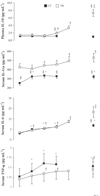
Flow cytometric acquisition and analyses. All samples were ac-quired on a FACSCalibur and analyzed using Cell Quest Pro software (BD Biosciences). For analysis of intranuclear NF-B, double gating on a forward vs. side scatter dot plot and FL2 PI staining were used to collect 3,000 PBMC and 5,000 PMN events and to quantify intranuclear NF-B expression in fresh whole blood and cultured samples (Fig. 2). Lymphocytes were determined using cell-surface staining characteristics gating side scatter and anti-CD14 APC, ex-cluding monocytes and neutrophils. Two-color gating, CD14 vs.
CD16, of the lymphocyte subset was used to determine CD16⫹subset
frequency. Total leukocyte counts were obtained from K2
EDTA-treated whole blood using a hematology analyzer (Coulter ACT diff 2)
and corrected for changes in blood volume incorporating hemo-globin and hematocrit values (19). Leukocyte subset counts (PMN, PBMC, and CD16⫹ lymphocytes) were obtained by multiplying
the corresponding population percentages obtained from FACS analysis by the total leukocyte count.
Statistical analyses. An ANOVA with one repeated factor (tem-perature) and one between factor (fitness) was calculated on the various immunologically and physiologically dependent measures sampled during the trial as well as a separate ANOVA comparison between baseline and Exh. In addition, a one-factor (fitness) ANOVA was used to compare physiologically dependent measures, such as BV, Tretolerated at Exh, TT, rate of Treincrease, and anthropometric
data. An ANOVA with two repeated factors (temperature and stimu-lus) and one between factor (fitness) was calculated on baseline and Exh samples stimulated in vitro. For all analyses, subject numbers (n) were as follows: from baseline to 38.5°C and Exh, n ⫽ 12 TR and 11 UT; at 39.0°C, n ⫽ 12 TR and 9 UT; and at 39.5°C, n ⫽ 11 TR. To correct for violations in the assumption of sphericity with the repeated factors, we applied the Huynh-Feldt correction to the F ratio. Post hoc comparisons were performed using a Newman-Keuls procedure to isolate specific group mean differences at each Treinterval and within
each group over temperature. All ANOVAs were performed with statistical software [StatSoft (2007) Statistica (data analysis software system), version 8.0, www.statsoft.com]. For all analyses, an ␣ level of 0.05 was used.
RESULTS
Group characteristics.Anthropometric characteristics were not different between groups (Table 1). All values are means ⫾ SE with range in parentheses. Maximum oxygen consumption expressed per kilogram of LBM [70 ⫾ 2 (63– 80) vs. 50 ⫾ 1 (45–52) ml 䡠kg LBM⫺1
䡠 min⫺1] and per unit of total mass [62 ⫾ 2 (54 –73) vs. 42 ⫾ 1 (37– 44) ml 䡠kg⫺1
䡠 min⫺1] as well as body fatness [9.6 ⫾ 1.0 (6 –15) vs. 15.8 ⫾ 1.7 (10 –24)%] were significantly different between the groups (TR vs. UT, respectively). Blood and plasma volumes were also signifi-cantly elevated in TR compared with UT subjects (Table 2).
Physiological response to EHS.Our EHS model produced significant increases in HR, Msk, and plasma volume shifts
(Table 3). The absolute metabolic cost of walking at 4.5 km/h and 2% elevation was not significantly different between the groups up to 39.0°C (15.23 ⫾ 0.1 ml 䡠kg⫺1
䡠 min⫺1); however, the average relative percentage of V˙O2peakduring the trial was
significantly elevated in UT compared with TR subjects (35.9 ⫾ 1.3 vs. 24.8 ⫾ 0.9% V˙O2peak, respectively). Resting
HR was significantly higher in UT compared with TR subjects; however, there were no differences observed in Tre(36.9 ⫾
0.1°C) at baseline between the groups. Of note, HR at exhaus-tion was not significantly different between groups at 160 ⫾ 3 beats/min or 81 ⫾ 1% of HRpeak, although still significantly
lower than HRpeak (196 ⫾ 2 beats/min). For absolute and
relative HR changes, there were significant fitness and temper-ature effects, greater for UT than for TR subjects. Because of lower Mskat baseline, change in Mskwas significantly greater
in UT compared with TR subjects for a given level of thermal strain. Tretolerated at Exh was higher in TR (39.7 ⫾ 0.1°C)
compared with UT subjects (39.1 ⫾ 0.1°C), which produced significantly longer TT in TR (162.5 ⫾ 11 min) compared with UT subjects (106 ⫾ 10 min). However, despite the longer TT in TR subjects, the rate of Treincrease (1.0 ⫾ 0.05°C/h) and
Fig. 2. Representative fluorescence histograms for NF-B FITC staining [1, unstained; 2, isotype (normal mouse IgG1); 3, NF-B p65; 4, NF-B p65 in
vitro LPS] for polymorphonuclear neutrophils (PMN; A) and peripheral blood mononuclear cells (PBMC; B). Data are from endurance-trained (TR) subject 3.
by 10.220.33.5 on January 9, 2017
http://ajpregu.physiology.org/
time between Tre sampling intervals were not significantly
different between groups. Reasons for trial termination con-sisted of six TR subjects attaining the ethical Tre cutoff of
40.0°C, one UT subject attaining the HR cutoff, and the remaining 16 subjects reaching physical exhaustion. In addi-tion, seven subjects (3 TR; 4 UT) also experienced nausea at trial termination. There was no relationship between endotoxin levels at Exh and symptoms of nausea. All osmolality values were within a normal range of 280 –300 mosmol/kgH2O,
indicating that all subjects were euhydrated at the beginning and throughout the trial. Sweat rates were greater in TR (1.2 ⫾ 0.09 kg/h) compared with UT subjects (0.87 ⫾ 0.06 kg/h), and body mass was significantly decreased post-EHS, but the change in body mass was ⬍2% (0.87 ⫾ 0.4 vs. 1.9 ⫾ 0.5% in UT and TR subjects, respectively), and no differences were observed between the groups. TR subjects experienced a sig-nificant plasma volume shift from baseline at 38.5°C, which remained for the duration of the trial, and were greater than values in UT subjects at Exh. In contrast, UT subjects did not experience a significant shift in plasma volume during the EHS trial (Table 3).
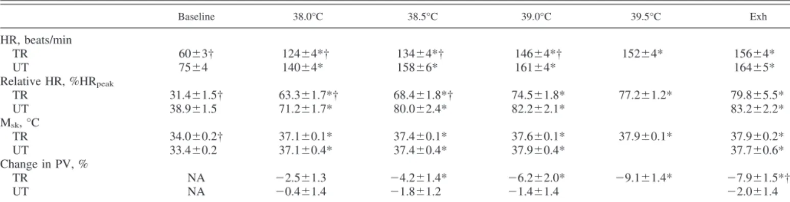
Total leukocytes and subset changes.EHS produced a tem-perature-dependent increase in the total leukocyte count and subset counts (⫻109 cells/l) (Fig. 3); however, the only
de-tected change in leukocyte distribution was within the CD16⫹
lymphocyte subset, with greater percentages observed in TR compared with UT subjects (Table 4).
Plasma endotoxin and LBP.Plasma endotoxin levels were not significantly different in TR compared with UT subjects at rest (trend P ⫽ 0.1); however, a significant group effect (UT ⬎ TR) was observed throughout the EHS trial. EHS produced significant increases in both plasma endotoxin and serum LBP levels, as depicted in Fig. 4. Resting plasma endotoxin was detectable in 11 subjects (4 TR; 7 UT), whereas at Exh, 18 of 23 subjects had a detectable increase in circulating endotoxin (9 TR, range 3.8 –16.5 pg/ml; 9 UT, range 3.8 –34 pg/ml). When 39.0°C was reached, a ⬎2-fold increase in circulating plasma endotoxin concentration was observed in UT compared with TR subjects.
Intranuclear NF-B translocation. A temperature-depen-dent elevation in the percentage of PBMC cells expressing NF-B in TR and UT subjects was observed when comparing baseline to 38.0°C during EHS; however, there were no further increases in the percentage of positive cells between 38.0 and
39.0°C in UT subjects (Table 4). Increases in NF-B in PBMC have been reported to correlate strongly with an increasing proportion of NK cells, a major source of nuclear p65 content, without intranuclear activation (69). In a whole body physical stress model, sympathetic-adrenomedullary system activation produces a redistribution of circulating lymphocytes, predom-inated by an increased mobilization of NK cells to the periph-eral blood (78). In the present study, a significantly greater percentage of CD16⫹ NK cells were present in TR compared
with UT subjects at baseline, contributing to the greater per-centage of PBMC positive for NF-B at baseline and during EHS (Table 4). CD16⫹ lymphocyte concentration accounted
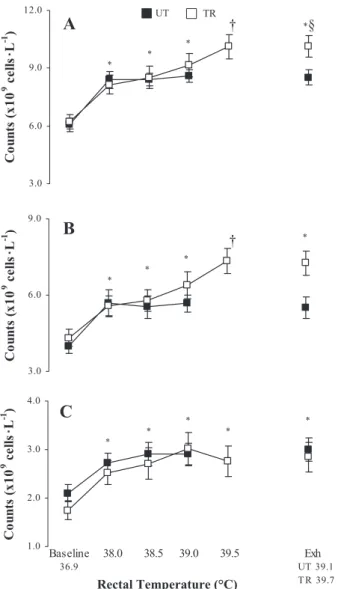
for less than one-half of the observed PBMC positive counts at baseline, indicating that other subsets, such as B and T cells and/or monocytes, might be contributing to the basal percent-ages. PBMC positive counts adjusted for NK cell mobilization (PBMC minus NK) produced significant increases at 38.0 and 38.5°C in UT subjects, whereas values in TR subjects did not significantly increase before 39.0°C (Fig. 5). There were no significant differences in mean fluorescence intensity (MFI) between TR and UT subjects at baseline in either PBMC or PMN. A significant main effect of temperature was observed in intranuclear NF-B MFI in PBMC, and a significant fitness ⫻ temperature interaction was observed in PMN MFI. LPS stim-ulation in vitro induced significant increases in NF-B trans-location in TR and UT subjects from baseline, although at Exh, significant increases in translocation were only observed in UT subjects. Furthermore, both groups showed an attenuation of NF-B translocation following LPS stimulation in PBMC, whereas in PMN, this was only reduced in TR subjects (Fig. 6).
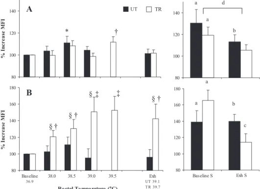
Circulating cytokines. Circulating levels of TNF-␣, IL-10, IL-6, and IL-1ra are depicted in Fig. 7. At rest, circulating cytokine profiles between TR and UT subjects were similar except for significantly greater levels of anti-inflammatory IL-1ra, which remained elevated in TR compared with UT subjects throughout the heat stress trial. EHS produced signif-icant increases in proinflammatory cytokine TNF-␣ at a lower absolute level of thermal strain in UT (38.0°C) compared with TR subjects (38.5°C). IL-6 increased consistently throughout the heat stress trial in both groups in a temperature-dependent manner, whereas anti-inflammatory IL-1ra significantly in-creased at 38.0 and 39.5°C in UT and TR subjects, respec-tively, but concentrations in UT subjects did not reach those in Table 1. Anthropometric characteristics of age, height, mass, LBM, AD, and AD:mass for TR and UT groups
Group Age, yr Height, cm Mass, kg LBM, kg AD, m
2
AD/Mass, m2䡠 kg⫺1䡠 10⫺2
TR 24⫾1 (18–31) 178⫾1 (170–189) 73.3⫾2.2 (60–89) 65.2⫾1.7 (55–76) 1.90⫾0.03 (1.78–2.11) 2.61⫾0.04 (2.3–2.9) UT 23⫾1 (18–32) 177⫾2 (167–190) 78.7⫾2.7 (64–92) 66.1⫾1.6 (56–78) 1.95⫾0.04 (1.83–2.17) 2.50⫾0.04 (2.4–2.8) Values are means ⫾ SE (range in parentheses) for age, height, mass, lean body mass (LBM), body surface area (AD), and surface-to-mass ratio (AD/mass)
in endurance-trained (TR) and untrained (UT) groups.
Table 2. Absolute and relative blood volume as determined using indocyanine green dye for TR and UT individuals
Group n BV, ml BV, ml/kg PV, liters PV, ml/kg
TR 11 7,565⫾282* (6,440–9,199) 104.2⫾5.7* (76.5–117.5) 4,673⫾200* (3,621–5,482) 64.4⫾3.6* (45.9–89.3) UT 10 6,675⫾191 (5,712–7,743) 84.0⫾3.1 (68.7–97.7) 4,057⫾100 (3,370–4,766) 51.0⫾1.9 (41.3–59.3)
Values are means ⫾ SE (range in parentheses) of absolute and relative blood (BV) and plasma volume (PV) in TR and UT individuals. *P ⬍ 0.05, between-group difference.
by 10.220.33.5 on January 9, 2017
http://ajpregu.physiology.org/
TR subjects. In contrast, an enhanced IL-10 concentration was observed only in TR subjects at Trevalues above 38.5°C.
DISCUSSION
During conditions of uncompensable EHS, endurance-trained individuals have been found to tolerate Tre ⱕ 40°C,
whereas untrained sedentary individuals succumb to EHS at much lower Trevalues, usually around 39.0°C (77). Therefore,
it was expected that in the current uncompensable EHS model, TR would tolerate a higher Tre at Exh. Traditionally, it has
been suggested that tolerance to higher levels of Treis due to
an increased cardiovascular and thermoregulatory stability as produced by hypervolemia associated with training (77). The current findings suggest an additional cellular link between intranuclear NF-B regulation and the translocation of endo-toxin, which underlie the greater heat tolerance in endurance-trained individuals. This study demonstrates for the first time that increases in circulating endotoxin correspond to NF-B translo-cation and inflammatory cytokine production at Trevalues below
40.0°C. Moreover, the inflammatory cascade is accompanied by a compensatory anti-inflammatory response during uncompensable EHS. These findings support the concept of a heat illness contin-uum, linking the pathophysiological progression from heat stress to exertional heat exhaustion and/or exertional heat stroke (40, 79).
Redistribution of splanchnic blood and intestinal permeabil-ity.As thermoregulatory mechanisms are activated, there is a redistribution of central blood volume to the cutaneous circu-lation, decreasing venous return, stroke volume, and mean arterial and venous pressures (71). To maintain cardiac output with a reduced stroke volume, HR is increased to meet the demand to maintain muscle and cutaneous blood flow. Accom-panying the sympathetic drive to increase HR is an equivalent sympathetic vasoconstriction of the splanchnic circulation such that corresponding relative intensities produce an equivalent reduction in visceral blood flow, independent of V˙O2peak(70).
Although a linear reduction in splanchnic blood flow is ob-served with increasing intensity, it does not immediately result in a compromised local oxygen demand.
A number of mechanisms are proposed for the breakdown of gastrointestinal barrier function, including the disruption of normal epithelial mucosa integrity and/or failure of epithelial physical tight junctions (20, 32). It is well documented that thermal loads above 41.5°C result in a rapid increase in
intestinal epithelial permeability and onset of heat stroke (4). Heating of Caco-2 monolayers from 37 to 41°C has been found to result in increased intestinal epithelial tight junction perme-ability (20), and temperatures as low as 38.3°C have been found to increase transepithelial electrical conductance, result-ing in paracellular permeability (51).
In addition to an increase in thermal load, adequate blood supply is critical for the maintenance of the gastrointestinal barrier (31, 72). Reduced oxygen supply impairs normal anti-inflammatory processes of the gut mucosa, resulting in in-creased NF-B activation (65) and iNOS (32) and/or TNF-␣ release (18), which appear to be essential components for the breakdown of tight junctions and mucosa during endotoxemia (32). As such, gut ischemia is a common aspect of critical illness and has been proposed as the “motor” of multiple organ failure (12).
Reductions in visceral blood flow have been reported at HR values below 90 beats/min during light exercise (57). Yet, it appears that a significantly higher intensity is required for induction of gut ischemia-related permeability (58, 59). For instance, a significant reduction of gastric mucosal perfusion and ischemia has been reported in untrained subjects after only 10 min of exercise at 80% HRpeak(162 beats/min) in a neutral
environment (58). Comparatively, Pals et al. (59) found that 60 min of running at 80% V˙O2peak(Tre, 39.6°C; HR, 180 beats/
min) in trained subjects (V˙O2peak, 57 ⫾ 2 ml 䡠kg⫺1䡠 min⫺1) was necessary to produce an increase in small intestine permeabil-ity compared with 40 and 60% intensities. Based on previous work by Rowell (70) and our current HR responses, UT subjects in the current study may have experienced a greater reduction in splanchnic blood flow compared with their TR counterparts, resulting in an earlier appearance of plasma endotoxin and TNF-␣. The observed reduction in HR in TR subjects can be related to the hypervolemia associated with endurance-training, since relative to improved ventricular function, elevated blood volume increases venous return, as well as preload and subsequent venous filling pressures, and consequently, increased left ventricular end-diastolic volumes lead to an increase in stroke volume and cardiac output. This progression allows workloads to be maintained at a lower submaximal HR (28). It is interesting to note that the appear-ance of plasma endotoxin corresponded to the same relative HR intensity between the groups, although at significantly different levels of thermal strain: 80% in UT and 75% in TR. Table 3. Physiological responses for TR and UT groups during exertional heat stress
Baseline 38.0°C 38.5°C 39.0°C 39.5°C Exh HR, beats/min TR 60⫾3† 124⫾4*† 134⫾4*† 146⫾4*† 152⫾4* 156⫾4* UT 75⫾4 140⫾4* 158⫾6* 161⫾4* 164⫾5* Relative HR, %HRpeak TR 31.4⫾1.5† 63.3⫾1.7*† 68.4⫾1.8*† 74.5⫾1.8* 77.2⫾1.2* 79.8⫾5.5* UT 38.9⫾1.5 71.2⫾1.7* 80.0⫾2.4* 82.2⫾2.1* 83.2⫾2.2* Msk, °C TR 34.0⫾0.2† 37.1⫾0.1* 37.4⫾0.1* 37.6⫾0.1* 37.9⫾0.1* 37.9⫾0.2* UT 33.4⫾0.2 37.1⫾0.4* 37.4⫾0.4* 37.9⫾0.4* 37.7⫾0.6* Change in PV, % TR NA ⫺2.5⫾1.3 ⫺4.2⫾1.4* ⫺6.2⫾2.0* ⫺9.1⫾1.4* ⫺7.9⫾1.5*† UT NA ⫺0.4⫾1.4 ⫺1.8⫾1.2 ⫺1.4⫾1.4 ⫺2.0⫾1.4
Values are means ⫾ SE. From baseline to 38.5°C and at exhaustion (Exh), n ⫽ 12 TR and 11 UT; at 39.0°C, n ⫽ 12 TR and 9 UT; and at 39.5°C, n ⫽ 11 TR. *P ⬍ 0.05, significantly different from baseline. †P ⬍ 0.05, between-group difference.
by 10.220.33.5 on January 9, 2017
http://ajpregu.physiology.org/
Furthermore, the present findings are consistent with the ob-served appearance of endotoxin in subjects following their completion of a half-marathon (mean Tre, 39.6°C; HR, 172
beats/min) (53) and suggest that the onset of gut permeability
may have occurred at lower intensities, perhaps even below the 80% HR threshold (⬃160 beats/min), as suggested by previous studies (58, 59).
Overall, HR responses did not approach maximum values in either of the groups, and a HR plateau occurred before Exh despite increasing sympathetic drive (data not shown), a re-sponse that may be reflective of a protective mechanism to maintain cardiac filling pressures (36). It is unlikely that progressive fluid loss contributed to the observed differences in HR responses or reasons for trial termination between the groups, since progressive dehydration and subsequent cardio-vascular drift were minimized by a regimented hydration schedule (⬍2% change in body weight). Previously, it was shown that a combined 1°C increase in Treand 4% loss of body
weight are required before significant cardiac dysfunction is observed (29).
Circulating plasma endotoxin. Quantification of endotoxin levels within the plasma and/or serum can be performed using the LAL (55). Historically, this assay has been problematic Fig. 3. Concentration (⫻109 cells/l) of total circulating leukocyte subsets
during external heat stress (EHS) between TR and untrained (UT) groups for total leukocytes (A), PMN (B), and PBMC (C). Values are means ⫾ SE. From baseline to 38.5°C and exhaustion (Exh), n ⫽ 12 TR and 11 UT subjects; at 39.0°C, n ⫽ 12 TR and 9 UT subjects; and at 39.5°C, n ⫽ 11 TR subjects. *P ⬍ 0.05, TR and UT are significantly different from baseline. †P ⬍ 0.05, TR significantly different from baseline to 38.5°C. §P ⬍ 0.05, between-group significance.
Fig. 4. Circulating plasma endotoxin and serum LPS binding protein (LBP) concentrations in TR and UT groups during EHS. Values are means ⫾ SE. From baseline to 38.5°C and Exh, n ⫽ 12 TR and 11 UT; at 39.0°C, n ⫽ 12 TR and 9 UT; and at 39.5°C, n ⫽ 11 TR. *P ⬍ 0.05, UT significantly different from baseline. †P ⬍ 0.05, TR significantly different from baseline. ‡P ⬍ 0.05, UT significantly different from baseline to 38.5°C. §P ⬍ 0.05, between-group significance.
Table 4. Percentage of PBMC positive for NF-B and CD16⫹lymphocytes during exertional heat stress for TR and UT groups Baseline 38.0°C 38.5°C 39.0°C 39.5°C Exh PBMC positive for NF-B, % TR 33.9⫾3.8† 43.8⫾3.9*† 49.6⫾7.3*† 55.5⫾8.0*† 63.0⫾6.6* 55.1⫾6.1*† UT 25.0⫾2.8 33.4⫾4.1* 36.3⫾4.7* 34.6⫾4.9* 33.0⫾4.6* CD16⫹lymphocytes, % TR 19.0⫾2.7† 36.7⫾4.1*† 38.3⫾4.7*† 35.0⫾4.6*† 36.7⫾3.8* 35.8⫾4.7*† UT 7.9⫾0.8 15.0⫾1.0* 14.0⫾1.1* 13.7⫾1.4* 13.8⫾1.5*
Values are means ⫾ SE. PBMC, peripheral blood mononuclear cells. From baseline to 38.5°C and at Exh, n ⫽ 12 TR and 11 UT; at 39.0°C, n ⫽ 12 TR and 9 UT; and at 39.5°C, n ⫽ 11 TR. *P ⬍ 0.05, significantly different from baseline. †P ⬍ 0.05, between-group difference.
by 10.220.33.5 on January 9, 2017
http://ajpregu.physiology.org/
because of the inhibition/enhancement properties of the plasma matrix. To remove matrix interference, samples are diluted and heat treated, and a 1–3--D-glucan-inhibiting buffer is
em-ployed before testing is conducted. Without the incorporation of these techniques, the LAL results can be difficult to inter-pret. Normal background levels without -glucan inhibition have been reported in clinical studies to occur at ⬃0.05 Eu/ml (55), which, depending on the potency of the endotoxin used as a standard, corresponds to a level from 5 to 10 pg/ml in healthy controls. Nadhazi et al. (52) reported measurable levels in all of their 116 healthy donors, including a subset of lower respond-ers who had a mean value of 0.05 Eu/ml and similar mean values of 0.06 Eu/ml that were reported in 10 student controls, ages 18 –22 yr (61). Others have reported lower resting mean
levels at baseline, with a range from undetectable to 2 pg/ml (⬃0.02 Eu/ml) (8, 33, 54, 56, 74). The decrease in the resting levels of endotoxin in recent studies may be attributed to the use of 1–3--D-glucan inhibitors. Other differences in
method-ology, such as heat treatment, medium for detection (plasma vs. serum) (55), and the potency of LPS used for standards, also may be contributing factors to variability in reported values. Another possible contributing factor to the variance in background values may be the nature of the subjects and individual variations in endotoxin-neutralizing capacity (88). Many of the earlier studies used age-matched normal healthy individuals as controls, whereas studies examining mild endo-toxemia with strenuous exercise have employed subject co-horts consisting of marathoners/triathletes (8, 33) or ultra-endurance athletes (54). A common characteristic of the latter is a considerably higher level of aerobic fitness than would be expected in an average healthy individual. Data from current studies support the notion that training adaptations contribute to lower levels of endotoxin as have been reported in this cohort of subjects (see below).
Systemic inflammatory activation and acute phase response.
As an inducible transcription factor, NF-B is activated by a variety of agents, including reactive oxygen species (ROS) (1), cytokines (39), and endotoxin (4), many of which may be related to the pathophysiological changes associated with strenuous exercise and/or exertional hyperthermia (49, 79). Moreover, ischemic and/or thermal stress can damage the intestinal barrier, provoking the release of endogenous “danger or damage” signals from degraded tissue or necrotic cells that further initiate NF-B activation (2). Our findings that sys-temic activation of NF-B and the appearance of TNF-␣ in the circulation occur at a Treof 38.0°C in UT subjects, preceding
significant increases in plasma endotoxin, is not surprising, since endotoxemia within the portal circulation can elicit NF-B activation and the production of inflammatory media-Fig. 5. Concentration (⫻109cells/l) of PBMC positive for NF-B corrected
for CD16⫹natural killer counts during EHS in TR and UT groups. Values are
means ⫾ SE. From baseline to 38.5°C and Exh, n ⫽ 12 TR and 11 UT; at 39.0°C, n ⫽ 12 TR and 9 UT; and at 39.5°C, n ⫽ 11 TR. *P ⬍ 0.05, UT significantly different from baseline. †P ⬍ 0.05, TR significantly different from baseline to 38.5°C.
Fig. 6. Percentage change in mean fluores-cence intensity (MFI) from baseline for PBMC (A) and PMN (B) positive for NF-B in vivo and following in vitro LPS stimulation (S; 100 ng/ml, 37°C, 30 min) in TR and UT groups during EHS. Values are means ⫾ SE. From baseline to 38.5°C and Exh, n ⫽ 12 TR and 11 UT; at 39.0°C, n ⫽ 12 TR and 9 UT; and at 39.5°C, n ⫽ 11 TR. *P ⬍ 0.05, TR and UT significantly different from baseline. †P ⬍ 0.05, TR significantly different from baseline. ‡P ⬍ 0.05, TR significantly different from baseline to 38.5°C. §P ⬍ 0.05, between-group difference.aP ⬍0.05, significant increase with
stimulation from baseline.bP ⬍0.05,
signifi-cant increase with stimulation from Exh.cP ⬍
0.05, baseline S significantly different from Exh S. dP ⬍ 0.05, main effect (baseline S
significantly different from Exh S).
by 10.220.33.5 on January 9, 2017
http://ajpregu.physiology.org/
tors in both the intestinal mucosa (65) and liver (14, 26, 63) before spillover into the systemic circulation (31). In fact, both the intestinal epithelial and liver are considered important sources of inflammatory mediators during endotoxemia (13, 63). Secretion of inflammatory mediators from ischemic intes-tinal Caco-2 cells has even been suggested to contribute to activation of circulating PBMC and PMN priming (42).
The acute phase protein LBP is primarily responsible for transporting endotoxin to immune effector cells and can en-hance sensitivity of monocytes/macrophages to LPS by more than 1,000-fold (75). Elevated plasma levels of LBP have been shown to correlate with poor outcome after thermal injury (21),
trauma (17), and in septic patients with infectious complica-tions (91). Peak values in such disease states have been reported as high as 200 g/ml (17). Release of LBP occurs primarily from hepatocytes and intestinal epithelial cells (35, 91) stimulated by IL-6, TNF-␣, IL-1, and/or glucocorticoids but not by endotoxin (86). Along with its ability to enhance endotoxin receptor binding, LBP at higher concentrations (50 – 80 g/ml) can hinder the LBP-CD14 interaction (91), releasing bound endotoxin to lipoproteins, such as HDL (37), enhancing hepatic uptake through LDL and VLDL (87) and contributing to endotoxin-neutralizing capacity (88).
As a consequence of its critical position early in the inflam-matory process, LBP has been proposed to contribute to the differences in in vitro LPS tolerance observed between trained and untrained individuals (22). Although differences in LBP concentration at baseline were not observed between our groups, LBP levels were significantly elevated in UT subjects at 39.0°C and Exh, suggesting the initiation of the acute phase response in these individuals. Despite our observed increases, LBP levels were still within the normal range (5–15 g/ml), and it remains to be seen whether these subclinical increases enhance LPS receptor sensitivity or contribute to the reduc-tion of in vitro LPS-induced NF-B translocareduc-tion observed post-EHS.
Circulating cytokine kinetics during SIRS are propagated by increased levels of the proinflammatory mediators TNF-␣ and 1, followed by anti-inflammatory increases in 6, IL-1ra, soluble TNF receptor (sTNFR), and IL-10 in a time-dependent manner (38). By comparison, the magnitude of the cytokinemia accompanying submaximal exercise is typically much milder and does not typically include the profound increases in proinflammatory mediators associated with many clinical conditions (62, 82). Of course, there are several studies that have documented subclinical increases in proinflammatory cytokines during strenuous exercise (84) and exertional hyper-thermia (60, 67, 81).
In the present study, we observed a greater increase in plasma endotoxin concentrations in UT (14.5 pg/ml) compared with TR subjects (8.08 pg/ml) at Exh, and these observed levels were comparable to those previously reported during mild endotoxemia (5–15 pg/ml) following strenuous endurance exercise (8, 33). In addition, the increases in TNF-␣ in the present study were comparable to those in studies employing a thermal clamping technique, which reported increases follow-ing 40 –90 min of cyclfollow-ing at 65–70% V˙O2peakin the heat (67,
81). Nevertheless, the observed increases in TNF-␣ during exertional hyperthermia are at least 100 times lower than levels seen in individuals suffering from acute heatstroke (4, 5) or following intravenous endotoxin injection (34).
Intravascular infusion of endotoxin (2 ng/kg–26 pg/ml) in healthy individuals (70 kg, 5.3-liter blood volume) has been associated with a decrease in circulating leukocytes, fever (i.e., an increase in Treof 2°C within 4 h), tachycardia (HR ⬎ 100
beats/min), headache, chills, and a ⬎100-fold increase in TNF-␣ (50 –1,000 pg/ml) within the first hour after injection (34). The relatively low TNF-␣ levels observed during exertion can be partly attributed to transient TNF-␣ kinetics and rapid clearance from the circulation, making its detection difficult (84), especially when samples are taken at the end of the exertional period. In addition, the anti-inflammatory response associated with strenuous exercise is a regulatory adaptation Fig. 7. Circulating plasma/serum concentrations of tumor necrosis factor
(TNF)-␣, IL-6, IL-1 receptor antagonist (IL-1ra), and IL-10 in TR and UT groups during EHS. Values are means ⫾ SE. From baseline to 38.5°C and Exh, n ⫽12 TR and 11 UT; at 39.0°C, n ⫽ 12 TR and 9 UT; and at 39.5°C, n ⫽ 11 TR. *P ⬍ 0.05 ,UT significantly different from baseline. †P ⬍ 0.05, TR significantly different from baseline. ‡P ⬍ 0.05, TR significantly different from baseline to 39.5°C. §P ⬍ 0.05, between-group difference.
by 10.220.33.5 on January 9, 2017
http://ajpregu.physiology.org/
that limits pathophysiological inflammatory responses (84) and may account for the relatively low TNF-␣ levels observed, despite endotoxemia (53). In the present study, the subclinical increase in TNF-␣ (⬍2-fold) was accompanied by a concom-itant increase in IL-6 and IL-1ra, plus a marked increase in IL-10 in TR subjects during EHS. IL-6 has been found to increase up to 100-fold (62) with strenuous exercise and can inhibit endotoxin-induced increases in TNF-␣ (80) as well as stimulate production of IL-1ra, IL-10 (82), and secretion of acute phase proteins in the liver (C-reactive protein and LBP) (62). Reciprocally, IL-10 can influence IL-1ra expres-sion in LPS-challenged leukocytes (11). High levels of epinephrine produced during exercise also may blunt endo-toxin-induced TNF-␣ release while potentiating IL-10 pro-duction (85). It is possible that increases in circulating proinflammatory mediators, such as TNF-␣ and IL-1 (67, 81), may be crossing a compromised blood-brain barrier during exercise in a warm environment (89), promoting physical exhaustion signaling in the brain (10) and contrib-uting to the differences in Tretolerated between our groups. Effects of training on endotoxin-mediated cytokinemia.
Knowledge of intravascular blood volume is an important factor when examining cardiovascular function and exercise performance in the heat. A major factor contributing to the increased V˙O2peak associated with endurance training is
im-proved cardiac function due to an increased blood volume (28, 45). As a result, hypervolemia enables endurance-trained indi-viduals to maintain workloads at a lower submaximal HR (28). Endurance-training associated increases (20%) in relative blood volume (ml/kg) contributed to a 10% reduction in the average relative metabolic cost of exercise, HR for a given workload, and splanchnic blood flow redistribution, shifting the relationship between endotoxin-mediated cytokinemia and Treto the right in these individuals (see Figs. 4, 5, and
7). This highlights an important secondary role of training-induced hypervolemia on monocytic inflammatory activa-tion during EHS.
There is increasing evidence that translocation of small amounts of endotoxin into the circulation occurs in healthy individuals during conditions such as heat stress, which can routinely stimulate host defenses (44). Athletes training for ultra-endurance competitions, such as a marathon or triathlon, appear to have elevated levels of anti-LPS IgG before a race (3, 33). This suggests that individuals participating in regular strenuous physical activity or endurance training may develop an improved endotoxin tolerance (15, 46, 81) due to small, repeated exposures to LPS (72), resulting in a form of self-immunization (3). An important modulator of endotoxin toler-ance is the repeated expression of glucocorticoids (90) and growth hormone (6), as well as anti-inflammatory cytokines and the induction of HSP in association with acute exercise (84). Although resting levels of TNF-␣, IL-6, and IL-10 were not different in the present study, increased IL-1ra was ob-served in TR subjects, and training has been associated with increased levels of sTNFR (38). Both of these proinflam-matory antagonists have been shown to enhance survival during septic conditions (62). Changes in surface expression of TLR-2 and -4 observed with training may also be impor-tant contributing factors in the improved endotoxin toler-ance following habitual physical activity (22).
Another important consideration is the observation that human plasma from different individuals can exhibit more than a 100-fold range in endotoxin-neutralizing capacity (88). En-durance training has been associated with increased circulating levels of anti-LPS IgG (3, 33), IgM (7), an accentuated NK cell mobilization (68), increased levels of HDL (41), and PMN hyperreactivity, all of which may be contributing to mediate endotoxin clearance and reduce inflammatory mediators such as TNF-␣ or C-reactive protein in our endurance-trained sub-jects (83). Since the chromogenic LAL is only able to measure the potency of free endotoxin and does not account for LPS bound to various lipoproteins (LBP, HDL, VLDL, LDL) or soluble/membrane CD14, the measured levels in the present study may represent only a fraction (⬃30%) of the total circulating LPS content (73). Therefore, differences in baseline circulating concentrations as well as improved endotoxin tolerance may be attributed to a greater endotoxin-neutral-izing capacity due to individual differences in lipoprotein profiles (41).
Induction of the stress response provides significant cyto-protection against various cellular stressors, providing mainte-nance of immune function (50). HSP72 accumulation has been found to complex with NF-B/IB, regulating inflammatory activation in the liver (14, 64), intestinal mucosa (65), and epithelium (43) during conditions of endotoxemia. A greater intracellular/circulating HSP72 induction (66) and increased HSF-1 DNA binding affinity (47) have been related to training and may contribute to the maintenance of gastrointestinal integrity at higher levels of thermal strain that was observed in our endurance-trained subjects. Likewise, impairment of tran-scriptional processes has been suggested in heat intolerance (49) and may hinder the ability to protect the gastrointestinal barrier against thermal and oxidative stressors in untrained, sedentary individuals.
Perspectives
Typically, the ability to tolerate higher levels of thermal load (⬃40.0°C) before circulatory compromise has been associated with a greater risk for the development of EHI in highly motivated endurance-trained individuals (27). Comparatively, sedentary individuals succumbing to EHS at much lower Tre
values are less prone to EHI, as long as cessation of exertion and removal from the environment occurs. However, as indi-cated in the present findings, sedentary untrained individuals under significant cardiovascular and thermoregulatory strain possess compromised gastrointestinal barrier integrity and in-flammatory activation at temperatures as low as 38.0°C. Since early symptoms of EHI are often misinterpreted or ignored, this may lead to a progression in EHI severity in unsuspecting individuals, driven in part by endotoxemia (40). Therefore, given the importance of the maintenance of barrier integrity and ensuing endotoxemia during EHS (4, 24, 72), the concept of endotoxin tolerance may be an important mechanism related to enhanced heat tolerance associated with endurance training. Furthermore, it is possible that transient endotoxin transloca-tion and the subsequent inductransloca-tion of the stress response with repeated training bouts may be an important mediator of antioxidant and anti-inflammatory cytoprotective adaptations accompanying habitual physical activity. As our understanding of the triggers and thresholds for the initiation of the
by 10.220.33.5 on January 9, 2017
http://ajpregu.physiology.org/
flammatory cascade and acute phase response during EHS improves, valuable insight is gained about the cellular mech-anisms contributing to improved heat tolerance and the regu-latory balance between proinflammatory and compensatory anti-inflammatory responses.
ACKNOWLEDGMENTS
We are indebted to D. Kerrigan-Brown, J. Pope, I. Smith, S. Petrongolo, R. Limmer, P. Lee, and C. Dann for technical assistance. The time and effort of the subjects in this investigation were greatly appreciated. We also thank Dr. N. Gledhill for guidance and recommendations for the measurement of blood volume.
GRANTS
This study was funded by a Defence Research and Development Canada Technology Investment Fund.
REFERENCES
1. Asehnoune K, Strassheim D, Mitra S, Kim JY, Abraham E. Involve-ment of reactive oxygen species in Toll-like receptor 4-dependent activa-tion of NF-kappaB. J Immunol 12: 2522–2529, 2004.
2. Bianchi ME. DAMPs, PAMPs, and alarmins: all we need to know about danger. J Leukoc Biol 81: 1–5, 2007.
3. Bosenberg AT, Brock-Utne JG, Gaffin SL, Wells MT, Blake GT. Strenuous exercise causes systemic endotoxemia. J Appl Physiol 65: 106 –108, 1988.
4. Bouchama A, Knochel JP. Heat stroke. N Engl J Med 346: 1978 –1988, 2002.
5. Bouchama A, Parhar RS, el-Yazigi A, Sheth K, al-Sedairy S. Endo-toxemia and release of tumor necrosis factor and interleukin 1 alpha in acute heatstroke. J Appl Physiol 70: 2640 –2644, 1991.
6. Bozzola M, De Amici M, Zecca M, Schimpff RM, Rapaport R. Modulating effect of human growth hormone on tumour necrosis factor-alpha and interleukin-1beta. Eur J Endocrinol 138: 640 – 643, 1998. 7. Camus G, Nys M, Poortmans JR, Venneman I, Monfils T,
Deby-Dupont G, Juchmes-Ferir A, Deby C, Lamy M, Duchateau J. Endo-toxaemia, production of tumour necrosis factor alpha and polymorphonu-clear neutrophil activation following strenuous exercise in humans. Eur J Appl Physiol79: 62– 68, 1998.
8. Camus G, Nys M, Poortmans JR, Venneman I, Monfils T,
Deby-Dupont G, Juchmes-Ferir A, Deby C, Lamy M, Duchateau J.Possible in vivo tolerance of human polymorphonuclear neutrophil to low-grade exercise-induced endotoxaemia. Mediators Inflamm 7: 413– 415, 1998. 9. Camus G, Poortmans J, Nys M, Deby-Dupont G, Duchateau J, Deby
C, Lamy M.Mild endotoxaemia and the inflammatory response induced by a marathon race. Clin Sci (Lond) 92: 415– 422, 1997.
10. Carmichael MD, Davis JM, Murphy EA, Brown AS, Carson JA,
Mayer EP, Ghaffar A. Role of brain IL-1 on fatigue after exercise-induced muscle damage. Am J Physiol Regul Integr Comp Physiol 291: R1344 –R1348, 2006.
11. Cassatella MA, Poortmans J, Leclercq R, Brasseur M, Duchateau J,
Newsholme EA.Interleukin 10 (IL-10) upregulates IL-1 receptor antag-onist production from lipopolysaccharide-stimulated human polymorpho-nuclear leukocytes by delaying mRNA degradation. J Exp Med 179: 1695–1699, 1994.
12. Chang JX, Chen S, Ma LP, Jiang LY, Chen JW, Chang RM, Wen LQ,
Wu W, Jiang ZP, Huang ZT.Functional and morphological changes of the gut barrier during the restitution process after hemorrhagic shock. World J Gastroenterol11: 5485–5491, 2005.
13. Chen D, Pan J, Du B, Sun D. Induction of the heat shock response in vivo inhibits NF-B activity and protects murine liver from endotoxemia-induced injury. J Clin Immunol 25: 452– 461, 2005.
14. Chen HW, Kuo HT, Wang SJ, Lu TS, Yang RC. In vivo heat shock protein assembles with septic liver NF-B/I-B complex regulating NF-B activity. Shock 24: 232–238, 2005.
15. Chen HI, Hsieh SY, Yang FL, Hsu YH, Lin CC. Exercise training attenuates septic responses in conscious rats. Med Sci Sports Exerc 39: 435– 442, 2007.
16. Chen YW, Chen SH, Chou W, Lo YM, Hung CH, Lin MT. Exercise pretraining protects against cerebral ischaemia induced by heat stroke in rats. Br J Sports Med 41: 597– 602, 2007.
17. Cunningham SC, Malone DL, Bochicchio GV, Genuit T, Keledjian K,
Tracy JK, Napolitano LM. Serum lipopolysaccharide-binding protein concentrations in trauma victims. Surg Infect (Larchmt) 7: 251–261, 2006. 18. Diebel LN, Liberati DM, Baylor AE 3rd, Brown WJ, Diglio CA. The pivotal role of tumor necrosis factor-alpha in signaling apoptosis in intestinal epithelial cells under shock conditions. J Trauma 58: 995–1001, 2005. 19. Dill DB, Costill DL. Calculation of percentage change in volumes of blood,
plasma, and red cells in dehydration. J Appl Physiol 37: 247–248, 1974. 20. Dokladny K, Moseley PL, Ma TY. Physiologically relevant increase in
temperature causes an increase in intestinal epithelial tight junction perme-ability. Am J Physiol Gastrointest Liver Physiol 290: G204 –G212, 2006. 21. Fang CW, Yao YM, Shi ZG, Yu Y, Wu Y, Lu LR, Sheng ZY.
Lipopolysaccharide-binding protein and lipopolysaccharide receptor CD14 gene expression after thermal injury and its potential mechanism(s). J Trauma53: 957–967, 2002.
22. Flynn MG, McFarlin BK. Toll-like receptor 4: link to the anti-inflam-matory effects of exercise? Exerc Sport Sci Rev 34: 176 –181, 2006. 23. Foulds S. Novel flow cytometric method for quantifying nuclear binding
of the transcriptional factor nuclear factor kappa B in unseparated human monocytes. Cytometry 29: 182–186, 1997.
24. Gathiram P, Gaffin SL, Brock-Utne JG, Wells MT. Time course of endotoxemia and cardiovascular changes in heat-stressed primates. Aviat Space Environ Med58: 1071–1074, 1987.
25. Gathiram P, Wells MT, Brock-Utne JG, Gaffin SL. Antilipopolysac-charide improves survival in primates subjected to heat stroke. Circ Shock 23: 157–164, 1987.
26. Gathiram P, Wells MT, Raidoo D, Brock-Utne JG, Gaffin SL. Portal and systemic plasma lipopolysaccharide concentrations in heat-stressed primates. Circ Shock 25: 223–230, 1988.
27. Gisolfi C, Robinson S. Relations between physical training, acclimatiza-tion, and heat tolerance. J Appl Physiol 26: 530 –534, 1969.
28. Gledhill N, Cox D, Jamnik R. Endurance athletes’ stroke volume does not plateau: major advantage is diastolic function. Med Sci Sports Exerc 26: 1116 –1121, 1994.
29. Gonzalez-Alonso J. Separate and combined influences of dehydration and hyperthermia on cardiovascular responses to exercise. Int J Sports Med 19, Suppl2: S111–S114, 1998.
30. Gonzalez-Alonso J, Crandall CG, Johnson JM. The cardiovascular challenge of exercising in the heat. J Physiol 586: 45–53, 2008. 31. Hall DM, Baumgardner KR, Oberley TD, Gisolfi CV. Splanchnic
tissues undergo hypoxic stress during whole body hyperthermia. Am J Physiol Gastrointest Liver Physiol276: G1195–G1203, 1999.
32. Han X, Fink MP, Uchiyama T, Yang R, Delude RL. Increased iNOS activity is essential for hepatic epithelial tight junction dysfunction in endotoxemic mice. Am J Physiol Gastrointest Liver Physiol 286: G126 – G136, 2004.
33. Jeukendrup AE, Vet-Joop K, Sturk A, Stegen JH, Senden J, Saris
WH, Wagenmakers AJ. Relationship between gastro-intestinal com-plaints and endotoxaemia, cytokine release and the acute-phase reaction during and after a long- distance triathlon in highly trained men. Clin Sci (Colch)98: 47–55, 2000.
34. Jorgensen VL, Ibsen M, Andresen L, Schulzke JD, Perner A. Effects of endotoxaemia on markers of permeability, metabolism and inflamma-tion in the large bowel of healthy subjects. Acta Anaesthesiol Scand 51: 1085–1092, 2007.
35. Kato A, Ogasawara T, Homma T, Saito H, Matsumoto K. Lipopo-lysaccharide-binding protein critically regulates lipopolysaccharide-in-duced IFN-beta signaling pathway in human monocytes. J Immunol 172: 6185– 6194, 2004.
36. Kawabata T, Suzuki T, Miyagawa T. Effect of blood volume on plasma volume shift during exercise. J Therm Biol 29: 775–778, 2004. 37. Kitchens RL, Thompson PA. Impact of sepsis-induced changes in
plasma on LPS interactions with monocytes and plasma lipoproteins: roles of soluble CD14, LBP, and acute phase lipoproteins. J Endotoxin Res 9: 113–118, 2003.
38. Krabbe KS, Reichenberg A, Yirmiya R, Smed A, Pedersen BK,
Bruunsgaard H.Low-dose endotoxemia and human neuropsychological functions. Brain Behav Immun 19: 453– 460, 2005.
39. Leon LR. Heat stroke and cytokines. Prog Brain Res 162: 481–524, 2007. 40. Lim CL, Mackinnon LT. The roles of exercise-induced immune system disturbances in the pathology of heat stroke : the dual pathway model of heat stroke. Sports Med 36: 39 – 64, 2006.
41. Lippi G, Schena F, Salvagno GL, Montagnana M, Ballestrieri F,
Guidi GC.Comparison of the lipid profile and lipoprotein(a) between
by 10.220.33.5 on January 9, 2017
http://ajpregu.physiology.org/
![Fig. 2. Representative fluorescence histograms for NF-B FITC staining [1, unstained; 2, isotype (normal mouse IgG 1 ); 3, NF-B p65; 4, NF-B p65 in vitro LPS] for polymorphonuclear neutrophils (PMN; A) and peripheral blood mononuclear cells (PBMC; B)](https://thumb-eu.123doks.com/thumbv2/123doknet/14163852.473555/5.904.69.441.637.1046/representative-fluorescence-histograms-unstained-polymorphonuclear-neutrophils-peripheral-mononuclear.webp)