Biomechanics of the cerebrum at finite strain:
tissue and cell level study
by
Thibault Philippe Prevost
S.M., Materials Science and Engineering
Massachusetts Institute of Technology, 2006
Ingenieur de l'Ecole Polytechnique
Ecole Polytechnique, 2004
Submitted to the Department of Materials Science and Engineering
in partial fulfillment of the requirements for the degree of
Doctor of Philosophy
at the
MASSACHUSETTS INSTITUTE OF TECHNOLOGY
September 2010
2010 Massachusetts Institute of Technology
All rights reserved
Signature of Author ...
Certified by
Certified by...
Accepted by ...
Signature redacted
Departr
9t of Mateials Science and Engineering
1
11 August 2010
Signature redacted
..
...
Simona Socrate
Principal Research Scientist
Signature redacted
Thesis Supervisor
...
Subra Suresh
Vannevar Bush Professor of Engineering
./7Thesis,8d'
2rvisor
...
Signature redacted
OF TJUL 1
02017
LIBRARIES
ARCHIVES
a
Biomechanics of the cerebrum at finite strain: a tissue and cell
level study
by
Thibault Philippe Prevost
Submitted to the Department of Materials Science and Engineering on 11 August 2010, in partial fulfillment of the
requirements for the degree of Doctor of Philosophy
Abstract
The present study addresses the large strain nonlinear mechanical response of the cere-bral cortex at the macroscopic tissue level and at the microscopic cell level. Unconfined uniaxial compression tests were conducted in vitro on cortical samples of porcine brains. The tests consisted of load-unload and relaxation segments to 50% nominal deformation at 0.01 to 10 s-1 strain rates. The tissue exhibited moderate volumetric compressibil-ity, marked hysteretic features, and substantial nonlinearities. Indentation tests - with displacement histories mirroring those imposed in compression - were performed on the cortex of porcine brains in vivo, in situ and in vitro, in order to assess and contrast the mechanical properties of the live and dead tissue. The tissue response shared similar qualitative nonlinear viscoelastic features under all testing conditions, although, quan-titatively, the response was found to be significantly stiffer in situ than in vivo. Test protocols were also developed at the neuronal cell level using atomic force microscopy. The response of individual somata to cyclic load-unload and relaxation test sequences was found to be nonlinear with time dependencies and hysteretic patterns similar to those measured at the tissue level. A large strain kinematics nonlinear continuum model was proposed to capture the features of the tissue and cell responses. The model was numer-ically implemented into a three-dimensional finite-element framework. The continuum formulation was found to successfully account for the main experimental observations gathered in vitro at the tissue and cell levels.
The present study provides novel insights into the tissue rheology in vivo, in situ and in vitro, at large strains, in the quasi-static and dynamic strain rate regime and reports the first body of observations on the large strain nonlinear viscoelastic properties of brain tissue in vivo. These observations could be directly compared to those pertaining to the tissue response in situ and in vitro, thereby providing a unique quantitative basis for further refinements of existing biomechanical models relying only on in vitro/situ measurements. The consistent set of mechanical data collected, and the constitutive
framework proposed at the tissue and cell levels might support the development of mul-tiscale numerical models to study traumatic brain injury.
Thesis Supervisor: Simona Socrate Title: Principal Research Scientist
Thesis Supervisor: Subra Suresh
Contents
Acknowledgments
1 Constitutive response of brain tissue in vitro
1.1 Overview . . . .
1.2 Preliminary remarks . . . .
1.3 Experiments . . . .
1.3.1 Literature review . . . . .
1.3.2 Test protocol - unconfined
1.3.3 Results . . . . 1.4 Constitutive model . . . . 1.4.1 Literature review... 1.4.2 Constitutive laws... 1.4.3 Model fit . . . . 1.4.4 Model predictions . . . . . 1.5 Discussion . . . . 1.6 Conclusions . . . .
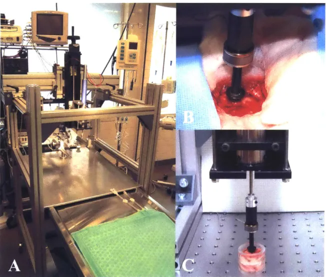
2 Brain tissue dynamics in vivo, 2.1 Overview . . . . 2.2 Rationale and background
2.3 Materials and Methods . .
zn uniaxial situ and compression in vitro 7 8 8 in vitro 9 10 10 12 16 20 20 22 29 30 32 37 38 38 39 41
2.3.1 2.3.2 2.3.3 2.3.4 2.4 Result 2.4.1 2.4.2 2.4.3 2.4.4 2.4.5 2.4.6 2.4.7 2.4.8 2.4.9 2.4.10 2.4.11 2.5 Discus Test apparatus . . . . Surgical procedures . . . . Indentation tests . . . . Tissue contact - tapping protocol . . . . s . . . .
Preliminary observations - effect of dura mater o response .... ... . .... ...
Tissue recovery after indentation . . . .
Test repeatability and rate order indifference . . . Location dependence . . . . Post-mortem time differences . . . .
Representative response in vivo . . . . Comparison with literature data . . . . Properties in vivo compared to properties in situ Properties in situ compared to properties in vitro Nonlinear rate dependencies . . . . Conditioning effects . . . . sion and conclusions . . . .
3 Large strain behavior of single cortical neurons 3.1 Overview . . . . 3.2 Background . . . .
3.3 Materials and methods . . . .
3.3.1 Cell Culture . . . .
3.3.2 Atomic Force Microscopy . . . .
3.3.3 Modeling: Finite Element Simulations 3.4 R esults . . . .
3.5 Discussion . . . .
4 Concluding remarks 86
A Brain tissue response: notes on numerical implementation of
constitu-tive model 88
B Brain tissue response: determination of material parameters 93
B.1 Preliminary parameter estimate . . . . 93
B.2 Automated Parameter Search Algorithm . . . . 94 B.3 Parameter estimates for "outlying" cases . . . . 96
C Summary of results obtained on hemorrhaged animals 99
C.1 Effect of dura mater on tissue response . . . . 99
C.2 Properties in vivo compared to properties in situ . . . . 99
C.3 Properties in vivo, in situ and in vitro compared between the hemorrhaged
and non hemorrhaged cases . . . . 102
D Preliminary assessment of tissue volumetric compliance in indentation
in vitro 105
Acknowledgments
My deepest gratitude goes to Dr. Simona Socrate - a great heart and a great mind
-without whom my research experience at MIT would have lacked much of its substance.
I am also truly indebted to Professor Subra Suresh, whose support was critical to the
successful completion of this work.
Special thanks to Dr. Hasan Alam, Dr. Marc de Moya and Dr. Guang Jin from the Massachusetts General Hospital who made the in vivo tests possible. I am especially grateful to Dr. Guang Jin for his patience and his support during the many hours of testing spent at the hospital.
I would also like to extend my gratitude to Dr. Asha Balakrishnan for contributing
greatly to the early stages of this project, and to Kristin Bernick for conducting the tests
in vitro on single neuronal cells.
My warmest thanks go finally to Peter Morley from the MIT Machine Shop, whose
help in the design of the mechanical apparatus was essential.
This work was supported by the US Army Research Office and Joint Improvised Explosive Devices Defeat Organization, under contract number W911NF-07-1-0035; the MIT Institute for Soldier Nanotechnologies, under contract number W911NF-07-D-0004;
Ecole
Nationale des Ponts et Chaussees (Universit6 Paris-Est, France); theComputa-tional Systems Biology Programme of the Singapore-MIT Alliance; and the Interdisci-plinary Research Group on Infectious Diseases at the Singapore-MIT Alliance for Re-search and Technology; and by the US Army Medical ReRe-search Material Command
Chapter 1
Constitutive response of brain tissue
in vitro
1.1
Overview
The dynamic behavior of porcine brain tissue, obtained from a series of in vitro obser-vations and experiments, is analyzed and described here with the aid of a large-strain, nonlinear, viscoelastic constitutive model. Mixed gray and white matter samples excised from the superior cortex were tested in unconfined uniaxial compression within 15 h post mortem. The test sequence consisted of three successive load-unload segments at strain rates of 1 s-1, 0.1 s1 and 0.01 s-1 followed by stress relaxation (N 25). The volumetric
compliance of the tissue was assessed for a subset of specimens (N 7) using video
exten-sometry techniques. The tissue response exhibited moderate compressibility, substantial nonlinearity, hysteresis, conditioning and rate dependence. A large-strain kinematics
nonlinear viscoelastic model was developed to account for the essential features of the tissue response over the entire deformation history. The corresponding material para-meters were obtained by fitting the model to the measured conditioned response (axial and volumetric) via a numerical optimization scheme. The model successfully captures the observed complexities of the material response in loading, unloading, and relaxation
over the entire range of strain rates. The accuracy of the model was further verified
by comparing model predictions with the tissue response in unconfined compression at
higher strain rate (10 s-) and with literature data in uniaxial tension. The proposed constitutive framework was also found to be adequate to model the loading response of brain tissue in uniaxial compression over a wider range of strain rates (0.01 s- to 3000
s_1), thereby providing a valuable tool for simulations of dynamic transients (impact,
blast/shock wave propagation) leading to traumatic brain injury.
1.2
Preliminary remarks
The study of the mechanical properties of brain matter - at the tissue-continuum level
- has been the focus of numerous investigations in the past four decades (see e.g. Refs [1-7]). Many investigators have concentrated their effort on characterizing the coupled
time and strain/stress dependencies inherent in the response of the tissue to externally applied mechanical transients - e.g. frontal or lateral impact of the head by rigid mass
[8-10], linear or angular acceleration pulses applied to the skull [11, 12], indentation of
the cortex surface [13]. Understanding how these loading/kinematic conditions applied to the organ boundary translate into local stress-strain states within the tissue continuum is challenging because the brain is, from a biomechanical perspective, a highly complex organ housing multiple "substructures" - e.g. brainstem, cerebellum, thalamus, cere-bral cortex, corpus callosum - associated with somewhat distinct mechanical properties [14-16]. Most biomechanical studies have been conducted in vitro although a few mea-surements have also been reported in vivo [17-21]. The results of different studies are at times difficult to reconcile due to the wide range of variation in experimental
proto-cols, including the species/age of the subjects (human, porcine, bovine, murine), loading
configurations (compression, shear, tension, indentation), loading histories (cyclic, stress relaxation, creep), and testing regimes (small/large strains or low/high strain rates). The collected data have facilitated the development of a large variety of constitutive models,
some of which have been shown to account for essential features of the tissue response [22-24] under selected testing conditions. Nonetheless, the integration of all the charac-teristic features of the large strain tissue response - hysteretic behavior, rate-dependence, nonlinearities, shear and volumetric behavior - into one single constitutive framework has
not been achieved thus far.
This study is a component of a multidisciplinary effort aimed at elucidating some key effects of primary blast on the central nervous system [25], and represents a first step towards the development of a predictive model for the response of brain tissue over an extensive range of strains and strain rates. A parallel effort is underway to assess differences between tissue properties in the in-vivo and in-vitro conditions, where the proposed constitutive formulation is employed to analyze results of indentation tests performed with a dynamic custom tool [26]. The first part of this chapter describes the experimental component of the study (test protocols and main results gathered on porcine cortical samples) while the second part addresses the modeling effort (constitutive laws, numerical implementation, model validation and predictions). Both are preceded by a brief literature review. Some of the limitations inherent in the current model formulation are discussed in the last section, guiding the effort for future model refinements. This work constitutes a preliminary step in the development of a comprehensive experimental data base and enhanced computational tools to be employed in support of a variety of clinical applications, as well as to elucidate mechanically mediated pathways leading to traumatic brain injury.
1.3
Experiments
1.3.1
Literature review
Mechanical tests on brain samples have been conducted mainly in the linear regime via small oscillatory deformations imposed on the tissue, in pure shear or in torsion, over a wide spectrum of frequencies [27-30]. In assessing the time/frequency dependence of the
linear viscoelastic properties of the tissue (i.e. storage and loss moduli), these studies have uncovered some important aspects of the tissue dynamics. The thrust of the results reported, nonetheless, suffer from a few limitations. First, most of the data so gathered lack consistency in that the magnitude of the measured scalar moduli varied at times
by more than 10-fold in relative terms from one study to another [31]. This lack of
consistency may be attributed to multiple factors including: inter-species/intra-species variations (e.g. animal breed, age, sex, inherent biological variability); differences in (1) post mortem testing time, (2) tissue storage and hydration conditions, (3) tissue prepara-tion/excision methods, (4) specimen neuroanatomical orientation, (5) temperature con-ditions, (6) interfacial testing conditions (degree of tissue friction/adherence/slipping on testing fixtures), (7) pre-conditioning effects prior to actual testing. Further, the scope of these investigations is limited per se to small deformations as the tissue has been shown to deviate from linearity at strains greater than 1% [32, 33]. More recently, the nonlin-ear features of the tissue response have been partly characterized in compression [34, 35], tension [36], and shear [23, 37]. While some of these studies measured the tissue response over more than two orders of strain rate magnitude, they were mostly focused on a lim-ited set of test histories (e.g. single load-ramp and stress relaxation tests or sinusoidal load-unload cyclic tests at low to medium strains), excluding any direct assessment of the tissue volumetric behavior. Some attempts have been made to retrieve quantitative in-formation on the tissue volumetric compliance [38, 39] but these attempts remain scarce and limited in scope, most investigators relying on incompressibility assumptions or spec-ulative arguments [34, 40, 41]. The experimental part of this doctoral work carried out in vitro aims to address some of the limitations noted in previous investigations via the systematic collection of experimental data on porcine cortical specimens in unconfined uniaxial compression comprising the following measurements: (1) nonlinear strain and strain-rate dependencies in load and unload over three orders of strain rate magnitude
(0.01-1 s-1), in the large deformation regime (up to 50% nominal strain), (2) long-term
com-pliance. A porcine model was preferred to other animal models because its gyrencephalic brain - architecturally close to the human brain - has been proven to share with the latter some similarities in terms of pre- and postnatal cerebral development relative to tissue growth, myelination and composition [42-46]. Swine models constitute, moreover,
an affordable alternative to more costly, ethically sensitive primate models.
1.3.2
Test protocol
-
unconfined uniaxial compression in vitro
Specimen preparation
Sixteen 6- to 18-month-old' swine brains (female, Yorkshire breed) were obtained from a
local vendor (Research 87 Inc., Boylston, Massachusetts), following a protocol approved
by the Committee on Animal Care at the Massachusetts Institute of Technology. The
brains were sectioned along the mid-sagittal plane and transferred on ice to the laboratory within three hours post mortem. Each hemisphere was rinsed upon delivery in phosphate-buffered saline (PBS, 10 mM phosphate buffer) and kept refrigerated in solution to limit tissue degradation. Shortly before testing, mixed gray and white matter samples were excised from the superior cortical region (frontal and parietal lobes2) and maintained hydrated in PBS during all subsequent steps. Preliminary investigations of the optimal sample size for uniaxial compression tests [48] showed that large samples - lying in the cubic centimeter range - yielded the most consistent results. All the data collected for this study were therefore obtained from samples that were, approximately, 25.4 mm x 25.4 mm in cross section and 9 mm in thickness (Figure 1-1A). The mechanical properties obtained shall be viewed as "homogenized" properties of the superior cortex (gray and white matter combined) although the samples tested were composed predominantly of
'Pigs having reached at least six months of age may be considered cerebrally mature [42], and were therefore selected to minimize age-related variability. Neonatal pigs have been shown to have significantly softer brains [31].
2No notable differences were observed in mechanical properties between the two cortical regions. This observation complements findings previously reported by Coats and Margulies [47] on local cortical gray matter homogeneities at the subcentimetric/millimetric tissue level.
A
25.4
mm
C;
B
0.5 0.4 - -- ---- --- - - --0.3 0.2 - --- -- - -- - -0.1 10-1 10 0 101 102U
10-1 100-J
w
Time Isi
101d
k
--
--
ii--[
Figure 1-1: Experimental protocol. (A) Cortical tissue sample. (B) Optical extensometry.
(C) Imposed strain history (logarithmic time scale). Each sample was subjected to a
sequence of three loading segments, at 1 s-1, 0.1 s-1 and 0.01 s- nominal strain rates respectively - each comprising five load-unload cycles to 50% nominal strain - followed
by a ramp-relaxation segment to 50% nominal strain held for 300 seconds with a ramp
0
0-0
-
--
-0 -- -- - - - -1000 800
1~
E 0 600 400 200D
10 2gray matter - accounting typically for ~70% of the sample mass. The experimental protocols were primarily designed so as to reduce the sources of experimental variability arising from tissue handling - which are many, given the delicate nature of the brain parenchyma. Separating intermingled gray and white matters of the superior cortex was found ill-suited for this study because the separation/cutting process implied further alterations of the tissue virgin state (via shearing or stretching) which were deemed too costly relative to the potential benefits.
Axial and lateral measurements
The samples (N = 25 from n = 16 Yorkshire sows) were tested in unconfined uniaxial compression on commercial testing machines equipped with 20 N load cells - ElectroForce
3200, Bose Corporation, Minnesota, USA (N = 11); Zwick Z2.5/TS1S, Ulm, Germany (N
= 14). Prior to testing, the platens were humidified with PBS to minimize friction at the
tissue-platen interface. All tests were conducted at room temperature (T = 210C) within
15 h post mortem.3 A preload of 0.02 N (corresponding to a compressive stress of -30
Pa) was imposed on each sample prior to testing in order to accurately determine the sample thickness. Each sample was subjected to a sequence of three loading segments, at 1 s-1, 0.1 s1 and 0.01 s-1 nominal strain rates respectively - each comprising five load-unload cycles to 50% nominal strain - followed by a ramp-relaxation segment to
50% nominal strain held for 300 seconds with a ramp rate of 1 s-1. Figure 1-1 illustrates
the imposed strain history (Figure 1-iC) and the resulting stress history (Figure 1-1D) for one representative tissue specimen. The transverse displacements were captured by
a Qimaging Retiga 1300 CCD camera equipped with a 200 mm f/4 Nikon lens (Figure
1-1B). Lateral stretch measurements were deemed sufficiently accurate only for a subset of specimens (N = 7) because most samples had somewhat irregular edges. Axial data
3 Samples were tested between 4 and 15 hours post mortem. No significant variations in tissue response
were noted in relation to post mortem test time differences. The responses measured were also found comparable to those reported by Tamura et al. [35] who conducted similar tests within a shorter time
1.4
1 e eTransverse stretch measured
-Axial stretch applied
0.8 0.6
0 20 40 60 80 100 120 140 160
Time [s]
Figure 1-2: Images of the tissue sample at increasing levels of axial deformation. The
lateral stretch measurements are obtained by visual extensometry.
(force and displacement) and lateral deformation images were acquired synchronously via
software Vic-Snap (Version 3.OD, Correlated Solutions, Inc). The lateral stretches were
subsequently assessed using a MATLAB image-analysis custom routine. The processed
data are shown for one representative specimen in Figure 1-2. Note that the nominal
axial strain values reported in Figures 1-1 and 1-2 were based on the platens' axial
displacement history. In practice, most of the specimens did not entirely recover their
initial configuration upon unloading: as the platens traveled back to their initial position
at the beginning of each loading cycle, the degree of adhesion between the platens and
the specimens was often insufficient to enforce complete recovery of the original specimen
height. Full contact was re-established at the end of each load-unload cycle (with a rise in the recorded load), and was lost at the end of the unloading ramps (as the load became negative, tracking the weak tensile adhesive forces between the top surface of the specimens and the platen).
1.3.3
Results
Stress-strain diagrams have been extracted from the representative data in Figure 1-1
for the three load-unload segments (at 1 s-1, 0.1 s-1 and 0.01 s-1) and are plotted in
Figure 1-3 together with the subsequent relaxation response. As shown in Figure 1-3, the tissue behavior in unconfined compression was intrinsically nonlinear at finite deformation
- both in the strain and strain rate domains. The tissue response exhibited marked hysteretic features over the range of strain rates considered for this study. Another notable feature was the substantial degree of "conditioning" associated with the first loading ramp, i.e. the dramatic increase in tissue compliance between the initial loading cycle and the subsequent cycles. Differences associated with softening effects between sequential ("conditioned") cycles at the same loading rate were much less dramatic. Conditioning effects reflected changes in tissue "state" occurring during the first cycle of loading through which the tissue might lose part of its interstitial fluid and/or undergo "irrecoverable" microstructural reorganization/reconfiguration (damage). To ascertain if conditioning was associated with a permanent change in tissue properties, a study was performed on a subset (N = 6) of tissue samples which were submitted to two series of five load-unload cycles (uniaxial compression to 50% nominal strain at 1 s-1) separated by a two-hour recovery period in PBS. To a large extent, the re-hydrated samples recovered their original response, with conditioning patterns highly similar to those measured on the virgin tissue, as illustrated in Figure 1-4 for one representative tissue sample. This observation substantiates the hypothesis that interstitial fluid diffusion plays a crucial role in the conditioning process, and that the tissue does not undergo substantial permanent damage in the first loading cycle. The essential features of the tissue response to the
10000 8000 6000 4000 2000-Load-unload
1
s-1 0 0 0.1 0.2 0.3 Nominal strain 5000 -4000 3000 ~2000-1000 -0 .4 __ 0' 5B
0
o
Load-unload 0.1 S-1 4~. 0.1 0.2 0.3 Nominal strain Load-unload 0.01 s-' 0.1 0.2 0.3 Nominal strain 4000 2 3000 2000 1000 0.4 0.I
Stress relaxation . I I 5 0 50 100 150 200 250 300 Timeis]
Figure 1-3: Representative cortical tissue response in unconfined compression. Data in
panels A, B, C, D are obtained sequentially on the same tissue sample (see corresponding
strain/stress-time histories for this representative sample in Figures 1-1C/1-1D).
'-eli
A
3500 3000 2500 -. 0.4 0.5 2000 1500 1000 500C
00
0, I12000 " 10000 -e- 1s- - virgin 8000 - 1s' -recovered 6000 -4000 2000 --- -- -- -- -- -- - --- - --00 0 0.1 0.2 0.3 0.4 0.5 Nominal strain
Figure 1-4: Representative cortical tissue response in unconfined compression. The sam-ple was submitted to two series of five load-unload cycles at 1 s-1 strain rate separated
by a two-hour recovery phase in PBS.
uniaxial compression loading protocol in Figure 1-1 were found to be consistent for all tested samples. The average response and standard deviation are shown in Figures
1-5A and 1-5B (nominal axial stress) and in Figure 1-5D (transverse stretch). For clarity
purposes, the stress-strain plots only display the first (virgin) loading ramp at 1 s1, the subsequent (conditioned) loading ramp at 1 s-1 and the first (conditioned) loading ramps at 0.1 s-land 0.01 s-1. The tissue response exhibited substantial volumetric compliance, with an average 12% volume reduction at an axial stretch of 0.5: AL 12%
[t5.5]
(N = 7 samples, n = 7x6 volumetric measurements taken from the sequences of
load-unload cycles at 0.1 s1 [X5] and 0.01 s1 [X1] strain rates). Data previously published
by Tamura et al. [35] for similar mixed gray/white matter samples from swine cortices
are also shown in Figure 1-5A. The (virgin) loading response measured in the Tamura study for a strain rate of 1 s-1 was found to be in good agreement with the results of the current study. Miller and Chinzei [34] also investigated the quasi-static response of similar tissue samples for the same animal model. The response of virgin specimens measured by Miller and Chinzei at a strain rate of 0.64 x 10⁻² S-1 is gathered in Figure
1-5C and it was found to be significantly stiffer than the average (conditioned) response
054 -- 0.1 0.1S 0.2 0.25 0.3 0.35 0.4 045 0.5 Nominal strain 4000 Relaxation, conditioned (N=25) 3000 *2000 1000
B
s
56 100 150 200 250 3 Time is]600O 1.4 - Load-unload cycles (N=7) -0- 0 Ols-I -Ramp 1, virgin (N=6)
C 5000 -V- 0.0064s-I -Ramp 1, virgin (N=13) Miller and Chinze1 4000 -0- O.Ols-I - Ramp 11, conditioned (N=25)
3000 --- - --- - 5 I 1.2
20001-C 0
C 0.05 0.1 0.15 02 0.25 0.3 0.35 04 D 0 50 100 I50
Nominal strain Time Isl
Figure 1-5: Average tissue response and standard
s-
1, 0.1 s-
1and 0.01 s-
1. (B) Relaxation. (C) Con
deviations. (A) Loading ramps at 1
litioning effects for the loading ramps
at 0.01 s1. (D) Transverse stretch history for the load-unload cycles.
12000r
10000-
8000-
6000--+ is-1 -Ramp 1, virgin (N=25)
--e- is-I -Ramp 2, conditioned (N=25)
-+ 0.1s- -Ramp 6, conditioned (N=25)
-0- 0.Ols-I -Ramp 11, conditioned (N=25 is-1-Ramp 1, virgin (N=9) Tamura
U)
A
-i--L
n,
-set of measurements was performed to confirm that this discrepancy might be primarily attributed to conditioning effects. Virgin tissue samples (N = 6) were subjected to five load-unload cycles at a strain rate of 10-2 s-1. The corresponding average first (virgin) loading response, shown in Figure 1-5C, was found to be comparable to that reported by
Miller and Chinzei.
1.4
Constitutive model
1.4.1
Literature review
Whether the brain should be considered - for continuum modeling purposes - as a single-phase viscous solid or as a dual-single-phase fluid-saturated solid is still the subject of differing viewpoints. The traditional dual phase approach follows the theory of consolidation (poroelasticity) originally formalized by Biot in the context of soil mechanics [49]. Al-though interstitial fluid diffusion and related transport mechanisms have long been recog-nized to play a crucial role in brain function [50], the poroelastic approach has received only marginal attention in the brain tissue biomechanics community [22, 51]. In the single phase approach, dissipation mechanisms and time dependencies in the tissue re-sponse are addressed in the framework of viscoelasticity. Most of the models following this approach are adaptations or combinations of three model subcategories: (1) linear viscoelastic models, based on cyclic loading, in which the tissue response is described by a set of two parameters, the storage and loss moduli [13, 28-30]; (2) small strain formula-tions for rheological models based on linear or nonlinear spring-dashpot combinaformula-tions [52,
53]; (3) large strain kinematics quasilinear or nonlinear viscoelastic models for which the
tissue response is decomposed into a hyperelastic component - Neo-hookean, Mooney-Rivlin or Ogden formulations are most common - and a dissipative component - e.g. multi-mode Prony series [5, 22-24, 54-56]. The linear and quasilinear models - mostly relevant in the small strain regime - are unable to fully capture the complexities of the tissue response at finite strains. In contrast, some of the large strain kinematics models
-integrating nonlinearities within their formulation - have been able to successfully repro-duce some specific features of the tissue deformation over fairly complex loading histories. Hrapko et al. [23] employed a multi-mode Mooney-Rivlin viscoelastic network in parallel with a nonlinear hyperelastic resistance to capture the simple shear response of white matter in load, unload and relaxation at strain rates of 1 s-1 to 1.5 s-1 up to 50% strain, using 16 material parameters. More recently, El Sayed et al. [24] developed a nonlinear elastic viscoplastic formulation to capture some of the hysteretic and dissipative charac-teristics of the tissue response in uniaxial tension and compression up to 50% nominal strain. The generalized framework proposed by El Sayed et al., in which an arbitrary number of viscoelastic networks are considered in parallel with a viscoplastic network, allowed the authors to fit a number of experimental findings with different configurations and numbers of constitutive elements. In most instances, the phenomenological models proposed in the literature rely on a fairly large number of material parameters validated over a limited range of loading regimes/histories.
The nonlinear visco-hyperelastic model developed here is also phenomenological in nature; it espouses the modeling "philosophy" pursued by previous investigators; and it idealizes the tissue as a single, homogeneous, isotropic phase. The proposed formulation combines the following advantages within a single framework: (1) it accounts for all the observed complexities inherent in the tissue response at low-to-medium strains and strain rates (i.e. nonlinearities, hysteresis, time-dependencies, volumetric behavior), (2) it is accurate and predictive over an extended range of strains and strain rates while requiring a relatively small number of material parameters, and (3) it relies on physically motivated arguments, where different constitutive elements of the model, and their corresponding parameters, can be conceptually associated with specific underlying mechanisms of de-formation. This empirical interpretation of modeling elements provides some guidance in the fitting process, as discussed in Appendix B.
1.4.2
Constitutive laws
A schematic (rheological) representation of the proposed model for the tissue response
is shown in Figure 1-6 with the corresponding 3D large kinematics deformation map. For the most part the notation used hereafter is that prevailing in modern continuum mechanics:
F., deformation gradient
V, - (F* - F left stretch tensor from polar decomposition of F*
VO(V) tissue volume at time 0 (time t)
V
10 =fiV
0 V20 = f2V0 = V" - V10 J det(F) = v J 2 = 2 2I,=j-2/3F
-FTE*
ln(V.) = In( F* F
T)1B.
=JN,F
*D
sym(L)
=(L+ L
T)W
=skw(L*)
= j(L* -L
) TA Th Td T' N, tr()1
TA tr(T)1 =T
- tr(T*)1 T = T* Vtr (T ) 2incompressible tissue volume fraction at time 0 compressible tissue volume fraction at time 0 volumetric macroscopic jacobian
volumetric jacobian for compressible tissue fraction
isochoric component of left Cauchy-Green strain tensor Hencky strain tensor
velocity gradient stretching tensor spin tensor Cauchy stress
hydrostatic component of Cauchy stress deviatoric component of Cauchy stress stress deviator
unit tensor in the direction of stress
The model comprises an elastic network (A) representing the instantaneous response of the tissue and a viscoelastic network (elements (B), (C), (D) and (E)) incorporating the dissipative contributions. Following Lee's decomposition [57], the total deformation gradient, F, applied to the tissue may be decomposed as:
F = F A FB
where F A and F B represent, respectively, the elastic (instantaneous) and viscoelastic
components of the tissue deformation. The viscous flow is assumed to be isochoric, hence the total volumetric Jacobian J may be expressed as: J -- det(F) = det(FA) - det(FB) =
det(FA). The viscoelastic response of the tissue is captured by the combination of a
nonlinear short-term viscous element (B) and a linear viscoelastic backstress network
(CDE). With regard to the backstress network, the viscoelastic deformation gradient FB
is further decomposed as:
FB = FC
'FD
where the linear viscous element (D) models the long-term relaxation of the backstress contribution. Both FC and F D are taken to be isochoric.
The total Cauchy stress TA in the tissue is decomposed into its hydrostatic and deviatoric components:
TA hT- -Td
where the hydrostatic component Th and the deviatoric Td component are physically associated with the deformation mechanisms prevailing in bulk and in shear, i.e. govern-ing the volume-mediated and shear-mediated portions of the tissue response respectively. Bulk-mediated response
Brain tissue is highly hydrated. Although interstitial fluid diffusion is not explicitly in-cluded in the current model, a simplified qualitative understanding of the role of hydrat-ing fluid in determinhydrat-ing the apparent volumetric tissue behavior is the foundation of the proposed formulation for the bulk response. Briefly, the undeformed tissue volume, V0
,
is partitioned into an incompressible fraction, fiV0 = V10, and a compressible fraction,
f2
V
0 = V2 , where fi + f2 = 1. The corresponding components of the deformed tissue(A)
A
...
(C.)
(B).----MME---(.---i(E
)
W.I.
(E)
(D)
F
F
D
cB
Initial (undeformed)
configuration
Current (deformed
configuratio
I -"< N)
ni-4
0
Fc
F
AIntermediate (relaxed)
configuration
Figure 1-6: Schematic of rheological model (top) and corresponding large kinematics deformation map (bottom).
a "bound" component, which does not diffuse freely under loading, and an "interstitial" component, which diffuses in response to hydrostatic loading, accommodating volumetric tissue deformation. The incompressible fraction of the volume, V1, is conserved, so that the corresponding volumetric Jacobian Ji = V remains constant (Ji - 1) throughout
the deformation history. The hydrostatic stress Th developed in the tissue is then as-sumed to be driven by the volumetric Jacobian of the compressible fraction, J2 = 2
V2
as:
Th = K -
n(J2) 1where the hydrostatic stress is characterized by a logarithmic dependence on the volumetric Jacobian, and K is the small-strain bulk modulus of the compressible fraction. As a consequence of the incompressibility constraint enforced for V, the macroscopic tissue volumetric Jacobian J = n can be expressed as:
J = fi + (1 - fi)J2
so that the hydrostatic stress may be restated in terms of the total volumetric Jacobian
J:
Th = K.- In-1
Estimates of typical hydration levels for brain tissue indicate that fluid accounts for approximately 80% of the brain mass [58]. Although the incompressible tissue fraction is effectively a material property, a fixed value fi ~ 0.8 is selected for the proposed model. This latter assumption is based on the notion that interstitial cerebrospinal fluid, which is free to diffuse throughout the tissue, only accounts for a small portion of the total water content. Phenomenologically, the proposed constitutive framework captures the
highly nonlinear resistance to volumetric deformation exhibited by the brain parenchyma.
implicitly assumes that the volumetric response of the tissue becomes infinitely stiff as the volumetric Jacobian approaches 0.8.
Shear-mediated response
The deviatoric component of the stress response, Td, is adapted from the freely-jointed 8-chain model developed for macromolecular elastic networks [59]:
Td - -t- - AL- L-1 ( A
-J
A
A
where J det(F) = det(FA B = J-2 F A*FT A~AA
A2 1 -A2 =tr(B ) = coth(#) -1
and po and AL are model parameters which scale with the initial shear modulus and the limiting extensibility (locking stretch) of the network. L denotes the Langevin function. The 8-chain model, originally developed for elastomeric materials, is based on statistical mechanics arguments. It assumes a weakly bonded network of randomly oriented chains for which the source of resistance to deformation, i.e. the change in Helmholtz free energy, is mainly governed by changes in configurational entropy of the reorienting chains. The adaptation of this model to brain tissue is motivated by the following considerations.
e The cortex is primarily composed of cells (neurons and glia) whose main constitutive
elements are actin filaments, neurofilaments and microtubules [58]; these latter components may be viewed - in a highly simplified representation at the mesoscale
level - as a loose "assembly" of randomly oriented "chains" sharing some similarities with the idealized statistical mechanics network envisioned for the 8-chain model.
" Preliminary force-displacement measurements performed on individual actin
fila-ments [60] suggest that the use of Langevin statistics may be suitable to account for the non-linear force-extension curves of these structural components.
" The tissue response - as measured experimentally - exhibits nonlinearities which are well captured by the inverse Langevin function. Further, some empirical evidence collected at the cell level [61] suggests that single neurons share similar nonlinear characteristics.
The time-dependent portion of the tissue deformation is defined via the evolution of the velocity gradient, L, imposed on the tissue:
L=F-F = -F
A A+ F
A FBFB FAF
=P
FA AF+
AF
LB AF= L
A A BF BBwhere L B B -F B denotes the viscoelastic velocity gradient in the relaxed
(un-loaded) configuration and LB
=
FA . LB F 1 may be interpreted as the "push-forwardvelocity gradient", i.e. the velocity gradient convected to the current (loaded) configura-tion (see kinematic map provided in Figure 1-6 and chapter 2 of Holzapfel [62] for more details on push-forward tensor operations). The velocity gradient LB may be further de-composed into its symmetric and skew-symmetric parts, LB = DB + WB ,where
hB
andWB denote the rate of viscous stretch and the viscous spin respectively. Following [63], the unloaded (relaxed) configuration may be made unique by specifying WB 0, and
thus the evolution of the viscoelastic component of the deformation gradient, FB, reduces to: FB =
F
A hB * F. The viscous stretching tensor DB is constitutively prescribed tofollow the direction of the deviator T' of the driving stress TB according to a nonlinear reptation-based scaling law, adapted from [64]:
n T' VifR T' TB
T
f )B DB B N B BT 0 - R v,2B7. 'B BtrT 2\
t'BTB2)
a2 fR= (avtr(FB-F )/3 -
1)2where o is a dimensional scaling constant ( -0 10- s-1). The reptation factor, fR, accounts for the increasing resistance to viscous flow observed in macromolecular
networks for increasing levels of accumulated viscous deformation. The factor a is a small constant introduced to eliminate the singularity at FB =1 [64], and is set to a = 0.005. The rate sensitivity exponent, n, and the strength parameter, o-, are material properties. The driving stress TB for the (short-term) nonlinear viscous element (B) is obtained as the difference between the Cauchy stress in the tissue, TA , and the backstress from the linear viscoelastic network (CDE) convected to the current configuration:
TB = TA -TC
=TA
- F A SC'FA
The 2nd Piola-Kirchhoff stress, Sc, in the elastic element (C) of the backstress net-work is taken to scale linearly with the Hencky strain4 of the corresponding deformation gradient Fc:
Sc =
2GoE=
2Go ln(Vc)=
2Go ln Fe .FT)1/21
where E. is deviatoric, due to the isochoric nature of F, [66], and Go is a mater-ial parameter representing the short-term shear modulus of the viscoelastic backstress network.
A parallel constitutive framework is followed to evaluate the driving stress for the
4For a discussion on the adequacy of the Hencky strain measure at finite strains, the reader is referred to [65].
long-term linear viscous element (D), as the difference between the stress Sc in the elastic element (C) and the long-term backstress SE in the elastic element (E):
S= S -- F - E T
where
E = 2GOED
=2Gcoln(VD)
2GoIn
FD FT)and G, is a material parameter related to the long-term (equilibrium) shear response of the viscoelastic backstress network. The flow-driven deformation mechanisms unfold-ing in the tissue at long times are controlled via the linear viscous element (D). The cor-responding velocity gradient
LB
in the elastically unloaded configuration is constitutively prescribed by settingVD=
0, and obtaining the stretching tensor, D D = FC D ' FBas
~ 1
D D SD
where the viscosity rj is a material parameter.
In summary, the proposed large strain kinematics model comprises eight material parameters: K, PO AL, n, Or, Go, Goo, r. Values of these parameters appropriate to capture the (conditioned) response of brain tissue are determined in the following sections
by fitting the proposed model to the measured response in unconfined compression.
1.4.3
Model fit
The constitutive model was implemented as a Fortran user-defined material subroutine in ABAQUS/Standard (Simulia, Providence, RI). Time-integration of the constitutive model is accomplished through an explicit scheme where the viscous stretching tensors are taken to be constant over each time increment. The model was fit to the conditioned tissue response using the representative set of axial data - i.e. axial data lying about the
average - as reported in Figures 1-1 and 1-3 along with the average lateral data shown in Figure 1-5D. The fit was initiated using the Nelder-Mead simplex method [67, 68] and subsequently optimized manually. The optimized material parameters were found to be: K = 800 Pa, po = 20 Pa, AL = 1.09, n = 3,
a,
= 25 Pa, Go = 4,500 Pa, Go. = 600Pa, r7 = 600,000 Pa.s. For a discussion on the material parameter determination and
the automated search algorithm, the reader is referred to Appendix B. The optimized simulation results are summarized in Figure 1-7. The proposed material formulation successfully captures the main features of the tissue response - volumetric compliance, hysteresis, long-time relaxation behavior and nonlinear strain/strain-rate dependencies spanning three orders of strain rate magnitude. Note that the load-unload response at different strain rates and the relaxation response were experimentally obtained (and numerically simulated) over a single sequential loading history. Some discrepancies in material response were encountered between model and experiment in the low strain rate, low strain regime (Figures 1-7C and 1-7D). These differences stemmed mainly from the fact that perfect adhesion was assumed in the numerical model between sample and upper platen at all times whereas, experimentally, contact between the sample and platen was lost at low strains, low rates in the final/initial portion of the unloading/reloading segments. Some artifacts could have been added to the simulation procedure in order to account for these loss of contact effects. However, given the high compliance of the tissue at low strains and low rates, the simpler "perfect adhesion" assumption was deemed sufficient for the purpose of this study.
1.4.4
Model predictions
To further assess the applicability/relevance of the proposed formulation, the predictions of the proposed model were compared with two sets of data collected respectively by Miller and Chinzei [36] in uniaxial tension at quasistatic strain rates5 and by the authors
5
Testing conditions and animal model used in the Miller and Chinzei study are nearly identical to the protocols followed in this study, allowing a direct comparison.
itt) 'K ,= 20 Pa Go X= 1.09 Tj K =800 Pa G Goa, 25 Pa n =3 Load-unload 0.1 S-1 0.1 0.2 0.3 0.4 Nominal strain 0.5 Stress relaxation 0 50 100 150 200 250 300 Time Isi = 4,500 Pa = 60,000 Pa.s = 600 Pa
A
C
* Data - Model fitE
G 0. -8000 6000 4000 2000 0 3000 2000 1000 0 .0 1.4 1.35 1.3 1.25 1.2 1.15 1.1 1.05 Load-unload I s-1B
0 0.1 0.2 0.3 0.4 0.5 Nominal strain 0 0.1 0.2 0.3 0.4 N i . I t i 0.5D
F
0 20 40 60 80 100 120 140 Time Is|Figure 1-7: Model fit to representative data set in unconfined uniaxial compression. (A)
Optimized material parameters. (B), (C), (D) and (E) Axial tissue response (measured
and simulated) over entire loading history. (F) Associated lateral deformation (measured
and simulated) over the five load-unload cycles at 0.1 s- strain rate and the first cycle
at 0.01 s-- strain rate. Weighted mean-squared error between simulated and measured
responses (see appendix for details) was found to be 3.5-10-.
31
G I . a 6000 4000 2000 0 0 5000 4000 3000 2000 1000 0 Load-unload O.0O s-I Iin unconfined uniaxial compression at 10 s-1 strain rate6. The results are reported in Figure 1-8. The loading and unloading response predicted by the model in uniaxial compression at 10 s1 strain rate is in good agreement with the experimental data. The responses predicted by the model in tension at low and medium strain rates are also in good agreement with those reported by Miller and Chinzei in the low-to-medium strain range. At larger strains however, the tissue response predicted by the model stiffened, as reported previously for other similar biological tissues [69, 70], while the response mea-sured by Miller and Chinzei softened. It may be postulated that the softening behavior observed by Miller and Chinzei at large tensile strains reflected the unfolding of failure mechanisms - which, given the delicate nature of brain tissue, are likely to occur within the tissue and/or at the platen interface for high levels of tensile deformation.7
1.5
Discussion
The experimental protocols developed in this study enabled the collection of a large, con-sistent pool of mechanical data gathered in vitro on macroscopic brain samples at finite strains (0-50%) in the low-to-medium strain rate regime (0.01-1 s- 1). Of particular in-terest are some quantitative features of the tissue response - e.g. volumetric compliance, large strain viscoelastic behavior upon unloading and reloading, combined nonlinearities in the strain and strain rate domains - which were not available from previous studies but which are highly relevant to guide the development of accurate rheological models. In particular, the experimental measurement of the unloading portion of the tissue response proved crucial in the identification of the relative contributions of elastic and viscoelastic mechanisms to the overall tissue resistance to deformation. The determination of these contributions in the modeling phase enabled the resulting model to predict tissue behavior 6The compression test protocol employed at 10 s 1 strain rate followed that detailed in the preceding section for the lower rates of deformation.
7
Unfortunately the Miller and Chinzei data do not include unloading and reloading segments, which could have provided a more conclusive interpretation of the tissue response.
- Model prediction * Data (N=4) - * S 6.e 0 0.1 0.2 0.3 Nominal strain 0.4 - Model prediction - Data by Miller (N=10) 4 8 12 Time Isi 1000 800 600 400 o 200 I' L~U U 16 20 0 0.1 - Model prediction - Data by Miller (N=11) 0.2 0.3 0.4 Time Isi
Figure 1-8: Model predictions compared with measured responses in unconfined uniaxial compression (A) and uniaxial tension (B and C). (A) Tissue response measured by the authors in compression to 50% nominal strain at 10 s- strain rate. (B) Average tissue response measured by Miller and Chinzei [36] in tension on 10 mm high specimens to 15%
nominal strain at 5 mm.min
1(-0.0083
s-1 nominal strain rate). (C) Average tissue
response measured by Miller and Chinzei [36] in tension on 10 mm high specimens to
50% nominal strain at 500 mm.min1 (-0.83 s-' nominal strain rate).
33 14000 12000 10000 8000 6000 4000 2000 0 W
A
200 0.5 160 1201 80 40 0 0 -1 CB
0.5 0.6-at a strain r-ate (10 s-
1) lying outside the original range on which the fit was performed.
The current study is only a first step towards the development of a model able to provide
accurate predictions for the tissue behavior over arbitrary strain histories. Among the
limitations of the study, it could be noted that: (1) the tissue behavior was only
char-acterized in one mode of deformation (unconfined uniaxial compression) over a range of
strain rates not exceeding 10 s-1; (2) lateral stretch measurements could be accurately
obtained only in the lower rate regime (0.01 to 0.1 s-
1) due to the limited capabilities of
the image acquisition system; (3) homogenized measurements may have been affected by
partial local discontinuities in the deformation field, as sulci were included,
8(4) partial
adhesion/decohesion of the specimens from the platens might have affected/altered the
measured tissue response at the end of the unloading ramps; (5) the tissue did not reach
full equilibrium at the end of the five minute relaxation period selected for this study,
suggesting that relaxation tests where the deformation is held over the course of 20-30
minutes
-
i.e. within a time window for which tissue degradation may still be neglected
-
are needed to measure equilibrium tissue response. Also note that the conditioned
response of the tissue was investigated and characterized according to a decreasing order
of deformation rates. Preliminary results (unpublished) seem to indicate that once a
.sample is deformed at a given rate it is "conditioned" for deformation at that rate and
at lower rates but it will still exhibit additional conditioning if tested subsequently at
higher rates. Altering the order of the loading segments in future studies may therefore
provide useful validation data for a model accounting for conditioning effects.
The proposed constitutive model successfully captures the main features of the large
strain tissue response, as measured in uniaxial compression and tension. While the
se-lected set of material parameters was optimized specifically for the conditioned tissue
response in the low to medium strain rate regime, the current formulation remains
gen-eral in nature and might be applied to model tissue response over more extensive ranges
'Care was taken to exclude results from any samples showing manifest opening/sliding at the sulci and/or inhomogeneous deformation (as attested by the images collected via the visual extensometer system).
of strain rates.9 To demonstrate this versatility, a preliminary fit of the model to the unconditioned (first loading ramp) tissue response measured at low to high rates of de-formation (0.01 to 3000 s-1) was conducted. The main results are reported in Figure 1-9. The set of material parameters found to capture the tissue response upon loading at
high rates was: K = 10 MPa, po = 10 kPa, AL = 1.03, n = 0.3, or = 2 kPa, Go = 6 kPa,
Go = 2 kPa, r7 = 1 kPa.s. Note that to capture the high-rate regime response the ma-terial parameters needed to be altered. This is to be expected as additional mechanisms of resistance to deformation might be operative at these rates. This observation mirrors findings in polymeric materials [71] where additional constitutive elements are required to account for the increased resistance to molecular motions in the high frequency regime. The bulk response of biological materials is also found to be substantially stiffer at high rates of deformation [72] as diffusion of interstitial fluid is drastically reduced. The pro-posed model, with the modified material parameters reported in Figure 1-9, efficiently captures the essential features of the unconditioned loading response of brain tissue over a range of strain rates from 0.01 to 3000 s-. While some of the fine details of the tissue response in the very low rate regime are unavoidably lost with this alternative set of parameters, the extensive range of strains and strain rates over which the tissue response is adequately captured highlights the potential value of this model for finite element simulations of loading transients leading to traumatic brain injury. Typical deformation histories of brain tissue under blunt impact or blast loading conditions present a very wide spectrum of strains and strain rates, with inhomogeneous fields characterized by steep spatial and temporal gradients. The availability of an accurate constitutive law to model the brain tissue response is a crucial requirement to assess local levels of strain, stress and strain rate associated with potential injury mechanisms. The proposed model represents a first step towards the attainment of this goal. Further model refinements are needed to address the time-dependence of the volumetric response associated with
9The proposed formulation was also found to be appropriate to model the response of rat neurons as measured via Atomic Force Microscope (AFM) indentation [61].
1 60
-1600
-40 'ls 1400 201
S- s0
0.01 S
1200
0
0.05 0.1 0.15 0.2 0.25 0.3 0.35 0.4 0.45 0.5
1000
Bovine (exp)
800
-
Porcine (exp)
3000 s
4 .5 600 - Model fit2000s
400
200
-1000
s-0
--
_-0
0.1
0.2
0.3
0.4
0.5
0.6
0.7
Nominal strain
Figure 1-9: Model fit to the tissue response measured upon loading in unconfined uniaxial
compression at nominal strain rates spanning six orders of magnitude (0.001 s-1 to 3000
s-
1). The sets of bovine and porcine data reported at high rates (1000 s
1to 3000 s-1)
have been obtained by the Chen group at Purdue University via Kolsky bar measurements
[7];
the porcine data shown for the low to medium strain rates (0.001 s-
1to 10 s-1) are
those collected by the authors. The optimized model parameters for the fit are: K
=
10
MPa, yo = 10 kPa, AL = 1.03, n = 0.3,
uO
= 2 kPa, Go = 6 kPa, G,, = 2 kPa,= 1 kPa.s. The dimensional factor was set to 1000 s-1, and the reptation factor fR to