0
BMI1 is a context-dependent tumor suppressor that is
a barrier to dedifferentiation in non-small cell lung
adenocarcinoma
by
Rachit Neupane
B.A. Biology
Bard College
Submitted to the Department of Biology
in partial fulfillment of the requirements for the degree of
Doctor of Philosophy
in Biology
at the Massachusetts Institute of Technology
May 2019
© Massachusetts Institute of Technology
All Rights Reserved
Signature of the Author _________________________________________________________
Rachit Neupane Department of Biology May 23, 2019 Certified by __________________________________________________________________ Jacqueline A. Lees Professor of Biology May 23, 2019 Accepted by __________________________________________________________________ Amy Keating Professor of Biology
Chairman, Graduate Committee
1
BMI1 is a context-dependent tumor suppressor that is a barrier to
dedifferentiation in non-small cell lung adenocarcinoma
by Rachit Neupane
Submitted to the Department of Biology on May 24, 2019 in Partial Fulfillment of the
Requirements for the Degree of Doctor of Philosophy in Biology
ABSTRACT
Predictive value is expected when preclinical models of disease are used for research. However, not all models appropriately mimic the disease progression or the treatment paradigm in the clinic. This thesis addresses an epigenetic regulator, Bmi1, which acts in stem cells to maintain their proliferative and self-renewal capacity primarily through silencing of the Ink4a/Arf locus.
Bmi1 has been proposed as a good therapeutic candidate in cancer because of its presumed
role in maintaining tumor propagating cells (TPCs). This conclusion is based on the observed tumor suppressive effects of Bmi1 deletion in in vitro cell culture models, in vivo transplant models, and autochthonous models in which Bmi1 was absent throughout development. However, to date, no one has assessed the consequences of deleting Bmi1 in existing
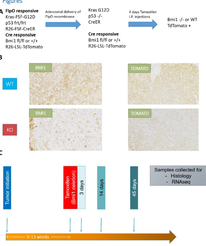
autochthonous tumors, to mimic patient treatment in the clinic. To accomplish this, we have generated a mouse model that allows induction of autochthonous lung adenocarcinoma, driven by oncogenic Kras and Tp53 loss (KP LUAD), and subsequent deletion of Bmi1 specifically within the tumor cells once more than half the tumors progress to grade 3 or higher. We confirmed that this model yielded Bmi1 loss that was tumor-specific and almost complete. We then aged tumor bearing mice for up to seven weeks post Bmi1 deletion to determine the impact on LUAD. Unexpectedly, Bmi1 deletion did not yield significant tumor suppression. Instead, gene expression analyses of Bmi1 deficient tumor cells revealed upregulation of a gastric gene expression program that is a known marker of lung tumor progression towards a more aggressive state in the KP LUAD model. Additionally, single cell sequencing showed that Bmi1 deficient tumors contained a higher frequency of cells that expressed previously described markers of TPCs and metastasis. We also extended these findings to colorectal cancer where we show that deletion of Bmi1 is not tumor suppressive in either in vitro organoids or
orthotopic transplants. Given these findings, we conclude that deletion, or inhibition, of BMI1 in existing tumors will be ineffective for cancer treatment in the contexts examined, and potentially deleterious because it can enable acquisition of alternate differentiation states that promote tumor progression.
Thesis supervisor: Jacqueline A. Lees Title: Professor of Biology
2
TABLE OF CONTENTS
ABSTRACT ... 1
TABLE OF CONTENTS ... 2
CHAPTER 1: INTRODUCTION ... 4
Cancer tumor propagating cells (TPCs) and their regulators ... 5
Polycomb Repressive Complex 1 ... 7
How PRC1 is targeted to chromatin sites ... 9
How PRC1 exerts its gene silencing function ... 12
PRC1’s role in self-renewal and differentiation ... 15
BMI1 ... 17
Bmi1 in stem cells ... 18
Bmi1 in cancer ... 23
Lung adenocarcinoma (LUAD) ... 28
LUAD in patients ... 28
Genetically engineered mouse models (GEMMs) of LUAD ... 30
Dedifferentiation as a route to KP LUAD progression ... 33
Bmi1 in lung cancer ... 35
Remaining questions ... 38
References ... 40
CHAPTER 2: BMI1 ACTS AS A TUMOR SUPPRESSOR IN HIGH GRADE LUNG ADENOCARCINOMA ... 49
Abstract ... 50
Introduction ... 51
Results ... 56
Discussion... 66
Figures ... 70
Materials and Methods ... 79
References ... 83
CHAPTER 3: DISCUSSION AND FUTURE DIRECTIONS ... 88
Review of Results ... 89
A cautionary note on epigenetic targets in cancer ... 93
3
Lung to Gut Specification ... 97
Future directions ... 99
Limitations of our mouse model ... 103
Concluding remarks ... 104
References ... 105
APPENDIX: BMI1 DELETION IS NOT TUMOR SUPPRESSIVE IN COLORECTAL CANCER ... 114
Abstract ... 115
Introduction ... 116
Results ... 118
Summary ... 122
Figures ... 123
Materials and Methods ... 126
4
CHAPTER 1: INTRODUCTION
5
Bmi1, a part of polycomb repressive complex 1 (PRC1), is a known epigenetic regulator of
self-renewal and proper differentiation of stem cells. There has been extensive interest in Bmi1 as a therapeutic candidate given its presumed role in self-renewal of tumor propagating cells, and the tumor suppressive effects observed upon Bmi1 deletion in cancer cell lines and mouse models of cancer. However, remarkably, the consequences of deleting Bmi1 in existing tumors has never been tested. In this chapter, I will briefly introduce tumor propagating cells and their roles in cancer progression, and then describe the broad mechanisms by which the polycomb complexes regulate gene expression and influence differentiation. I will then focus on Bmi1, describing its known functions in stem cell biology and differentiation, and its role in cancer. Finally, I will describe lung cancer development and progression, and genetically engineered mouse models that enable its study, and are the major context in which I have investigated
Bmi1’s role.
Cancer tumor propagating cells (TPCs) and their regulators
Cancer is a huge medical unmet need. In the United States alone, cancer will claim an estimated 600,000 lives in 2019 (Siegel et al., 2019). This estimated death toll varies largely by cancer tissue of origin. However, the trend of worsening 5-year survival rate as cancer stage at diagnosis increases is very consistent for all cancer types. Cancers that have spread to distant sites have worse prognosis than locally restricted tumors or regionally disseminated cancer. This is largely because most local tumors and lymph node metastases can be surgically
resected, but treatment of distant metastases relies upon non-surgical cancer therapies. These non-surgical therapies are usually less effective than surgical resection because cancer cells
6
often develop resistance, leading to relapse. Consequently, cancer cells that are able to spread to distant secondary organ sites, and cells that are chemoresistant and cause relapse, are the main culprits of cancer-related deaths.
It has been postulated since the 90s that not all cancer cells are equal, and that a hierarchy exists amongst cancer cells that is reminiscent of the stem cell hierarchy (Bonnet & Dick, 1997). According to this model, only a subpopulation of cancer cells have the potential to divide indefinitely and to give rise to all cell types within the tumor. These cells have been dubbed cancer stem cells or tumor propagating cells (TPCs). TPCs are functionally defined by their ability to produce tumors upon serial transplantations, similar to reconstitution assays for stem cells. As an extension of this functional definition, TPCs are also thought to be responsible for forming metastases at distant sites, and contributing to tumor relapse post-therapy (reviewed in Desai et al., 2019). The first identification of TPC in human cancer was in the context of acute myeloid leukemia (Bonnet & Dick, 1997; Lapidot et al., 1994). These cells were rare in the population, but could be enriched by specific cell surface markers, and were able to generate leukemia when transplanted into immunocompromised mice, (Bonnet & Dick, 1997; Lapidot et al., 1994). Since then, TPCs have been identified in various human solid cancers including breast cancer (Al-hajj et al., 2003), brain cancer (Singh et al., 2003), pancreatic cancer (C. Li et al., 2007), colon cancer (O’Brien et al., 2007), and ovarian cancer (S. Zhang et al., 2008). Since TPCs are responsible for cancer relapse and metastases, which account for most cancer-related deaths, there has been a big push in the field to understand TPC biology and hopefully exploit TPC vulnerabilities for cancer therapy.
7
One approach to understanding TPC biology has been to investigate the role of genes and pathways important for maintaining stemness potential in normal stem cells. This has been fueled by the observation that poorly differentiated aggressive human tumors tend to gain gene expression signatures reminiscent of embryonic stem cells, including core pluripotency genes such as Nanog, Oct4, Sox2, and c-Myc (Ben-Porath et al., 2008). Moreover, signaling pathways that have been well-established to be important in normal stem cells, such as
Hedgehog, Notch, and Wnt, are all exploited in various TPCs (reviewed in Desai, Yan, & Gerson,
2019). Numerous studies have also established the role of epigenetic regulators in maintaining the self-renewal potential and undifferentiated state of stem cells (reviewed in Avgustinova & Benitah, 2016). These epigenetic regulators are of key interest in TPC biology. One such epigenetic regulator is BMI1, which is a component of polycomb repressive complex 1 (PRC1). In advanced cancer, PRC1-regulated genes with documented importance in stem cell biology are preferentially repressed by hypermethylation, suggesting that they have acquired a more stem-like state (Ben-Porath et al., 2008; Widschwendter et al., 2007). This has fueled interest in the field of cancer biology to understand the role of PRC1 and Bmi1 in TPCs. This first requires a thorough understanding of role of PRC1 and Bmi1 in normal cellular context, which is detailed in the sections below.
Polycomb Repressive Complex 1
Even though all the cells in an organism have the same genetic material, they are able to control the genetic expression patterns to create diversity in cell types. These patterns include the genes that are turned on, and equally importantly, the genes that are turned off. The
8
initiation and maintenance of these genetic expression patterns are controlled by both transcription factors that dictate lineage specificity and epigenetic modulators that control chromatin structure and histone modifications. One group of such epigenetic modulators are the polycomb proteins, which were first identified as regulators of expression of Hox gene expression in Drosophila melanogaster via a screen for body segmentation patterning during development (Lewis, 1978). Subsequent studies established the existence of two different polycomb complexes with different core functions. Polycomb repressive complex 1 (PRC1) has the enzymatic function of placing a single ubiquitin molecule on histone H2A at K119 amino acid (Cao, Tsukada, & Zhang, 2005; de Napoles et al., 2004; Ve, 2005; H. Wang et al., 2004). Additionally, PRC1 has a non-enzymatic role in compacting polynucleosomes (Francis, Kingston, & Woodcock, 2004). Polycomb Repressive Complex 2 (PRC2), in contrast, acts to methylate histone 3 at K27 (Cao et al., 2002; Czermin et al., 2002; Kuzmichev et al., 2002; Müller et al., 2002). In this section, I will primarily focus on PRC1 with additional discussion on PRC2 whenever pertinent to the function of PRC1.
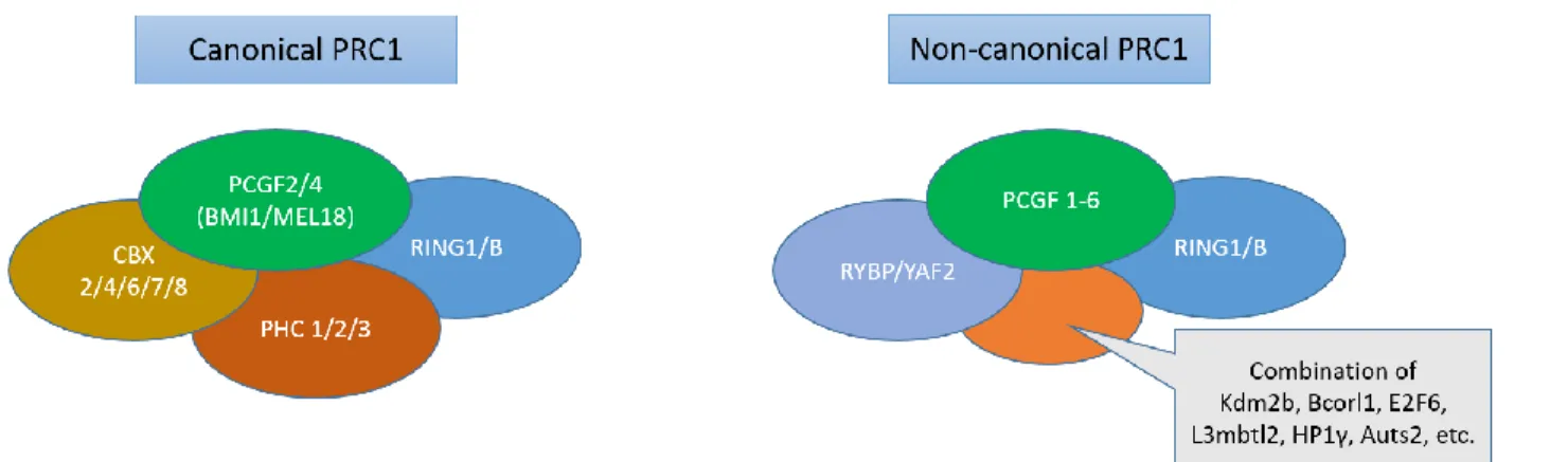
The catalytic core of PRC1 is comprised of an E3 ligase, RING1A or RING1B, along with one of the PCGF(1-6) subunit (Buchwald et al., 2006; Gao et al., 2012; Kloet et al., 2016; Li et al., 2006). This catalytic core binds to many combinations of auxiliary proteins to give rise to variations in PRC1 complex structure and biological function. One diversification of function comes from competition between CBX proteins and RYBP or YAF2 in binding to the C terminal domain of RING 1A/B (Wang et al., 2010). Based on the participating binding partner, PRC1 is broadly classified into canonical or non-canonical PRC1 complexes (Figure 1.1) (Gao et al., 2012; Kloet et al., 2016). The canonical PRC1 contains CBX proteins along with polyhomeotic-like proteins
9
(PHC 1/2/3) and either PCGF2 (MEL18) or PCGF4 (BMI1). The non-canonical PRC1 complex contains RYBP or YAF2 together with any of the six known PCGF proteins, including BMI1. The catalytic activity of PRC1 is specific to the non-canonical PRC1 complex containing RYPB or YAF2, suggesting a catalysis-independent role for canonical PRC1 (Blackledge et al., 2014; Tavares et al., 2012). Importantly, despite the variation within PRC1, its function mostly revolves around repressing gene expression. It is still a field of intense research to understand how these complexes target chromatin sites and how they exert their gene silencing effect. Since BMI1 is part of both canonical and non-canonical PRC1, much of the mechanistic and functional analyses of PRC1 is relevant to BMI1. Below I will discuss our understanding of PRC1, which is directly applicable to our understanding of BMI1.
Figure 1.1. Schematic illustrating components of the two variants of polycomb repressive complex 1 (PRC1): canonical PRC1 with CBX subunit, and non-canonical PRC1 with RYBP or YAP2 subunit. Canonical PRC1 contains either PCGF2 (MEL18) or PCGF4 (BMI1), and one of the PHC subunits. Non-canonical PRC1 contains any one of the 6 PCGF subunits. Different variants of non-canonical PRC1 is dictated by additional binding partners shown in the grey box.
How PRC1 is targeted to chromatin sites
The first understanding of how PRC1 is targeted to genes came from the observation that PRC1 and PRC2 are often colocalized to polycomb domains containing H2AK119ub1 and H3K27me3
10
marks (reviewed in Blackledge et al., 2015). Subsequent studies in D. melanogaster showed that PRC1 can be recruited to the H3K27me3 mark, which is laid down by PRC2, and this recruitment is mediated by the chromobox containing subunits (CBX) in PRC1 (Figure 1.2) (Min et al., 2003; L. Wang et al., 2004). Based on our current knowledge, this PRC1 recruitment mechanism can only be applicable to canonical PRC1 complex, as CBX proteins are limited to this variant of PRC1 (Gao et al., 2012; Kloet et al., 2016). Notably, loss of PRC2 doesn’t abolish PRC1 occupancy in mouse ES cells, and the levels of H2AK119Ub are unchanged (Tavares et al., 2012), supporting the existence of a different recruitment mechanism for PRC1, presumably non-canonical PRC1. Moreover, while these observations explained how PRC1 is targeted to genes, it did not shed any light on how PRC2 is recruited in the first place.
In D. melanogaster, early evidence demonstrated that polycomb complexes are targeted to the genome by polycomb repressive elements (PREs) (reviewed in Steffen & Ringrose, 2014). However, PRE equivalents do not appear to exist in vertebrates, leading to an ongoing search for the mechanism(s) of PRC recruitment (reviewed in Blackledge et al., 2015). For a few
examples, transcription factors have been shown to recruit polycomb complexes to specific loci in the genome (Figure 1.2) (Arnold et al., 2013; Dietrich et al., 2012; Gao et al., 2012; M. Yu et al., 2012). However, it is unclear whether this is a general targeting mechanism for polycomb complexes. There is also evidence that non-coding RNAs can target PRC2, but whether this mechanism is example-specific or general to this complex is still highly debated (Figure 1.2) (reviewed in Brockdorff, 2013).
In absence of locus-specific targeting mechanisms for polycomb complexes, there has been a lot of research in examining the generic targeting mechanisms for recruitment. Early insight
11
into a generic mechanism came from the observation that polycomb complexes occupied sites are enriched for CpG islands devoid of activating motifs (Figure 1.2) (Ku et al., 2008; Mikkelsen et al., 2007). This correlative evidence was supported by sufficiency experiment in vivo in which an artificial CpG island was able to recruit polycomb complex (Mendenhall et al., 2010). Recent evidence that KDM2B, a component of PRC1, is able to bind non-methylated CpG dinucleotides through its zinc finger CXXC DNA binding domain provides a potential mechanism for
recruitment of PRC1 to non-methylated CpG islands (Farcas et al., 2012; He et al., 2013; Wu, Johansen, & Helin, 2013). Notably, KDM2B is part of a variant of non-canonical PRC1 containing PCGF1 (Gao et al., 2012; Kloet et al., 2016), thus providing the first mechanism for recruitment of non-canonical PRC1 to the genome. Similarly, JARID2, a known component of PRC2, has been shown to bind to GC-rich DNA, providing an analogous mechanism for PRC2 targeting and the subsequent recruitment of canonical PRC1 (Figure 1.2; G. Li et al., 2010).
Another mechanism of polycomb targeting has been proposed to be promiscuous binding of PRC2 to RNA (Figure 1.2; Davidovich et al., 2015; Davidovich et al., 2013). This model suggests that PRC2 randomly samples the genome and binds to nascent RNA to increase its local
concentration at sites of transcription (Kaneko et al., 2013). PRC2 is then either excluded by the productive transcription machinery and histone marks at transcriptionally active sites, or retained at transcriptionally silent regions with short abortive RNAs (Kanhere et al., 2010; reviewed in Klose et al., 2013). This is consistent with evidence that cessation of transcription precedes PRC2 recruitment and deposition of H3K27me3 marks (Hosogane et al., 2013; Riising et al., 2014). An alternate possibility is that polycomb complexes could be recruited to
12
PRC2 at target sites upon loss of G9A, a histone methyltransferase for H3K9 (Mozzetta et al., 2014). The PRC2-catalyzed H3K27me3 marks could then recruit PRC1 for the establishment of polycomb domains at these transcriptionally silent loci, which would inhibit further stochastic activation of transcription and enable a locking in of the silenced state.
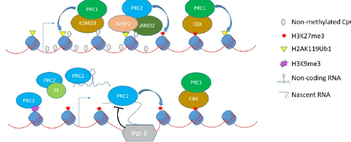
Figure 1.2. Schematic illustrating different modes of recruitment of PRC1 and PRC2 to the chromatin. The top schematic illustrates recruitment of non-canonical PRC1 to non-methylated CpG islands via interaction facilitated by the KDM2B subunit, and subsequent monoubitinylation of H2AK119. PRC2 is recruited either by non-methylated CpG islands via JARID2 or by H2AK119Ub1 via AEBP2/JARID2, leading to subsequent methylation of H3K27. This H3K27me3 mark can further recruit canonical PRC1 via its CBX subunit. The bottom schematic illustrates other modes of PRC2 recruitment, including: other repressive marks on histones, transcription factors, non-coding RNA, or short abortive nascent RNA while being inhibited by productive transcription. After recruitment, PRC2 places the H3K27me3 mark, which subsequently recruits canonical PRC1 to the chromatin.
How PRC1 exerts its gene silencing function
Since PRC1’s enzymatic activity to ubiquitinate H2AK119 was first discovered, there has been interest in understanding how this histone mark is coupled with repressive mechanisms. There are some hints that H2AK119ub1 restrains poised polymerase at the promoter (Figure 1.3)
13
(Stock et al., 2007), however, no direct mechanisms have been elucidated to date (reviewed in Simon & Kingston, 2013). Evidence for the ubiquitin mark having a repressive role comes from studies where enzymatic activities of RING proteins were specifically ablated. In the context of mouse ES cells, mutation of RING protein led to deregulation of many of the PRC1 target genes (Endoh et al., 2012). However, classic PRC1 targets such as Hox genes were only partially deregulated suggesting that other mechanisms of repression are in play.
Evidence for another repression mechanism came from the finding that PRC1 is able to
compact nucleosomal arrays (Figure 1.3) even in the absence of histone tails, suggesting this is independent of PRC1’s enzymatic function (Francis et al., 2004). This compaction is able to resist chromatin remodeling by other chromatin remodelers such as SWI/SNF (King, Francis, & Kingston, 2002). Importantly, subsequent studies showed that this chromatin compaction is able to repress genes such as Hox genes in vivo, and it remains intact in the context of
catalytically inactive RING1B (Eskeland et al., 2010). Instead, chromatin compaction has been attributed to the PHC subunit of PRC1, which can catalyze auto polymerization via its SAM domain (Isono et al., 2013). Accordingly, loss of PHC leads to loss of PRC1 selectively at sites marked by H3K27me3. This leads to a model in which CBX protein mediated recruitment to H3K27me3 and SAM mediated polymerization act together to maintain polycomb domains at silenced genes.
14
Figure 1.3. Schematic representing modes of gene repression by PRC1. The monoubiquitination mark placed by PRC1 on H2AK119 can restrain poised polymerases, while PRC1 mediated compaction can block access to the transcriptional machinery.
Our current understanding of polycomb biology can be summarized in the following model. PRC2 can be targeted to specific loci by transcription factors, non-coding RNA, or accumulation of PRC2 at transcriptionally silent loci by random genome sampling. Retention of PRC2 at these loci then results in deposition of methylation marks at H3K27. PRC2 can recognize the
H3K27me3 through its EED subunit, which also acts as an allosteric activator of the enzymatic activity of PRC2 (Margueron et al., 2009). This has been proposed to enable spreading of the H3K27me3 mark along the genome and also as a mechanism for maintaining these marks during replication (Hansen et al., 2008). The H3K27me3 marks are also recognized by CBX proteins in canonical PRC1. This leads to recruitment of canonical PRC1, which can then polymerize using its PHC subunit to compact the chromatin and establish the polycomb repressive domain. As an alternate mechanism, the canonical PRC1 are recruited to non-methylated CpG islands via its KDM2B subunit. Upon recruitment to chromatin, this complex deposits ubiquitin marks on H2AK119 to establish the repressive domain. Recently a new feedback loop has been elucidated where PRC1 mediated monoubiquititylation of H2AK119 is
15
able to recruit PRC2 and subsequently deposit H3K27me3 mark (Blackledge et al., 2014; Cooper et al., 2014). This could be driven by a variant of PRC2 containing AEBP2 and JARID2, which has been shown to bind to the H2AK119ub mark in vitro (Kalb et al., 2014). Thus, de novo
recruitment of non-canonical PRC1 could lead to recruitment of PRC2 and subsequent recruitment of canonical PRC1. It is still unclear whether this mechanism is in play in vivo.
PRC1’s role in self-renewal and differentiation
To understand the functional relevance of the repressive function of PRC1, the major
components of PRC1, RING1A and RING1B, were deleted in vivo and assessed for phenotype. Both homozygous and heterozygous deletions of RING1A led to anterior transformation and defect in axial skeletal patterning, however, the mice were still viable (del Mar Lorente et al., 2000). This suggests that RING1A is important for late stages of murine development. In contrast, deletion of RING1B/RNF2 resulted in a more severe developmental phenotype that is characterized by gastrulation arrest (Voncken et al., 2003). Notably, this is similar to the
phenotype seen in PRC2 deficiency that results from the deletion of EED, EZH2, or SUZ12 (Faust et al., 1998; Faust et al., 1995; O’Carroll et al., 2001; Pasini et al., 2004). In contrast to RING1A, no phenotype is observed upon heterozygous deletion of RING1B. These results tell us that the targets of RING1A and RING1B complexes are not entirely overlapping.
Regardless of phenotype of deletion occurring in the early or late stages of development, RING1A and RING1B are both important to ES cell potency. RING1B deletion in ES cells leads to deregulation of repressed genes that are part of differentiation pathways and organismal development (van der Stoop et al., 2008). These genes exhibit promoters that are bivalent and
16
also CpG-rich. However, binding studies for RING1B in ES cells revealed that only a small subset of the genes bound by RING1B are derepressed. Moreover, these ES cells maintain the
expression of pluripotency regulators such as OCT4 and NANOG. This suggests that additional mechanisms are used to repress most genes that are bound by RING1B. The additional
mechanism could very well be other variants of PRC1, because complete loss of PRC1 by deleting both RING1A and RING1B leads to loss of self-renewal of ES cells and eventual
differentiation (Endoh et al., 2008). This tells us that even though RING1A KO mice have defects in later stages of development, RING1A still contributes to gene regulation in ES cells.
In addition to the maintenance of self-renewal in ES cells, PRC1 is also important for proper differentiation. Upon differentiation of ES cells, chromatin states and chromatin bound proteins are stabilized leading to more heterochromatin formation and gene silencing (Mattout &
Meshorer, 2010; Meshorer et al., 2006). Simultaneously, we also observe upregulation of members of canonical PRC1 such as CBX2, CBX4, CBX8, PHC2, and BMI1 (Kloet et al., 2016; Morey et al., 2012). Since canonical PRC1 lack enzymatic activity and repress genes by mediating chromatin compaction, their upregulation as ES cell programs are downregulated makes sense. Direct evidence that PRC1 subunits MEL18, RYBP and PCGF6 dictate mesodermal lineage specification further confirms the role of PRC1 in proper differentiation (Morey et al., 2015; Ujhelly et al., 2015; Zdzieblo et al., 2014).
Much of the descriptive role of PRC1 in stem cells and differentiation come from studies
conducted in ES cells and in utero. Outside of this context, there are very few studies looking at the role of PRC1 in adult stem cells, and most of these delete components of PRC1 prenatally and then assess the effect on adult stem cells in postnatal animals (reviewed in Avgustinova &
17
Benitah, 2016). It is important to ask whether postnatal tissue lineage restricted stem cells still requires PRC1 for their self-renewal and multipotency maintenance. New insights were
revealed when RING1A and RING1B were deleted simultaneously in adult intestinal stem cells (Chiacchiera, Rossi, Jammula, Chiacchiera, et al., 2016). Upon deletion of PRC1, intestinal stem cells were unable to maintain their self-renewal and tissue homeostasis. Mechanistically, deletion of PRC1 led to derepression of many non-intestine lineage-specific transcription factors that negatively affect WNT/b-CATENIN pathway activity, which is essential for intestinal stem cells maintenance (Chiacchiera et al., 2016). This provided new insights into the ability of PRC1 to repress non-lineage genes and provide lineage commitment. Similar insight into PRC1 being important to maintain lineage identity and repress non-lineage genes have been
recapitulated in experiments deleting other subunits of PRC1, such as CBX4 (Mardaryev et al., 2016). Notably, most of the remaining studies on PRC1 function in adult stem cells come from deleting Bmi1 in various contexts.
BMI1
Bmi1 (B-cell-specific Moloney murine leukemia virus integration site 1) was originally discovered
as an oncogene that cooperated with Myc in driving B cell and T cell lymphomagenesis (Ygal Haupt et al., 1993; van Lohuizen, Verbeek, et al., 1991). Based on sequence similarity, it was quickly realized that Bmi1 was a member of the posterior sex combs (psc) gene family identified in Drosophila melanogaster (Brunk et al., 1991; van Lohuizen et al., 1991). Psc is a PRC1
component that is required to appropriately regulate Hox genes expression and segmentation patterning in D. Melanogaster (Paro, 1990; Struhl & Akam, 1985; Wedeen et al., 1986). This led
18
to analyses of the role of BMI1 in vertebrate development through the generation of germline
Bmi1 knockout (KO) mice (N. M. van der Lugt et al., 1994). These Bmi1 KO mice were viable but
displayed three major phenotypes: a profound hematopoiesis defect, cerebellar neurological abnormalities, and posterior transformation of the axial skeleton. Mice with overexpression of
Bmi1 were subsequently generated, and these animals demonstrated the converse axial
skeletal phenotype; anterior transformation (Alkema et al., 1995). These axial skeleton phenotypes are accounted for by deregulation of Hox genes along the strict boundaries
required for proper morphogenesis, and BMI1 was shown to be involved in repression of subset of the Hox genes (Alkema et al., 1995; N. M. T. van der Lugt et al.,1996).
Bmi1 in stem cells
The focus of Bmi1 research shifted towards the phenotype in hematopoiesis and neurogenesis in germline KO mice when it was shown that Bmi1 was highly expressed in the most primitive bone marrow cells and the expression was minimal in mature blood cells (J Lessard et al.,1998). This led to the hypothesis that Bmi1 is important for maintenance of the hematopoietic and neuronal stem cell compartments in both prenatal and postnatal animals. As a first step in this direction, it was shown that Bmi1 deficient mouse embryonic fibroblasts (MEFs) and lymphoid cells had increased levels of both p16 (Ink4a) and p19 (Arf) expression (Jacobs et al., 1999).
Ink4a and Arf are two major tumor suppressor genes, expressed from the same locus (Cdkn2a),
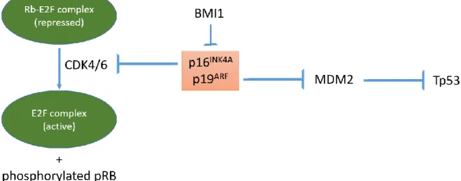
which regulate cellular senescence and apoptosis (Figure 1.4; Ivanchuk et al., 2001; Sharpless & DePinho, 1999; Sharpless & Chin, 2003). INK4A is an inhibitor of cell cycle. It inhibits the binding of CYCLIND to CDK 4/6, which results in mono-phosphorylation of the retinoblastoma protein,
19
pRB. Mono-phosphorylated pRB can bind to E2F and represses E2F mediated transcription. This leads to G1 phase arrest and senescence. ARF, on the other hand, inhibits MDM2, which would have ubiquitinated TP53 and led to its degradation. Inhibition of MDM2 stabilizes the tumor suppressor TP53, which can induce cell cycle arrest, senescence, or apoptosis (Ivanchuk et al., 2001; Sharpless & DePinho, 1999). Importantly, loss of the Ink4a/Arf locus in vivo was able to largely rescue the Bmi1 KO hematopoietic and neurological phenotypes, again confirming that BMI1 plays a critical role in repressing Ink4a/Arf, and controlling cell cycle, senescence, and cell survival (Jacobs, Kieboom, et al., 1999).
Figure 1.4. Schematic illustrating the BMI1 and INK4A/ARF axis. BMI1 represses two potent tumor suppressors INK4A and ARF that are expressed from the same locus. INK4A is an inhibitor of CDK4/6, which is required for phosphorylation of pRB and subsequent activation of E2F complex. ARF is an inhibitor of MDM2, which ubiquitinates TP53 for degradation.
Following the stem cell hypothesis, further research was conducted in the hematopoietic and neuronal lineages, because this is where the defects were observed in Bmi1 KO animals. Bmi1 KO animals were found to have normal number of hematopoietic stem cells (HSCs) in the fetal
20
liver but reduced HSC numbers in postnatal animals (Park et al., 2003). This suggests that the hematopoietic phenotype is not because of problem of lineage specification early in
development, but rather after HSCs have been established in the fetal liver. Transplant experiments demonstrated that both fetal liver HSCs and bone marrow HSCs from Bmi1 KO mice were only transiently able to contribute to hematopoiesis, indicating a defect in self-renewal. Indeed, Bmi1 KO HSCs are impaired in self-renewal and proliferation and have increased levels of Ink4a/Arf expression (Julie Lessard & Sauvageau, 2003; Park et al., 2003). The first description of Bmi1 KO mice and its hematopoietic phenotype had noted that Bmi1 KO mice had decreased number of hematopoietic cells in bone marrow, and the vacant space was filled by adipocytes. Thus, it was possible that the hematopoietic phenotype could be cell autonomous or microenvironment dependent (N. M. van der Lugt et al., 1994). Subsequent studies showed that both are true. Specifically, loss of Ink4a/Arf rescued the HSC self-renewal phenotype in Bmi1 KO mice, but the bone marrow microenvironment remained impaired and it resulted in sustained depletion of HSCs in postnatal animals (Oguro et al., 2006). Accordingly, the bone marrow of Bmi1 KO recipients could not support transplanted Bmi1 WT HSCs (Oguro et al., 2006). Conversely, overexpression of Bmi1 in mice caused HSCs to have enhanced self-renewal ability, reinforcing the role of Bmi1 in maintaining self-self-renewal capability in stem cells (Iwama et al., 2004; A. Rizo et al., 2008).
Similar to the role of Bmi1 in HSCs, Bmi1 is also important in neural stem cells. Bmi1 has been shown to be important for self-renewal capability but not the survival or differentiation of neural stem cells (Molofsky et al., 2003). The reduced self-renewal capability upon Bmi1 loss is also largely accounted for by increased expression of Ink4a/Arf, as loss of Ink4a partially
21
rescues the phenotype (Bruggeman et al., 2005; Molofsky et al., 2005). This phenotype is restricted to neural stem cells, as neuronal progenitor cells do not exhibit a phenotype upon
Bmi1 loss (Molofsky et al., 2003). The role of Bmi1 is also conserved in epithelial tissues. Bmi1
KO mice are able to undergo normal lung development, but putative bronchioalveolar stem cells (BASCs) have impaired self-renewal capacity and proliferation in vitro, and post injury in
vivo (Dovey et al., 2008). This phenotype is also partially rescued by loss of Arf (Dovey et al.,
2008). Similarly, Bmi1 KO mice have a mammary epithelium growth defect that is rescued upon loss of Ink4a/Arf (Pietersen et al., 2008). Other examples of Bmi1 KO phenotype in stem cells include a self-renewal defect in satellite cells, which are the resident stem cells of skeletal muscle (Robson et al., 2011). Upon Bmi1 loss, the stem cell pool decreases and progenitor cells pool increases. The remaining stem cells also enter a pre-senescence state and are unable to proliferate even when stimulated by high serum in vitro. This is reminiscent of the phenotype seen in stem cells of geriatric mice, which is caused by de-repression of Ink4a (Sousa-Victor et al., 2014).
Even though Bmi1 is important for self-renewal of stem cells in multiple tissue types, its expression is not restricted to just the stem cells. In mammary glands, Bmi1 is expressed in all cells, with the expression highest in luminal cells (Pietersen et al., 2008). Similarly, Bmi1 is expressed in both basal and non-proliferative suprabasal cells in the epidermis (K. Lee et al., 2008). In the intestine, Bmi1’s expression is present in both the stem cells at the bottom of the crypt and most of the transient amplifying cells (Itzkovitz et al., 2011). The first hint consistent with this expanded expression was the finding that Bmi1 is important for subsequent fate specification. In HSCs and multipotent progenitors (MPPs), genes that regulate B cell lineage
22
development, such as Ebf1 and Pax5, are repressed by bivalent domains. Loss of Bmi1 results in activation of these genes and aberrant lymphoid specification, resulting in increased B cell differentiation (Oguro et al., 2010). Since Bmi1 is important for maintenance of bivalent domains controlling lineage specification, it is plausible that Bmi1 needs to stay expressed not only in stem cells, but also in multipotent or bipotent progenitors. Similar phenotypes are also seen in the intestinal tissue where loss of Bmi1 perturbs the normal balance of absorptive and secretory lineages (Lopez-Arribillaga et al., 2015; Maynard & Lees, unpublished observation). The role of Bmi1 in repressing Ebf1 and Pax5 also establishes a role for Bmi1 beyond repression of the Ink4a/Arf locus. It was noted early on that even though deletion of Ink4a/Arf rescued some of the phenotypes of germline Bmi1 KO mice, the double KO mice were still smaller in size than WT mice (Jacobs et al., 1999) and the early lethality phenotype of Bmi1 loss remains (Molofsky et al., 2005). Careful examination of the neurological and hematopoietic phenotype showed that loss of Ink4a/Arf yielded only partially rescue, even though the self-renewal phenotype of stem cells were almost fully rescued in vitro (Molofsky et al., 2003; Oguro et al., 2006). This is at least partially explained by the role of Bmi1 in fate specification: specifically, transplant of Bmi1 and Ink4a/Arf double KO bone marrow cells into WT recipient mice showed that these cells still retained the Bmi1 KO differentiation defect of early lymphoid specification and increased differentiation towards B cell lineage (Oguro et al., 2010). These observations establish that Bmi1 has additional targets beyond Ink4a/Arf that are also important for maintenance of self-renewal capability in stem cells.
23
Bmi1 in cancer
Since Bmi1’s discovery through its ability to cooperate with Myc in inducing lymphomagenesis, its role has been studied extensively in the context of cancer. Bmi1’s oncogenic potential was first identified in mice with forced expression of Bmi1 in lymphoid compartment, which
resulted in mostly T-cell lymphoma (Y Haupt et al., 1993). Combining Bmi1 and Myc transgenes resulted in pre-B and B cell lymphoma (Y Haupt et al., 1993). Mutational analysis of Bmi1 showed that the N terminal RING domain and central portion of BMI1 protein is essential for the lymphomagenesis phenotype, while the C terminal region is dispensable (Alkema et al., 1997). This result is clearer under the light shed by structural studies on BMI1-RING1B complex, where BMI1 and RING1B interact using the RING domain, and the central part of BMI1 loops around RING1B to hug it (Buchwald et al., 2006; Li et al., 2006). In contrast, the C terminal region of BMI1 acts as a negative regulatory domain, and thus its deletion causes BMI1 to have increased oncogenic function (Yadav et al., 2010). The function of Bmi1 as an oncogene and a cooperating partner in Myc-driven lymphomagenesis comes primarily from its ability to repress the Ink4a/Arf locus and block Myc-induced apoptosis (Jacobs, Scheijen, et al., 1999a). In the context of MEFs, expression of Myc also results in upregulation of Arf and leads to apoptosis, which is inhibited by Bmi1 (Jacobs, Scheijen, et al., 1999a). This first description of oncogenic role of Bmi1 by repressing Ink4a/Arf locus shaped the next decade of research on Bmi1’s function in cancer.
Identification of Bmi1 as an oncogene that represses two very important tumor suppressor genes, Ink4a and Arf, led to the hypothesis that cancer cells could employ upregulation or
24
amplification of Bmi1 as a mechanism to repress Ink4a/Arf. This led to the hunt for expression patterns and amplification of BMI1 in human cancers. Since the field was focused on Bmi1’s role in lymphomagenesis until that point, it made sense for researchers to look at human hematological malignancies. Early research showed that BMI1 locus amplification is present, but not common, in human mantle cell lymphoma and non-Hodgkin lymphoma (Beà et al., 2001). However, some hematopoietic malignancies still upregulate BMI1 expression regardless of amplification status. Contrary to results in mice, BMI1 and INK4A/ARF expression levels did not show a simple inverse relationship in human cancer (Beà et al., 2001). Despite this finding, the expression levels of BMI1 correlated with an increase in grade of the malignancy, at least in the case of B-cell non-Hodgkin lymphomas (van Kemenade et al., 2001). The expression of BMI1 also correlated with KI67 expression, which is a marker of proliferating cells. This was even more impressive given that normal dividing B-cells do not express BMI1 (van Kemenade et al., 2001).
In addition to lymphomas, increased levels of BMI1 in human tumor samples compared to normal tissue of same origin has been shown in multiple solid tumors including
medulloblastomas (Leung et al., 2004), gastrointestinal tumors, pituitary and parathyroid adenomas (Sánchez-Beato et al., 2006), chronic myeloid leukemia (CML) (Mohty et al., 2007), neuroblastoma (Nowak et al., 2006), and many other tumor types (reviewed in Sauvageau & Sauvageau, 2010). Consistent with previously described literature, BMI1 was also found to be higher in advanced cancer, as in the case of CML, where advanced-phase patients had higher expression of BMI1 than chronic-phase patients (Mohty et al., 2007). As expected from increased expression in advanced cancer, BMI1 mRNA level was also found to be a marker of
25
poor prognosis in various tumors types such as CML (Mohty et al., 2007), bladder cancer (Qin et al., 2009), pediatric brain cancer (Farivar et al., 2013), esophageal squamous cell carcinoma (Hwang et al., 2014), and many others.
It was inferred that since BMI1 expression was higher in cancer compared to normal tissues, and this increase correlated with increased grade, BMI1 must be contributing to cancer progression. However, it is also possible that BMI1 is a tumor suppressor that cancer cells upregulate with increased oncogenic insult to try and mitigate the damage. This alternative hypothesis is similar to the observation in murine model of non-small cell lung cancer (NSCLC) that tumor suppressive Ink4a/Arf expression is upregulated during the transition from adenoma to adenocarcinoma (Feldser et al., 2010). The role of BMI1/Bmi1 has been addressed using human and murine cancer cell lines, and murine models. Early studies showed that Bmi1 is important for proliferation and survival of leukemia cells in murine models (Julie Lessard & Sauvageau, 2003). This result has been reiterated in numerous other cancer cell lines including glioma (Godlewski et al., 2008), gastric carcinoma (Li et al., 2010), multiple myeloma (Jagani et al., 2010), cervical cancer (Chen et al., 2011), breast cancer (Xu et al., 2011), Ewing sarcoma (Hsu & Lawlor, 2011) and many more. Genetically engineered mouse models (GEMMs) also support the notion that Bmi1 is oncogenic. In a hepatocellular carcinoma model, forced expression of Bmi1 in purified hepatic stem/progenitor cells was sufficient to initiate
tumorigenesis when these cells were transplanted in mice (Chiba et al., 2007). In a melanoma model, Bmi1 had no effect on the primary tumor but it enhanced dissemination of tumor cells and metastatic growth (Ferretti et al., 2016). This is consistent with the observation in human cancers that Bmi1’s expression increases with tumor progression. Conversely, germline deletion
26
of Bmi1 impaired the dissemination of tumor cells in the melanoma model (Ferretti et al., 2016). Importantly, these tumor suppressive effects of Bmi1 deletion in germline or tissue specifically during development has been observed in other GEMMs of cancer as well. In murine pancreatic cancer models, Bmi1 is required for initiation of neoplasia (Bednar et al., 2015). In the context of Apc loss driven murine intestinal cancer models, Bmi1 KO animals had lower tumor numbers and burden compared to WT animals. This phenotype was dependent upon Bmi1’s function to repress Arf, as combining Arf loss in the intestinal cancer model partially rescued the Bmi1 KO phenotype (Maynard et al., 2013).
Given Bmi1’s positive role in maintaining self-renewal potential in stem cells and Bmi1’s positive role in cancer, both via Ink4a/Arf locus repression, it was not long before researchers started to focus on Bmi1’s role in TPCs. Given that the first description of TPCs was in leukemia (Bonnet & Dick, 1997), the first studies were conducted in leukemia tumor propagating cells (L-TPCs). Bmi1 KO in L-TPCs arrested proliferation and yielded signs of apoptosis, and the L-TPCs were unable to maintain their undifferentiated state or transplant potential (Julie Lessard & Sauvageau, 2003). As with the Bmi1 deficient HSCs, the L-TPCs also showed upregulation of
Ink4a/Arf (Aleksandra Rizo et al., 2009). Examination of the role of Bmi1 during reprogramming
of normal cells to L-TPCs also showed its importance at early differentiation stages (Yuan et al., 2011). Bmi1 KO granulocyte/macrophage progenitors (GMPs) could be transformed by
introduction of oncogenes but fewer L-TPCs were retained and these showed increased differentiation. Moreover, when transplanted into recipient mice, these cells could not
establish leukemia. Interestingly, this phenotype was only partially rescued upon Ink4a/Arf loss, and it revealed derepression of lineage inappropriate transcription factors including Tbx15
27
(Yuan et al., 2011). Moreover, reintroduction of Bmi1 could not rescue the phenotype, indicating that the effect of Bmi1 loss is permanent, most likely pushing the cells to a more differentiated state from which L-TPCs cannot arise (Yuan et al., 2011).
Bmi1’s role in TPCs have also been established in solid cancers. One such example is human
colorectal cancer. The standard assay of TPC frequency is limiting dilution in vivo, where cells are transplanted at various doses into mice and the frequency of tumor development is
measured. Using this assay, knockdown of BMI1 was shown to decrease TPC frequency in both cancer cell lines and primary tumor samples from patients (Kreso et al., 2014). Similarly, in a murine model of head and neck squamous cell carcinoma (HNSCC), Bmi1 positive tumor cells were shown to have higher enrichment for TPCs compared to Bmi1 negative tumor cells in a limited dilution assay (D. Chen et al., 2017). These Bmi1 positive tumor cells were also enriched in the tumor upon treatment of cisplatin, suggesting that they are more chemoresistant, which is one of the hallmarks of TPCs.
Even though most of the literature is consistent with the role of Bmi1 in tumor promotion through its ability to repress Ink4a/Arf locus, there is evidence for alternate mechanisms. In a murine model of pancreatic cancer, loss of Bmi1 is tumor suppressive. However, this phenotype is independent of Ink4a/Arf and the data point to reactive oxygen species dysregulation instead (Bednar et al., 2015). Similarly, Bmi1 deficient immortalized GMPs have low replating efficiency in colony forming assays that is independent of Ink4a/Arf status (Yuan et al., 2011). This
phenotype is phenocopied by overexpression of Tbx15, a lineage inappropriate transcription factor that is upregulated upon loss of Bmi1 (Yuan et al., 2011). This suggests that Bmi1’s role of
28
repressing lineage specifying transcription factors is also important for TPC potential, similar to repression of Ebf1 and Pax5 being important for HSC function (Oguro et al., 2010).
Lung adenocarcinoma (LUAD)
LUAD in patients
According to the Cancer Facts & Figures published in 2019, lung and bronchus cancer is the second leading cause of estimated new cancer cases, and the first leading cause of estimated cancer deaths, in the United States. Even though lung and bronchus cancer accounts for only 12.9% of total newly diagnosed cancer cases, it accounts for 23.5% of total estimated cancer deaths, making it a huge unmet medical need (Siegel et al., 2019). According to the National Cancer Institute’s Surveillance, Epidemiology, and End Results Program (SEER), the five-year survival rate for lung and bronchus cancer is 19.4%. This survival rate is very dependent on the stage at which the disease was diagnosed; if at the localized tumor stage, the 5-year survival rate is 57.4%, but diagnosis at the distant metastasis stage, which is true for more than half of lung cancer patients, decreases the 5-year survival rate to a meager 5.2% (SEER, 2019).
Even though the national statistics lump lung and bronchus cancer into one category, lung cancer is a collection of various different malignancies (Chen et al., 2014; Reck et al., 2013). Based on histological differences, lung cancer can be broadly classified into non-small cell lung cancer (NSCLC) and small cell lung cancer (SCLC). Even within NSCLC, there are various
subtypes, with adenocarcinoma and squamous cell carcinoma predominating, and a small fraction of NSCLC being categorized as large-cell carcinoma or other miscellaneous categories (Howlader et al., 2019). Sequencing analyses of large numbers of adenocarcinomas and
29
squamous cell carcinomas showed that the recurring mutations, amplifications, and deletions differ greatly between the two subtypes, reinforcing the histological classifications as different malignancies (Campbell et al., 2016).
Lung adenocarcinoma (LUAD) arises mostly in distal lung and is associated with the marker NKX2.1/TTF1. It is characterized by glandular histology and/or the presence of mucin (Davidson et al., 2013). IASLC/ATS/ERS classification of adenocarcinoma, based on invasiveness, breaks down the adenocarcinoma subtype into adenocarcinoma in situ (AIS), minimally invasive adenocarcinoma (MIC), and invasive adenocarcinoma, with the first two having effectively 100% 5-year survival rate post resection. Invasive adenocarcinoma is further subdivided based on histological patterns, such as lepidic, acinar, papillary, micropapillary, and solid (Travis et al., 2011). This is important because certain histological features, such as micropapillary, are associated with poor prognosis (Travis, 2011).
In recent years, a lot of effort has been placed on understanding the molecular drivers of LUAD and in using the information about genetic alterations to inform therapeutic approaches. Comprehensive profiling from The Cancer Genome Atlas (CGA) Research Network has provided extensive insights in that regard (Collisson et al., 2014). TP53 has been identified as the most mutated gene (46%), closely followed by KRAS (33%). Interestingly, EGFR mutations in 14% of patients were mutually exclusive to KRAS mutations, suggesting that there is a strong selective pressure to activate the EGFR-RAS pathway in LUAD. Identification of EGFR mutations led to the use of EGFR inhibitors in lung cancer. Other targeted therapies approved for LUAD target
alterations in BRAF and ALK. The CGA study also show that LUAD has a relatively high
30
neoepitopes, some of which are recurrent among patients (Campbell et al., 2016).
Consequently, there is great interest in the use of immune checkpoint inhibitors in LUAD, and to date four anti-PD1/PD-L1 antibodies have received FDA approval for treatment of LUAD (CRI, 2019).
Genetically engineered mouse models (GEMMs) of LUAD
With the detection of vast array of genetic alterations in human LUAD comes the challenge of identifying driver mutations in cancer versus passenger mutations that have little functional consequences. This is particularly challenging for patients with smoking history who have an especially high mutational burden. Researchers have relied on preclinical models to validate putative driver mutations and also assess the therapeutic efficacy of targeting these alterations. GEMMs have been widely used as relevant preclinical model of cancer. GEMMs allow for
generation of autochthonous tumors that arise and progress in their own native
microenvironment, thus, recapitulating a more complete picture of tumor biology. Conditional alleles and site-specific recombinase systems such as Cre-loxP system and Flp-frt system made possible GEMMs in which candidate driver genes are expressed at the endogenous level in any cancer type (reviewed in Branda & Dymecki, 2004). These systems allow for whole body or tissue specific deletion or activation of any gene of interest, by either flanking exons with recognition sites for the recombinase, or inserting flanked STOP cassettes that can be removed by the recombinase, respectively. Newer generation versions of these technologies include the ability to temporally control the deletion or activation of any gene by allowing a small molecule to control recombinase activity. The most notable example is the CreERT2 system, where the
31
Cre recombinase nuclear import and activity is controlled by 4-hydroxy-tamoxifen (reviewed in Branda & Dymecki, 2004). Coupling different recombinases and inducible systems can allow for temporal and spatial control over genetic alterations. For instance, tumor initiation can be triggered by one recombinase system, while deletion of another gene of interest in established tumors can subsequently be triggered by a second inducible recombinase system.
In the field of LUAD, multiple GEMMs have been established that use various recurring mutations in patient samples as the driver mutations. One of the most commonly used
mutations to model LUAD is the Kras mutation. The first model to recapitulate the endogenous level of expression of mutated Kras was a GEMM with a latent Kras allele that activates via a spontaneous recombination event and predominantly gives rise to lung cancer (Johnson et al., 2001). This quickly gave way to GEMMs with an inducible allele of Kras carrying a G12D
mutation, which is the most frequent KRAS mutation seen in patients (Jackson et al., 2001; Meuwissen et al., 2001). In this model, expression of this Kras-G12D allele is prevented by the presence of a STOP cassette, which can be excised at the flanking loxP sites using the Cre recombinase. The Cre recombinase can be easily introduced using intratracheal delivery of adenoviral or lentiviral particles encoding for Cre recombinase (DuPage et al., 2009). Cre mediated excision of the STOP cassette leads to activation of the Kras-G12D allele and a subset of the affected cells give rise to mostly adenoma, which can progress at low frequency and with long latency to adenocarcinoma. This model is made more aggressive by combining mutant
Tp53 alleles with the Kras mutation (Jackson et al., 2005), both of which are relevant mutations
in patients (Collisson et al., 2014). The resulting Kras and Tp53 mutant (KP) tumors progress much faster than Kras (K) only tumors, and recapitulate advanced human LUAD. Tumors of all
32
grades and lymph node metastases are seen in the majority of animals, with few animals progressing to distant metastases.
The development of GEMMs that recapitulate advanced human LUAD faithfully created the ability to study the various stages of tumor progression, and to dissect pathways important for tumor development, progression, and dissemination. One early insight from the KP GEMM was that, even though tumorigenesis is driven by mutant Kras, there is still amplification in MAPK signaling during the adenoma to adenocarcinoma transition (Feldser et al., 2010). This is consistent with the previous findings that activation of Kras-G12D alone in different epithelial tissue types leads to proliferation but not detectable phosphorylated ERK (Tuveson et al., 2004), and that K only GEMMs rarely progress to adenocarcinoma (Jackson et al., 2001; Meuwissen et al., 2001). Moreover, activation of the ARF/TP53 pathway and downstream cell cycle arrest or apoptosis is limited to the amplified MAPK signaling setting (Feldser et al., 2010). KP GEMMs have also been vital in describing changes associated with metastases. Using an extracellular matrix (ECM) array, researchers identified a panel of ECM molecules (Fibronectin, Laminin, Galectin-3, and Galectin-8) that are enriched in metastases but not present in primary tumors (Reticker-Flynn et al., 2012). This study also identified SPP1/osteopontin as the ECM molecule that is upregulated in high grade tumor cells and metastases (Reticker-Flynn et al., 2012). Another key insight from KP GEMM of LUAD has been the identification of markers for TPCs. Careful orthotopic serial transplantation experiments have revealed markers such as SCA1, CD24, NOTCH, and ITGB4 as key markers that enrich for functional TPCs (Curtis et al., 2010; Lau et al., 2014; Y. Zheng et al., 2013). These insights have added to the repertoire of functional markers and therapeutic target candidates for LUAD.
33
To understand the genetic alterations important for tumor progression, with the hope of expanding on the mutational analysis from patient samples, a lot of effort was put into characterizing the mutational profile of KP LUAD (McFadden et al., 2016). However, to the researchers’ surprise, the mutational burden in murine KP primary tumors and cell lines was much lower than that seen in patient samples. This highlights one of the limitations of the GEMM to model LUAD: there is less pressure on tumor cells to acquire and select for
advantageous mutations, since the tumorigenesis is driven by a powerful oncogene and loss of a major tumor suppressor. Perhaps unsurprisingly, KP LUAD have little to no lymphocyte
infiltration, possibly owing to the low mutational burden and lack of neoantigens (Cheung et al., 2008; DuPage et al., 2011). This highlights another major limitation of the GEMM.
Dedifferentiation as a route to KP LUAD progression
Since not all tumors in the KP GEMM LUAD model acquire the ability to metastasize, this provides the perfect opportunity to assess differences in malignant and non-malignant tumor that otherwise have the same driver mutations. Researchers were able to identify NKX2.1/TFF-1 as a suppressor of malignant progression in this model (Winslow et al., 2011). This is consistent with the previous finding that NKX2.1 expression correlates with better prognosis in lung cancer patients (Berghmans et al., 2006). However, this finding was somewhat controversial, because frequent amplification of NKX2.1 is seen in patient samples, and oncogenic functions have been established for Nkx2.1 in cell lines (Kendall et al., 2007; Kwei et al., 2008). Results from KP GEMM clearly established that Nkx2.1 controls the differentiation status and metastatic
34
HMGA2, which is normally restricted to the embryonic lineage. HMGA2 has been previously shown to be a marker of poor prognosis, and in vitro data suggests a pro-tumorigenic role (Y. S. Lee & Dutta, 2007; Sarhadi et al., 2006). Collectively, these data suggest that LUAD progresses from NKX2.1 positive to a NKX2.1 negative and HMGA2 positive state. By extension of this logic, perhaps the presence of Nkx2.1 amplification in human tumors reflects the fact that it
promotes the early stages, but not progression, of human LUAD.
Further insights into the role of Nkx2.1 in LUAD came from the study where Nkx2.1 was deleted in KP tumors (Snyder et al., 2013). This led to expression of a latent gastric transcriptional program that is marked by expression of HNF4a, ONECUT2 and other gut lineage genes (Snyder et al., 2013). This latent gastric lineage is repressed by NKX2.1 through sequestration of
FOXA1/2 and AP1 transcription factors at pulmonary genes (Maeda et al., 2012; Snyder et al., 2013). Loss of HNF4a on top of NKX2.1 loss leads to upregulation of HMGA2, suggesting a linear pathway for tumor progression in the KP LUAD model. Additional factors, such as FOXA2 and CDX2, have also been found to cooperate with NKX2.1 in inhibiting malignant progression of LUAD, further revealing regulatory nodes involved in metastasis (Li et al., 2015).
The KP LUAD model appropriately recapitulates different stages of LUAD progression, without the need for additional driver mutations to drive those transitions. This strongly suggests that epigenetic changes are responsible for promoting tumor progression. The NKX2.1/HMGA2 axis, combined with previous studies showing a correlation between dedifferentiated status and worse survival in lung cancer patients, raises the possibility that epigenetic changes might enable acquisition of a more dedifferentiated status as a mechanism of tumor progression (Liu, Kho, Kohane, & Sun, 2006). This is also consistent with the presence of recurring mutations in
35
epigenetic modulators in human LUAD patients (Collisson et al., 2014). Thus, this LUAD. model provides a perfect setting to examine the role of the Bmi1 epigenetic regulator in tumor development and progression.
Bmi1 in lung cancer
The correlation of Bmi1 expression levels and NSCLC prognosis has been studied since 2001. First, in NSCLC mostly of squamous subtype, BMI1 mRNA levels in patient tumors was shown to inversely correlate with Ink4a/Arf levels (Vonlanthen et al., 2001). Subsequent independent studies, including a meta study, showed that high BMI1 mRNA levels in LUAD is correlated with worse overall survival, larger tumor size, poor differentiation, and distant metastasis (Meng et al., 2012; Zhang et al., 2017; Zhang et al., 2014). To assess the putative oncogenic role of BMI1
in vitro, multiple studies assessed the phenotypic effect of shRNA mediated knockdown of BMI1
in human cell lines, A549 (Ink4a mutant) and SPC-A1 (Ink4a WT), and found that BMI1
knockdown was tumor suppressive regardless of the Ink4a status (Meng et al., 2012; Q. Yu et al., 2007; X. Zheng et al., 2014). Potential mechanisms for this tumor suppression were touted to be downregulation of AKT pathway, upregulation of cell cycle inhibitors p21/p27, and/or cell cycle arrest before the S phase.
The importance of Bmi1 in lung cancer has been recapitulated in GEMMs of LUAD driven by different genetic alterations. In a study conducted in our lab and others, Bmi1 deficiency was shown to be tumor suppressive by launching Kras driven tumors in germline Bmi1 KO animals (Figure 1.5; Dovey et al., 2008). The Bmi1 KO animals had both reduced tumor number and impaired progression compared to the WT controls, suggesting a role for Bmi1 at tumor
36
initiation. This phenotype could be partially rescued upon loss of Arf, corroborating previous conclusions that Bmi1 imparts its oncogenic function through repression of Ink4a/Arf locus (Jacobs et al., 1999b). Notably, the tumor suppressive phenotype of Bmi1 KO also correlates with bronchioalveolar stem cells (BASCs) exhaustion, in either the presence or absence of oncogenic stimuli. This phenotype is partially rescued by deletion of Arf, or deletion of p57 (Dovey et al., 2008; Zacharek et al., 2011). A similar tumor suppressive effect of Bmi1 KO was also observed in a LUAD model driven by loss of Cebpa (Yong et al., 2016), and C-RAF driven model of NSCLC (Becker et al., 2009), suggesting a driver mutation agnostic function of Bmi1. These findings fit with the prevailing view that Bmi1 is required to enable stem cells and TPCs function, primarily via suppression of Ink4a/Arf.
Since BMI1 has been implicated in repression of Ink4a/Arf locus during development in utero, it is possible that the central role of Ink4a/Arf in these Bmi1 deficient GEMM reflects the lack of proper silencing of Ink4a/Arf locus during development, as shown by high expression levels of
Ink4a/Arf in Bmi1 deficient postnatal stem cells compared to WT controls (Dovey et al., 2008).
To confirm that Bmi1 deletion can be a good therapeutic approach, it is essential to assess this in animals that have developed in normal Bmi1 WT condition with appropriate Ink4a/Arf silencing.
Previous work from our lab tested the consequences of tumor specific deletion of Bmi1 at the time of tumor initiation in adult animals in both K only, and KP, LUAD models (Figure 1.5; Karl et al., unpublished data). These studies described a profound tumor suppressive phenotype in early stage tumors that resulted in significant extension of survival in both models upon
37
mice had similar number of tumors. In contrast, the tumors originating in Bmi1 KO state were significantly smaller compared to WT tumors and this difference was driven by slower
proliferation rate in Bmi1 KO tumors. Interestingly, this proliferation defect was evident at the grade 2 stage but not at grade 3, suggesting that tumor cells eventually adapt to Bmi1 loss. Of note, there was no evidence of differential Ink4a/Arf expression between Bmi1 KO and WT tumors, antithetical to previous literature.
Gene expression analysis of individual tumors revealed a cell cycle defect in grade 2 Bmi1 KO tumors but not grade 3, consistent with the observed grade-specific proliferation defects. In addition, Bmi1 KO tumors also had upregulation of lineage inappropriate genes irrespective of grade, consistent with previously identified function of Bmi1 to repress non-lineage genes (Yuan et al., 2011). Some of the Bmi1 KO tumors were positive for HMGA2, a marker of tumor progression in the KP LUAD model (Snyder et al., 2013; Winslow et al., 2011). Surprisingly, staining of serial sections of tissue with NKX2.1 revealed that some of these HMGA2 expressing tumor cells were also positive for NKX2.1. Since these two markers are mutually exclusive with each other during tumor progression, the double positivity of these tumor cells suggested that this might result from alterations in gene reguation upon Bmi1 loss rather than being a sign of tumor progression. However, since HMGA2 positive tumors accounted for a small fraction of total tumors, this could not account for the apparent adaptation phenotype we observe. This study was unable to establish the mechanisms that underlie the tumor suppressive phenotype upon Bmi1 loss, or the subsequent adaptation to Bmi1 loss.
38
Remaining questions
Since most cancer patients are diagnosed when the cancer is already aggressive, assessing the impact of Bmi1 deficiency on KP LUAD by deleting Bmi1 in either the germline or at the time of initiation of tumor does not mimic the clinical treatment paradigm. This provides strong
rationale for assessing the role of Bmi1 in already established, aggressive tumors. Additionally, there is evidence in the literature that BMI1 expression is high in the early stages of primary tumors, but is downregulated in matched lymph node metastases (Xiong et al., 2015),
suggesting that Bmi1 may play different roles during tumor initiation versus tumor progression. Since most of the studies in GEMMs has focused on Bmi1’s role during tumor initiation, we feel there is a huge need to address its role in existing tumors.
In this thesis, I address the role of Bmi1 during tumor progression in KP LUAD, where the tumor bearing animals have developed in utero with normal Bmi1 expression, and the tumors
progressed through early stages of tumorigenesis with normal Bmi1 expression as well (Figure 1.5). My data show that once the tumors reach higher grades, Bmi1 functions as a tumor suppressor by limiting the dedifferentiation of tumor cells, which is necessary for malignant progression. Thus, Bmi1 plays a context dependent role in tumorigenesis, and targeting Bmi1 in LUAD might have an unwanted effect in aggressive cancer.
39
Figure 1.5. Schematic summarizing the work done in our lab to assess the impact of Bmi1 deletion in GEMMs of LUAD. Deletion of Bmi1 in germline leads to development of animal in utero in the absence of BMI1. Launching Kras driven LUAD in this context shows that Bmi1 deficiency causes tumor suppression, which is mediated by Ink4a/Arf derepression. In adult animals that developed with normal Bmi1 levels, deletion of Bmi1 at the time of tumor initiation causes tumor suppression of early tumor grades without differential expression of Ink4a/Arf. This tumor suppressive phenotype subsides by the time tumors reach grade 3. Our current model assesses the impact of Bmi1 deletion in established high grade KP LUAD tumors.
40
References
Atlasi, Y., & Stunnenberg, H. G. (2017). The interplay of epigenetic marks during stem cell differentiation and development. Nature Reviews Genetics, 18(11), 643–658.
https://doi.org/10.1038/nrg.2017.57
Avgustinova, A., & Benitah, S. A. (2016). Epigenetic control of adult stem cell function. Nature
Reviews Molecular Cell Biology, 17(10), 643–658. https://doi.org/10.1038/nrm.2016.76
Avgustinova, A., Symeonidi, A., Castellanos, A., Urdiroz-Urricelqui, U., Solé-Boldo, L., Martín, M., … Benitah, S. A. (2018). Loss of G9a preserves mutation patterns but increases chromatin accessibility, genomic instability and aggressiveness in skin tumours. Nature Cell Biology,
20(12), 1400–1409. https://doi.org/10.1038/s41556-018-0233-x
Bankhead, P., Loughrey, M. B., Fernández, J. A., Dombrowski, Y., McArt, D. G., Dunne, P. D., … Hamilton, P. W. (2017). QuPath: Open source software for digital pathology image analysis. Scientific Reports, 7(1), 16878. https://doi.org/10.1038/s41598-017-17204-5 Barletta, J. A., Perner, S., Iafrate, A. J., Yeap, B. Y., Weir, B. A., Johnson, L. A., … Chirieac, L. R.
(2009). Clinical significance of TTF-1 protein expression and TTF-1 gene amplification in lung adenocarcinoma. Journal of Cellular and Molecular Medicine, 13(8b), 1977–1986. https://doi.org/10.1111/j.1582-4934.2008.00594.x
Beà, S., Tort, F., Pinyol, M., Puig, X., Hernández, L., Hernández, S., … Campo, E. (2001). BMI-1 gene amplification and overexpression in hematological malignancies occur mainly in mantle cell lymphomas. Cancer Research, 61(6), 2409–2412. Retrieved from
http://www.ncbi.nlm.nih.gov/pubmed/11289106
Bednar, F., Schofield, H. K., Collins, M. A., Yan, W., Zhang, Y., Shyam, N., … Pasca di Magliano, M. (2015). Bmi1 is required for the initiation of pancreatic cancer through an Ink4a-independent mechanism. Carcinogenesis, 36(7), 730–738.
https://doi.org/10.1093/carcin/bgv058
Ben-Porath, I., Thomson, M. W., Carey, V. J., Ge, R., Bell, G. W., Regev, A., & Weinberg, R. A. (2008). An embryonic stem cell–like gene expression signature in poorly differentiated aggressive human tumors. Nature Genetics, 40(5), 499–507.
https://doi.org/10.1038/ng.127
Brien, G. L., Valerio, D. G., & Armstrong, S. A. (2016). Exploiting the Epigenome to Control Cancer-Promoting Gene-Expression Programs. Cancer Cell, 29(4), 464–476.
https://doi.org/10.1016/J.CCELL.2016.03.007 Broad Institute. (n.d.). Morpheus. Retrieved from
https://software.broadinstitute.org/morpheus
Camolotto, S. A., Pattabiraman, S., Mosbruger, T. L., Jones, A., Belova, V. K., Orstad, G., … Snyder, E. L. (2018). FoxA1 and FoxA2 drive gastric differentiation and suppress squamous