Publisher’s version / Version de l'éditeur:
Two-dimensional multi-spectral digitization and three-dimensional modelling of easel paintings, 1, 2008
READ THESE TERMS AND CONDITIONS CAREFULLY BEFORE USING THIS WEBSITE. https://nrc-publications.canada.ca/eng/copyright
Vous avez des questions? Nous pouvons vous aider. Pour communiquer directement avec un auteur, consultez la première page de la revue dans laquelle son article a été publié afin de trouver ses coordonnées. Si vous n’arrivez pas à les repérer, communiquez avec nous à PublicationsArchive-ArchivesPublications@nrc-cnrc.gc.ca.
Questions? Contact the NRC Publications Archive team at
PublicationsArchive-ArchivesPublications@nrc-cnrc.gc.ca. If you wish to email the authors directly, please see the first page of the publication for their contact information.
NRC Publications Archive
Archives des publications du CNRC
This publication could be one of several versions: author’s original, accepted manuscript or the publisher’s version. / La version de cette publication peut être l’une des suivantes : la version prépublication de l’auteur, la version acceptée du manuscrit ou la version de l’éditeur.
Access and use of this website and the material on it are subject to the Terms and Conditions set forth at
Two-dimensional multi-spectral digitization and three-dimensional
modelling of easel paintings
Lahanier, C.; Aitken, G.; Pillay, R.; Beraldin, Jean-Angelo; Blais, François;
Borgeat, Louis; Cournoyer, Luc; Picard, Michel; Rioux, Marc; Taylor, John;
Breuckmann, B.; Colantoni, P.; de Deyne, C.
https://publications-cnrc.canada.ca/fra/droits
L’accès à ce site Web et l’utilisation de son contenu sont assujettis aux conditions présentées dans le site LISEZ CES CONDITIONS ATTENTIVEMENT AVANT D’UTILISER CE SITE WEB.
NRC Publications Record / Notice d'Archives des publications de CNRC:
https://nrc-publications.canada.ca/eng/view/object/?id=8a85ca56-6577-4954-adfc-3714784e19ee https://publications-cnrc.canada.ca/fra/voir/objet/?id=8a85ca56-6577-4954-adfc-3714784e19ee
National Research Council Canada Institute for Information Technology Conseil national de recherches Canada Institut de technologie de l'information
Two-dimensional multi-spectral digitization
and three-dimensional modelling of easel
paintings *
Lahanier, C., Aitken, G., Pillay, R., Beraldin, J.-A., Blais, F., Borgeat, L., Cournoyer, L., Picard, M., Rioux., M., Taylor, J., and Breuckmann, B., Colantoni, P., de Deyne, C.
2005
* 14th Triennial Meeting the Hague, Rome: International Council of Museums (ICOM), 2005
Copyright 2008 by
National Research Council of Canada
Permission is granted to quote short excerpts and to reproduce figures and tables from this report, provided that the source of such material is fully acknowledged.
Title
The 2D multi-spectral digitisation and the 3D modelling of easel paintings.
Authors
Christian Lahanier1, Geneviève Aitken1, Angelo Beraldin2, François Blais2, Louis Borgeat2,
Bernd Breuckmann3, Philippe Colantoni4, Luc Cournyer2, Christophe de Deyne5, Michel
Picard2, Ruven Pillay1, Marc Rioux2, and John Taylor2
Adresses
1.- Centre de recherche et de restauration des musées de France, Palais du Louvre, Porte des Lions, 14, quai François Mitterrand, 75001 Paris Cedex01, France
2.- Institut de technologie de l'information, Conseil national de recherches Canada, 1200 Chemin Montréal, Édifice M-50, Ottawa, Ontario, K1A 0R6, Canada
3.- Breuckmann Gmbh, Torenstrasse 14, 88709 Meersburg, Germany
4.- Laboratoire LIGIV, Université Jean Monnet, 10, rue Barrouin/ 42000 Saint-Étienne, France
5.- Pébéo, BP106, avenue du Pic de Bertagne, 13881 Gémenos Cedex France
lahanier.christian@culture.fr, genevieve.aitken@culture.fr, angelo.beraldin@nrc.ca,
francois.blais@nrc-cnrc.gc.ca, louis.borgeat@nrc.ca, bernd.breuckmann@breuckmann.com,
colantoni@couleur.org, luc.cournyer@nrc.ca, cdedeyne@pebeo.com, michel.picard@nrc.ca,
ruven@free.fr, marc.rioux@nrc.ca, john.taylor@nrc.ca
Abstract
This paper presents the usefulness and utility of three complementary digitisation techniques for the dynamic rendering of colour, roughness and shape of easel paintings.
The 2D multi-spectral digitisation and the 3D modelling of paintings are used for colour accuracy rendering and measurement, colour characterisation, pigment identification, for high definition visualisation of the painting and also for the measurement of the shape of the support and the paint-layer roughness.
The experimentation was performed on several paintings. The results show the advantages of each technique. Several viewers were developed to handle multi-spectral images and 3D models.
Keywords
Multi-spectral images, 3D model, easel painting, colour chart, viewer, reflectance spectral curve, roughness, shape
Introduction
The 2D multi-spectral digitisation and the 3D modelling of easel paintings are used for colour accuracy, colour rendering and measurement, colour characterisation, pigment identification; they are used also for high definition visualisation and measurement of the shape of the support and of the paint-layer roughness.
Three kinds of equipment were used for this study :
- the CRISATEL camera for high definition image capture and high accuracy colour reproduction (using a multi-spectal system based on optical filters). This technology was developed by Lumière Technology as part of the CRISATEL project,
- the High-Resolution Colour Laser Scanner and the Large Volume of View or “Big Scan” Laser Scanner from NRC, Canada
The same experimentation was made on a painting entitled "a Dame en prière". The comparison of the results shows the advantages of each technique.
A "Dame en prière"
This oil painting on wood (fig.10), dated between 1575 and 1599, belongs to the Louvre museum collections (inv RF2090). This small painting (310 x 215 mm) is part of a larger composition as we can see on the X ray (fig. 1a). Two figures appear on the left side and on the upper part of the painting (fig. 1e and fig. 1f). The wood support was cut on its left and upper sides for an unknown reason and the paint layer was modified to mask the figures belonging to the original composition. The face and the ruff of the lady were also repainted. They contain more lead white than the other figure on her right side. The right side of the panel, without a white preparation made of calcium carbonate, absorbs less X Rays (fig. 1a and fig. 1d). The dark paint layer made of chromium green (a XIX century pigment) applied on the ground and on the left arm of the lady is visible on the IR photograph (fig 1d). Curiously the painting was enlarged on its right side. A varnish was applied on the paint layer after the modification of this panel. Few restorations appear on top of the varnish (fig 1c). Pentimento are visible on the X Ray radiograph along the right hand of the lady (fig 1g). We chose this painting because of its good state of conservation, and for the changed composition as well as for the measurement of the relief of the decoration of the lady's costume.
The multi-spectral imaging system (2), (3)
Optimisation of the lighting source and of the parameters of the camera
Figure 1: 1a the X Ray overview, 1b the raking light photograph, 1c the ultraviolet fluorescence
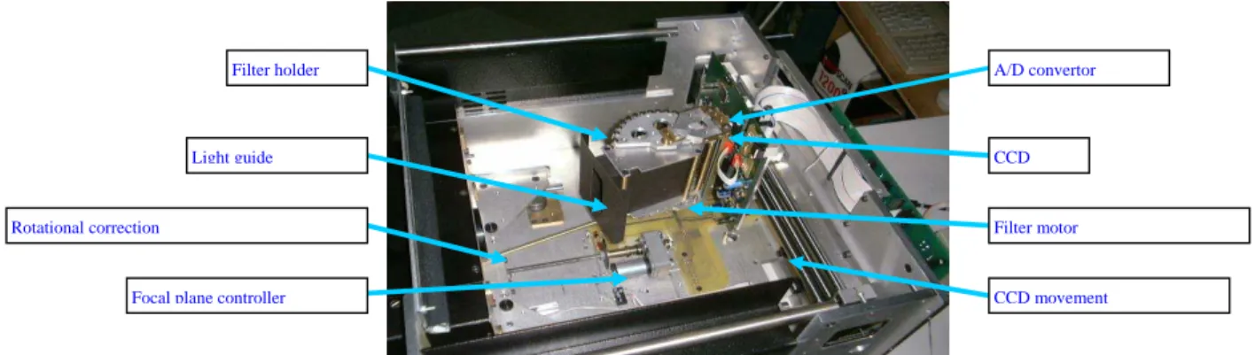
High speed and high definition digital capture need intense and uniform lighting limited to the area viewed by the CCD array. To comply with admissible light exposure standards, a new process for non-destructive lighting of easel paintings has been developed by Lumière
Technology. Four HQI lamps (metal halide lights) with an electronic stabiliser, set at the focal point of an elliptical mirror, project a 5cm by 3m line beam at 2 metres distance, producing a uniform light of one hundred thousand lux . The projector is motorised and the beam
movement is synchronised by computer with the moving CCD array in the associated camera fig. 2). The linear light beam produces a spatially homogeneous light on the painting (4).
figure 2 : the inside view of the CRISATEL camera
Before an acquisition session, the multispectral system is calibrated with a Labsphere grey scale in order to determine a set of optimal parameters for each channel : offset and gain of the CCD amplifier, exposure time, etc. The spatial distribution of the lighting onto the plane of the painting is recorded by digitising a custom non-glossy white board in the same position as the painting under invertigation(5).
The corrections of the 13 multispectral images are made in a post-processing stage by taking into account
- the black level and gain for each individual CCD element, - the spatial heterogeneity of the lighting (topographic correction),
- the resizing of the 13 images because of the change in the focal plane distance for each filter (geometric correction).
Spectral Reconstruction (6)
The spectral reconstruction was initially based on a method of Mixture Density Network colour chart calibration. This method uses a neural network to determine a transformation matrix. This matrix transforms the input channels to an estimation of the spectral curve. The neural network "learns" by comparing the spectra of the several calibration charts measured using a spectrophotometer to the camera channel responses. However, we have determined that there is a sufficient number of colour channels to allow us to use a simpler and sufficiently reliable method based on bicubic interpolation. The neural network technique can be better utilised for tasks such as pigment identification.
Colour Reconstruction
In order to generate a colour image, the deduced spectrum needs to be weighted by the desired illuminant (eg D65 daylight) and by the standardised CIE xyz relative spectral trisimulus responses. The result gives the colour in the XYZ colour space.
The XYZ image then needs to be transformed into a format for viewing and storage. Normally into CIELAB or sRGB.
Filter holder
Light guide
Rotational correction
Focal plane controller CCD movement
CCD
Filter motor A/D convertor
Reference Targets
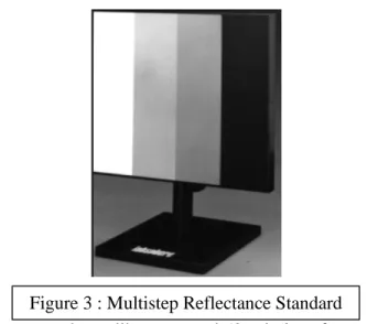
A new four-level reference Labsphere greyscale multistep contrast target (SRT-MS-050)has
enabled us to improve the accuracy of the normalisation calibration (fig. 3). A best-fit gradient is determined from all four targets and a more accurate gain and offset can be determined. Having references at different reflectivities also allows us to scan very dark paintings using a brighter lighting that would saturate the old 99% Labsphere target. The calibration software previously developed was consequently upgraded to handle any future number and combination of different normalisation references.
This new software was used to calibrate around 50 paintings from the Thomas Henry Museum. These are paintings that had already been previously scanned using the Lumiere Technology RGB Jumboscan camera. The results of the multi-spectral colour calibration show a marked improvement over the original RGB acquisitions.
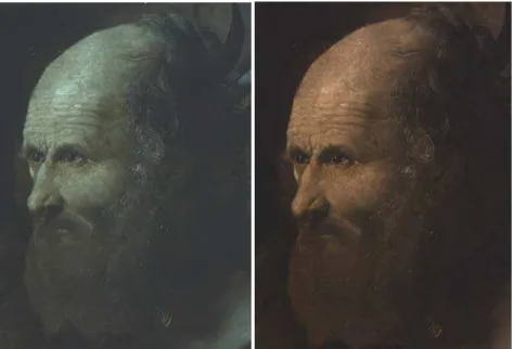
Example improvement in colour reconstruction for “CONVERSION OF SAINT
AUGUSTINE” by ANGELICO Fra Giovanni da Fiesole (fig. 4a et 4b)
Figure 3 : Multistep Reflectance Standard
Pigmented Pébéo Charts for the camera control and pigment identification
The first chart is composed of three parts containing 81 coloured patches and 36 grey patches. The colours are mixed with an acrylic medium to increase the speed of the drying. The first block of colour is not covered with a varnish; the second block is covered by a matt varnish and the third block with a glossy varnish. The measures made directly on the colour patches by means of a colorimeter show that the glossy varnish increases the contrast and reduces the brightness (fig. 5).
Charts containing pure pigments used by artist have also been prepared for pigment
identification. All those pigments are used pure and mixed with white lead, and grinded in a clear acrylic binder. The paints obtained are applied on a white card, 76 µm thick, then assembled on the chart.
The multi-spectral dynamic viewers
Visual interpretation of multispectral imagery generally requires a high level of knowledge about the radiative scattering, absorption, and emission properties of pigments. In order to facilitate the comprehension and the manipulation of such images, software was developed to display and process the reflectance information.
IIPImage
IIPImage is an Open Source multi-platform client/server system designed for the remote
viewing of extremely high resolution images. The system can handleCIELAB, multispectral
as well as 16 bit images.
SpectraViewer
The tool uses a custom-made engine able to quickly access information contained in multispectral images (6 GB images). The software works on different operating systems (Windows, Linux, UNIX, MacOS X) and is able to run on standard computers.
There are two viewing modes :
- a view of the 13 multi-spectral images (images in grey levels) using thumbnails (fig. 6a). - the spectral reflectance curve of any selected pixel, (bottom fig. 6a).
La mire de pigments purs Pébéo
Conservation Restoration Innovation Systems for Image capture and digital Archiving to enhance
Training Education and lifelong Learning
sans vernis avec un vernis mat avec un vernis brillant
81 plages colorées
2 mires : l’une avec un liant à l’huile l’autre avec liant acrylique
36 plages grises
The main window on the right side (Fig. 6b) shows a detail of the overview in full resolution by selecting a region of interest (ROI) in the preview images.
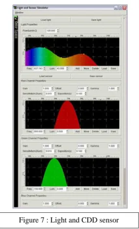
The software also includes a module which simulates the interaction of the painting with any source of light for a CCD sensor.
The light source can be selected:
- from a preset illuminant (A, C, B, E, D50, D55, D65, D75, D95, F2, F6, F7, F8, F10, F11, F12)
- interactively, with control points which define by interpolation the shape of the corresponding spectrum (Fig. 7).
The CCD sensor is modeled using these 3 sensitivity spectral curves, which can be defined by the user (fig. 7).
Once the properties of the light are defined, we can build and visualize in 3D the resulting colored stimuli (in XYZ, xyY, CIELAB and CIELUV color spaces) generated by the light/material interaction, and generate a RGB image resulting from the interaction of the color stimuli previously calculated with the virtual CDD sensor.
The reconstruction of the spectral reflectance curves in the visible and the near infra-red spectrum for each pixel of the multi-spectral images allows a more general colour
representation which is independent from the light spectral distribution and from the camera used for the multi-spectral image acquisition (fig. 8). This representation can be used for different purposes, such as high fidelity colour reproduction of paintings.
Simulated image from a virtual CCD sensor
Simulated colors in CIELAB color space
The 3D digitisation of paintings
In combination with a variety of computer graphics applications, the high-resolution 3D imaging of paintings offers a significant new analytical tool to conservators curators, and art historians, which provides some new and unique types of information which otherwise is not obtainable using traditional techniques. The high-resolution 3D image data contains
information that can be used for display, comparison, measurement and analysis applications. For example, in order to study artists’ technique, curators can zoom in and interactively examine small features such as brush stroke details or signatures that can be difficult to study on the actual paintings. Conservators can monitor and measure crack pattern formations as well as the overall shape of canvas and panel paintings. To measure optically the topography of an object, many techniques have been devised. In the case of easel paintings, optical triangulation based upon fringe patterns projection (pattern of well defined periodic fringes) and laser scanner-based synchronised scanning geometry have shown great potential in the field.
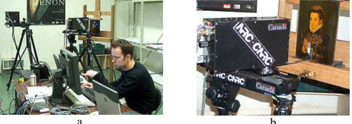
In 2004, the C2RMF tested two systems for the scanning of Dame en prière. One was the synchronised colour laser scanner developed by the National Research Council of Canada and the other was optical triangulation technique based on pattern projection from Breuckmann GmbH in Germany. These two non-destructive optical based triangulation systems were tested for painting 3D modelling and analysis.
NRC 3D Technology
NRC brought two of most advanced 3D imaging systems to the C2RMF (fig. 10) for the scan tests. In addition to scanning Dame en prière, four additional paintings were scanned which included three oil on canvas paintings by Renoir, Femme nue dans un paysage, Gabrielle et
Jean et Portrait d’un jeune homme et d’une jeune fille from the Orangerie museum and Vue de Venise from the Thomas Henry Museum. Some of these paintings were digitised front and
rear to measure the curvature of the support and the surface relief due to brush stroke or impasto structure as well as craquelure of the paint-layer.
One of NRC the systems, the High Resolution Colour Laser Scanner was developed for the simultaneous digitisation of the shape and colour of paintings. The system scans a small (less than 100 micron diameter) “white” laser spot from a RGB (red, green, blue) laser source over the complete surface of the painting. The low power laser light is safe for scanning works of art. The triangulation based detection system simultaneously records the shape (x,y,z) measurements and the colour (R,G,B) reflectance from the spot on the painting in perfect registration (8), (9).
With the maximum resolution configuration, the system provides a lateral spatial (x and y) resolution of 50 µm (0.050 mm) and a depth uncertainty of 10 µm (0.010mm). This resolution is sufficient to record brush stroke details as well as to examine, measure and compare
craquelure and other surface features.
The second scanner used in this project was the Large Volume of View or “Big Scan” Laser Scanner. This system is also a triangulation-based scanner, which uses a monochrome laser for the rapid high-resolution digitisation of larger objects. It was used to record the complete overall shape of the paintings for future monitoring of dimensional changes and features such as warping or distortion of the wood panel. At optimal resolution configuration, the system provides a lateral spatial (x and y) resolution of 200 µm (0.20 mm) and a depth uncertainty of 100 µm (0.1 mm).
NCR 3D Viewer
An important issue brought by the use of high resolution scanners is the difficulty to manage and visualise the very large data-sets they produce. For example, displaying the entire model for a medium-size painting such has “Femme nue dans un paysage ” would represent a significant challenge even to the latest professional 3D visualization systems. By using advanced multi-resolution display techniques, the NRC visualisation software allows to interactively explore the full detail of extremely large data-sets on commodity PC hardware.
a
b
Figure 9: (a) View of the scanner and control set up used at the C2RMF. The Large Volume or “Big Scan” scanner is shown on the tripod on the left and the High–Resolution Colour Scanner is shown mounted on a linear translation stage between two tripods in the center. (b) Detail of the High-Resolution Colour Scanner scanning Dame en Priere
For applications such has this one, the software offers numerous specific functionalities to help conservators make the best of the scanned data-set. Colour can be virtually removed from the painting to analyse only its shape, light sources can be positioned arbitrarily around the painting to produce effects such has virtual raking light. These source can be customised to remove specular components, to change their colour in order separate their contributions to the image, etc. Alternatively, depth can be displayed instead of colour and light reflection to
help analyse the thickness and global shape of thepainting. Also, high resolution 2D images
can be generated for printing when interactive exploration of the models lead to interesting discoveries. All figures of NRC data presented in the paper where produced using this interactive application.
This visualisation software is integrated in the more global framework of the DIMENSION (Distributed Multimedia Environment for Synchronous Interaction Over a Network) project (7) at NRC, Through this framework, models such as the paintings presented in this paper can be displayed using high resolution virtual reality set-ups, and explored collaboratively by remotely located participants.
Preliminary Results
To illustrate the type of 3D image data recorded on paintings, some preliminary results of selected details of Dame en prière and Femme Nue Dans un Paysage by Renoir are presented here. It is important to note that the actual examination of the image data for professional conservation and curatorial study is normally performed by interactive examination using the 3D viewing software. Using interactive examination conservators and curators can zoom in
and interactively examine, compare and measure features such as brush stroke details or craquelure. This will be demonstrated during the ICOM-CC conference.
In examining the following images, it is important to note that a unique feature of the technology for the examination of paintings with varnished surfaces, is that the shape data
recorded by the scanner originates from the immediate surface of the paint layer, under the varnish, rather than from the varnish surface. This results in a detailed high-resolution recording of the surface relief or 3D structure of the paint layer from brush stroke details as well as craquelure formations due to ageing. No other technique captures this type of information.
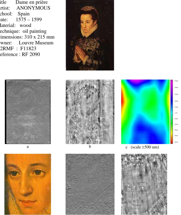
Title Dame en prière Artist: ANONYMOUS School: Spain
Date: 1575 – 1599 Material: wood Technique: oil painting Dimensions: 310 x 215 mm Owner: Louvre Museum C2RMF : F11823
Reference : RF 2090
a b c (scale ±500 um)
d e f
Figure 10: 3D image of Dame en prière scanned at a depth resolution of 10 microns. (a) Shaded monochrome image illustrating surface relief due to brush stroke features and grain structure of wood panel, (b) grey coded depth image of panel in which light areas are raised and dark areas recessed, (c) colour coded image illustrating the overall shape of the slightly warped panel, (d) colour detail of the face showing craquelure, (d) grey coded depth image which illustrates the shape of the painting in the face area due to paint layers and panel shape. Note that the darker the area – the closer the feature is to the viewer. (e and f) Shaded
monochrome image of the face areas showing brush stoke details of the eyes, the mouth and few craquelure.
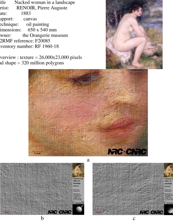
Title Nacked woman in a landscape Artist: RENOIR, Pierre Auguste Date: 1883
Support: canvas Technique: oil painting Dimensions: 650 x 540 mm Owner: the Orangerie museum C2RMF reference: F20085
Inventory number: RF 1960-18
Overview : texture = 26,000x23,000 pixels and shape = 320 million polygons
a
b
c
Figure 11: Detail of the face area scanned at a lateral resolution of 50 microns. (a) Colour image showing the cracks network. (b and c) Shaded monochrome image of the same area showing the craquelure and the canvas relief due to the thin paint layer. The light sphere indicates the direction of the light source.
Title Gabrielle and Jean Artist: RENOIR, Pierre Auguste Date: 1895 - 1896
Support: canvas Technique: oil painting Dimensions: 650 x 540 mm Owner: the Orangerie museum C2RMF reference: F20028
Inventory number: RF 1963-13
Overview : texture = 26,000x23,000 pixels and shape = 320 million polygons
a
b
c
Figure 12: Detail of the chin and the shoulder area of Jean scanned at a lateral resolution of 50 microns. (a) Colour image showing the brush stroke formation. (b) Shaded monochrome image of the same area showing the enhanced brush stroke details. The light sphere indicates the direction of the light source
.
Topometric Metrology
The Dame en prière has also been analyzed by means of a topometric system based on optical
triangulation. This technique enables the high resolution 3-dimensional digitisation of paintings.
The painting to be analysed is illuminated by a special projection unit with fringe patterns; a digital camera records the images of the illuminated area with the projected fringe patterns.
The optical axes of projection unit and camera define an angle 1 (figure 13).
For this project, an triTOS-HE-100 system from Breuckmann GmbH, Germany, has been used. The measuring area of this system is about 80 x 60 mm per view. It is equipped with a colour camera with a resolution of 1,280 x 1,024 pixels, which offers a spatial resolution of about 16 pixels per mm or 60 microns point distance, respectively. Therefore, the whole painting is digitised with around 15 million pixels.
In order to achieve both a high depth resolution of about 2 microns and an unambiguous data analysis, a special sequence of fringe patterns, known as the Grey-code and phase-shift-technique (10), is projected for each view and recorded by the camera. The colour of the painting is recorded in additional images, using the same homogenous and diffuse
illumination for all different views. The acquisition of all images of a single view takes a few seconds.
The complete digitisation of the whole painting has been done with 30 views, which are partly overlapping. The whole data acquisition was carried out manually within 90 minutes.
This can be reduced to about 10 to 15 minutes by using an automated X,Y-table. The alignment of the different views has been carried out by means of the 3D-structure of the painting in the overlapping areas.
The Breuckmann 3D viewer
This viewer allows the visualisation of both 2D and 3D images. In the 2D-domain, it is possible to visualise directly the grey-level or colour images of the recorded views. In the 3D-domain, one can display the 3D-data (polygon meshes) of all the recorded images as well as of the merged data set. Display modes are point clouds, shading plots or wireframes. In addition, the corresponding texture and / or colour can be displayed.
The 3D-data may be orientated with respect to a given co-ordinate system. Scaling of the different axis and the calculation and visualisation calculate of cross sections is also possible.
Moreover, an InspectModule allows the quantitative analysis of 3-dimensional image features, such as distances, flatness, angles etc.
Figure 2 shows the 3D-structure (roughness) of the whole painting and the corresponding colour image; both images result from the mapping of all 30 single views. A cross section, showing the relief of the painting, is shown in figure 3. Please note, that the Z-scale in this plot is ±100 microns, only, whereas the X-scale is about 66 mm.
Fig 15 : cross section along indicated line
Conclusion
Multispectral digitisation and 3D modelling of easel paintings open new fields and techniques to investigate ageing, painting techniques, colour accuracy and high quality printing. To perform accurate colorimetry on a painting requires multi-spectral digitisation. In order to accurately measure shape and roughness necessitates 3D modelling. The association of the different digital techniques gives the user to access to new ways of analysis and research for the conservation and the study of painting material.
The scientific museum community would benefit from adopting common technologies and techniques for high resolution imaging as has already been done for indexing concepts and metadata. This could include common image formats, image analysis and visualisation tools. Several records and large size digital prints will be presented at the next ICOM-CC meeting.
Acknowledgements
We thank the European Commission for their support in the frame of the CRISATEL project and the Department of Foreign Affairs and International Trade of the Government of Canada for their assistance to NRC in this project.
Bibliography
(8) Louis Borgeat, Guy Godin, Jean-François Lapointe and Philippe Massicotte,
Collaborative visualization and interaction for detailed environment models, Proc. of the Tenth International Conference on Virtual Systems and Multimedia, VSMM 2004, 17-19 Nov, Ogaki City, Japan.
(11) Breuckmann, B. : Bildverarbeitung und optische Messtechnik in der industriellen Praxis; Franzis-Verlag München ( 1993 ), ISBN-3-7723-4861-0
(2) CRISATEL Conservation Restauration Innovation Systems for Image capture and digital Archiving to enhance Training Education and lifelong Learning, (programme IST2-1999 n°20163, septembre 2001 - août 2004)
(2) CRISATEL Conservation Restauration Innovation Systems for Image capture and digital Archiving to enhance Training Education and lifelong Learning, (programme IST2-1999 n°20163, septembre 2001 - août 2004)
(3) CRISATEL http://www.crisatel.jussieu.fr
(10) G.Godin, J.A. Beraldin, J. Taylor, L. Cournoyer, M. Rioux, S. El-Hakim, R. Baribeau, F. Blais, P. Boulanger, J. Domey, et M. Picard, Active Optical 3D Imaging for Heritage
Applications, IEE Computer Graphics and Applications, September October 2002, pp.24-36
(4) C. Lahanier, G. Alquié, P. Cotte, C. Christofides, C. de Deyne, R. Pillay, D. Saunders, F Schmitt, CRISATEL : High definition spectral digital imaging of paintings with simulation of
varnish removal. ICOM-CC 13th Triennial Meeting, Rio de Janeiro, 22-27 September 2002
vol 1, pp. 295-300.
(5) Ch. Lahanier, F. Schmitt, P. Le Bœuf and G. Aitken, Multi-spectral Digitisation and 3D Modelling of Paintings and Objects for Image Content Recognition, Image Classification and Multimedia Diffusion.An Ontology Access to the C2RMF Database and Library using the CIDOC-CRM, International Conference of Museum Digitization, Antiquities, Painting and Calligraphy, November 17-18, 2003, National Palace Museum, Taiwan, pp. 157-201
(1) Elisabeth Martin et Michel Dubus, La vie des œuvres, numéro spécial de connaissance des arts, février 1995
(6) Alejandro Ribes, Francis Schmitt, Ruven Pillay and Christian Lahanier, Calibration, Spectral Reconstruction and Illuminant Simulation for CRISATEL : an Art Painting Multispectral Acquisition System.
(9) Marc Rioux, Color 3-D Electronic Imaging of the Surface of the Human Body, CNRC-NRC 38344, SPIE Proceedings, Automatic Systems for the Identification and Inspection of Humas, San Diego, CA July 28-29, 1994 Vol.2277. pp.42-54