ORIGINAL ARTICLE
Genetic and microbial factors modulating the ubiquitin
proteasome system in in
flammatory bowel disease
Isabelle Cleynen,
1Emilie Vazeille,
2,3Marta Artieda,
4Hein W Verspaget,
5,6Magdalena Szczypiorska,
4Marie-Agnès Bringer,
2Peter L Lakatos,
7Frank Seibold,
8Kirstie Parnell,
9Rinse K Weersma,
6,10Jestinah M Mahachie John,
11,12Rebecca Morgan-Walsh,
13Dominiek Staelens,
1Ingrid Arijs,
1Gert De Hertogh,
14Stefan Müller,
15Atilla Tordai,
16Daniel W Hommes,
5,6,17Tariq Ahmad,
9Cisca Wijmenga,
6,18Sylvia Pender,
13Paul Rutgeerts,
1Kristel Van Steen,
11,12Daniel Lottaz,
19Severine Vermeire,
1Arlette Darfeuille-Michaud
2,3▸ Additional material is published online only. To view please visit the journal online (http://dx.doi.org/10.1136/ gutjnl-2012-303205). For numbered affiliations see end of article.
Correspondence to Dr Isabelle Cleynen, Targid— Translational Research Center for Gastrointestinal Disorders, IBD Leuven, Herestraat 49, O&N1, Box 701, Leuven 3000, Belgium; isabelle.cleynen@ med.kuleuven.be; http://www. ibdase.org and Dr Arlette Darfeuille-Michaud, M2iSH, Inserm 1071, CBRV, 28 Place Henri Dunant, Clermont-Ferrand 63000, France; arlette.darfeuille-michaud@ udamail.fr
Equal contribution of IC and EV
Equal contribution of SV and AD-M
Received 29 June 2012 Revised 22 August 2013 Accepted 23 August 2013 Published Online First 3 October 2013
To cite: Cleynen I, Vazeille E, Artieda M, et al. Gut 2014;63:1265–1274.
ABSTRACT
Objective Altered microbiota composition, changes in immune responses and impaired intestinal barrier functions are observed in IBD. Most of these features are controlled by proteases and their inhibitors to maintain gut
homeostasis. Unrestrained or excessive proteolysis can lead to pathological gastrointestinal conditions. The aim was to validate the identified protease IBD candidates from a previously performed systematic review through a genetic association study and functional follow-up.
Design We performed a genetic association study in a large multicentre cohort of patients with Crohn’s disease (CD) and UC fromfive European IBD referral centres in a total of 2320 CD patients, 2112 UC patients and 1796 healthy controls. Subsequently, we did an extensive functional assessment of the candidate genes to explore their causality in IBD pathogenesis.
Results Ten single nucleotide polymorphisms (SNPs) in four genes were significantly associated with CD: CYLD, USP40, APEH and USP3. CYLD was the most significant gene with the intronically located rs12324931 the strongest associated SNP (pFDR=1.74e-17, OR=2.24 (1.83
to 2.74)). Five SNPs in four genes were significantly associated with UC: USP40, APEH, DAG1 and USP3. CYLD, as well as some of the other associated genes, is part of the ubiquitin proteasome system (UPS). We therefore determined if the IBD-associated adherent-invasive Escherichia coli (AIEC) can modulate the UPS functioning. Infection of intestinal epithelial cells with the AIEC LF82 reference strain modulated the UPS turnover by reducing poly-ubiquitin conjugate accumulation, increasing 26S proteasome activities and decreasing protein levels of the NF-κB regulator CYLD. This resulted in IκB-α degradation and NF-κB activation. This activity was very important for the pathogenicity of AIEC since decreased CYLD resulted in increased ability of AIEC LF82 to replicate intracellularly.
Conclusions Our results reveal the UPS, and CYLD specifically, as an important contributor to IBD pathogenesis, which is favoured by both genetic and microbial factors.
INTRODUCTION
Crohn’s disease (CD) and ulcerative colitis (UC) rep-resent two distinct forms of chronic inflammation of
the gastrointestinal tract, collectively called IBD. Although the categories of underlying pathogenic factors (genetic, environmental, microbial and immunological factors) are roughly the same, they are evidently clinicopathologically distinct with dif-ferences in disease location, behaviour and severity;1 and with different pathogenic mechanisms, which to date are not entirely clear. Genome-wide associ-ation studies (GWAS) and meta-analyses have identi-fied 99 susceptibility loci.2–4 More recently a meta-analysis of CD and UC GWAS, with extensive validation of significant findings and a combined total of more than 75 000 cases and controls brought the total number to 163 IBD loci: 110 for
both CD and UC, 30 CD-specific and 23
UC-specific.5 Still, these loci explain only 15%– 23% of the heritability to these diseases, and for
Signi
ficance of this study
What is already known on this subject?
▸ Alterations in the composition and diversity of the gut microbiota play a key role in the establishment and maintenance of IBD. ▸ Increased numbers of mucosa-associated
adherent-invasiveEscherichia coli (AIEC) are observed in patients with Crohn’s disease (CD). ▸ Proteases and protease inhibitors tightly
regulate microbiota composition, immune response and intestinal barrier function to maintain gut homeostasis.
▸ Pivotal to the host inflammatory responses to commensal and pathogenic bacteria is the NF-κB pathway which is largely regulated by the ubiquitin proteasome system (UPS). The main function of the UPS is the degradation of unneeded or damaged proteins.
▸ CYLD is a de-ubiquitinase and is part of the UPS. It mainly acts as a negative regulator of NF-κB to protect the host from an over-reactive inflammatory response.
▸ The expression of CYLD is significantly downregulated in the intestine of CD patients.
Cleynen I, et al. Gut 2014;63:1265–1274. doi:10.1136/gutjnl-2012-303205 1265
In
flammatory bowel disease
group.bmj.com
on July 10, 2014 - Published by
gut.bmj.com
many of the loci the exact causal gene(s) have not yet been identified.
Alterations in the composition and diversity of the gut micro-biota have shown to play a key role in IBD aetiology.6Also, in patients with CD and UC, high concentrations of bacteria forming a biofilm on the surface of the gut mucosa,7 and increased numbers of mucosa-associated Escherichia coli are observed.8 9 These bacteria, called adherent-invasive Escherichia coli (AIEC), colonise the ileal mucosa of CD patients.10 They are able to adhere to and invade intestinal epithelial cells, and to survive and highly replicate within macrophages leading to the secretion of large amounts of TNF-α.11 12Gut colonisation triggered by adhe-sion and invaadhe-sion of AIEC is accompanied by a breakdown of the mucosal barrier, sustaining the inflammatory response.13
Besides altered microbiota composition, changes in immune responses and impaired intestinal barrier functions are observed in IBD.14 Each of these features is controlled by proteases and protease inhibitors in order to maintain gut homeostasis. Unrestrained or excessive proteolysis can lead to pathological gastrointestinal conditions: increased proteolytic activity has been found in the secretions of colonic biopsy samples from patients with IBD.15Exactly which proteases and inhibitors are involved in the context of IBD is currently unknown. In a previ-ously performed and published systematic review of protease (inhibitor) genes in IBD, we ranked the known protease (inhibi-tor) genes according to the amount of evidence present in litera-ture for linkage and/or association of their genomic location with IBD.16 In the present study, we are following up on the identified candidates from that systematic review through a genetic association study in CD and in UC on the common var-iants in the top-ranked genes and functional analyses on the most promising candidate genes to explore their candidacy as
causal genes and understand their possible mode of action in IBD pathogenesis.
MATERIALS AND METHODS Genetic association study
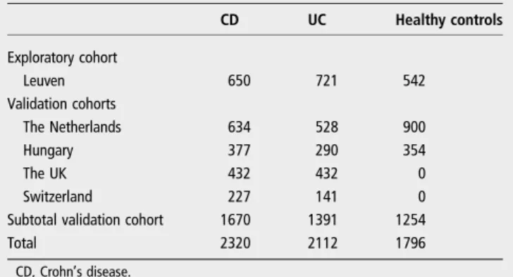
A genetic association study was performed in two stages. First, 182 tagging SNPs in 23 protease (inhibitor) genes (table 1) were genotyped in an exploratory cohort (University Hospital Leuven, Belgium) of 650 CD and 721 UC patients, and 542 healthy con-trols (HC) using Sequenom. The candidate genes were selected based on the results from the previously published systematic review.16Nominally significant SNPs (puncorrected<0.05) were then validated in an independent cohort constituting a total of 1670 CD patients, 1391 UC patients and 1254 HC from four European countries: the Netherlands (ICC/Dutch), Hungary, the UK (Exeter) and Switzerland (table 2) using KASPar chemistry (KBioscience). An FDR corrected p (pFDR)<0.05 was considered statistically significant.
Detailed information on the study samples, SNP selection and statistical analysis for the genetic association study is provided in online supplementary materials and methods. Overall patient characteristics are given in table S1.
Functional studies with AIEC reference strain LF82 Cell culture
The intestinal epithelial cell line T84 (ATCC, CCL-248) was maintained in an atmosphere containing 5% CO2at 37°C in the culture medium recommended by ATCC.
Bacterial strain and culture
E coli strain LF82, isolated from a chronic ileal lesion of a CD patient, was used as the AIEC reference strain.8Isogenic mutants
Signi
ficance of this study
What are the new
findings?
▸ Polymorphisms in CYLD, USP40, APEH and USP3 genes are significantly associated with CD. The strongest association was withCYLD, a de-ubiquitinating enzyme and key regulator of the NF-κB pathway, located adjacent to the well-established CD susceptibility geneNOD2.
▸ Polymorphisms in USP40 and USP3 genes, and several SNPs located at 3p21 (USP4/DAG1/APEH/MST1) are significantly associated with UC.
▸ We show that AIEC bacteria modulate the UPS turnover by reducing the accumulation of poly-ubiquitin conjugates, increasing 26S proteasome activities and decreasing the protein level of the NF-κB regulator CYLD, in turn leading to IκB-α degradation and subsequent NF-κB activation. ▸ The effect of AIEC bacteria on CYLD protein levels is very
important for the pathogenicity of AIEC bacteria since decreasedCYLD resulted in an increased ability of AIEC LF82 to replicate intracellularly.
How might it impact on clinical practice in the
foreseeable future?
▸ Our results indicate that proteasome inhibitor therapy targeting the UPS could be efficient in IBD. A specific example for future use in IBD could be bortezomib, which was thefirst therapeutic proteasome inhibitor tested in humans and which is successfully used in cancer treatment.
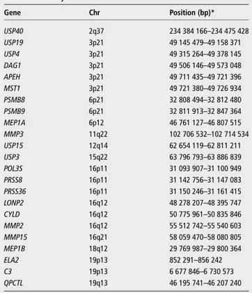
Table 1 Overview selected candidate genes for the genetic association study Gene Chr Position (bp)* USP40 2q37 234 384 166–234 475 428 USP19 3p21 49 145 479–49 158 371 USP4 3p21 49 315 264–49 378 145 DAG1 3p21 49 506 146–49 573 048 APEH 3p21 49 711 435–49 721 396 MST1 3p21 49 721 380–49 726 934 PSMB8 6p21 32 808 494–32 812 480 PSMB9 6p21 32 811 913–32 847 364 MEP1A 6p12 46 761 127–46 807 515 MMP3 11q22 102 706 532–102 714 534 USP15 12q14 62 654 119–62 811 211 USP3 15q22 63 796 793–63 886 839 POL3S 16p11 31 093 907–31 100 949 PRSS8 16p11 31 142 756–31 147 083 PRSS36 16p11 31 150 246–31 161 415 LONP2 16q12 48 278 207–48 395 747 CYLD 16q12 50 775 961–50 835 846 MMP2 16q12 55 512 742–55 540 603 MMP15 16q21 58 059 470–58 080 805 MEP1B 18q12 29 769 987–29 800 364 ELA2 19p13 852 291–856 242 C3 19p13 6 677 846–6 730 573 QPCTL 19q13 46 195 741–46 207 240 *Coordinates in NCBI GRCh37.
of LF82 deleted for theFimH, lpfA, FliC, OmpA, OmpC, OmpF and thelpfA genes17–21were also used in this study.
siRNA tr3ansfection
The siRNA oligonucleotides CYLD-si2 (CGCTGTAA
CTCTTTAGCAT) were used in this study.22 For control siRNA treatment, siRNAs targeting luciferase (TTCUCCGAAC GTGTCACGT) were used. Cells were transfected with siRNAs using Oligofectamine (Invitrogen) according to the manufacturer’’s protocol.
Plasmids and transfection
T84 cells were used to amplify CYLD by PCR with the platinum Taq DNA polymerase (Invitrogen) according to the manufac-turer’s instructions. The PCR reaction was performed with the primers, CYLDF1 (50-GACAGCATGGACACCACGTT-30) and CYLDR1 (50-TTGCCTTTCCCGATGACCC-30), designed on the full sequence of CYLD (transcript ID=ENST00000569418) and with the following temperature cycle: 94°C for 2 min fol-lowed by 40 cycles of 94°C for 30 s, annealing at 50°C for 30 s, and 72°C for 3 min. A final extension incubation of 72°C for 5 min was followed by 5 min at 5°C. The PCR product was purified with the Nucleospin extract II kit (Macherey-Nagel) and cloned into pCR2.1 vector with the TA Cloning Kit (Invitrogen) according to the manufacturer’s instructions. The resulting plasmid was digested with HindIII and XbaI, and the excised fragment was ligated into pEGFP-C1 cut with the same enzymes. T84 cells were transfected with plasmids pEGFP-C1
or pEGFP-C1-CYLD (100nM) and Lipofectamine 2000
(Invitrogen) according to the manufacturer’s protocol.
Invasion and intracellular survival assays
For adhesion assays, monolayers were infected for 3 h at a multiplicity of infection (MOI) of 10 bacteria per cell. Bacterial invasion of T84 cells was performed using the gentamycin pro-tection assay.11 At 1 and 24 h postinfection, the number of intracellular bacteria was determined by counting the number of colony-forming units. To determine the effect of MG132 on bacterial adhesive and invasive abilities, T84 cells were infected for 3 h at an MOI of 10 in the presence of MG132 (25mM) or vehicle (dimethyl solfoxide, DMSO, 0.2%). For bacterial inva-sion assay, T84 cells were washedfive times in phosphate buffer saline (PBS) after 3 h infection in presence of MG132 or vehicle. The number of intracellular bacteria was determined by counting the number of colony-forming units without adding MG132 or vehicle during the postinfection period. One
representative dataset of at least three independent experiments was presented where appropriate.
Chymotrypsin-like activities of 26S proteasome
Monolayers were infected for 3 h at an MOI of 100 or 200 bac-teria per cell and washed twice with ice-cold PBS and then scraped in ice-cold HEPES buffered saline (150 mmol/L NaCl, 5 mmol/L KCl and 10 mmol/L HEPES, pH 7.4).22 Cells were pelleted (14 000 g for 20 s at room temperature) and then resus-pended in ice-cold lysis buffer (10 mmol/L Tris ( pH 7.4), 5 mmol/L MgCl2 and 50 U/mL deoxyribonuclease and ribo-nuclease, plus complete protease inhibitor cocktail; Roche Molecular Biochemicals). Protein concentrations were deter-mined by Bradford assay, and samples stored at−80°C until use. Chymotrypsin-like activity of the proteasome was determined by measuring the hydrolysis of the fluorogenic substrates succinyl-Leu-Leu-Val-Tyr-7-amido-4-methylcoumarin (AMC) (Sigma). To measure peptidase activity, 15mL of lysate contain-ing 20μg protein was added to 60 μL medium containing 50 mM Tris ( pH 7.5), 10 mM MgCl2, 1 mM dithiothreitol and 300μM succinyl-Leu-Leu-Val-Tyr-7-AMC. The peptidase activity was determined by measuring the accumulation of the fluoro-genic cleavage product (AMC) using the FLX800 fluorimeter (Bio-Tek) with 380 and 440 nm excitation and emission wave-lengths, respectively. The difference between arbitrary fluores-cence units recorded with or without 40 or 100μmol/L of the proteasome inhibitor MG132 (Affiniti) in the reaction medium was calculated, and thefinal data were corrected by the amount of protein in the reaction. The time course for the accumulation of AMC after hydrolysis of the substrate was analysed by linear regression to calculate peptidase activities, that is, the slopes of bestfit of accumulated AMC versus time. Values were expressed in relative fluorescence unit per microgram of protein per minute. One representative dataset of at least three independent experiments was presented where appropriate. At each experi-ment, monolayers treated with MG132 (25mM) or vehicle (DMSO, 0.2%) were treated like the uninfected or infected cells.
Western blotting
Monolayers were infected for 3 h at an MOI of 100 in presence or not of MG132 (25mM) or vehicle (DMSO, 0.02%) and washed twice with ice-cold PBS and then scraped in NP40 lysis buffer (50 mmol/L Tris HCl, 150 mmol/L NaCl, 40 mmol/L EDTA, 1% NP40, pH 7.5 plus complete protease inhibitor cocktail; Roche Molecular Biochemicals) containing 5 mmol/L N-ethyl-maleimide to protect Ub protein conjugates. Whole cell extracts were subjected to SDS-PAGE on 12% or 7.5% (wt/vol) acrylamide gels. Protein concentrations were determined by Bradford assay. Western immunoblotting was performed accord-ing to the procedure of Towbin.23 Proteins were electroblotted onto nitrocellulose membranes (Amersham International). The membranes were immunoblotted for CYLD (rabbit anti-CYLD, 1:1000, Thermo scientific), IkB-a (Rabbit anti- IkB-a, 1:200, Santa Cruz Biotechnology Inc),β-actin (rabbit anti-β-actin, Santa Cruz Biotechnology Inc.) and for polyUb chains (anti-FK1, 1:1000, Affiniti) Immunoreactants were detected using horse-radish peroxidase-conjugated antirabbit or antimouse immuno-globulin G antibody, ECL reagents (Amersham Biosciences) and autoradiography. Image J software was used to estimate protein quantity. Results are expressed as protein amount relative to β-actin, or normalised against the amount of proteins (deter-mined following Ponceau Red staining) for poly-ubiquitinated proteins.
Table 2 Number of individuals per dataset
CD UC Healthy controls Exploratory cohort Leuven 650 721 542 Validation cohorts The Netherlands 634 528 900 Hungary 377 290 354 The UK 432 432 0 Switzerland 227 141 0
Subtotal validation cohort 1670 1391 1254
Total 2320 2112 1796
CD, Crohn’s disease.
Cleynen I, et al. Gut 2014;63:1265–1274. doi:10.1136/gutjnl-2012-303205 1267
In
flammatory bowel disease
group.bmj.com
on July 10, 2014 - Published by
gut.bmj.com
Confocal microscopy for NF-κB and poly-ubiquitin conjugates localisation
T84 cells were plated at a concentration of 2.105 per cm2in 24 well plates containing sterile cover slips and grown at 37°C for 48 h. Cells were infected at an MOI of 100 in presence or not of MG132 (25mM) or vehicle (DMSO, 0.2%). After infection, the cells were washed four times with PBS prewarmed to 37°C andfixed onto the cover slips by incubation in 3.7% formalde-hyde for 15 min. Cells were then washed with PBS and permea-bilised by incubation in 0.1% Triton for 15 min at room temperature. The cover slips were blocked in blocking buffer (5% decomplemented serum, 3% bovine serum albumin (BSA), 0.025% triton) during 2 h at room temperature. Antibody to the NF-κB p65 subunit or FK1 (1:500) was added in blocking buffer and incubated for 2 h at room temperature. The cover slips were washed six times with PBS and incubated with Hoechst solution (final concentration of 0.5 μg/mL, Sigma) and CY3 antibody (1:300) during 1 h in the dark. Cover slips were then washed six times each with PBS and mounted onto slides using Fluorescent mounting medium (mowiol). The nuclear translocation of p65 was imaged using a Zeiss LSM 510 META confocal microscope equipped with a Zeiss 37°C incubation system. Images were analysed using the Zeiss LSM Image Examiner.
Statistical analysis
Data were analysed by Student’ t test. A p≤0.05 was considered statistically significant. Data are expressed as the means±SEM.
RESULTS
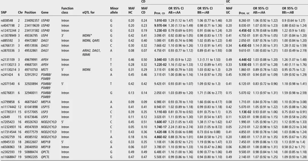
Genetic association study of protease (inhibitor) genes
Of the 182 tagging SNPs in 23 candidate genes, 26 SNPs were nominally significant in the exploratory cohort. With one SNP deleted from further analysis because it failed quality control (QC), wefinally had 25 SNPs with genotyping results in both the exploration and validation cohorts. Association results of these 25 SNPs in the combined cohort (2320 CD, 2112 UC and 1796 HC) are listed in table 3. Ten SNPs were significantly asso-ciated ( pFDR<0.05) with CD. They were located in CYLD (16q12), USP40 (2q37), APEH (3p21) and USP3 (15q22). Five SNPs were significantly associated with UC, being located in USP40 (2q37), DAG1 and APEH (both on 3p21), and USP3 (15q22).
In CD, the most significant finding was for rs12324931 with a combined p value of 1.74e-17 (OR=2.24 (1.83 to 2.74)). Rs12324931 is located intronically in the CYLD gene. Three other SNPs in CYLD (2 intronic, and 1 upstream)—all in low linkage disequilibrium (LD) with rs12324931 (r2<0.1) but with moderate LD between them (0.5<r2<0.7)—were also asso-ciated with CD: rs17314544 ( pFDR=1.42e-08, OR=0.76 (0.66 to 0.88)), rs2302759 ( pFDR=4.86e-02, OR=0.88 (0.76 to 1.01)) and rs7205423 ( pFDR=1.60e-07, OR=1.23 (1.05 to 1.43)). Haplotype analysis showed a significant association of this 3-SNP block with CD ( pomnibus=2.69e-14). For none of the associated CYLD SNPs, a clear function is eminent. They are non-coding and are not identified as being expression quantita-tive trait loci (eQTLs) (table 3) or transcription factor binding sites.CYLD is located on chromosome 16q12, 9 kb downstream of the well-established CD susceptibility geneNOD2. We there-fore tested whether the observed signals in CYLD were acting independently or were driven by the three major NOD2 CD-risk variants. Genotypes were obtained for rs2066844
( p.R702W), rs2066845 ( p.G908R) and rs2066847
( p.L1007fsinsC). Overall, relatively low LD was observed between theNOD2 and CYLD SNPs with an average r2=0.10, but there is a high correlation between rs2066844 and rs12324931: r2=0.83. Conditional on rs2066844, rs2066845 and rs2066847, a significant independent association was
shown for rs12324931: p=5.76e-5, p=2.03e-17 and
p=4.36e-18, respectively. When including all three NOD2 mutations in a single conditional logistic regression test, rs12324931 was still significantly associated (p=2.92e-05). Also, when stratifying based on NOD2 carrier status, NOD2-negative patients (not carrying any of the three NOD2 risk alleles) showed a significant association with rs12324931 ( p=0.001, OR=4.05 (1.68 to 9.73)). None of theCYLD SNPs were associated with UC.
A detailed analysis of the other associated SNPs is provided in table 3 and online supplementary results (also seefigure S1 and tables S2-S4). Inherent to the way the candidate protease (inhibitor) genes studied here were selected, we found that also the other associated proteases in this study are located near known susceptibility genes:ATG16L1 for USP40 and MST1 for the genes located at 3p21 (USP4, APEH, DAG1). Although the results of the genetic studies cannot prove that these genes are definitely causal, we could show that there are probably multiple variants in these loci (allelic heterogeneity) that are important in IBD pathogenesis (see online supplementary results).
Importance of the role of CYLD in CD as revealed by CD-associated bacteria
The genetic results point towards the implication of CYLD in IBD pathogenesis. We therefore next assessed if this gene indeed could be functionally involved. It has been reported that some bacteria and viruses are able to modulate CYLD.23–25We there-fore first investigated if mucosa-associated bacteria known to abnormally colonise the gastrointestinal tract of IBD patients would also be able to modulate CYLD. As AIEC are the proto-type bacteria recognised worldwide as abnormally colonising the gut of IBD patients, we tested the effect of the AIEC refer-ence strain LF82 on CYLD in T84 intestinal epithelial cells. In AIEC LF82 infected T84 cells, 40% decreased CYLD protein levels were observed ( p<0.00001) (figure 1A). CYLD is shown to be significantly downregulated in the intestine of CD patients.26 As CYLD could also act as a negative regulator for immune and inflammatory response against invading bac-teria,27 28we tested if a decrease in CYLD expression affects the ability of AIEC LF82 to invade and replicate intracellularly. Transfection of T84 cells withCYLD siRNA (figure 1B) resulted in a significant increase in the ability of AIEC LF82 to invade ( p<0.0001) and replicate intracellularly ( p<0.01), compared with cells transfected with scramble siRNA (taken as 100%), with values reaching 151% and 223% (figure 1C,D). In con-trast, we observed that overexpression of CYLD in T84 cells transfected with cloned CYLD cDNA into PEGFP-C1 vector did not modify the level of internalised bacteria as shown at 1 h postinfection but significantly decreased by 22.7% the number of intracellular bacteria at 24 h postinfection ( p=0.005 vs T84 cells transfected with the empty vector) (figure 1E–G). Altogether, these results indicated that a decrease in CYLD protein level can favour the invasion and the intracellular repli-cation of AIEC bacteria.
Importance of the role of the UPS in CD
CYLD, as well as many of the other genes where we found evi-dence for association with IBD (USP40, USP3, USP4, APEH), is part of the ubiquitin proteasome system (UPS). Its main function
Table 3 Significant associations with CD and UC in the combined cohort
CD UC
SNP Chr Position Gene
Function
class eQTL for
Minor allele MAF HC MAF CD pFDR, CD OR 95% CI AB><AA OR 95% CI BB><AB MAF UC pFDR, UC OR 95% CI AB><AA OR 95% CI BB><AB rs838548 2 234092357 USP40 Intron G 0.20 0.24 1.01E-03 1.29 (1.12 to 1.47) 1.06 (0.77 to 1.46) 0.20 8.26E-01 1.06 (0.92 to 1.22) 0.9 (0.64 to 1.27) rs4047198 2 234119639 USP40 Intron G 0.20 0.23 9.97E-04 1.30 (1.13 to 1.49) 0.98 (0.71 to 1.36) 0.20 8.03E-01 1.07 (0.93 to 1.23) 0.88 (0.63 to 1.24) rs12472244 2 234131582 USP40 Intron G 0.23 0.19 1.23E-03 0.79 (0.69 to 0.91) 0.91 (0.66 to 1.24) 0.20 4.45E-02 0.78 (0.68 to 0.89) 1.22 (0.9 to 1.65) rs13078949 3 49336795 USP4 30 WDR6* G 0.42 0.41 3.09E-01 0.92 (0.80 to 1.05) 0.98 (0.83 to 1.17) 0.41 4.75E-01 0.91 (0.79 to 1.05) 1.01 (0.84 to 1.20) rs885592 3 49472887 DAG1 Intron WDR6, QARS G 0.42 0.40 1.08E-01 0.85 (0.74 to 0.98) 1.02 (0.85 to 1.22) 0.40 2.05E-01 0.87 (0.76 to 1.01) 1.00 (0.84 to 1.20) rs6766131 3 49513936 DAG1 Intron C 0.30 0.32 7.66E-02 1.10 (0.96 to 1.26) 1.13 (0.91 to 1.41) 0.34 6.45E-03 1.14 (1.00 to 1.31) 1.28 (1.02 to 1.59) rs3870336 3 49532861 DAG1 Intron ARIH2, DAG1,
RBM6 A 0.08 0.07 4.75E-01 0.93 (0.77 to 1.12) 0.89 (0.41 to 1.93) 0.08 9.61E-01 1.00 (0.83 to 1.21) 1.03 (0.49 to 2.19) rs2131109 3 49679991 APEH Intron T 0.46 0.50 3.04E-03 1.05 (0.9 to 1.22) 1.3 (1.11 to 1.53) 0.49 4.44E-02 1.03 (0.88 to 1.20) 1.26 (1.07 to 1.48) rs11130213 3 49687301 APEH Intron T 0.28 0.32 1.22E-02 1.16 (1.02 to 1.33) 1.12 (0.89 to 1.41) 0.33 5.93E-03 1.11 (0.97 to 1.28) 1.40 (1.11 to 1.76) rs11130214 3 49710750 MST1 50 WDR6 G 0.31 0.29 3.11E-01 0.90 (0.79 to 1.03) 1.03 (0.82 to 1.3) 0.29 1.86E-01 0.89 (0.78 to 1.02) 0.95 (0.75 to 1.2) rs241424 6 32912912 PSMB8/
PSMB9
Intron A 0.45 0.46 3.11E-01 1.00 (0.86 to 1.16) 1.14 (0.97 to 1.35) 0.45 9.39E-01 0.94 (0.81 to 1.09) 1.09 (0.92 to 1.29) rs2071540 6 32920894 PSMB8/
PSMB9 5
0 T 0.42 0.42 9.42E-01 0.93 (0.81 to 1.07) 1.09 (0.92 to 1.3) 0.41 6.12E-01 0.83 (0.72 to 0.96) 1.18 (0.98 to 1.41) rs9276831 6 32940011 PSMB8/
PSMB9 Intron G 0.13 0.14 2.05E-01 1.03 (0.89 to 1.20) 1.71 (1.06 to 2.77) 0.15 5.07E-02 1.13 (0.97 to 1.31) 1.59 (0.98 to 2.59) rs3799858 6 46875647 MEP1A Intron A 0.09 0.09 6.98E-01 0.93 (0.78 to 1.10) 1.66 (0.66 to 4.17) 0.08 1.71E-01 0.84 (0.70 to 1.00) 1.10 (0.39 to 3.08) rs11174442 12 61041898 USP15 Intron A 0.41 0.41 8.94E-01 1.02 (0.89 to 1.18) 0.99 (0.83 to 1.18) 0.42 5.07E-01 1.05 (0.91 to 1.22) 1.05 (0.88 to 1.26) rs17765311 15 61577005 USP3 Intron C 0.39 0.35 1.11E-03 0.81 (0.70 to 0.92) 0.91 (0.75 to 1.1) 0.35 1.13E-02 0.93 (0.81 to 1.06) 0.75 (0.61 to 0.91) rs2649 15 61673646 USP3 Intron T 0.11 0.12 3.02E-01 1.11 (0.95 to 1.30) 1.01 (0.54 to 1.87) 0.11 9.32E-01 0.98 (0.83 to 1.15) 1.09 (0.58 to 2.05) rs7205423 16 49326763 NOD2/CYLD 50 C 0.45 0.51 1.60E-07 1.23 (1.05 to 1.43) 1.38 (1.17 to 1.62) 0.47 1.99E-01 1.05 (0.90 to 1.21) 1.12 (0.95 to 1.33) rs12324931 16 49347659 NOD2/CYLD Intron C 0.05 0.10 1.74E-17 2.24 (1.83 to 2.74) 3.03 (1.05 to 8.72) 0.05 2.21E-01 1.22 (0.98 to 1.52) 0.88 (0.23 to 3.34) rs17314544 16 49377579 NOD2/CYLD Intron T 0.43 0.36 1.42E-08 0.76 (0.66 to 0.88) 0.73 (0.6 to 0.88) 0.41 4.85E-01 0.90 (0.78 to 1.04) 1.03 (0.86 to 1.24) rs2302759 16 49385102 NOD2/CYLD Intron A 0.18 0.16 4.86E-02 0.88 (0.76 to 1.01) 0.84 (0.58 to 1.21) 0.20 1.80E-01 1.17 (1.01 to 1.35) 0.95 (0.67 to 1.35) rs9945133 18 28022607 MEP1B 50 G 0.33 0.35 1.10E-01 1.06 (0.92 to 1.21) 1.19 (0.96 to 1.47) 0.33 7.45E-01 0.99 (0.86 to 1.13) 1.13 (0.91 to 1.4) rs6506963 18 28040950 MEP1B Intron A 0.06 0.07 3.78E-01 1.10 (0.90 to 1.33) 1.16 (0.47 to 2.86) 0.06 8.28E-01 1.08 (0.88 to 1.31) 0.58 (0.2 to 1.71) rs2302593 19 50888474 QPCTL Intron G 0.49 0.49 6.48E-01 1.16 (1.00 to 1.36) 0.90 (0.77 to 1.06) 0.49 9.50E-01 0.98 (0.84 to 1.14) 1.00 (0.85 to 1.18) rs11668847 19 50902205 QPCTL Intron G 0.47 0.47 5.50E-01 0.99 (0.86 to 1.16) 0.94 (0.80 to 1.10) 0.49 2.14E-01 1.07 (0.92 to 1.25) 1.09 (0.93 to 1.28)
Bold: significant associations. *ExonQTL.
AA, homozygous for the wild-type allele; AB, heterozygous; BB, homozygous for the minor allele; CD, Crohn’s disease; eQTL, expression quantitative trait loci; HC, healthy controls; MAF, minor allele frequency.
Cle ynen I, et al . Gut 2014; 63 :1265 – 1274. doi:10.11 36/gutjnl-2012 -303205 1269
In
fl
amma
tory
bo
w
e
l
d
isea
se
group.bmj.com on July 10, 2014 - Published by gut.bmj.com Downloaded fromis to degrade unneeded or damaged proteins, but the proteasome machinery is, as is CYLD, also a key regulator of the NF-κB pathway.29 30We tested the effect of the AIEC reference strain LF82 on the UPS of T84 intestinal epithelial cells by determining
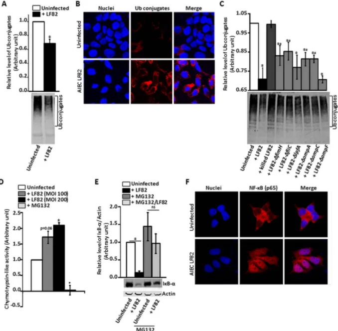
the level of accumulation of poly-ubiquitinated (Ub) proteins, which is the result of all three major UPS activities (ubiquitinase, de-ubiquitinase and proteasome activities). T84 cells were infected with AIEC LF82 bacteria during 3 h. We determined the Figure 1 Interrelation between CYLD
and adherent-invasiveEscherichia coli (AIEC) LF82 infection. T84 cells were infected with AIEC LF82 bacteria at a multiplicity of infection (MOI) of 100, and CYLD levels in T84 cells were measured by western blotting with antiserum against CYLD andβ-actin as an internal control (A). T84 cells were transfected withCYLD siRNA, total proteins loaded and immunoblotted with antibody against CYLD and β-actin as an internal control (B). T84 transfected cells were infected with AIEC LF82 at an MOI of 10. The number of internalised bacteria at 1 h postinfection (C) and internalised bacteria at 24 h/1 h postinfection (D) were determined. T84 cells were transfected with pEGFP-C1-CYLD vector, total proteins loaded and immunoblotted with antibody against CYLD andβ-actin as an internal control (E). T84 transfected cells were infected with AIEC LF82 at an MOI of 10. The number of internalised bacteria at 1 h postinfection (F) and internalised bacteria at 24 h/1 h postinfection (G) were determined. Results are expressed as the percentage of intracellular bacteria within transfected T84 cells relative to untransfected T84 cells, defined as 100%. Data are means±SEM for≥3 independent experiments. *p<0.05.
level of accumulation of Ub proteins by using an antibody detect-ing multi-ubiquitin chains (-K29-, L63- and -K48-linked chains). Infection of T84 cells with AIEC LF82 bacteria significantly decreased the level of Ub conjugates by 46% ( p<0.001) com-pared with non-infected cells (figure 2A,B). We also observed that the decrease in Ub conjugates in AIEC LF82 infected T84 cells is an active bacterial process since this was no longer found when experiments were performed with heat-killed bacteria (figure 2C). To further investigate which LF82 bacterial factor was involved in decreased Ub proteins, we performed experi-ments with various LF82 mutants affected in their abilities to
interact with T84 cells. We observed that when T84 cells were infected with mutants LF82-ΔfimH, LF82-ΔfliC, LF82-ΔompA and LF82-ΔompC, which have decreased ability to adhere and to invade T84 cells, the decrease in Ub conjugates was not as high as observed with wild-type LF82 bacteria. In contrast, when T84 cells were infected with LF82-ΔlpfA and LF82-ΔompF mutants, which are still able to replicate within T84 cells, the level of Ub proteins in infected cells was not significantly different from that of LF82 infected cells (figure 2C). Ub proteins are recognised and degraded by the 26S proteasome, which contains a chymotrypsin-like proteolytic activity in the β-subunits of its
Figure 2 Regulation of ubiquitin proteasome system and NF-κB pathways by adherent-invasive Escherichia coli (AIEC) LF82 infection on T84 cells. T84 cells were infected with AIEC LF82 bacteria and isogenic mutants of LF82 deleted for theFimH, lpfA, FliC, OmpA, OmpC, OmpF and the lpf genes at a multiplicity of infection of 100 or 200. The formation of high molecular weight biotinylated-ubiquitin (Ub) conjugates was determined by western blotting (A,C) and immunefluorescence staining (B) by using an antibody specifically recognising poly-Ub chains. Representative
immunoblots are shown. Chymotrypsin-like activities of the 26S proteasome (D) were determined by measuring the accumulation of thefluorogenic cleavage product (amido-4-methylcoumarin). Values were expressed in relativefluorescence unit per microgram of protein per minute. IκB-α protein level was determined by western blotting with antiserum against IκB-α and β-actin as an internal control (E). Results are expressed as protein amount relative toβ-actin. NF-κB was localised in T84 cells infected with AIEC LF82 bacteria by immune fluorescence staining (F). The accumulation of Ub conjugates (A,C), chymotrypsin-like activities (C) and IκB-α protein levels (D) are means±SEM for ≥3 independent experiments. *p<0.05 versus uninfected or killed LF82 infected T84 cells, #p<0.05 versus LF82 infected T84 cells.
Cleynen I, et al. Gut 2014;63:1265–1274. doi:10.1136/gutjnl-2012-303205 1271
In
flammatory bowel disease
group.bmj.com
on July 10, 2014 - Published by
gut.bmj.com
proteolytic core complex.31To see if the decrease in the accumu-lation of Ub proteins as observed in the presence of AIEC LF82 is in part explained by an effect on 26S proteasome proteolytic function, we compared the levels of chymotrypsin-like activity in AIEC LF82 infected T84 cells with non-infected cells. AIEC LF82 infection induced an MOI-dependent increase in chymotrypsin-like activity in T84 cells: 1.71-fold ( p=0.06) increase at an MOI of 100, and 2.1-fold ( p<0.05) at an MOI of 200 (figure 2D). This suggested that AIEC LF82 bacteria are able to increase the basal proteolytic level of the 26S proteasome, and consequently increase its ability to degrade Ub conjugates. In response to extracellular stimuli, the NF-κB inhibitor IκB-α is poly-ubiquitinated and degraded by the 26S proteasome, leading to the activation of NF-κB.32We tested if, in accordance with the induction of the 26S proteasome, AIEC LF82 infection favours IκB-α degradation and subsequently activates the NF-κB pathway. AIEC LF82 infection induced a significant (p<0.001) 7.7-fold decrease in IκB-α protein level in T84 cells compared with non-infected cells. This was not observed in the presence of MG132 (figure 2E). Similarly, we observed a nuclear transloca-tion of the NF-κB p65 subunit in AIEC LF82-infected T84 cells (figure 2F).
DISCUSSION
The most strongly associated protease was CYLD, which was specific for CD. It encodes a cytoplasmic de-ubiquitinating enzyme and one of its main functions is to protect the host from an over-reactive inflammatory response by negatively regu-lating NF-κB.30Interestingly, a role for CYLD in IBD pathogen-esis can be suggested as it was identified as one of the most significantly downregulated genes in the intestine of CD patients as shown using microarrays.26 In addition, cyld−/− mice dis-played more severe intestinal inflammation and intestinal tumourigenesis, compared with wild-type controls.33 it is important to note the chromosomal location ofCYLD adjacent toNOD2 on 16q12. The (functional) link of NOD2/CARD15 to IBD has been firmly and reproducibly established: there are several well-characterised polymorphisms in NOD2 leading to different capacities of the protein products to regulate NF-κB-mediated inflammatory responses to bacterial compo-nents in the gut, thus providing a causal explanation for the observed association with the disease.34 CD GWAS and meta-analyses have reported strong association on 16q12,3 4 which is attributed to NOD2. Still, Hampe et al found that a robust association signal in this region remains after stratification
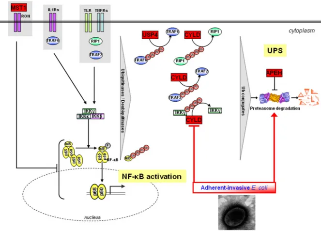
Figure 3 Proposed working model. Several of the genes found to be associated with Crohn’s disease (CD) and/or UC in this study have been shown to negatively regulate NF-κB: MST1 is a negative regulator of LPS-induced NF-κB activation.43CYLD can de-ubiquitinate several of the NF-κB signalling components such as TRAF2/TRAF6 and Nemo (IKK-γ) and toll-like receptors (TLR), particularly TLR2. USP4 de-ubiquitinates TRAF2/TRAF6 in the context of TNF-α- and IL1-β-induced NF-κB activation,44and targets TAK1 to downregulate TNF-α-induced NF-κB activation.45APEH is a negative effector of proteasome activity. While in normal healthy conditions commensal bacteria limit NF-κB signalling through some of these pathways, in individuals harbouring variant alleles leading to an aberrant expression or altered function of CYLD, USP4, MST1 and/or APEH, activation of NF-κB is no longer counteracted, resulting in aberrant immune responses and hence inflammation. In addition, CD-associated
pathogenic adherent-invasiveEscherichia coli bacteria can modulate ubiquitin proteasome system (UPS) turnover, increase 26S proteasome activities, IκB-α degradation (directly through the UPS and by negatively regulating CYLD) and subsequently NF-κB activation. The contribution of the UPS in IBD pathogenesis as revealed in this study could thus be modulated by both genetic and microbial factors.
by NOD2 polymorphisms.35 In the study reported here, we show a significant association of CD with CYLD, which appears to be independent of the association of the three majorNOD2 variants. In accordance with ourfindings, a recent re-analysis of the WTCCC1 CD data using a multimarker approach also revealed a significant association of CYLD independent from NOD2.36 The study by Rivas et al37 where they resequenced some of the genes identified through GWA studies suggests add-itional low frequencyNOD2 risk alleles. To further disentangle the independent contribution of CYLD and to find putative causal (functional) variants within theCYLD gene, the complete region needs resequencing. In addition, we found a significant association of USP40, the 3p21 gene region (USP4/APEH/ DAG1/MST1) and USP3 with both CD and UC. It should be noted however that the observed association results require further interrogation and conditional analysis of data from a dense SNP set within these and neighbouring LD blocks before firm conclusions can be drawn.
Although the genetic studies cannot prove that these genes are definitely causal (also see online supplementary results), we do show functional evidence for an important role of CYLD, and the UPS, in IBD pathogenesis.
Several lines of evidence suggest that one of the hallmarks of IBD is an alteration in the composition and diversity of the gut microbiota.6 Pathogenic bacteria like AIEC colonise the ileal mucosa of CD patients10 and favour a breakdown of the mucosal barrier, further sustaining the inflammatory response.13 In addition to our genetic results which point towards the impli-cation of CYLD in IBD pathogenesis, CYLD has been shown to be downregulated in the intestine of CD patients.26 CYLD, a key regulator of the NF-κB pathway, can de-ubiquitinate several of the NF-κB signalling components such as TRAF2/TRAF6 and Nemo (IKK-γ), which would otherwise lead to the degradation of IκB-α by the proteasome.30 38 39 In our functional studies, we show that the pathogenic CD-associated AIEC LF82 bac-teria, like some other bacteria and viruses,23–25 are able to modulate CYLD protein levels (hence a way to induce NF-κB signalling), in turn leading to an increased or a decreased ability of AIEC LF82 to invade and replicate within intestinal epithelial cells depending on whether CYLD expression is decreased or increased respectively. Of note, the levels of AIEC replication within epithelial cells with decreased CYLD expression were similar to those seen in epithelial cells with decreased expression of autophagy-related proteins ATG16L1 or IRGM (immunity-related GTPase family M protein), for which the encoding genes are mutated in patients with CD.40 41
CYLD, as well as many of the other genes where we found evidence for association with IBD (USP40, USP3, USP4, APEH), is part of the UPS. The main function of the UPS is to degrade unneeded or damaged proteins, but the proteasome machinery is, as is CYLD, also a key regulator of the NF-κB pathway. In the same way as for CYLD, we speculated that AIEC bacteria could modulate the UPS to induce an inflammatory response. We show that the pathogenic AIEC LF82 bacteria decrease the accumulation of poly-ubiquitin conjugates and increase 26S pro-teasome activity in intestinal epithelial cells, indicating that AIEC bacteria stimulate ubiquitin-proteasome proteolysis. Experiments with various LF82 mutants indicated that this process is dependent on the ability of the bacteria to survive and/or replicate within host cells. Pivotal to the host in flamma-tory responses to commensal and pathogenic bacteria is the NF-κB pathway which is largely regulated by the UPS.42 In response to extracellular stimuli, the NF-κB inhibitor IκB-α is poly-ubiquitinated and degraded by the 26S proteasome leading
to NF-κB activation.32 We show that AIEC bacteria induce proteasome-dependent degradation of IκB-α, leading to a nuclear translocation of the NF-κB p65 subunit. AIEC bacteria can thus induce an inflammatory response involving the NF-κB pathway, which is modulated by the effect of AIEC bacteria on the 26S proteasome activity.
In summary, we believe that the genetic data together with the functional data we provide here promote the case for the candidacy of CYLD as being—at least one—of the causal genes in the NOD2-CYLD gene region. Our studies identify the UPS as a major and common (both CD and UC) contributor to IBD pathogenesis. In the light of the findings of this study, we propose a working model presented infigure 3 showing that the major contribution of the UPS in IBD pathogenesis could have two origins, genetic and/or microbial, both of which being largely recognised as favouring the development and mainten-ance of IBD.
Author affiliations
1Department of Clinical and Experimental Medicine, TARGID, KU Leuven, Leuven, Belgium
2Clermont Université, Inserm U1071, Université d’Auvergne, INRA USC 2018, Clermont-Ferrand, France
3Centre Hospitalier Universitaire, Clermont-Ferrand, France 4Progenika Biopharma, S.A., Derio, Spain
5Department of Gastroenterology and Hepatology, Leiden University Medical Center, Leiden, The Netherlands
6Dutch Initiative on Crohn and Colitis (ICC)
71st Department of Medicine, Semmelweis University, Budapest, Hungary 8Department of Gastroenterology, Spitalnetz Bern, Switzerland 9Peninsula Medical School, University of Exeter & Plymouth, Exeter, UK 10Department of Gastroenterology and Hepatology, University Medical Center Groningen and the University of Groningen, Groningen, The Netherlands 11Systems and Modeling Unit, Montefiore Institute, University of Liège, Liège, Belgium
12Bioinformatics and Modeling, GIGA-R, University of Liège, Liège, Belgium 13Clinical and Experimental Sciences, Faculty of medicine, University of Southampton, Southampton, UK
14Department of Morphology and Molecular Pathology, University Hospital Gasthuisberg, Leuven, Belgium
15Department of Clinical Research, University of Bern, Bern, Switzerland 16Hungarian National Blood Transfusion Service, Molecular Diagnostics, Budapest, Hungary
17Division of Digestive Diseases, Inflammatory Bowel Diseases Center, UCLA, Los Angeles, USA
18Department of Genetics, University Medical Center Groningen and the University of Groningen, Groningen, The Netherlands
19Department of Rheumatology, Clinical Immunology and Allergology, University Hospital of Bern, Inselspital, Switzerland
Acknowledgements The authors thank Vera Ballet for patient inclusion and database management; and Tamara Coopmans, Willem-Jan Wollants, Sophie Organe, Karolien Claes and Nooshin Ardeshir Davani for technical and scientific support for sample selection, DNA extraction, and genotyping.
Contributors Study concept and design: IC, EV, MA, SV, AD-M. Acquisition of data: IC, EV, HWV, MB, PLL, FS, KP, RKW, RM-W, DS, IA, AT, DWH, TA, CW, SP, PR, SV, AD-M. Analysis and interpretation of data: IC, EV, MA, HWV, JMJ, IA, KVS, SV, AD-M. Drafting of the manuscript: IC, EV, MA, SV, AD-M. Critical revision of the manuscript for important intellectual content: IC, EV, MA, HWV, MS, MB, PLL, FS, KP, RKW, JMJ, RM-W, DS, GDH, IA, SM, AT, DWH, TA, CW, SP, PR, DL, KVS, SV, AD-M. Statistical analysis: IC, EV, MA, JMJ, KVS. Obtained funding: PR, DL, SV, AD-M. Study supervision: DL, SV, AD-M.
Funding The research leading to these results has received funding from the European Community’s Seventh Framework Programme (FP7) under grant agreement no 200931 ( project IBDase) and from Centre Hospitalier Universitaire de Clermont-Ferrand and Association François Aupetit.
Competing interests IC and IA are postdoctoral fellows and SV a clinical researcher of the Fund for Scientific Research (FWO) Flanders, Belgium. RKW is supported by a clinical fellowship grant (90.700.281) from the Netherlands Organization for Scientific Research (NWO). JMJ and KVS are funded by the Belgian BioMAGNet Network, an IAP Phase VI/4 Program initiated by the Belgian State Science Policy Office, and by the IST Program of the European Community under the PASCAL2 Network of Excellence (IST-2007-216886). AD-M is funded by Ministre de
Cleynen I, et al. Gut 2014;63:1265–1274. doi:10.1136/gutjnl-2012-303205 1273
In
flammatory bowel disease
group.bmj.com
on July 10, 2014 - Published by
gut.bmj.com
l’Enseignement Supérieur et de la Recherche, Inserm (U1071), by INRA (USC-2018) and by grants from the Association F. Aupetit.
Patient consent Obtained.
Ethics approval The ethical boards of each separate recruiting institution approved the study.
Provenance and peer review Not commissioned; externally peer reviewed.
REFERENCES
1 Carter MJ, Lobo AJ, Travis SP. Guidelines for the management of inflammatory bowel disease in adults. Gut 2004;53(Suppl 5):V1–16.
2 Anderson CA, Boucher G, Lees CW, et al. Meta-analysis identifies 29 additional ulcerative colitis risk loci, increasing the number of confirmed associations to 47. Nat Genet 2011;43:246–52.
3 Barrett JC, Hansoul S, Nicolae DL, et al. Genome-wide association defines more than 30 distinct susceptibility loci for Crohn’s disease. Nat Genet 2008;40:955–62.
4 Franke A, McGovern DP, Barrett JC, et al. Genome-wide meta-analysis increases to 71 the number of confirmed Crohn’s disease susceptibility loci. Nat Genet 2011;42:1118–25.
5 Jostins L, Ripke S, Weersma RK, et al. Host-microbe interactions have shaped the genetic architecture of inflammatory bowel disease. Nature 2012;491:119–24. 6 Joossens M, Huys G, Cnockaert M, et al. Dysbiosis of the faecal microbiota in
patients with Crohn’s disease and their unaffected relatives. Gut 2011;60:631–17. 7 Swidsinski A, Ladhoff A, Pernthaler A, et al. Mucosalflora in inflammatory bowel
disease. Gastroenterology 2002;122:44–54.
8 Darfeuille-Michaud A, Neut C, Barnich N, et al. Presence of adherent Escherichia coli strains in ileal mucosa of patients with Crohn’s disease. Gastroenterology 1998;115:1405–13.
9 Martin HM, Campbell BJ, Hart CA, et al. Enhanced Escherichia coli adherence and invasion in Crohn’s disease and colon cancer. Gastroenterology 2004;127:80–93. 10 Darfeuille-Michaud A, Boudeau J, Bulois P, et al. High prevalence of
adherent-invasive Escherichia coli associated with ileal mucosa in Crohn’s disease. Gastroenterology 2004;127:412–21.
11 Boudeau J, Glasser AL, Masseret E, et al. Invasive ability of an Escherichia coli strain isolated from the ileal mucosa of a patient with Crohn’s disease. Infect Immun 1999;67:4499–509.
12 Glasser AL, Boudeau J, Barnich N, et al. Adherent invasive Escherichia coli strains from patients with Crohn’s disease survive and replicate within macrophages without inducing host cell death. Infect Immun 2001;69:5529–37. 13 Carvalho FA, Barnich N, Sivignon A, et al. Crohn’s disease adherent-invasive
Escherichia coli colonize and induce strong gut inflammation in transgenic mice expressing human CEACAM. J Exp Med 2009;206:2179–89.
14 Kaser A, Blumberg RS. Autophagy, microbial sensing, endoplasmic reticulum stress, and epithelial function in inflammatory bowel disease. Gastroenterology 2011;140:1738–47.
15 Cenac N, Andrews CN, Holzhausen M, et al. Role for protease activity in visceral pain in irritable bowel syndrome. J Clin Invest 2007;117:636–47.
16 Cleynen I, Juni P, Bekkering GE, et al. Genetic evidence supporting the association of protease and protease inhibitor genes with inflammatory bowel disease: a systematic review. PLoS One 2011;6:e24106.
17 Boudeau J, Barnich N, Darfeuille-Michaud A. Type 1 pili-mediated adherence of Escherichia coli strain LF82 isolated from Crohn’s disease is involved in bacterial invasion of intestinal epithelial cells. Mol Microbiol 2001;39:1272–84. 18 Chassaing B, Rolhion N, de, et al. Crohn disease—associated adherent-invasive
E. coli bacteria target mouse and human Peyer’s patches via long polar fimbriae. J Clin Invest 2011;121:966–75.
19 Claret L, Miquel S, Vieille N, et al. Theflagellar sigma factor FliA regulates adhesion and invasion of Crohn disease-associated Escherichia coli via a cyclic dimeric GMP-dependent pathway. J Biol Chem 2007;282:33275–83.
20 Dreux N, Denizot J, Martinez-Medina M, et al. Point mutations in FimH adhesin of Crohn’s disease-associated adherent-invasive Escherichia coli enhance intestinal inflammatory response. PLoS Pathog 2013;9:e1003141.
21 Rolhion N, Carvalho FA, Darfeuille-Michaud A. OmpC and the sigma(E) regulatory pathway are involved in adhesion and invasion of the Crohn’s disease-associated Escherichia coli strain LF82. Mol Microbioly 2007;63:1684–700.
22 Stegmeier F, Sowa ME, Nalepa G, et al. The tumor suppressor CYLD regulates entry into mitosis. Proc Natl Acad Sci USA 2007;104:8869–74.
23 An J, Mo D, Liu H, et al. Inactivation of the CYLD deubiquitinase by HPV E6 mediates hypoxia-induced NF-kappaB activation. Cancer Cell 2008;14:394–407. 24 Koga T, Lim JH, Jono H, et al. Tumor suppressor cylindromatosis acts as a negative
regulator for Streptococcus pneumoniae-induced NFAT signaling. J Biol Chem 2008;283:12546–54.
25 Lim JH, Stirling B, Derry J, et al. Tumor suppressor CYLD regulates acute lung injury in lethal Streptococcus pneumoniae infections. Immunity 2007;27:349–60. 26 Costello CM, Mah N, Hasler R, et al. Dissection of the inflammatory bowel disease
transcriptome using genome-wide cDNA microarrays. PLoS Med 2005;2:e199. 27 Lim JH, Jono H, Koga T, et al. Tumor suppressor CYLD acts as a negative regulator
for non-typeable Haemophilus influenza-induced inflammation in the middle ear and lung of mice. PLoS One 2007;2:e1032.
28 Pan ZK, Fisher C, Li JD, et al. Bacterial LPS up-regulated TLR3 expression is critical for antiviral response in human monocytes: evidence for negative regulation by CYLD. Int Immunol 2011;23:357–64.
29 Lim JH, Ha UH, Woo CH, et al. CYLD is a crucial negative regulator of innate immune response in Escherichia coli pneumonia. Cell Microbiol 2008;10:2247–56.
30 Sun SC. CYLD: a tumor suppressor deubiquitinase regulating NF-kappaB activation and diverse biological processes. Cell Death Differ 2010;17:25–34.
31 Saeki Y, Tanaka K. Assembly and function of the proteasome. Methods Mol Biol 2012;832:315–37.
32 Viatour P, Merville MP, Bours V, et al. Phosphorylation of NF-kappaB and IkappaB proteins: implications in cancer and inflammation. Trends Biochem Sci
2005;30:43–52.
33 Zhang J, Stirling B, Temmerman ST, et al. Impaired regulation of NF-kappaB and increased susceptibility to colitis-associated tumorigenesis in CYLD-deficient mice. J Clin Invest 2006;116:3042–9.
34 Hugot JP, Zouali H, Lesage S. Lessons to be learned from the NOD2 gene in Crohn’s disease. Eur J Gastroenterol Hepatol 2003;15:593–7.
35 Hampe J, Frenzel H, Mirza MM, et al. Evidence for a NOD2-independent susceptibility locus for inflammatory bowel disease on chromosome 16p. Proc Natl Acad Sci USA 2002;99:321–6.
36 Elding H, Lau W, Swallow DM, et al. Dissecting the genetics of complex inheritance: linkage disequilibrium mapping provides insight into crohn disease. Am J Hum Genet 2011;89:798–805.
37 Rivas MA, Beaudoin M, Gardet A, et al. Deep resequencing of GWAS loci identifies independent rare variants associated with inflammatory bowel disease. Nat Genet 2011;43:1066–73.
38 Kovalenko A, Chable-Bessia C, Cantarella G, et al. The tumour suppressor CYLD negatively regulates NF-kappaB signalling by deubiquitination. Nature 2003;424:801–5. 39 Trompouki E, Hatzivassiliou E, Tsichritzis T, et al. CYLD is a deubiquitinating enzyme
that negatively regulates NF-kappaB activation by TNFR family members. Nature 2003;424:793–6.
40 Brest P, Lapaquette P, Souidi M, et al. A synonymous variant in IRGM alters a binding site for miR-196 and causes deregulation of IRGM-dependent xenophagy in Crohn’s disease. Nat Genet 2011;43:242–5.
41 Lapaquette P, Glasser AL, Huett A, et al. Crohn’s disease-associated
adherent-invasive E. coli are selectively favoured by impaired autophagy to replicate intracellularly. Cell Microbiol 2010;12:99–113.
42 Wang J, Maldonado MA. The ubiquitin-proteasome system and its role in inflammatory and autoimmune diseases. Cell Mol Immunol 2006;3:255–61. 43 Nikolaidis NM, Gray JK, Gurusamy D, et al. Ron receptor tyrosine kinase negatively
regulates TNFalpha production in alveolar macrophages by inhibiting NF-kappaB activity and Adam17 production. Shock 2010;33:197–204.
44 Xiao N, Li H, Luo J, et al. Ubiquitin-specific protease 4 (USP4) targets TRAF2 and TRAF6 for deubiquitination and inhibits TNFalpha-induced cancer cell migration. Biochem J 2012;441:979–86.
45 Fan YH, Yu Y, Mao RF, et al. USP4 targets TAK1 to downregulate TNFalpha-induced NF-kappaB activation. Cell Death Differ 2011;18:1547–60.
doi: 10.1136/gutjnl-2012-303205
2014 63: 1265-1274 originally published online October 3, 2013
Gut
Isabelle Cleynen, Emilie Vazeille, Marta Artieda, et al.
inflammatory bowel disease
the ubiquitin proteasome system in
Genetic and microbial factors modulating
http://gut.bmj.com/content/63/8/1265.full.html
Updated information and services can be found at:
These include:
Data Supplement
http://gut.bmj.com/content/suppl/2013/10/03/gutjnl-2012-303205.DC1.html"Supplementary Data"
References
http://gut.bmj.com/content/63/8/1265.full.html#ref-list-1This article cites 45 articles, 9 of which can be accessed free at:
service
Email alerting
the box at the top right corner of the online article.
Receive free email alerts when new articles cite this article. Sign up in
Collections
Topic
(889 articles)
Crohn's disease
Articles on similar topics can be found in the following collections
Notes
http://group.bmj.com/group/rights-licensing/permissions
To request permissions go to:
http://journals.bmj.com/cgi/reprintform
To order reprints go to:
http://group.bmj.com/subscribe/