Protective effect of dietary n-3 polyunsaturated fatty acids on myocardial
resistance to ischemia-reperfusion injury in rats
Sabrina Zeghichi-Hamri
a,b, Michel de Lorgeril
a,⁎
, Patricia Salen
a, Mohamed Chibane
b,
Joël de Leiris
a, François Boucher
a, François Laporte
ca
Cœur & Nutrition, TIMC-IMAG CNRS 5525, Université Joseph Fourier, Faculté de Médecine, Domaine de la Merci, 38706 La Tronche, France
b
Laboratoire 3BS, Faculté des sciences de la nature et de la vie. Université de Béjaia, Algérie
c
Département de Biochimie, Pharmacologie et Toxicologie, Hôpital Universitaire de Grenoble, 38706 La troche, France Received 1 September 2010; revised 3 October 2010; accepted 13 October 2010
Abstract
Dietary n-3 polyunsaturated fatty acids (PUFA) reduce coronary heart disease (CHD) complications, such as chronic arrhythmia and sudden cardiac death. Improved myocardial resistance to ischemia-reperfusion injury results in smaller myocardial infarction, which is a major factor in the occurrence of CHD complications. We hypothesized that a specific dietary fatty acid profile (low in saturated and n-6 PUFA but high in plant and marine n-3 PUFA) may improve myocardial resistance to ischemia-reperfusion injury and reduce infarct size. To test this assumption, we used a well-defined rat model of myocardial infarction. Based on our results, in comparison to a diet that is high in either saturated or n-6 PUFA but poor in plant and marine n-3 PUFA, a diet that is low in saturated fats and n-6 PUFA but rich in plant and marine n-3 PUFA results in smaller myocardial infarct size (Pb .01). The effects of the 3 diets were also examined by analyzing the fatty acid composition of plasma, erythrocyte cell membranes, and the phospholipids of myocardial mitochondria. The results show a great accumulation of n-3 PUFA and a parallel decrease in arachidonic acid, the main n-6 PUFA, in plasma, cell membranes, and cardiac mitochondria (Pb .0001). We conclude that improved myocardial resistance to ischemia-reperfusion may be one of the critical factors explaining the protective effects of dietary n-3 PUFA against CHD complications in humans. In addition to increasing n-3 PUFA intake, an optimal dietary pattern aimed at reducing cardiovascular mortality should include a reduction of the intake of both saturated and n-6 PUFA. © 2010 Elsevier Inc. All rights reserved.
Keywords: Myocardial infarction; n-3 PUFA; n-6 PUFA; Arachidonic acid; Saturated fatty acids, Mediterranean diet; Rat Abbreviations: AA, arachidonic acid; CHD, coronary heart disease; LV, left ventricular; MED, mixed marine and plant
n-3 polyunsaturated fatty acids; n-3, n-3 polyunsaturated fatty acids; n-6, n-6 polyunsaturated fatty acids; RZ, risk zone.
1. Introduction
Dietary fats play an important role in coronary heart disease (CHD) complications[1-4]. Apart from their effects on atherosclerosis, thrombosis, and cardiac arrhythmia[1-4],
it is still unclear whether specific dietary fatty acid profiles modulate the resistance of the myocardium to ischemia and reperfusion injury. This is a critical issue because myocardial resistance to ischemia-reperfusion is a major determinant of myocardial infarct size, which is a causal factor in the development of CHD complications, such as cardiac pump failure and fatal ventricular arrhythmia, which are the main causes of cardiac death in humans. Therefore,
Nutrition Research 30 (2010) 849–857
www.nrjournal.com
⁎ Corresponding author. Tel.: +33 476 63 74 71; fax: +33 476 63 71 52. E-mail address:michel.delorgeril@ujf-grenoble.fr(M. de Lorgeril). 0271-5317/$– see front matter © 2010 Elsevier Inc. All rights reserved. doi:10.1016/j.nutres.2010.10.010
it is critical to identify which specific fatty acid profile may be optimal in improving myocardial resistance to ischemia-reperfusion injury.
The main hypothesis of this study was that a fatty acid profile low in animal and plant saturated fats, trans-fatty acids, and plant n-6 polyunsaturated fatty acids (n-6 PUFA) but rich in both plant and marine n-3 polyunsaturated fatty acids (n-3 PUFA) results in improved myocardial resistance and smaller infarct size in a rat model of myocardial infarction. We also examined the fatty acid changes in plasma, red cell membranes, and cardiac mitochondria resulting from the tested diet. Myocardial resistance to ischemia-reperfusion was assessed using a well-defined model of myocardial infarction in rats.
2. Methods and materials
This study was conducted with the approval of the local animal ethics committee and in accordance with the Guide for the Care and Use of Laboratory Animals, National Academic Press, Washington, DC, 1996, and the European Council Directive 86/609/EEC on the care and use of laboratory animals (OJ L 358). The protocols were performed under license from the French Ministry of Agriculture (license no. A380727).
2.1. Experimental approach
To assess its protective effects, the experimental diet was
compared with 2 “control” diets. The 3 diets had similar
energy and fat content but different fatty acid profiles. The first group of rats (PO) received palm oil (an oil rich in saturated fats but poor in n-3 and n-6 PUFA), in addition to a standard diet described in previous studies[5,6]. The second group (SO) received sunflower oil (low in saturated fats and n-3 PUFA but extremely rich in n-6 PUFA), in addition to the standard diet. The third group (MED) received a mixture of plant and marine n-3 PUFA (low in saturated and n-6 PUFA but rich in n-3 PUFA), in addition to the standard diet. Besides their effect on the heart, the 3 diets were analyzed in terms of fatty acid composition in plasma, erythrocyte cell membranes, and the phospholipids of cardiac mitochondria. We used erythrocytes because of their short half-life (compatible with dietary protocols in animals) and because they are known to reflect the fatty acid composition of
cardiac cell membranes [7]. We analyzed the fatty acid
composition of the major phospholipids in cardiac mito-chondria because mitomito-chondrial phospholipids are thought to play a central role in myocardial resistance to ischemia-reperfusion[8-11].
2.2. Animals and experimental diets
Male Wistar rats (IFFA-Credo, L'Abresle, France) were used throughout this investigation. All animals received a standard laboratory solid chow diet (regime A04, UAR, Villemoisson-sur-Orge, France) and water ad libitum. The
chow diet was a conventional (and standardized) low-fat rodent diet with 67% of energy as carbohydrates, 23% as proteins, 10% as fats, and 4 g of fiber per 100 g of foods. All rats (n = 48 for blood and cardiac lipid measurements and n = 42 for cardiac experiments) were fed dietary fat supplements at the same time on a daily basis via gavage for 8 weeks.
The animals were randomly divided into 3 groups according to their dietary supplements. The animals in the
PO group were supplemented with 650 μL of palm oil
(Palmella, München, Germany). Those of the SO group were
supplemented with 650 μL of sunflower oil (Lesieur,
Asnières sur Seine, France), and those in the MED group
were supplemented with 650μL of a mixture of plant and
marine n-3 PUFA (Mixalpha, Synergia, Beaune-sur-Arzon, France). Mixalpha is a mixture of linseed and fish oils. The fatty acid compositions of the palm, sunflower, and
Mixalpha oils are shown inTable 1. The dose of 600 mg
of Mixalpha was calculated to bring the final diet of the MED group ratio of 18:2n-6/18:3n-3 close to 1, which is the optimal ratio for adequate conversion of 18:3n-3 into very long-chain n-3 PUFA. The food intake in each group was checked daily and the rats were weighed weekly. The rats were housed under conditions of constant temperature, humidity, and standard light-dark cycle.
2.3. Cardiac experiments
The preparation and perfusion of rat hearts were carried
out according to methods described elsewhere [5,6,12].
Briefly, the rats were anaesthetized with pentobarbital sodium (Sanofi, Paris, France; 40 mg/kg, IP) and adminis-tered heparin (Sigma, St. Louis, MO, USA; 100 UI/rat, IV). The hearts were excised, washed in cold Krebs-Henseleit buffer, suspended, and canulated via the aorta and perfused at a constant pressure of 9.81 kPa (1 m H2O) using the Langendorff mode with Krebs-Henseleit crystalloid buffer [5,6,12](containing, in millimoles per liter: NaCl 118; KCl
Table 1
Fatty acid composition of PO, SO, and Mixalpha oils fed to rats Fatty acids (percentage of total) PO SO Mixalpha Saturated 14:0 1.1 – 0.1 16:0 49.5 5.8 3.9 18:0 4.9 4.7 1.9 Monounsaturated 18:1n-9 35.0 27.5 9.5 16:1n-7 0.2 0.1 0.3 18:1n-7 0.6 0.5 0.6 Polyunsaturated Total n-6 8.1 60.6 12.3 18:2n-6 8.1 59.6 11.4 20:4n-6 - 0.8 0.4 Total n-3 0.1 0.2 71.3 18:3n-3 0.1 0.1 47.4 20:5n-3 – 0.1 7.4 22:5n-3 – – 1.5 22:6n-3 – – 15.0
4.75, NaHCO3 25, MgSO4.7 H2O 1.19, KH2PO4 1.18, CaCl2.H2O 1.36, and glucose 11.1) and equilibrated with a
mixture of O2/CO2 (95%/5%) at 37°C, pH 7.4. After
removing the sinus node, the heart was paced at 5 Hz (300 beats per minute [bpm]) via a monopolar electrode that was connected to a stimulator (6021 SRI, UK) and placed on the left atrial wall.
Left ventricular (LV) pressure was measured with a transducer (Statham P23ID; Gould, El Segundo, France) that was connected to a water-filled noncompliant ultrathin balloon, which was introduced into the LV cavity with a volume adjusted to preset a baseline end-diastolic pressure of 4 mm Hg[5,6,12]. A 5-0 silk snare was passed under the left coronary artery close to its root. After a 15-minute equilibration period and normoxic perfusion, the left coronary artery was occluded by tightening the snare for 30 minutes and then reperfused for 120 minutes. All hearts were kept at 37°C in a thermostatically controlled glass chamber throughout the experimental protocol. For each heart, coronary flow was measured and myocardial function was recorded after 15 minutes stabilization and then every 10 minutes. After 120 minutes of reperfusion and retightening of the coronary snare, a solution of Evans Blue was injected through the aorta to delineate the nonstained risk zone (RZ). The hearts were then cut into 6 to 7 cross-sectional slices of 1 mm thickness. The slices were incubated in 1% triphenyltetrazolium chloride in a sodium phosphate buffer at 37°C for 20 minutes to stain the viable cells in the RZ. The volume of the infarct and of the RZ was calculated using image software (NIH AutoExtractor 1.51). The RZ was expressed as a percentage of total ventricular volume and the infarct size as a percentage of the RZ.
Cardiac mitochondria were isolated as described [13].
Briefly, cardiac tissue was minced, washed, suspended in 10 mL of an isolation medium, subjected to mild trypsin digestion for 30 minutes at 4°C, and then diluted in the
isolation medium [14] containing bovine serum albumin
(BSA) and trypsin inhibitor. The homogenate was centri-fuged for 10 minutes at 600g and the supernatant was decanted and centrifuged at 8000g for 15 minutes to obtain the mitochondrial pellet. The upper layer was discarded and the tightly packed dark pellet was resuspended and washed 3 times. The mitochondria were purified on a discontinuous gradient consisting of 6% Percoll, followed by 17% and 35% metrizamide[15]. After 30 minutes of centrifugation at 50 000g, a narrow band was recovered to sediment mitochon-dria for 10 minutes at 8000g.
2.4. Fatty acid analyses
Plasma lipids and erythrocyte phospholipids were extracted in hexane/isopropanol. Fatty acids were first methylated; and after extraction with hexane, methyl esters were separated and quantified by gas chromatography using a 6850 Series gas chromatograph (Agilent Technologies). Methyl ester peaks were identified by comparing their
retention times to those of a standard mixture. Each fatty acid was expressed as a percentage of total fatty acids.
Mitochondrial lipids were extracted in hexane/isopropa-nol and dissolved in chloroform/methahexane/isopropa-nol containing cholesterol acetate as an internal standard. After lipid extraction, mitochondrial phospholipids were separated from the total lipid extract by thin layer chromatography on silica gel plates with chloroform/methanol/water as a solvent. Phosphatidylcholine, phosphatidylethanolamine, and cardiolipin spots were scraped directly into test tubes, dissolved in chloroform/methanol, evaporated under N2, and
mixed with 0.1 mg/mL heptadecanoic acid[16]. The method
used for blood fatty acids analysis was then applied in a similar way.
2.5. Statistical analyses
Data are expressed as means ± SEM. Measurements were analyzed by analysis of variance (ANOVA) with between-group differences tested by post hoc application of Tukey test. For all tests, Pb .05 was considered significant. 3. Results
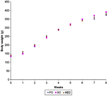
After 8 weeks of supplementation, the SO group was slightly heavier than the PO and MED groups, although the differences did not achieve statistical significance (Fig. 1). In the lipid measurement experiments, 2 animals in the SO group were excluded from the analysis of cardiac phospho-lipids because of technical problems. In the cardiac experiments, 3 animals were lost at the time of the ischemia-reperfusion experiments (technical difficulties) and another 5 were excluded because no ischemic zone was formed (failure to pass the silk snare under the artery), which left 13, 10, and 11 rats in the PO, SO, and MED
groups, respectively, for infarct size analyses. As shown in Table 2, there was a borderline significant difference between the 3 groups for standard food but no difference for total fat intakes. The main differences between groups regarded saturated, n-3 PUFA, and n-6 PUFA. Blood lipids and plasma fatty acids are shown inTable 3. Total and high-density lipoprotein cholesterol were significantly lower in the MED group, whereas triglycerides were not significantly different. In regard to plasma fatty acids, the main
differences between groups were for total n-6 (P b .0001)
and arachidonic acid (AA, 20:4n-6; Pb .0001), which were
strikingly lower in the MED group. Total plasma n-3 PUFA and each individual n-3 PUFA also were notably higher in
the MED group (P b .0001). Erythrocyte fatty acid
Table 2
Fat intakes (standardized chow diet + supplements) of rats in the 3 dietary groups PO SO MED P Chow diet (mg/d per rat) 630 ± 11.7 696 ± 6.9 603 ± 10.5 b.05 Fat supplement (mg/d per rat) 600 600 600 – Total fats (% total energy) 17.33 ± 0.1 17.02 ± 0.1 17.25 ± 0.1 NS Saturated (mg/d per rat) 464.6 ± 2.4 ⁎ 208.0 ± 1.4 159.5 ± 2.1 b.0001 Polyunsaturated (mg/d per rat) Total n-6 374.3 ± 6.1 723.4 ± 3.6† 386.8 ± 5.4 b.0001 18:2n-6 371.2 ± 6.0 713.5 ± 3.5 378.4 ± 5.4 b.0001 20:4n-6 1.9 ± 0.0 ⁎ 6.5 ± 0.0 4.2 ± 0.0 b.0001 Total n-3 44.0 ± 0.8 49.2 ± 0.5 469.6 ± 0.7‡ b.0001 18:3n-3 24.5 ± 0.4 27.7 ± 0.3 307.4 ± 0.4‡ b.0001 20:5n-3 6.9 ± 0.1 8.2 ± 0.1 51.1 ± 0.1‡ b.0001 22:5n-3 1.3 ± 0.1 1.6 ± 0.1 10.2 ± 0.1‡ b.0001 22:6n-3 11.3 ± 0.2 12.5±0.1 100.9 ± 0.2‡ b.0001 Means ± SEM. n = 16 per group. NS indicates not significant.
⁎ P b .0001 vs SO and MED.
† Pb .0001 vs PO and MED. ‡ Pb .0001 vs PO and SO.
Table 3
Blood lipids and plasma fatty acids
PO SO MED P
Blood lipids (g/L)
Total cholesterol 0.53 ± 0.04 0.56 ± 0.03 0.41 ± 0.02 .001 HDL cholesterol 0.30 ± 0.01 0.33 ± 0.01 0.25 ± 0.01 .001 Triglycerides 1.50 ± 0.20 1.36 ± 0.25 1.00 ± 0.06 .13 Plasma fatty acids (%)
14:0 0.81 ± 0.05 0.70 ± 0.05 0.68 ± 0.04 .11 16:0 21.7 ± 0.46 ⁎ 19.3 ± 0.39 19.3 ± 0.58 .001 18:0 5.73 ± 0.26 6.19 ± 0.19 5.62 ± 0.19 .11 Total n-6 42.7 ± 1.15 49.3 ± 1.34 36.2 ± 0.89† b.0001 18:2n-6 26.2 ± 0.78 28.2 ± 0.66‡ 25.8 ± 0.61 .03 20:4n-6 14.9 ± 0.91 19.1 ± 0.95 9.32 ± 0.41† b.0001 Total n-3 5.90 ± 0.17 4.84 ± 0.14 21.0 ± 0.71† b.0001 18:3n-3 1.08 ± 0.09 0.97 ± 0.03 4.40 ± 0.15† b.0001 20:5n-3 0.74 ± 0.03 0.55 ± 0.02 6.95 ± 0.36† b.0001 22:5n-3 0.55 ± 0.03 0.52 ± 0.02 2.37 ± 0.10† b.0001 22:6n-3 3.22 ± 0.13 2.57 ± 0.12 6.98 ± 0.28† b.0001 Mean ± SEM. HDL indicates high-density lipoprotein.
⁎ P b .01 vs SO and MED.
† Pb .0001 vs PO and SO. ‡ Pb .05 vs PO and MED.
Table 4
Erythrocyte fatty acid composition of rats in the 3 dietary groups
PO SO MED P Saturated (%) 14:0 0.23 ± 0.01 0.25 ± 0.02 0.24 ± 0.01 .80 16:0 25.1 ± 0.33 24.0 ± 0.23 24.7 ± 0.35 .10 18:0 11.6 ± 0.38 12.9 ± 0.31 11.6 ± 0.45 .07 Polyunsaturated Total n-6 (%) 45.7 ± 0.51 47.7 ± 0.44 36.8 ± 0.44 ⁎ b.0001 18:2n-6 11.8 ± 0.25 12.3 ± 0.25 12.7 ± 0.34 .08 20:4n-6 31.0 ± 0.47 31.9 ± 0.35 22.4 ± 0.39 ⁎ b.0001 Total n-3 (%) 6.37 ± 0.17 4.83 ± 0.13 15.9 ± 0.42 ⁎ b.0001 18:3n-3 0.09 ± 0.02 0.06 ± 0.02 0.57 ± 0.02 ⁎ b.0001 20:5n-3 0.47 ± 0.02 0.25 ± 0.02 4.93 ± 0.20 ⁎ b.0001 22:5n-3 1.51 ± 0.05 1.18 ± 0.04 3.91 ± 0.14 ⁎ b.0001 22:6n-3 4.18 ± 0.13 3.22 ± 0.09 6.49 ± 0.18 ⁎ b.0001 Means ± SEM. ⁎ P b .0001 vs PO and SO. Table 5
Fatty acid composition of mitochondrial phospholipids in myocardial cells of rats in the 3 dietary groups (% of total fatty acids)
PO SO MED P Phosphatidylcholine Total saturated 45.0 ± 0.91 45.0 ± 1.27 41.6 ± 0.69 ⁎ b.05 Total n-6 40.7 ± 0.97† 38.8 ± 1.18 38.0 ± 0.60 .15 18:2n-6 17.6 ± 0.61 20.11 ± 0.64 21.2 ± 0.75 b.005 20:4n-6 21.9 ± 0.71 17.5 ± 0.69 15.7 ± 0.89‡ b.0001 Total n- 3 3.51 ± 0.20 3.64 ± 0.13 11.2 ± 0.40‡ b.0001 18:3n-3 0.02 ± 0.01 0.07 ± 0.01 0.41 ± 0.03‡ b.0001 20:5n-3 0.17 ± 0.02 0.10 ± 0.01 1.13 ± 0.06‡ b.0001 22:5n-3 0.77 ± 0.06 0.82 ± 0.04 2.13 ± 0.09‡ b.0001 22:6n-3 2.55 ± 0.14 2.65 ± 0.11 7.49 ± 0.34‡ b.0001 Phosphatidylethanolamine Total saturated 44.1 ± 1.11† 36.3 ± 0.13 38.8 ± 1.08 b.0001 Total n-6 31.3 ± 0.84 36.1 ± 1.01§ 21.6 ± 0.73 b.0001 18:2n-6 8.87 ± 0.51 12.1 ± 0.63 8.93 ± 0.59 b.005 20:4n-6 20.4 ± 0.55 22.0 ± 0.45 11.7 ± 0.29‡ b.0001 Total n-3 16.6 ± 0.62 19.6 ± 0.96 34.0 ± 1.70‡ b.0001 18:3n-3 0.01 ± 0.01 0.06 ± 0.01 0.46 ± 0.03‡ b.0001 20:5n-3 0.20 ± 0.01 0.18 ± 0.015 1.24 ± 0.05‡ b.0001 22:5n-3 1.41 ± 0.1 1.68 ± 0.07 2.70 ± 0.13‡ b.0001 22:6n-3 15.0 ± 0.54 17.6 ± 0.90 29.6 ± 1.59‡ b.0001 Cardiolipin Total saturated 11.95 ± 0.42† 5.54 ± 0.26 5.58 ± 11.80 b.0001 Total n-6 81.13 ± 0.62† 89.24 ± 0.40 86.63 ± 0.28 b.0001 18:2n-6 78.64 ± 0.60† 87.81 ± 0.40 84.86 ± 0.31 b.0001 20:4n-6 0.72 ± 0.02† 0.59 ± 0.02 0.54 ± 0.02 b.0001 Total n-3 0.97 ± 0.03 0.69 ± 0.05 2.86 ± 0.17‡ b.0001 18:3n-3 0.21 ± 0.01 0.15 ± 0.01 1.53 ± 0.12‡ b.0001 20:5n-3 0.08 ± 0.01 0.06 ± 0.01 0.34 ± 0.01‡ b.0001 22:5n-3 0.25 ± 0.007 0.21 ± 0.01 0.45 ± 0.03‡ b.0001 22:6n-3 0.43 ± 0.02 0.28 ± 0.02§ 0.54 ± 0.04 b.0001 Means ± SEM.
⁎ P b .05, MED vs PO and SO.
† Pb .0001 vs SO and MED. ‡ Pb .0001 vs PO and SO § Pb .0001 vs PO and MED.
composition is shown inTable 4. There was no significant difference between groups for total saturated or total PUFA. The main difference between groups was in total n-6 PUFA
and total n-3 PUFA (P b .0001). In addition, in the MED
group, 20:4n-6 was lower (P b .0001) and 20:5n-3 was
higher (P b .0001) than those in the PO and SO groups, so
that the 20:4n-6/20:5n-3 ratio was 66, 127, and 4.5 for the
PO, SO, and MED groups, respectively (Pb .0001).Table 5
shows the fatty acid composition of the 3 main phospholipids of cardiac mitochondria. The main differences between groups were for total and individual n-3 PUFA (which were consistently higher in the MED group) and for 20:4n-6, which was lower in the MED group as compared to PO and SO (with the exception of cardiolipin).
In phosphatidylcholine and phosphatidylethanolamine, the main differences between groups were for total and individual n-3 PUFA and for 20:4n-6. In cardiolipin, saturated fatty acids were consistently higher in the PO group. 3.1. Cardiac experiments
At baseline and throughout the ischemia-reperfusion period, there was no significant difference between groups in hemodynamics. In particular, left ventricular function and coronary flow at baseline (normoxic perfusion) did not differ
between groups (Table 6A). Likewise, after 30- and
120-minute reperfusion (Table 6B and C), we found no significant difference between groups for coronary flow, diastolic pressure, and left ventricular developed pressure (LVDevP). The RZ was not different in the 3 groups (Fig. 2): 48.1% ± 1.3%, 48.3% ± 2.3%, and 48.2% ± 3.2% in the PO, SO and MED groups, respectively. In contrast, infarct size was significantly different between groups with 37.7% ± 2.1%, 32.9% ± 1.5%, and 28.6% ± 1.7 % of the RZ in the PO, SO,
and MED groups, respectively (ANOVA P b .01). As
compared with PO and SO, infarct size was smaller by
24.1% (Pb .05) and 13.1% (P b .05), respectively, in the
MED group. When PO and SO— the 2 groups with low n-3
PUFA — were pooled together and compared to MED
(which had high n-3 PUFA), infarct size was still
significantly smaller (P b .05) with 35.3% ± 1.4% for
PO + SO and 28.6% ± 1.7% for MED. In contrast, when the 2 low n-6 PUFA groups, PO and MED, were pooled together and compared to the high n-6 PUFA SO group, there was no significant difference.
4. Discussion
This study shows that an increased intake of n-3 PUFA combined with a low intake of n-6 PUFA and saturated fatty
Table 6
Left ventricular function in rats of the 3 dietary groups
PO SO MED P
(A) After 15-min stabilization (baseline)
Coronary flow (mL/min) 15.3 ± 0.3 15.1 ± 0.5 14.1 ± 0.4 NS Diastolic pressure (mm Hg) 4.2 ± 0.1 3.9 ± 0.1 4.4 ± 0.1 NS LVDevP (mmHg) 140 ± 9 132 ± 10 124 ± 7.1 NS +dP/dt (mm Hg/s) 3545 ± 106 3462 ± 150 3313 ± 148 NS −dP/dt (mm Hg/s) 2122 ± 101 2198 ± 152 2100 ± 97 NS (B) After 30-min ischemia and 30-min reperfusion
Coronary flow (% baseline) 79.9 ± 1.4 81.5 ± 2.5 80.8 ± 3.0 NS Diastolic pressure (mm Hg) 27.7 ± 1.4 26.6 ± 2.5 25.6 ± 2.2 NS LVDevP (% baseline) 63.7 ± 2.5 66.3 ± 30 70.2 ± 2.5 NS +dP/dt (% baseline) 61.7 ± 3.1 71.8 ± 4.4 71.4 ± 3.4 NS −dP/dt (% baseline) 75.8 ± 3.7 79.2 ± 3.6 77.7 ± 3.3 NS (C) After 120-min reperfusion
Coronary flow (% baseline) 58.4 ± 2.1 63.6 ± 2.7 58.1 ± 3.4 NS Diastolic pressure (mm Hg) 36.9 ± 1.5 33.8 ± 3.4 36.2 ± 2.4 NS LVDevP (% baseline) 46.3 ± 1.9 46.5 ± 3.9 46.4 ± 2.4 NS +dP/dt (% baseline) 47.5 ± 2.6 53.1 ± 3.5 53.5 ± 2.8 NS −dP/dt (% baseline) 54.0 ± 4.3 59.0 ± 2.4 55.5 ± 4.1 NS Abbreviation: LVDevP, left ventricular developed pressure.
Values are means ± SEM.
acids will result in smaller myocardial infarct size. This may result in a decreased risk of major complications after acute myocardial infarction (ie, cardiac pump failure and fatal arrhythmia). In this study, we used an isolated heart model that allowed us to study the response of the myocardium independently from other organs, neurologic brain-heart connections, and blood components, such as circulating cells, platelets, hormones, or cytokines. Although these blood components are theoretically influenced by dietary fats and can by themselves influence the myocardial response to ischemia-reperfusion, the ex vivo isolated heart model was preferred to an in vivo model to be able to specifically study the myocardial response independently from these blood components and from neurologic connections.
4.1. Clinical implication
In this study, we examined the effects of a dietary fatty acid profile with high plant and marine n-3 PUFA, low saturated fatty acids and n-6 PUFA, and without trans-fatty acids. This indicates that the observed protection against ischemia-reperfusion injury probably resulted from a complex interplay between several lipid factors and not from a single nutrient. This is a common problem encountered in both experimental and clinical nutrition because any change in the diet of experimental animals or humans is multifactorial. When comparing diets with similar energy content, the reduction of one nutrient results in a proportional increase in one or several others [17]. It is often difficult to identify one individual factor as more active than the others and to decide whether one particular change is more important than any other. In this study, the hearts from rats fed a high saturated fat diet (PO group) were less resistant than those in the 2 other groups. Are the effects due to saturated fats, n-6 PUFA, or n-3 PUFA? On the basis of our study, we can only conclude that the n-3-rich fatty acid profile resulted in smaller myocardial infarct size compared to 2, rather typical, Western dietary patterns that were rich in either n-6 PUFA or saturated fats.
This study also suggests that for the same amounts of total fats and saturated fats, the hearts of rats receiving n-3 PUFA were more resistant than those receiving increased amounts of n-6 PUFA. Although the difference was small, this may be of clinical relevance because high n-6 intake has been encouraged for many years in Western countries to replace saturated fats in the context of cholesterol-lowering diets [18]. Therefore, this study shows that the n-6-rich dietary strategy is not optimal in terms of myocardial resistance to ischemia-reperfusion injury and CHD complications. This actually confirms previous clinical trials demonstrating no obvious beneficial effect of n-6–rich cholesterol-lowering diets, although these were low in saturated fats [18]. Thus, to decrease the risk of CHD complications, the optimal dietary strategy would be to decrease both saturated fats and n-6 PUFA and to increase n-3 PUFA, as also suggested by
clinical studies [2,3,19]. This is of critical importance at a time when the association of n-6 PUFA and saturated fatty acid intakes with CHD complications is being called into question[20-23].
Although not identical to the traditional Mediterranean diet, the n-3–rich diet tested here is similar (in terms of saturated and polyunsaturated fats) to the experimental diet tested in the Lyon Diet Heart Study as a secondary prevention of CHD, where a striking protection against
CHD complications was observed[2,3]. Thus, part of the
cardiac protection following the adoption of a Mediterranean diet may be due to smaller myocardial infarct size.
4.2. Respective roles of n-3, n-6, and saturated fatty acids In this study, the group of rats with high plant and marine n-3 PUFA had the best resistance to ischemic insult. Were plant and marine n-3 PUFA the main mediators of that effect? This would not be unexpected as the changes in
n-3 PUFA concentrations — in plasma, cell membranes,
and cardiac mitochondria— were, quantitatively, the most
important biological changes in that study. However, although a number of experimental studies have been conducted to assess the effects of plant or marine n-3 PUFA on the ischemic myocardium and their ability to
prevent CHD complications [10,24-39], the results have
been conflicting.
One problem comes from the variability of the protocols and techniques used in these studies. Some studies did not specifically study myocardial resistance to regional ische-mia-reperfusion but rather the effects on global ischemia-reperfusion, whereas others examined the effects of ischemia-reperfusion on ventricular arrhythmia rather than
on myocardial injury and necrosis [25,28,31,32,36,38].
Some studies were not designed to assess the effect on infarct size because the duration of ischemia was too short to induce measurable cell necrosis [10,30,35,38]. In others, there was no clear description of the tested diets (specifically, the diet in the control group) or of the biochemical changes induced by the tested diets (specifically, in cardiac mitochondria) [26,30,33,34,37]. In fact, mitochondria are thought to play an important role in myocardial resistance to ischemia-reperfusion[8-11], but only a few studies actually tested the modulation of myocardial resistance to regional ischemia-reperfusion by dietary fatty acids[29,33,35,39].
Oskarsson et al[35]used an in vivo model and showed
that marine n-3 PUFA reduce infarct size in a canine model of ischemia-reperfusion. However, the total amounts of fat were not similar in the experimental and control groups, leaving the possibility that the smaller infarct size was not the result of a protection specifically induced by marine n-3
PUFA. Zhu et al [33] also used an in vivo model and
assumed that this effect on infarct size was due to circulating platelets (rather than to an increased myocardial resistance). In fact, only 2 studies of transient regional ischemia-reperfusion with appropriate control groups used an
experimental ex vivo model similar to ours [29,39]. In the study by Force et al[29], marine n-3 PUFA did not actually reduce infarct size, whereas in the study by Abdukeyum et al [39], a striking (close to 80%) reduction of infarct size was reported. In that study (as in ours), the mechanism of protection resided within the myocardium because there were no neurologic brain-heart connection and no blood component, such as circulating cells, platelets, hormones, or cytokines, in the isolated heart perfusate. Thus, it appears that our present data stand midway between the negative
results of Force et al [29] and the positive results of
Abdukeyum et al [39]. How can we reconcile these
contrasting data?
In the 3 studies, the experimental cardiac apparatus is very similar, using the Langendorff mode with Krebs-Henseleit buffer as perfusate. In the study by Force et al, the duration of ischemia was 40 minutes followed by 2 hours of reperfusion, whereas in Abdukeyum's and ours, the duration of ischemia was only 30 minutes followed by 2-hour reperfusion. A longer ischemic period (even only a 10-minute difference) may partly explain the differences in the results. It should be recognized that without collateral blood flow, the longer the ischemic period, the less likely any salvage becomes apparent during reperfusion. Thus, the 40-minute ischemia may have been too long to allow significant salvage in Force's study[29].
Can different dietary protocols explain the different infarct sizes in the 3 studies? The supplemental fats were similar in the 3 studies. In terms of amounts, however, fish oil represented 22% by weight of the total diet in Force's
study [29]compared with 7% in the study by Abdukeyum
and 6% in ours. In fact, the very high intake in polyunsaturated fatty acids in Force's study may have induced deleterious side effects, such as an increased oxidative stress[40]. Thus, the lack of protection in Force's study may be explained by the duration of ischemia and an excess in polyunsaturated fatty acids. Therefore, our study
supports that of Abdukeyum et al[39]in showing that n-3
PUFA increase myocardial resistance to ischemia-reperfu-sion injury and result in smaller infarct size.
The difference between the 2 studies regarding the
extent of protection — the between-group difference in
infarct size— may have several explanations. One of them
may be purely statistical (a chance effect) as the small number of rats in each group of Abdukeyum's study (n = 6 vs n = 10-13 in our study) may have led to an overestimation of the protective effect of n-3 PUFA.
Another explanation is that in the present study, the hearts were paced at 300 bpm, whereas spontaneous heart rates in Abdukeyum's study were 182 to 203 bpm. The ischemic insult was, therefore, more severe in our study, and this may have allowed less tissue salvage and a smaller between-group difference in infarct size. For the same reason (high rate pacing), we did not investigate the effects of n-3 PUFA on the incidence of ventricular arrhythmia and cannot confirm the arrhythmia data reported in
Abdukeyum's study because the hearts were beating spontaneously [39].
This also may be the main reason why, in contrast with Abdukeyum's study[39], we did not find any difference in left ventricular function between the 3 groups after ischemia-reperfusion, despite differences in infarct size. In our study, and because of the more severe ischemia, resistant hearts (with smaller infarct size) may have developed a kind of myocardial“hibernation” (a more or less reversible state of reduced contractility) that may have masked the better recovery of postischemic function as compared to less resistant hearts with a larger infarct size.
Another difference between our study and Abdukeyum's study was that we used a mixture of plant and marine n-3 PUFA, whereas Abdukeyum et al used marine n-3 PUFA exclusively. As Abdukeyum et al did not report fatty acid levels, we cannot compare the n-3 PUFA concentrations in the 2 studies. One question is whether the addition of plant n-3 PUFA may have been beneficial. A few studies have
suggested that ALA— the main plant source of n-3 — may
have a protective effect on the heart[24,32,41]or the brain [42], but no study examined the specific effect of ALA on infarct size. In our study, ALA was considerably higher in the plasma, erythrocyte membranes, and mitochondrial phospholipids of the MED rats compared to that in the PO and SO rats. However, the relative contribution of ALA to total fatty acids remained very small. In addition, there was no group with ALA supplementation and without marine n-3 PUFA because the study was designed to study a global fatty acid profile.
Beyond a specific and protective effect of n-3 PUFA, the association of ALA and marine n-3 PUFA may have been protective by inducing a massive reduction in AA levels in cell membranes and mitochondrial phospholipids. Although they exhibited levels of linoleic acid (the precursor of AA) similar to those of the PO rats, the MED rats had considerably lower AA levels (30%-50% lower) than both PO and SO rats. This suggests that ALA supplementation induced a strong inhibition of the endogenous synthesis of AA from its precursor linoleic acid, a result that was not unexpected[43,44].
The next question is whether such a decrease in AA levels may have a role in the protection observed in our study. Arachidonic acid is often presented as a major player in CHD complications[45]and probably responsible for cell damage after ischemia-reperfusion[45]. A massive accumulation of AA metabolites, such as lipoxygenase and cytochrome P450 epoxygenase, has been reported in the postischemic
myocardium [46,47]. It is noteworthy that part of the
detrimental effect of AA may be related to competition with the 20-carbon n-3 fatty acid (eicosapentaenoic acid [EPA]), in several pathways potentially contributing to ischemia-reperfusion injury[46-48]. Because EPA may be protective and AA detrimental, the AA/EPA ratio may hypothetically be more relevant than AA concentrations. In this context, we note striking differences between groups in the AA/EPA
ratio: 66, 127, and 4.5 in the PO, SO, and MED groups, respectively. However, there is no correlation between the AA/EPA ratio and infarct size in the 3 groups, as the highest AA/EPA ratio was not associated with the largest infarct size. This suggests that other factors are involved. Saturated fatty acids, which are significantly lowered in the cardiac mitochondria of MED rats as compared with those of PO rats, may play a role. As a matter of fact, certain saturated fatty acids, in particular, palmitic acid [49], have been involved in myocardial apoptotic cell death. Further studies are required to examine these issues.
The present experiments suggest that, as stated in our primary hypothesis, a MED dietary fatty acid profile induces a significant myocardial resistance to ischemia-reperfusion as compared with diets enriched in n-6 and/or saturated fatty acids. In addition, a diet rich in 6 fatty acids (but poor in n-3 PUFA and saturated fatty acids) seems to be better, in terms of myocardial resistance, than a diet rich in saturated fats but poor in both n-3 PUFA and n-6 PUFA. This may have major clinical implications.
Acknowledgment
This study was supported by a grant from the Institut de Recherche sur les Boissons.
References
[1] Burr ML, Fehily AM, Gilbert JF, Rogers S, Holliday RM, Sweetnam PM, et al. Effect of changes in fat, fish and fibre intakes on death and myocardial reinfarction: diet and reinfarction trial (DART). Lancet 1989;2:757-61.
[2] de Lorgeril M, Renaud S, Mamelle N, Salen P, Martin JL, Monjaud I, et al. Mediterranean alpha-linolenic acid–rich diet in secondary prevention of coronary heart disease. Lancet 1994;343:1454-9. [3] de Lorgeril M, Salen P, Martin JL, Monjaud I, Delaye J, Mamelle N.
Mediterranean diet, traditional risk factors, and the rate of cardiovas-cular complications after myocardial infarction: final report of the Lyon Diet Heart Study. Circulation 1999;99:779-85.
[4] GISSI-Prevenzione Investigators. Dietary supplementation with n-3 polyunsaturated fatty acids and vitamin E after myocardial infarction: results of the GISSI-Prevenzione trial. Lancet 1999;354:447-55. [5] Guiraud A, de Lorgeril M, Boucher F, de Leiris J. Cardioprotective
effect of chronic low dose ethanol drinking: insights into the concept of ethanol preconditioning. J Mol Cell Cardiol 2004;36:561-6. [6] Guiraud A, de Lorgeril M, Zeghichi S, F Laporte F, Salen P, Saks V,
et al. Interactions of ethanol drinking with omega-3 fatty acids in rats. Potential consequences for the cardiovascular system. Br J Nutr 2004; 29:1-8.
[7] Harris WS, Sands SA, Windsor SL. Omega-3 fatty acids in cardiac biopsies from heart transplantation patients. Correlation with erythro-cytes and response to supplementation. Circulation 2004;110:1645-9. [8] Gustafsson AB, Gottlieb RA. Heart mitochondria: gates of life and
death. Cardiovasc Res 2008;77:334-43.
[9] Murphy E, Steenbergen C. Mechanisms underlying acute protection from cardiac ischemia-reperfusion injury. Physiol Rev 2008;88: 581-609.
[10] Pepe S, McLennan PL. Cardiac membrane fatty acid composition modulates myocardial oxygen consumption and postischemic recovery of contractile function. Circulation 2002;105:2303-8.
[11] Uchiyama T, Engelman RM, Maulik N, Das DK. Role of Akt signaling in mitochondrial survival pathway triggered by hypoxic precondition-ing. Circulation 2004;109:3042-9.
[12] Toufekstian MC, de Lorgeril M, Nagy N, Salen P, Boucher F, de Leiris J, et al. Chronic dietary intake of plant-derived anthocyanins protects the rat heart against ischemia-reperfusion injury. J Nutr 2008;138: 747-52.
[13] Saks VA, Kuznetsov AV, Kupriyanov VV. Creatine kinase of rat heart mitochondria. The demonstration of functional coupling to oxidative phosphorylation in an inner membrane-matrix preparation. J Biol Chem 1985;260:7757-64.
[14] Hara A, Radin B. Lipid extraction of tissues with a low toxicity solvent. Anal Bioch 1978;90:420-6.
[15] Storrie B, Madden E. Isolation of subcellular organelles. Methods in Enzymology. Academic Press 1990;182:203-25.
[16] Comte J, Gautheron D, Peypoux F, Michel G. Lipid composition and endogenous respiration of pig heart mitochondria. Lipid 1971;6:882-8. [17] de Lorgeril M, Salen P. Fish and n-3 fatty acids for the prevention and treatment of coronary heart disease: nutrition is not pharmacology. Am J Med 2002;112:316-9.
[18] de Lorgeril M, Salen P. Diet as preventive medicine in cardiology. Curr Opin Cardiol 2000;15:364-70.
[19] Ramsden CE, Faurot KR, Carrera-Bastos P, Cordain L, De Lorgeril M, Sperling LS. Dietary fat quality and coronary heart disease prevention: a unified theory based on evolutionary, historical, global, and modern perspectives. Curr Treat Options Cardiovasc Med 2009;11:289-301. [20] Harris WS, Mozaffarian D, Rimm E, et al. Omega-6 fatty acids and risk
for cardiovascular disease: a science advisory from the American Heart Association Nutrition Subcommittee of the Council on Nutrition, Physical Activity, and Metabolism; Council on Cardiovascular Nursing; and Council on Epidemiology and Prevention. Circulation 2009;119:902-7.
[21] Choo J, Ueshima H, Curb JD, et al. Serum n-6 fatty acids and lipoprotein subclasses in middle-aged men: the population-based cross-sectional ERA-JUMP Study. Am J Clin Nutr 2010;91:1195-203. [22] Katan MB. Omega-6 polyunsaturated fatty acids and coronary heart
disease. Am J Clin Nutr 2009;89:1283-4.
[23] Siri-Tarino PW, Sun Q, Hu FB, Krauss RM. Meta-analysis of prospective cohort studies evaluating the association of saturated fat with cardiovascular disease. Am J Clin Nutr 2010;91:535-46. [24] Al-Khalifa A, Maddaford TG, Chahine MN, Austria JA, Edel AL,
Richard MN, et al. Effect of dietary hempseed intake on cardiac ischemia-reperfusion injury. Am J Physiol Regul Integr Comp Physiol 2007;292:R1198-1203.
[25] Billman G, Kang J, Leaf A. Prevention of sudden cardiac death by dietary pureω-3 polyunsaturated fatty acids in dogs. Circulation 1999; 99:2452-7.
[26] Culp B, Lands W, Lucchesi B, Pitt B, Romson J. The effect of dietary supplementation of fish oil on experimental myocardial infarction. Prostaglandins 1980;20:1021-31.
[27] Demaison L, Sergiel J, Moreau D, Grynberg A. Influence of the phospholipid n-6/n-3 polyunsaturated fatty acid ratio on the mito-chondrial oxidative metabolism before and after myocardial infarction. Biochim Biophys Acta 1994;1227:53-9.
[28] Engelbrecht AM, Engelbrecht P, Genade S, Niesler C, Page C, Smuts M, et al. Long-chain polyunsaturated fatty acids protect the heart against ischemia/reperfusion-induced injury via a MAPK dependent pathway. J Moll Cell Cardiol 2005;39:940-54.
[29] Force T, Malis CD, Guerrero JL, Varadarajan GS, Bonventre JV, Weber PC, et al. n-3 fatty acids increase postischemic blood flow but do not reduce myocardial necrosis. Am J Physiol 1989;257: H1204-1210.
[30] Hock C, Beck D, Bodine R, Reibel D. Influence of dietary n-3 fatty acids on myocardial ischemia and reperfusion. Am J Physiol Heart Circ Physiol 1990;259:1518-26.
[31] McLennan PL, Bridle TM, Abeywardena MY, Charnock JS. Comparative efficiency of n-3 and n-6 polyunsaturated fatty acids in
modulating ventricular fibrillation threshold in marmoset monkeys. Am J Clin Nutr 1993;58:666-9.
[32] McLennan PL, Dallimore JA. Dietary canola oil modifies myocardial fatty acids and inhibits cardiac arrhythmia in rats. J Nutr 1995;125: 1003-9.
[33] Zhu BQ, Sievers RE, Sun YP, et al. Is the reduction of myocardial infarct size by dietary fish oil the result of altered platelet function? Am Heart J 1994;127:744-55.
[34] Ogita H, Node K, Asanuma H, Sanada S, Takashima S, Minamino T, et al. Eicosapentanoic acid reduces myocardial injury induces by ischemia and reperfusion in rabbits hearts. J Cardiovasc Pharmacol 2003;41:964-9.
[35] Oskarsson H, Godwin J, Gunnar R, Thomas J. Dietary fish oil supplementation reduces myocardial infarct size in a canine model of ischemia and reperfusion. J Am Coll Cardiol 1993;21:1280-5. [36] Takeo S, Nasa Y, Tanonaka K, Yabe K, Nojiri M, Hayashi M, et al.
Effects of long term treatment with eicosapentaenoic acid on the heart subjected to ischemia/reperfusion and hypoxia/reoxygenation in rats. Mol Cell Biochem 1998;188:199-208.
[37] Yanagisawa A, Matsukura T, Aoki N, Miyagawa M, Satoh K, Metori K, et al. Protection of the rat myocardium from ischemic injury by dietary lamprey oil. Ecosanoids 1988;1:93-100.
[38] Yang B, Saldeen TGP, Nichols WW. Dietary fish oil supplementation attenuates myocardial dysfunction and injury caused by global ischemia and reperfusion in isolated rat hearts. J Nutr 1993;123: 2067-74.
[39] Abdukeyum GG, Owen AJ, McLennan PL. Dietary (n-3) long-chain polyunsaturated fatty acids inhibit ischemia and reperfusion arrhyth-mia and infarction in rat heart not enhanced by ischemic preconditioning. J Nutr 2008;138:1902-9.
[40] Bloomer RJ, Larson DE, Fisher-Wellman KH, Galpin AJ, Schilling BK. Effect of eicosapentaenoic and docosahexaenoic acid on resting
and exercise-induced inflammatory and oxidative stress biomarkers: a randomized, placebo controlled, cross-over study. Lipids Health Dis 2009;19(8):36.
[41] Fiaccavento R, Carotenuto F, Minieri M, Masuelli L, Vecchini A, Bei R, et al. Alpha-linolenic acid-enriched diet prevents myocardial damage and expands longevity in cardiomyopathic hamsters. Am J Pathol 2006;169:1913-24.
[42] Heurteaux C, Laigle C, Blondeau N, Jarretou G, Lazdunski M. Alpha-linolenic acid and riluzole treatment confer cerebral protection and improve survival after focal brain ischemia. Neuroscience 2006;137: 241-51.
[43] Brenna T. Efficiency of conversion ofα-linolenic acid to long chain n-3 fatty acids in man. Curr Opin Clin Nutr Metab 2002;5:127-32. [44] Sprecher H, Luthria DL, Mohammed BS, Baykousheva SP.
Re-evaluation of the pathways for the biosynthesis of polyunsaturated fatty acids. J Lipid Res 1995;36:2471-7.
[45] Hjelte LE, Nilsson A. Arachidonic acid and ischemic heart disease. J Nutr 2005;135:2271-3.
[46] Oe H, Kuzuya T, Oshida S, Nishida M, Tada M. Calcium overload and cardiac myocyte cell damage induced by arachidonate lipoxygenation. Am J Physiol 1994;267:H1396-1402.
[47] Adamek A, Jung S, Dienesch C, Laser M, Ertl G, Frantz S. Role of 5-lipoxygenase in myocardial ischemia-reperfusion injury in mice. Eur J Pharmacol 2007;571:51-4.
[48] Gross GJ, Falck JR, Gross ER, Isbell M, Moore J, Nithipatikom K. Cytochrome P450 and arachidonic acid metabolites: role in myocardial ischemia/reperfusion injury revisited. Cardiovasc Res 2005;68:18-25.
[49] Ostrander DB, Sparagna GC, Amoscato AA, McMillin JB, Dowhan W. Decreased cardiolipin synthesis corresponds with cytochrome c release in palmitate-induced cardiomyocyte apoptosis. J Biol Chem 2001;276:38061-7.