Afferents Integration and Neural Adaptive Control of Breathing
byChung Tin
B.Eng., Mechanical Engineering, The University of Hong Kong, 2002 S.M., Mechanical Engineering, Massachusetts Institute of Technology, 2004
SUBMITTED TO THE DEPARTMENT OF MECHANICAL ENGINEERING IN PARTIAL FULFILLMENT OF THE REQUIREMENTS FOR THE DEGREE OF
DOCTOR OF PHILOSOPHY IN MECHANICAL ENGINEERING AT THE
MASSACHUSETTS INSTITUTE OF TECHNOLOGY JUNE 2011
@ 2011 Massachusetts Institute of Technology. All rights reserved.
Signature of A uthor ... ... . .... ...* .. ... ... Dep a ent of Mechanical Engineering
May 19,2011
C ertified by ... ... .... . ...
/
Chi-Sang PoonPrincipal Research Scientist of Health Sciences & Technology Thesis Supervisor
C ertified by
...
9
. - .. ...
y.... . ...
...
Neville Hogan Sun Jae Professor of Mechanical Engineering Professor of Brain and Cognitive Sciences TesiComnuittee Chair
A ccepted by ... . ... ... David E. Hardt Ralph E. and Eloise F. Cross Professor of Mechanical Engineering Chairman, Committee on Graduate Students
Afferents Integration and Neural Adaptive Control of Breathing
by
Chung Tin
Submitted to the Department of Mechanical Engineering On May 19, 2011, in Partial Fulfillment of the
Requirements for the Degree of
Doctor of Philosophy in Mechanical Engineering Abstract
The respiratory regulatory system is one of the most extensively studied homeostatic systems in the body. Despite its deceptively mundane physiological function, the mechanism underlying the robust control of the motor act of breathing in the face of constantly changing internal and external challenges throughout one's life is still poorly understood.
Traditionally, control of breathing has been studied with a highly reductionist approach, with specific stimulus-response relationships being taken to reflect distinct feedback/feedforward control laws. It is assumed that the overall respiratory response could be described as the linear sum of all unitary stimulus-response relationships under a Sherringtonian framework. Such a divide-and-conquer approach has proven useful in predicting the independent effects of specific chemical and mechanical inputs. However, it has limited predictive power for the respiratory response in realistic disease states when multiple factors come into play. Instead, vast amounts of evidence have revealed the existence of complex interactions of various afferent-efferent signals in defining the overall respiratory response.
This thesis aims to explore the nonlinear interaction of afferents in respiratory control. In a series of computational simulations, it was shown that the respiratory response in humans during muscular exercise under a variety of pulmonary gas exchange defects is consistent with an optimal interaction of mechanical and chemical afferents. This provides a new understanding on the impacts of pulmonary gas exchange on the adaptive control of the exercise respiratory response. Furthermore, from a series of in-vivo neurophysiology experiments in rats, it was discovered that certain respiratory neurons in the dorsolateral pons in the rat brainstem integrate central and peripheral chemoreceptor afferent signals in a hypoadditive manner. Such nonlinear interaction evidences classical (Pavlovian) conditioning of chemoreceptor inputs that modulate the respiratory rhythm and motor output. These findings demonstrate a powerful gain modulation function for control of breathing by the lower brain.
The computational and experimental studies in this thesis reveal a form of associative learning important for adaptive control of respiratory regulation, at both behavioral and neuronal levels. Our results shed new light for future experimental and theoretical elucidation of the mechanism of respiratory control from an integrative modeling perspective.
Thesis Supervisor: Chi-Sang Poon
Principle Research Scientist of Health Sciences & Technology
Thesis Committee:
Neville Hogan (Thesis Committee Chair) Sun Jae Professor of Mechanical Engineering Professor of Brain and Cognitive Sciences
Kamal Youcef-Toumi
Acknowledgements
Earning a PhD. Degree is hard, especially from MIT. It would have been impossible for me without the help and support of many people throughout the process.
I would like to first thank my advisor, Dr. Chi-Sang Poon, who offered me the
opportunity to pursue a research project totally distinct from my previous background. Furthermore, his advice and guidance throughout these years have paved the road for me towards becoming an independent scientist. I would also like to thank Prof. Neville Hogan and Prof. Kamal Youcef-Toumi for their invaluable feedback and guidance in shaping the thesis in its best possible form.
Members of Poon Lab have been wonderful. Dr. Gang Song has taught me all the neuroscience, physiological and experimental techniques that open my road towards biological research. Thank to Dr. Yunguo Yu for teaching me the skills for performing multielectrode recording, as well as for being a good friend. I am indebted to Dr. Ping Yu and Martha Adams for their generous help on carrying out the experiments. Dr. Paula Feinberg-Zadek has been very kind to help me proofread my thesis writing. I would also like to thank all the past and present members of Poon Lab for their friendship and support all these years. It has been a great time working with all of you.
I must thank our beloved Leslie Regan at the Graduate Office of Mechanical Engineering for her help and care in many occasions through my life as a graduate student in MIT.
I would like to thank the Croucher Foundation and American Heart Associative for
their generous financial support for my graduate study.
I have been very lucky to meet so many fantastic friends here who have kept me
moving toward the finish line. My special thank to Sally Kwok and Edmond Lee and their baby Natalie. Sally and Edmond have been like brother and sister to me and they treat me like one of their family. Natalie has been one of the best cheerleaders for me since she was born. I owe countless emotional, as well as nutritional, support from them. My special thanks also go to Samuel Au, who has been both a mentor and a friend to me. Thank to Sam for helping me to settle down in MIT when I first came. We have had enormous sharing both in science and in life over the years. I will always remember the fun times when we went playing soccer, snowboarding and hiking together.
My life here would have been a very different story without the friendship from all of
you: Patrick Sit, Louis Wong, Ivy Lee, Raymond Lam, Ming Yan Poon, Preston Li, Albert Kong, Albert Lok, David Choa, Ricky Tong, Victor Yeung and many more. Thanks for your company and support and fun.
Thanks to my friends in Hong Kong and anywhere in the world. Thanks for being my friends and for caring about me since the day we met.
I owe my deepest gratitude to my parents. They have always offered me the best they could since the very first day and they support unconditionally every move I have made in life. I can never thank you enough.
Chung Tin Cambridge, Massachusetts May 2011
Table of Contents
Chapter 1 Introduction...15 1.1 THESIS OUTLINE ... 18 1.2 REFERENCES... 19 Chapter 2 Background ... 21 2.1 RESPIRATORY FEEDBACK ... 21 2.1.1 Mechanical Feedback... 22 2.1.2 Chemical Feedback... 232.2 BRAINSTEM RESPIRATORY CONTROL CENTER... 23
2.2.1 Dorsal respiratory group (DRG)... 24
2.2.2 Ventral respiratory group (VRG)... 24
2.2.3 Pontine respiratory region... 25
2.3 Traditional view of respiratory control and limitations ... 26
2.4 Evidence of intelligent respiratory control... 27
2.4.1 Nonassociative learning in the chemical and mechanical feedback pathways 28 2.4.2 Clinical implications from models of nonassociative learning... 29
2.4.3 Associative interaction of vagal and carotid chemoafferent inputs... 30
2.5 Summary ... 30
Chapter 3
Integrative and Reductionist Approaches to Modeling of
Control of Breathing...
37
3.1 INTRO D U CTION ... 37
3.2 REDUCTIONIST VIEW OF BIOLOGICAL MODELING ... 38
3.2.1 The Physiome Project and Multiscale Model... 39
3.3 ENGINEERING AND PHYSICS VIEW OF INTEGRATIVE MODELING.. 41
3.4 TOP-DOWN (INTEGRATIVE) VS BOTTOM-UP (REDUCTIONIST) APPROACH TO BIOLOGICAL MODELING ... 42
3.5 CRITERIA OF A GOOD MECHANISTIC MODEL ... 44
3.6 CONTROL OF BREATHING: REDUCTONISM VS INTEGRATION... 45
3.6.1 Limitations of classical reflex models: a case for sensorimotor integration45 3.6.2 Optimal sensorimotor integration in respiratory control ... 47
3.6.3 Hebbian feedback covariance learning model of respiratory motor control49 3.6.4 Cheyne-Stokes breathing from different engineering control perspectives 54 3.7 REFEREN CES... 56
Chapter 4
Mechanical-Chemical Interaction underlies Optimal
Respiratory Control in Gas Exchange Defects ... 614.1 INTRODU CTION ... 61
4.2 BACKG ROU ND ... 63
4.2.1 Traditional notion of dead space... 63
4.3 GENERALIZED GAS EXCHANGE EQUATION AND OPTIMAL RESPIRATORY CONTROL FOR PULMONARY GAS EXCHANGE DEFECTS.. 64
4.3.1 Notion of dead space in gas exchange equation ... 64
4.3.2 Role of dead space in optimal respiratory control ... 67
4.4 THE OPTIMIZATION MODEL (MODIFIED FROM POON'S MODEL)... 68
4.5 GAS EXCHANGE EQUATION UNDER DIFFERENT PULMONARY GAS EXCHANGE DEFECTS... 72
4.5.1 Congestive heart failure ... 72
4.5.2 Right-to-left shunts ... 73
4.6 THE EFFECT OF EXTERNAL DEAD SPACE, PARALLEL DEAD SPACE
AND SHUNT ON OPTIMIZATION OF RESPIRATORY CONTROL ... 81
4.6.1 Congestive heart failure ... 81
4.6.2 Right-to-left shunts ... 83
4.6.3 External Dead Space ... 84
4.7 DISCUSSION ... 86
4.7.1 Optimization as a framework for afferent interaction in homeostatic control 87 4.7.2 Different mechanisms underlying isocapnic augmented exercise hyperpnea in CHF and shunt ... 88
4.7.3 Mechanisms of potentiation of exercise ventilatory response by added dead space: Optimization vs. short term modulation ... 89
4.7.4 Influence of chemical afferent signal on breathing pattern revealed by comparing airway CO2 and dead space loading ... 89
4.8 CONCLUSIONS... 90
Appendix A: Numerical simulation for external dead space ... 91
4.9 REFERENCES... 95
Chapter
5
Central-peripheral Chemoreceptors Interaction Revealed a
Form of Pavlovian Conditioning ... 1015.1 INTRODUCTION... 101 5.2 METHODS... 103 5.2.1 Animal preparation ... 103 5.2.2 Microelectrode arrays ... 104 5.2.3 Recording ... 104 5.2.4 Protocols ... 106 5.2.5 Data analysis ... 106 5.2.6 Histology... 108 5.3 RESULTS... 109
5.3.1 Interaction of hypercapnia and hypoxia in phrenic motor output... 109
5.3.2 Neuronal recording loci ... 110
5.4 DISCUSSION ... 126
5.4.1 Controversy about chemoreceptors interaction in phrenic motor output.. 126
5.4.2 Recording loci of respiratory neurons... 127
5.4.3 Role of dl-pons in chemoafferent signaling... 127
5.4.4 Associative learning (Pavlovian conditioning) in respiratory chemoreflex control 128 5.4.5 Implications for respiratory rhythmogenesis, respiratory instability/apnea and acclimatization ... 130
5.5 CONCLUSIONS... 131
5.6 REFERENCES... 132
Chapter 6 Conclusions and Future W ork ... 139
6.1 FUTURE WORKS... 140
6.1.1 Modeling ... 140
6.1.2 Experimental... 142
List of Figures
Figure 3.1 Two views for respiratory control (a) Classical reflex model assumes additive, reducible and superposable characteristics of chemical, mechanical and exercise stimuli;
(b) The optimization model integrates various afferent-efferent signals in a single model
to characterize the complex interactions among these signals. (Poon et al., 2007)... 46
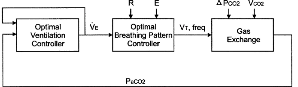
Figure 3.2 Simplified block diagram of the optimization model of respiratory control. Sensorimotor feedback signals are integrated by an intelligent controller which produces optimal ventilatory drive and breathing pattern that are most cost-effect for meeting metabolic demands, subject to the constraints imposed by the mechanical plant and chemical plant. The functional block "G" represents the transmission gains for
sensorim otor integration. ... 53
Figure 3.3 Hebbian Feedback Covariance Control Paradigm ... 53
Figure 3.4 Limit cycle a) Stable, b) unstable, c) semi-stable (Slotine & Li, 1991)... 55
Figure 4.1 Expired CO2 trace with illustration of "dead space". Total "dead space"
normally comprises alveolar and airway dead space... 65
Figure 4.2 Block diagram of optimal respiratory CO2 control ... 67
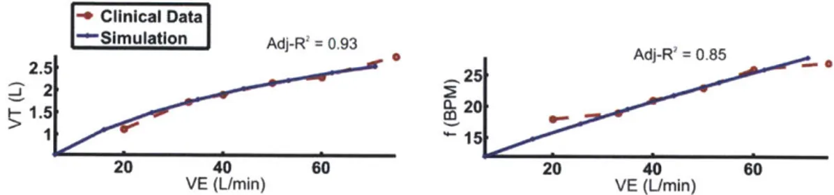
Figure 4.3 Simulated breathing pattern fitted to clinical data to determine parameter values for mechanical work index ... 71
Figure 4.4 Illustration of parallel dead space, VD, and the corresponding expired CO2
Figure 4.5 Illustration of right-to-left shunt and the corresponding expired CO2 trace.... 74
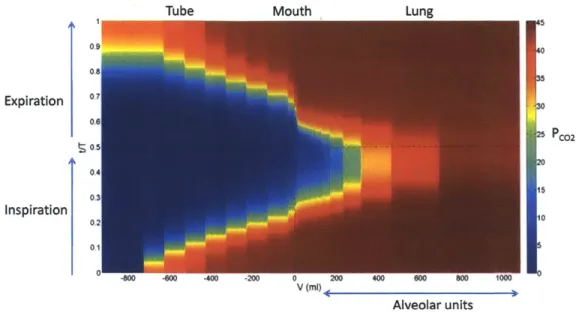
Figure 4.6 Distribution of PCo2 along the airway over a breath cycle in the presence of an
external dead space. Tube volume VDex = 1L... 77
Figure 4.7 Expired CO2 trace at mouth (red) and at end of tube (blue). Two components
of APco2 are illustrated... 78
Figure 4.8 Change of APco2 as a function of external dead space volume. (A) 200ml; (B)
600m l; (C ) 1000m l...79
Figure 4.9 Dependence of APco2 on breathing pattern (1) rapid shallow (dashed lines); (2)
normal (solid line) and (3) slow deep (dash-dot lines) with constant total ventilation. (Red lines represent expired CO2 trace at mouth and blue lines represent expired CO2 trace at
end of tube.) ... 79
Figure 4.10 (A) APco2 as a function of total ventilation and breathing frequency; (B)
APco2 as a function of VE at three levels of VCO2 (VT and breathing frequency are
obtained from minimization of Eq.4.12). (VD, = 1L). ... 80
Figure 4.11 Simulated exercise respiratory response of CHF patient compared with normal subject. CHF patients demonstrated augmented ventilation, with a rapid shallow pattern. The dashed lines are clinical data adopted from (Wasserman, K., Y.-Y. Zhang, et al., Circulation, 1997.). Prediction of breathing pattern is characterized by adj-R2 values.
... 82
Figure 4.12 (A) Simulated exercise respiratory response of two levels of right-to-left shunt compared with normal. Increased shunt fraction is associated with augmented ventilation. (B) Change of APco2 as a function of shunt fraction and metabolic CO2. ... 84
Figure 4.13 Simulated exercise respiratory response of external dead space loading compared with normal and inhaled CO2. External dead space resulted in a stronger
augmented exercise ventilatory response than inhaled CO2. Both cases results in
hypercapnia with normal breahting pattern. ... 86
Figure 4.14 Compartment model indicating pulmonary gas exchange defect problem under present study. ... 87
Figure 4.15 Schematic diagram of external dead space simulation... 94
Figure 5.1 A. Local field potential recording in one channel of multielectrode array. B.
PCA decomposition of signals in (A) indicating that the neuron cluster is readily
Figure 5.2 Response of phrenic discharge amplitude and timing components to hypoxia under varying CO2 backgrounds in one rat... 110
Figure 5.3 Recording loci. (A) Electrolytic lesion was made at the end of the experiment to mark the recording site. (B) Loci of 4 types of respiratory-related neurons in KFN and LPBN exhibiting hypoadditive C0 2- 02 interaction... 111
Figure 5.4 A sample pure-I neuron (A) Firing pattern of neuron presented as perievent raster referenced to respiratory cycle; (B) Hypoxic response (separated into inspiratory and expiratory components) under different CO2 background showing hypoadditive
interaction; (C) Time-stamped spike trains representaiton of hypoxic response under different CO 2 background... 115
Figure 5.5 A sample E-I neuron (A) Firing pattern of neuron presented as perievent raster referenced to respiratory cycle; (B) Hypoxic response (separated into inspiratory and expiratory components) under different CO2 background showing hypoadditive
interaction; (C) Time-stamped spike trains representaiton of hypoxic response under different CO 2 background... 116
Figure 5.6 A sample eE neuron (A) Firing pattern of neuron presented as perievent raster referenced to respiratory cycle; (B) Hypoxic response (separated into inspiratory and
expiratory components) under different CO2 background showing hypoadditive
interaction; (C) Time-stamped spike trains representaiton of hypoxic response under different CO 2 background... 117
Figure 5.7 A sample wE/lateE neuron (A) Firing pattern of neuron presented as perievent raster referenced to respiratory cycle; (B) Hypoxic response (separated into inspiratory and expiratory components) under different CO2 background showing hypoadditive
interaction; (C) Time-stamped spike trains representaiton of hypoxic response under different CO 2 background... 118
Figure 5.8 Mutliexponential curve fitting to breath-by-breath phrenic data, using the same time constants over three different CO2 background. Results suggested that
hypercapnic input altered only the gains of the learning dynamics but had little influence on its tim e course... 120
Figure 5.9 Mutliexponential curve fitting to breath-to-breath neuronal firing (top: E-I inspiratory firing; bottom: wEFlate-E expiratory firing) data, using the same time
constants over three different CO2 background. Results suggested that hypercapnic input
altered only the gains of the learning dynamics but had little influence on its time course.
Figure 5.10 Left panels: Averaged gains of multiexponential curve fitting (normalized to normocapnic condition) for phrenic and neuronal activity over three different CO2
background during hypoxia stimulation. Right panels: Correlation between gains of phrenic TI/TE and I/E-neurons mutliexponential curve fitting. (A) Phrenic T1 vs.
I-neurons (n=12); (B) Phrenic TE vs. E-I-neurons (n=7). * indicates statistically significant
difference from normal C02 condition (p<0.05)...
124
Figure 5.11 Left panels: Averaged gains of multiexponential curve fitting (normalized to normocapnic condition) for phrenic and neuronal activity over three different CO2
background during post-hypoxia recovery. Right panels: Correlation between gains of phrenic TI/TE and I/E-neurons mutliexponential curve fitting. (A) Phrenic T1 vs.
I-neurons (n=12); (B) Phrenic TE vs. E-I-neurons (n=7). * indicates statistically significant difference from normal C02 condition (p<0.05)... 125
Chapter 1 Introduction
The essence of living organism survival is the maintenance of a stable constant internal environment of the body. From glucose to temperature, water to oxygen, the body continually strives to regulate all its physiological states in the face of various disturbances. Claude Bernard, the founder of modern physiology, coined the concept of milieu intirieur, and he suggested that:
The constancy of the internal environment is the condition that life should be free and independent... So far from the higher animal being indifferent to the external world, it is on the contrary in a precise and informed relation with it, in such a way that its equilibrium results from a continuous and delicate compensation, established as by the most sensitive of balances. (Bernard, 1878, 1974)
It would take nearly fifty years before this concept of biological stability received renewed attention. To describe this concept, physiologist Walter B. Cannon coined the term homeostasis in 1926 in his seminal work, The Wisdom of the Body (Cannon, 1932), which presented the first notions of a biological automatic controller. However, the body's high-dimensional organization and sophisticated dynamical interactions largely preclude thorough understanding of such a robust regulation mechanism (Somjen, 1992; Dworkin, 1993; Poon & Siniaia, 2000).
The respiratory system is probably one of the most extensively studied physiological systems. Current understanding of respiratory control is built upon a variety of classic respiratory reflex responses such as CO2 or hypoxic chemoreflex, lung inflation
and deflation reflexes and exercise hyperpnea reflex, etc. Such Sherringtonian stimulus-response analyses are useful in predicting the independent effects of specific chemical and mechanical inputs, from a traditional reductionist view. All of these reflex responses have been extensively studied and, for the most part, independently of one another for the sake of simplicity. However, the predictive power of these reflex models is limited when multiple factors come into play together. A major limitation of the classical chemoreflex model is that it cannot explain the isocapnia hyperpnea response during exercise. This discrepancy has led to the general postulation of a distinct "exercise stimulus" that feeds forward to the chemoreflex feedback loop as an "exercise reflex" (Grodins, 1950). However, despite extensive explorations for over a century, the putative "exercise
stimulus" has remained elusive to this date (Forster, 2000).
In contrast to the assumption of afferents independence of the classical Sherringtonian reflex model, considerable evidence indicates that distinct classes of respiratory afferent inputs might interact with one another when applied concomitantly. For example, it has long been recognized that lung stretch receptors activation may interact with chemical drive in control of inspiratory and/or expiratory muscle activity (Kelsen et al., 1977; Mitchell et al., 1982; Mitchell et al., 1990; Ainsworth et al., 1992). Such afferent interactions have potential significance in certain clinical applications such as positive end-expiratory pressure (PEEP) or continuous positive airway pressure
(CPAP), or certain disease states such as chronic obstructive pulmonary disease or lung
transplant, where the resultant exaggeration (due to lung inflation) or total absence (deafferentation) of vagal afferent inputs may alter the chemoreflex regulation of breathing. Such interactions also have important physiological implications in that they indicate a much more complex central processing of these respiratory afferent inputs than the conventional reflex models imply.
Presently, there is limited information about how afferent information arising from chemoreceptors and mechanoreceptors are processed and integrated centrally and
how they may interact with one another. Baji6 et al. (1994) reported that peripheral chemoreceptor and pulmonary stretch receptor inputs interact linearly in modulating certain medullary neurons in the dorsal respiratory group and caudal ventral respiratory group in dogs, and a nonadditive interaction in other caudal VRG neurons. Tonkovic-Capin et al. (2000) showed that the interaction between arterial CO2 tension and
pulmonary stretch receptor-mediated modulations of caudal medullary, expiratory bulbospinal neuron activity is mainly additive, but synergism between Paco2 and
excitatory inputs is also present. However, little is known about how these afferent inputs affect the activities of other pontomedullar neurons when presented together and how such interactions may influence respiratory activity.
The mammalian lower brain has traditionally received little attention in regard to its intelligence. However, as evidenced by the precision, robustness, versatility and reliability in physiological control, the traditional reductionist view is inadequate in explaining the common complex scenario of physiological control especially when multiple factors come into play. In fact, the respiratory system demonstrates sustained capability of adaption and memory as in the higher brain (reviewed in (Eldridge & Millhorn, 1986a); (Poon & Siniaia, 2000)). Examples of such processes are numerous and include responses to the following inputs: carotid sinus nerve and carotid chemoreceptors (Eldridge, 1974; Eldridge & Gill-Kumar, 1978; Millhorn et al., 1980; Zhang & Mifflin, 1995; Mifflin, 1997), hypoglossal nerve (Jiang et al., 1991), hypoxia (Fregosi, 1991b; Georgopoulus et al., 1992; Bisgard & Neubauer, 1995; Bach & Mitchell,
1996), and vagal nerve fibers from pulmonary stretch receptors (Stanley et al., 1975;
Karczewski et al., 1976).
In this thesis, we explore the interactions of respiratory afferents and their implications for intelligent respiratory control from both experimental and modeling aspects. We show that nonlinear interactions between respiratory afferents provide a consistent picture over a wide range of experimental and physiological scenarios, that contradicts the assumptions of additivity, reducibility and superposition characteristics in the classical reflex model. Such interactions have been emphasized in the rapidly growing fields of systems biology and systems medicine, which are important in
advancing our understanding of how our body works as an integrated system (Ahn et al.,
2006b, a).
1.1 THESIS OUTLINE
Chapter 2 provides a review of the organization of the respiratory system, anatomically and functionally. We provide evidence that respiratory control is not merely a simple reflex but possess more sophisticated computation.
Chapter 3 contrasts integrative and reductionist approaches in biological system modeling, with particular emphasis on the respiratory system. This chapter sets the stage for the integrative approach taken in this thesis.
Chapter 4 investigates the interaction of mechanical and chemical feedback signals from a computational approach. We hypothesize that an optimal respiratory control model provides consistent prediction for the exercise respiratory control under various pulmonary gas exchange defective conditions by integrating both chemical and mechanical respiratory signals. The optimization framework is shown to provide a convenient mathematical tool to predict a wide range of respiratory responses with a conceptually simple but generalized theory.
Chapter 5 investigates the interaction of peripheral and central chemoreceptors using an experimental approach. We test the influence of the CO2 background on the
subsequent hypoxic response in both phrenic motor output and dorsolateral pons respiratory neurons activity. Furthermore, we demonstrate the important role of dorsolateral pons in the C0 2-02 interaction. We also show that gain modulation of
hypoxic response by central input represents a form of associative learning in respiratory control. Several clinical implications regarding this interaction are suggested.
1.2 REFERENCES
Ainsworth DM, Smith CA, Johnson BD, Eicker SW, Henderson KS & Dempsey JA.
(1992). Vagal modulation of respiratory muscle activity in awake dogs during
exercise and hypercapnia. JAppl Physiol 72, 1362-1367.
Bach KB & Mitchell GS. (1996). Hypoxia-induced long-term facilitation of respiratory activity is serotonin dependent. Respir Physiol 104, 253-260.
Bajic J, Zuperku EJ, Tonkovic-Capin M & Hopp FA. (1994). Interaction between chemoreceptor and stretch receptor inputs at medullary respiratory neurons. The American journal ofphysiology 266, R1951-1961.
Bernard C. (1878, 1974). Lectures on the phenomena of life common to animals and plants. (Lecons sur les phinomenes de la vie communs aux animaux et aux
vegitaux.). Thomas, Springfield, Ill.
Bisgard GE & Neubauer A. (1995). Peripheral and central effects of hypoxia. In Regulation of Breathing, ed. Dempsey JA & Pack Al, pp. 617-668. Marcel Dekker, Inc., N.Y. Basel, Hong Kong.
Cannon WB. (1932). The wisdom of the body. W.W. Norton & Company, New York,.
Dworkin BR. (1993). Learning and Physiological Regulation. University of Chicago Press, Chicago.
Eldridge FL. (1974). Central neural respiratory stimulatory effect of active respiration. J
Appl Physiol 37,723-735.
Eldridge FL & Gill-Kumar P. (1978). Lack of effect of vagal afferent input on central neural respiratory afterdischarge. JAppl Physiol 45, 339-344.
Eldridge FL & Millhorn DE. (1986). Oscillation, gating, and memory in the respiratory control system. In Handbook of Physiology: The Respiratory System, ed. Fishman AP, Cherniack NS & Widdicombe JG, pp. 93-114. Amer. Phys. Soc., Bethesda.
Forster HV. (2000). Exercise hyperpnea: where do we go from here? Exerc Sport Sci Rev
28, 133-137.
Fregosi RF. (1991). Short-term potentiation of breathing in humans. J Appl Physiol 71,
892-899.
Georgopoulus D, Giannouli E, Tsara V, Argiropoulou P, Patakas D & Anthonisen N.
(1992). Respiratory short-term poststimulus potentiation (after-discharge) in
Grodins FS. (1950). Analysis of factors concerned in regulation of breathing in exercise. Physiol Rev 30, 220-239.
Jiang C, Mitchell GS & Lipski J. (1991). Prolonged augmentation of respiratory discharge in hypoglossal motoneurons following superior laryngeal nerve stimulation. Brain Res 538, 215-225.
Karczewski WA, Budzinska K, Gromysz H, Herczynski R & Romaniuk JR. (1976). Some response of the respiratory complex to stimulation of its vagal and mesencephalic inputs. In Respiratory Centers and Afferent System, ed. Duran E,
pp. 107-115. INSERM.
Kelsen SG, Altose MD & Cherniack NS. (1977). Interaction of lung volume and chemical drive on respiratory muscle EMG and respiratory timing. J Appl Physiol 42, 287-295.
Mifflin S. (1997). Short-term potentiation of carotid sinus nerve inputs to neurons in the nucleus of the solitary tract. Respir Physiol 110.
Millhorn DE, Eldridge FL & Mitchell GS. (1980). Prolonged stimulation of respiration
by a new central neuronal mechanism. Respir Physiol 41, 87-103.
Mitchell GS, Cross BA, Hiramoto T & Scheid P. (1982). Interactions between lung stretch and PaCO2 in modulating ventilatory activity in dogs. J Appl Physiol 53,
185-191.
Mitchell GS, Douse MA & Foley KT. (1990). Receptor interactions in modulating ventilatory activity. The American journal ofphysiology 259, R911-920.
Poon CS & Siniaia MS. (2000). Plasticity of cardiorespiratory neural processing: classification and computational functions. Respir Physiol 122, 83-109.
Somjen GG. (1992). The missing error signal - regulation beyond negative feedback. News Physiol Sci 7, 184-185.
Stanley NN, Altose MD, Cherniack NS & Fishman AP. (1975). Changes in strength of lung inflation reflex during prolonged inflation. JAppl Physiol 38, 474-480.
Tonkovic-Capin M, Zuperku EJ, Stuth EA, Bajic J, Dogas Z & Hopp FA. (2000). Effect of central CO(2) drive on lung inflation responses of expiratory bulbospinal neurons in dogs. American journal ofphysiology 279, R1606-1618.
Zhang W & Mifflin SW. (1995). Expiratory amino-acid receptors contribute to carotid sinus and vagus nerve evoked excitation of neurons in the nucleus of the tractus solitarius. JAuton Nerv Syst 55, 50-56.
Chapter 2 Background
The respiratory control system is a multi-input multi-output nonlinear system (Poon, 2000). The system comprises a mechanical plant (respiratory mechanics and muscles) and a chemical plant (pulmonary gas exchange) that are controlled by respiratory neurons in the medulla and pons regions of the brainstem. Extensive studies over the past century have accumulated an enormous collection of knowledge about the respiratory system. Translating these scientific findings into clinical practice becomes critical, as in many other biological research fields (Saijo, 2002; Mankoff et al., 2004; Horig et al., 2005).
2.1 RESPIRATORY FEEDBACK
The two major inputs to the respiratory control system for normal autonomic respiratory regulation are the mechanical and chemical feedbacks. These two sensory modalities play the most significant role in modulating the respiratory rhythm in terms of tidal volume and phase durations (i.e. inspiratory and expiratory duraitons). These feedback pathways are also critical for proper responses during various acute or chronic stressful conditions that occur during exercise or chronic lung disease, for example.
2.1.1 Mechanical Feedback
The motor act of breathing rhythmically inflates/deflates the lungs by altering the pleural pressure. The change in lung volume is sensed by slowly adapting pulmonary stretch receptors (SARs) in the airways. These receptors transmit information to the dorsal respiratory neurons and pontine nuclei via myelinated afferent fibers of the vagus nerve (Ezure et al., 1998). The SARs are phasically stimulated by lung expansion and exhibit small tonic activities throughout the respiratory cycle. The classic Hering-Breuer inflation reflex (HBIR) describes the inhibitory effect of lung inflation and subsequent SAR activation on inspiration (Breuer, 1868b; Breuer, 1868a). This 'reflex' is presumed to protect the lungs from overexpansion. Recent studies have revealed that the fictive HBIR in rats undergoes dynamical changes in the form of non-associative learning (habituation and desensitization) that provides necessary temporal and frequency filtering. The dynamics are modulated by NMDA (N-methyl-D-aspartate) receptors (Poon et al., 2000a; Siniaia et al., 2000a; Poon & Siniaia, 2000b; Poon & Young, 2006b; MacDonald et al., 2009).
Other mechanical sensory receptors include the irritant and J-type receptors (Taylor et al., 1989). The irritant receptors, or rapidly adapting receptors (RARs), are located in the lung epithelium, and respond to noxious chemical or mechanical agents. RARs activity is also transmitted via myelinated fibers of the vagus nerve, and is less sensitive to lung inflation compared to SARs. Stimulation of RARs causes broncho-constriction, coughing, sneezing and general increases in ventilation.
The third receptor type called J-receptors are unmyelinated C-fiber vagal nerve endings that respond to vascular distension and fluid increases in the interstitium. Activation of these J-receptors can cause hyperpnea and dyspnea. Due to the high activation threshold of these unmyelinated C-fibers, small current stimulation of the vagus nerve is unlikely to activate them.
2.1.2 Chemical Feedback
The primary function of the respiratory system is to regulate blood gas concentration. There are two classes of chemoreceptors that continuously monitor the blood chemistry states, peripherally and centrally.
Two peripheral chemoreceptors exist: carotid and aortic bodies. The carotid bodies are located high in the neck at the bifurcation of the common carotid arteries. These receptors sense changes in arterial Po2, Pco2, and pH. Carotid body activity is
transmitted to the brainstem via a small branch of the glossopharyngeal nerve called the carotid sinus nerve (CSN) (Black & Torrance, 1967) and also referred to as Hering's nerve. Carotid body is stimulated mainly by a decrease in Po2 and increases in H*/Pco2.
In human, the aortic bodies play a smaller role in peripheral detection and do not respond to changes in Po2.
Unlike the peripheral chemoreceptors, the loci of central chemoreceptor are still being debated. A number of brainsten regions have been shown to be CO2/H+ sensitive, including ventrolateral medullary surface, nucleus of solitary tract, locus coeruleus, medullary raphe, retrotrapezoid nucleus (RTN), rostroventrolateral medullar, pre-B6tzinger's Complex (pre-BQtC), cerebellar fastigial nucleus (reviewed in Dean & Nattie, 2010). In recent years, RTN at the ventrolateral medullar surface is suggested to be the principal site of intracranial C02/H+ chemoreception (Guyenet et al., 2008). Their tonic activity likely supplies a general background excitation to the respiratory central pattern generator (RCPG) (Duffm, 1991).
2.2
BRAINSTEM RESPIRATORY CONTROL CENTER
Respiratory neurons are densely populated in three main brainstem areas: the dorsal respiratory group (DRG), the ventral respiratory group (VRG) and the pontine respiratory group (PRG).
2.2.1 Dorsal respiratory group (DRG)
The DRG is comprised of mainly inspiratory neurons located in the ventrolateral subnucleus of the nucleus of the solitary tract (NTS) (Cohen & Feldman, 1984). Extensive monosynaptic excitatory projections to the phrenic nucleus in the spinal cord are identified from the neurons in the DRG (Fedorko et al., 1983; de Castro et al., 1994).
Furthermore, the NTS is the principal site of terminations of respiratory-related sensory afferents, including SARs, RARs, and peripheral chemoreceptors. Inhibitory projection to the B6tzinger's Complex (B6tC) and retrotrapezoid nucleus (RTN) have been identified. Commissural subdivision of NTS relays neurons for peripheral chemoreceptors and it provides important excitation to VRG (Takakura et al., 2006; Takakura et al., 2007).
2.2.2
Ventral respiratory group (VRG)
The VRG is a column of respiratory neurons in the ventrolateral medulla. It is commonly divided into the following subdivisions: para-facial respiratory group (pFRG), B6tzinger's Complex (B6tC), pre-B6tzinger's Complex (pre-BQtC), rostral VRG (primarily inspiratory neurons) and caudal VRG (primarily expiratory neurons). The inspiratory (I-Aug) neurons in rostral VRG project monosynaptically to motoneurons in the phrenic nucleus that innervate the diaphragm while expiratory (E-Aug) neurons in caudal VRG innervate the abdominal and expiratory intercostal motoneurons. The BtC consists of a large population of expiratory neurons that provides extensive inhibitory projections within VRG, to NTS and to the phrenic nucleus.
Pacemaker-like neurons have been identified in pre-BtC (Smith et al., 1991) and pFRG (Onimaru et al., 1987, 1988; Onimaru & Homma, 2003) which are thought to generate inspiratory and expiratory rhythms respectively. How the two rhythm generators coordinate with each other to set the respiratory rhythm remains unclear. Our recent
modeling work suggests that they interact in a two-way "handshake" process via a sequence of excitation-reverse inhibition-postinhibitory rebound excitation (Wittmeier et
al., 2008).
2.2.3
Pontine respiratory region
Respiratory neurons are mostly found in the dorsolateral (dl-pons) and the ventrolateral (vl-pons) pontine areas.
2.2.3.1 Dorolateral pons
Two major structures constituting the dl-pons are the K6lliker-Fuse nucleus (KFN) and the parabrachial nucleus (lateral and medial), commonly termed the "pneumotaxic center" (Lumsden, 1923a). The dl-pons has extensive reciprocal projections with VRG,
NTS and the phrenic motor nucleus. Using single-unit extracellular recording in combination with juxtacellular labeling, the functional taxonomy and topographic distribution of pneumotaxic respiratory neurons in the rat dl-pons have been systematically characterized (Song et al., 2006). Six subtypes of respiratory neurons, phasic or phase-spanning have been identified.
The pneumotaxic center is traditionally thought to play an ancillary role in inspiratory-expiratory phase transition by providing a fail-safe mechanism for inspiratory off-switch (IOS) secondary to vagal proprioceptive feedback. Recently, the role of dl-pons in mediating chemoreflex resdl-ponses has been increasingly recognized (Foumier & Kinkead, 2008; Nuding et al., 2009; Song & Poon, 2009a, 2009b; Bonis et al., 2010; Boon & Milsom, 2010).
2.2.3.2 Ventrolateralpons/AS area
A5 is not a well-defined anatomical structure, it includes a noradrenergic cell
expiration (Jodkowski et al., 1997). Furthermore, the vl-pons has an important role in shaping the post-hypoxic depression of respiration (Dick & Coles, 2000).
2.3 TRADITIONAL VIEW OF RESPIRATORY CONTROL
AND LIMITATIONS
Traditionally, control of breathing has been understood in terms of a set of stimulus-response relationships presumably reflecting various afferent feedback/feedforward control mechanism. Such Sherringtonian stimulus-response analyses are useful in predicting the independent effects of specific chemical and mechanical inputs under well-controlled experimental conditions. However, less attention has been put on the interaction of these inputs, that the response when different stimuli are presented simultaneously.
A long unresolved problem in respiratory control is the isocapnic exercise
hyperpnea, defined as an automatic increase in respiratory ventilation geared to metabolic demands with near constancy of arterial CO2 and pH levels. The
chemoreflex/mechanoreflex feedback model implies the existence of a feedforward signal from an exercise-related input. However, despite extensive search over the past century, none of the hypothesized feedforward mechanisms have been demonstrated conclusively as the true "exercise stimulus" obligatory to exercise hyperpnea (Mateika & Duffin, 1995; Ward, 2000; Eldridge et al., 2006; Secher et al., 2006; Waldrop et al, 2006; Yu & Poon, 2006).
Furthermore, the available evidence reveals a distinct multiplicative (synergistic) component in the ventilatory response to concomitant exercise and hypercapnia such that
CO2 responsiveness is potentiated during exercise (Poon & Greene, 1985; Poon, 1989;
Mitchell & Babb, 2006). Ventilatory response to chemical or exercise input is also potentiated by increases in physiological dead space and shunt (e.g. in congestive heart failure patients). Such sensorimotor integration characteristics are ignored in the
oversimplified reflex model which mistakenly considers all respiratory inputs as additive to the "exercise stimulus"
Clearly, the deceptively mundane motor function of breathing is not a trivial task of "as-easy-as-breathing". Breathing is regulated by the brain continually throughout life without fail in the face of constant physiologic and environmental challenges, which can easily surpass the most robust man-made controller. Accumulating evidences show that the respiratory control system undergoes more intelligent computation involving various memory, adaptation and gating mechanisms (Eldridge & Millhorn, 1986a; Poon &
Siniaia, 2000).
2.4 EVIDENCE OF INTELLIGENT RESPIRATORY
CONTROL
Two forms of intelligent behavior evidenced in respiratory control are nonassociative and associative learning.
Habituation and sensitization are two common forms of nonassociative learning that are exhibited in many neuronal structures of mammalian and invertebrate nervous systems (Thompson & Spencer, 1966; Groves & Thompson, 1970; Poon, 1996a; Cohen et al., 1997). According to the classic Dual-Process Theory (Groves & Thompson, 1970), habituation is a progressive reduction in physiological response that occurs upon repetitive application of the same stimulus. On the other hand, sensitization is the progressive amplification of a response following a repetitive stimulus. In simple invertebrate nervous systems, habituation
and sensitization have been shown to result from activity-dependent phasic depression and enhancement of synaptic efficacy respectively (Kandel, 1978; Cohen et al., 1997).
Associative learning is the learning process that is dependent on the pairing of two inputs. Two forms of associative learning have been extensively studied: operant conditioning and classical (Pavlovian) conditioning.
In operant conditioning, an individual modifies an action-outcome association by reinforcement/punishment. On the other hand, classical (or Pavlovian) conditioning involves repeatedly pairing an unconditioned stimulus (which unfailingly evokes a reflexive response) with another previously neutral stimulus (which does not normally evoke the response). Upon learning, response occurs both to the unconditioned stimulus and to the other, originally neutral stimulus (now referred to as the "conditioned stimulus"). The response to the conditioned stimulus is termed a conditioned response.
2.4.1 Nonassociative learning in the chemical and mechanical feedback
pathways
Recent studies have demonstrated various forms of nonassociative learning that process mechanical and peripheral chemical feedback afferent signals in respiratory control (Siniaia et al., 2000a; Young et al., 2003b; Poon & Young, 2006a). For instance, the inspiratory duration is modulated by peripheral chemical feedback through short-term potentiation (STP) of the inspiratory motor activity which can be modeled as leaky integrators that are only intact during inspiratory phrase but not expiratory phrase (or they are logically gated to inspiratory circuitry). On the other hand, the expiratory circuit is modulated by a hypoxia-induced short-term depression (STD), with post-stimulus rebound, of respiratory frequency mediated by neurons in the ventrolateral pons.
Such STD behavior is also revealed in the classic Hering-Breuer inflation reflex through vagal modulation of the expiratory circuitry. Blockade of N-methyl-D-aspartate receptors (NMDA-R) has been shown to slow the response of these learning processes (Poon et al., 1999b; Siniaia et al., 2000b).
STP can function to nullify the steady-state errors in homeostatic regulation and STD may help to filter out any large and undesired tonic bias and hence, recalibrate the
controller's sensitivity to new operating states. NMDA-R may serve to adaptively adjust the optimal response speed of such nonassociative learning mechanisms.
2.4.2 Clinical implications from models of nonassociative learning
These nonassociative learning mechanisms have important implications for respiratory control in various respiratory disorders (Young, 2002). During hypoxia, the initial increase in respiratory frequency may be compensated by the adaptation in the primary and secondary chemical feedback pathways that act to restore the expiration time. In the absence of such adaptation, it can be predicated that prolonged hypoxia may provoke unremitting tachypnea along with impaired pulmonary gas exchange due to premature emptying of the lungs, resulting in an adverse departure from homeostasis with increased breathing effort.
In certain chronic obstructive pulmonary diseases such as emphysema, bronchitis and asthma characterized by over-inflation of the lungs, pontine desensitization could protect against possible apnea resulting from the Hering-Breuer reflex due to augmented vagal feedback. Alternatively, the pontine respiratory center may be viewed as a "fail-safe" mechanism for averting apneusis in the event of vagal feedback breakdown, which accompanies certain chronic or acute restrictive lung diseases.
Our recent report showed that overexpression of either sensitization or desensitization of the Hering-Breuer inflation reflex occurs in two contrasting mutant mouse models of Rett syndrome. Rett syndrome is an autism-spectrum disorder caused
by mutations in the X-linked gene encoding methyl-CpG-binding protein 2 (MECP2)
(Amir et al., 1999). Mecp2 mutant mice exhibit either spontaneous repetitive apnea (Viemari et al., 2005; Poon & Song, 2007; Stettner et al., 2007; Abdala et al., 2010) or tachypnea (Song et al., 2011) depending on whether the Hering-Breuer reflex is sensitized or desensitized. Another important translational application is our recent demonstration that abolition of desensitization by pontine lesion in vagi-intact rats may disrupt the entrainment of the respiratory rhythm to mechanical ventilation (MacDonald et al., 2007).
2.4.3 Associative interaction of vagal and carotid chemoafferent inputs
Associative learning in respiratory control similar to classical studies in Aplysia (Glanzman, 1995) and cerebellum (Thompson, 1988) has not been widely investigated. However, preliminary data in our previous studies indicate that these vagal mechanical and carotid chemoafferent inputs may also interact associatively, evidencing strong chemical-mechanical interaction. Importantly, the adaptation to carotid sinus nerve (CSN) input was greatly attenuated by vagal input but not vice versa, suggesting that the vagal pathway may inhibit the CSN pathway but not the other way around.
A possible neural correlate for such associative interaction is suggested by our
preliminary data. These date show that late-E neurons (expiratory neurons firing in the late expiratory phase with an augmenting pattern (Song et al., 2006)) in dl-pons receive convergent inhibitory vagal input and excitatory CSN input, such that any CSN adaptation effect on TE mediated by these neurons would be necessarily suppressed by vagal feedback.
Previous modeling study demonstrated that a Hebbian feedback covariance learning rule is a plausible mechanism to achieve optimal respiratory control by correlating mechanical and chemical feedback in order to adapt the controller gain (Poon, 1996c).
2.5
SUMMARY
Section 2.4 shows evidence that the respiratory control system undergoes various form of learning processes. It is apparent that the traditional simple reflex model does not provide the full picture of the system behavior. Instead, a wide variety of complex respiratory behaviors may arise as a learned behavior from sophisticated computation in the brainstem. In this thesis, we demonstrate how afferent interactions may contribute to various
2.6 REFERENCES
Abdala AP, Dutschmann M, Bissonnette JM & Paton JF. (2010). Correction of
respiratory disorders in a mouse model of Rett syndrome. Proc Natl A cad Sci U S
A 107, 18208-18213.
Amir RE, Van den Veyver IB, Wan M, Tran CQ, Francke U & Zoghbi HY. (1999). Rett syndrome is caused by mutations in X-linked MECP2, encoding methyl-CpG-binding protein 2. Nat Genet 23, 185-188.
Black AMS & Torrance RW. (1967). Chemoreceptor effects in the respiratory cycle. J Physiol 189, 59-61P.
Bonis JM, Neumueller SE, Krause KL, Kiner T, Smith A, Marshall BD, Qian B, Pan LG
& Forster HV. (2010). The pontine respiratory group, particularly the
Kolliker-Fuse nucleus, mediates phases of the hypoxic ventilatory response in unanesthetized goats. JAppl Physiol 108, 1321-1335.
Boon JA & Milsom WK. (2010). The role of the pontine respiratory complex in the response to intermittent hypoxia. Respir Physiol Neurobiol 171, 90-100.
Breuer J. (1868a). Self-steering of respiration through the nerves vagus. In Breathing: Hering-Breuer Centenary Symposium, ed. Porter R, pp. 365-394. Churchhill, London.
Breuer J. (1868b). Self-steering of respiration through the nerves vagus.
Cohen MI & Feldman JL. (1984). Discharge properties of dorsal medullary inspiratory neurons: relation to pulmonary afferent and phrenic efferent discharge. J Neurophysiol 51, 753-776.
Cohen TE, Kaplan SW, Kandel ER & Hawkins RD. (1997). A simplified preparation for relating cellular events to behavior: mechanisms contributing to habituation, dishabituation, and sensitization of the Aplysia gill-withdrawal reflex. Journal of Neuroscience 17,2886-2899.
de Castro D, Lipski J & Kanjhan R. (1994). Electrophysiological study of dorsal
respiratory neurons in the medulla oblongata of the rat. Brain Research 639,
49-56.
Dean JB & Nattie EE. (2010). Central C02 chemoreception in cardiorespiratory control. Journal of Applied Physiology 108, 976.
Dick TE & Coles SK. (2000). Ventrolateral pons mediates short-term depression of respiratory frequency after brief hypoxia. Respir Physiol 121, 87-100.
Duffin J. (1991). A model of respiratory rhythm generation. NeuroReport 4, 1215-1218.
Eldridge FL & Millhorn DE. (1986). Oscillation, gating, and memory in the respiratory control system. In Handbook of Physiology: The Respiratory System, ed. Fishman AP, Cherniack NS & Widdicombe JG, pp. 93-114. Amer. Phys. Soc., Bethesda.
Eldridge FL, Morin D, Romaniuk JR, Yamashiro S, Potts JT, Ichiyama RM, Bell H, Phillipson EA, Killian KJ, Jones NL & Nattie E. (2006). Supraspinal locomotor centers do/do not contribute significantly to the hyperpnea of dynamic exercise in humans. JAppl Physiol 100, 1743-1747.
Ezure K, Tanaka I & Miyazaki M. (1998). Pontine projections of pulmonary slowly adapting receptor relay neurons in the cat. NeuroReport 9, 411-414.
Fedorko L, Merrill EG & Lipski J. (1983). Two descending medullary inspiratory pathways to phrenic motoneurones. Neuroscience Letters 43, 285-291.
Foumier S & Kinkead R. (2008). Role of pontine neurons in central 0(2) chemoreflex during development in bullfrogs (Lithobates catesbeiana). Neuroscience 155,
983-996.
Glanzman DL. (1995). The cellular basis of classical conditioning in Aplysia californica--it's less simple than you think. Trends Neurosci 18, 30-36.
Groves PM & Thompson RF. (1970). Habituation: a dual-process theory. Physiological Reviews 77, 419-450.
Guyenet PG, Stornetta RL & Bayliss DA. (2008). Retrotrapezoid nucleus and central chemoreception. The Journal of Physiology 586, 2043-2048.
Horig H, Marincola E & Marincola FM. (2005). Obstacles and opportunities in translational research. Nature medicine 11, 705-708.
Jodkowski JS, Coles SK & Dick TE. (1997). Prolongation in expiration evoked from ventrolateral pons of adult rats. JAppl Physiol 82, 377-381.
Kandel ER. (1978). A Cell-Biological Approach to Learning. Society for Neuroscience, Bethesda, MD.
Lumsden T. (1923). Observations on the respiratory centres in cat. J Physiol London 57,
153-160.
MacDonald SM, Song G & Poon CS. (2007). Nonassociative learning promotes respiratory entrainment to mechanical ventilation. PLoS ONE 2, e865.
MacDonald SM, Tin C, Song G & Poon CS. (2009). Use-dependent learning and memory of the Hering-Breuer inflation reflex in rats. Exp Physiol 94, 269-278.
Mankoff SP, Brander C, Ferrone S & Marincola FM. (2004). Lost in Translation: Obstacles to Translational Medicine. J Transl Med 2, 14.
Mateika JH & Duffin J. (1995). A review of the control of breathing during exercise. Eur
J Appl Physiol Occup Physiol 71, 1-27.
Mitchell GS & Babb TG. (2006). Layers of exercise hyperpnea: modulation and plasticity. Respir Physiol Neurobiol 151, 251-266.
Nuding S, Segers L, Shannon R, O'Connor R, Morris K & Lindsey B. (2009). Central and peripheral chemoreceptors evoke distinct responses in simultaneously recorded neurons of the raph6-pontomedullary respiratory network. Philosophical
Transactions of the Royal Society B: Biological Sciences 364, 2501. Onimaru H, Arata A & Homma I. (1987). LOCALIZATION OF RESPIRATORY
RHYTHM-GENERATING NEURONS IN THE MEDULLA OF BRAIN-STEM SPINAL-CORD PREPARATIONS FROM NEWBORN RATS. Neuroscience Letters 78, 151-155.
Onimaru H, Arata A & Homma I. (1988). PRIMARY RESPIRATORY RHYTHM GENERATOR IN THE MEDULLA OF BRAIN-STEM SPINAL-CORD PREPARATION FROM NEWBORN RAT. Brain Research 445,314-324.
Onimaru H & Homma I. (2003). A Novel Functional Neuron Group for Respiratory Rhythm Generation in the Ventral Medulla. The Journal of Neuroscience 23,
1478-1486.
Poon C-S. (1996a). Synaptic Plasticity and Respiratory Control. In Bioengineering Approaches to Pulmonary Physiology and Medicine, ed. Khoo MCK, pp. 93-113.
Plenum, Los Angeles.
Poon C-S & Greene JG. (1985). Control of exercise hyperpnea during hypercapnia in humans. JAppl Physiol 59, 792-797.
Poon C-S & Siniaia MS. (2000b). Plasticity of cardiorespiratory neural processing: classification and computational functions. Respir Physiol 122, 83-109.
Poon C-S, Siniaia MS & Young DL. (2000a). High-pass filtering of carotid-vagal
influences on expiration in rat: role of NMDA receptors. Neurosci Letters 284,
Poon C-S & Young D. (2006a). Nonassociative learning as gated neural integrator and differentiator in stimulus-response pathways. Behavioral and Brain Functions 2,
29.
Poon C-S & Young D. (2006b). Nonassociative learning as gated neural integrator and differentiator in stimulus-response pathways. Behavioral and Brain Functions 2,
29.
Poon CS. (1989). Effects of inspiratory resistive load on respiratory control in hypercapnia and exercise. JAppl Physiol 66, 2391-2399.
Poon CS. (1996b). Self-tuning Optimal Regulation of Respiratory Motor Output by
Hebbian Covariance Learning. Neural Netw 9, 1367-1383.
Poon CS. (2000). Respiratory models and control. In Biomedical Engineering Handbook, 2nd edn, ed. Bronzion JD, pp. 161. CRC Press, Boca Raton, Fl.
Poon CS & Siniaia MS. (2000). Plasticity of cardiorespiratory neural processing: classification and computational functions. Respir Physiol 122, 83-109.
Poon CS, Siniaia MS, Young DL & Eldridge FL. (1999). Short-term potentiation of carotid chemoreflex: an NMDAR-dependent neural integrator. Neuroreport 10,
2261-2265.
Poon CS & Song G. (2007). Habituation, desensitization and sensitization of the Hering-Breuer reflex in normal and Mecp2-/y knockout mice. J Physiol 584, 359-360; author reply 361.
Saijo N. (2002). Translational study in cancer research. Internal medicine (Tokyo, Japan) 41, 770-773.
Secher N, Poon C-S, Ward S, Whipp B & Duffin J. (2006). Supraspinal locomotor centers do/do not contribute significantly to the hyperpnea of dynamic exercise in humans. JAppl Physiol 100, 1417-1418.
Siniaia MS, Young DL & Poon C-S. (2000a). Habituation and desensitization of the Hering-Breuer reflex in rat. J Physiol 523.2, 479-491.
Siniaia MS, Young DL & Poon C-S. (2000b). NMDA receptor-dependent short-term depression of neurotransmission from vagal C-fibers in NTS. In Soc Neurosci Annual Conf.
Smith J, Ellenberger H, Ballanyi K, Richter D & Feldman J. (1991). Pre-Botzinger complex: a brainstem region that may generate respiratory rhythm in mammals. Science 254, 726-729.
Song G & Poon C-S. (2009a). Lateral parabrachial nucleus mediates shortening of expiration during hypoxia. Respiratory physiology & neurobiology 165, 1-8.
Song G & Poon C-S. (2009b). Lateral parabrachial nucleus mediates shortening of expiration and increase of inspiratory drive during hypercapnia. Respiratory physiology & neurobiology 165, 9-12.
Song G, Tin C, Giacometti M & Poon C-S. (2011). Habituation without NMDA receptor-dependent desensitization of Hering-Breuer apnea reflex in a Mecp2+/- mutant mouse model of Rett syndrome. Frontiers in Integrative Neuroscience
(Accepted).
Song G, Yu Y & Poon CS. (2006). Cytoarchitecture of pneumotaxic integration of respiratory and nonrespiratory information in the rat. J Neurosci 26, 300-310.
Stettner GM, Huppke P, Brendel C, Richter DW, Gartner J & Dutschmann M. (2007). Breathing dysfunctions associated with impaired control of postinspiratory activity in Mecp2-/y knockout mice. The Journal ofphysiology 579, 863-876.
Takakura AC, Moreira TS, West GH, Gwilt JM, Colombari E, Stornetta RL & Guyenet
PG. (2007). GABAergic Pump Cells of Solitary Tract Nucleus Innervate
Retrotrapezoid Nucleus Chemoreceptors. Journal of Neurophysiology 98,
374-381.
Takakura ACT, Moreira TS, Colombari E, West GH, Stornetta RL & Guyenet PG.
(2006). Peripheral chemoreceptor inputs to retrotrapezoid nucleus (RTN)
C02-sensitive neurons in rats. The Journal of Physiology 572, 503-523.
Taylor AE, Rehder K, Hyatt RE & Parker JC. (1989). Clinical Respiratory Physiology. Saunders, Philadelphia.
Thompson RF. (1988). The neural basis of basic associative learning of discrete behavioral responses. Trends Neurosci 11, 152-155.
Thompson RF & Spencer WA. (1966). Habituation: A model phenomenon for the study of neuronal substrates of behavior. Psychological Review 73, 16-46.
Viemari JC, Roux JC, Tryba AK, Saywell V, Burnet H, Pena F, Zanella S, Bevengut M, Barthelemy-Requin M, Herzing LB, Moncla A, Mancini J, Ramirez JM, Villard L
& Hilaire G. (2005). Mecp2 deficiency disrupts norepinephrine and respiratory
systems in mice. JNeurosci 25, 11521-11530.
Waldrop TG, Iwamoto GA & Haouzi P. (2006). Point:Counterpoint: Supraspinal
locomotor centers do/do not contribute significantly to the hyperpnea of dynamic exercise. JAppl Physiol 100, 1077-1083.
Ward SA. (2000). Control of the exercise hyperpnoea in humans: a modeling perspective. Respir Physiol 122, 149-166.
Wittmeier S, Song G, Duffin J & Poon C-S. (2008). Pacemakers handshake synchronization mechanism of mammalian respiratory rhythmogenesis. Proceedings of the National Academy of Sciences 105, 18000-18005.
Young DL. (2002). Neural adaptive mechanisms in respiratory regulation: theory and experiments. In Mechanical Engineering. Massachusetts Institute of Technology, Cambridge.
Young DL, Eldridge FL & Poon CS. (2003). Integration-differentiation and gating of carotid afferent traffic that shapes the respiratory pattern. JAppl Physiol 94,
1213-1229.
Yu Y & Poon C-S. (2006). Critique of 'Control of arterial PCO2 by somatic afferents'. J Physiol 572, 897-898.
Chapter 3 Integrative and
Reductionist Approaches to
Modeling of Control of Breathing'
3.1 INTRODUCTION
Systems biology is a classic discipline which has its root when Norbert Weiner (Wiener, 1948) first coined the term "cybernetics". Engineers have a long history of getting inspirations from biology. The wings of Icarus might be considered one of the first "flying machine" models of birds. Although it is just a Greek myth, it shows human's innate appreciation for modeling biological designs. Today, mathematical or computer modeling of biological systems is used to improve our understanding of biological phenomena in their full complexity. The traditional approach adopted by biologists for analyzing biological systems is via a strategy of reductionism (Lazebnik 2002; Sorger 2005; Strange 2005; Ahn, Tewari et al. 2006; Ahn, Tewari et al. 2006; Pugh and Andersen 2008). A complementary approach is via the method of integrative modeling based on physical principles. This integrative framework will likely facilitate the collaboration of researchers from such diverse disciplines as biology, chemistry,