HAL Id: hal-03037735
https://hal.archives-ouvertes.fr/hal-03037735
Submitted on 3 Dec 2020
HAL is a multi-disciplinary open access
archive for the deposit and dissemination of
sci-entific research documents, whether they are
pub-lished or not. The documents may come from
teaching and research institutions in France or
abroad, or from public or private research centers.
L’archive ouverte pluridisciplinaire HAL, est
destinée au dépôt et à la diffusion de documents
scientifiques de niveau recherche, publiés ou non,
émanant des établissements d’enseignement et de
recherche français ou étrangers, des laboratoires
publics ou privés.
Polyoxazolines based lipid nanocapsules for topical
delivery of antioxidants
L. Simon, V. Lapinte, L. Lionnard, N. Marcotte, M. Morille, Abdel
Aouacheria, K. Kissa, J.M. Devoisselle, S. Begu
To cite this version:
L. Simon, V. Lapinte, L. Lionnard, N. Marcotte, M. Morille, et al.. Polyoxazolines based lipid
nanocap-sules for topical delivery of antioxidants. International Journal of Pharmaceutics, Elsevier, 2020, 579,
pp.119126. �10.1016/j.ijpharm.2020.119126�. �hal-03037735�
1
Polyoxazolines Based Lipid Nanocapsules for Topical Delivery of Antioxidants
1
2
L. Simon1, V. Lapinte1, L. Lionnard2, N. Marcotte1, M. Morille1, A. Aouacheria2, K. Kissa3, J.M. Devoisselle1,
3
S. Bégu1*
4
5
1: ICGM, Univ Montpellier, CNRS, ENSCM, Montpellier, France
6
2: ISEM, Univ Montpellier, CNRS, EPHE, IRD, Montpellier, France
7
3 : DIMNP, Univ Montpellier, CNRS, Montpellier, France
8
9
10
Abstract:
Nano-sized lipid formulations offer a great potential for topical delivery of active compounds to treat11
and prevent human skin damages. Of particular importance is the high loading of hydrophobic molecules, the
12
long-term stability and the auspicious penetration capacity especially reached when using lipid nanocapsules
13
(LNC). Unfortunately, their formation currently relies on a phase inversion process that only operates when
14
using a poly(ethylene glycol) (PEG) based surfactant belonging to the controversial PEG family that was subject
15
of clinical awareness. The present study proposes an alternative to this overused polymer in formulations by
16
designing LNC made of harmless amphiphilic polyoxazolines (POx). Implementing a short sonication step in the
17
process allowed well-defined spherical nanoparticles of ~30 nm to be obtained. The structure of the so called
18
LNC POx was composed of an oily core surrounded by a rigid shell of phospholipids and POx, which ensures a
19
high stability over time, temperature, centrifugation and freezing. Encapsulation of the natural quercetin
20
antioxidant led to a drug loading three times higher than for LNC constituted of PEG (LNC PEG). The
21
antioxidant activity of loaded LNC POx was tested on mice fibroblasts and human keratinocytes after exposure
22
to free radicals from peroxides and UVB irradiation, respectively. The radical scavenging capacity of quercetin
23
loaded in the LNC POx was preserved and even slightly enhanced compared to LNC PEG, highlighting the POx
24
value in nanoformulations.
25
Key words: Antioxidant, Lipid nanocapsules, PEG free, Polyoxazolines, Skin protection, Topical delivery
26
27
28
*Manuscript
2
Graphical abstract:
29
30
31
3
1.
Introduction
32
Environmental factors such as UV radiations, pollution or tobacco have a negative impact on human skin by
33
accelerating skin aging and potentially leading to skin cancer (Krutmann et al., 2017). Human skin acts as a
34
barrier against this hostile environment thanks to its unique structure and composition. The main protective
35
layer, stratum corneum (SC), ensures stiffness and permeability to the skin due to corneocyte cells embedded in
36
a lipid matrix (Elias, 1983). The vital barrier function of the SC then becomes a hurdle to overcome when
37
delivery of active compound (AC) to the deepest layer of the skin is concerned (e.g. the imiquimod with
38
antimitotics properties). Indeed, only small (less than 500 Da) uncharged lipophilic (logP 1-3) molecules are able
39
to go through the SC (Roberts et al., 2017), whereas the largest or charged ones need to be formulated for a
40
successful delivery through the skin. Topical formulations of lipid nano-sized vectors are one of the preferred
41
solutions since their size allows for enhanced penetration (Hatahet et al., 2016). As a matter of fact, solid or
42
polymeric nanoparticles and colloidal lipid nanocarriers loaded with AC were shown to induce therapeutic
43
effect, crossing through the SC to reach their site of action (Roberts et al., 2017). Formulating the nanovector
44
with lipid-based colloids increases the epidermis penetration thanks to a fluidization mechanism of the SC lipid
45
barrier. Among the colloidal formulations developed, solid lipid nanoparticle (SLN), nanostructured lipid carrier
46
(NLC), liposome and lipid nanocapsule (LNC) are increasingly exploited due to their enhanced efficiency
47
(Roberts et al., 2017). It has to be mentioned that adding chemical penetration enhancers (CPE) to the topical
48
formulations facilitates the passive diffusion of the AC once into the deepest layer of the skin (Dragicevic et al.,
49
2015).
50
The structure of the main lipid-based formulations used for topical delivery of AC is schematically
51
depicted in Table 1. Also listed are the composition of the lipid formulations, some physical parameters and the
52
advantages and drawbacks of the corresponding nanovector. Liposomes, the oldest known lipid-based
53
formulation systems, consist of a lipid bilayer of phospholipids delimiting an inner aqueous core. Such structure
54
allows liposomes to encapsulate either hydrophilic or hydrophobic active pharmaceutical ingredients that can
55
diffuse through the skin layers, after the liposomes have adsorbed on the skin surface and merged with the lipid
56
matrix (Sala et al., 2018). Contrary to liposomes, SLN, NLC and LNC possess an oily core. SLN are composed
57
of a solid lipid core at room temperature that is stabilized by a polymer shell, whereas the NLC core is made of a
58
mixture of liquid and solid lipids surrounded by a surfactant layer (Schäfer-Korting et al., 2007). SLN and NLC
59
interact with the SC and create a lipid rearrangement facilitating drug penetration (Garcês et al., 2018). Only
60
LNC possesses an oily liquid core surrounded by a rigid surfactant and phospholipid membrane (Huynh et al.,
61
2009).62
63
64
65
66
67
68
69
70
71
4
72
Table 1: Properties and features of NLC, SLN, LNC and liposome formulations
73
74
Abbreviations: HPH: High pressure homogenizer, PIT: Phase inversion process, DL: Drug loading
75
76
Among all those lipid based nanoformulations, LNC demonstrates higher performances for topical
77
delivery of a model compound as AC. Indeed, they allow similar permeation as SLN and NLC with a reduced
78
intradermal drug accumulation and exhibit a better stability and a higher loading efficiency (Abdel-Mottaleb et
79
al., 2011). LNC are usually formed by a low energy process using the phase inversion method, which was
80
initially developed by Heurtault et al. (Heurtault et al., 2002). It relies on the properties of the nonionic surfactant
81
polyethylene glycol (15)-hydroxystearate, which possesses a temperature-dependent hydrophilic-lipophilic
82
balance (HLB) allowing emulsions to switch from oil in water to water in oil (Anton et al., 2007). After three
83
heating and cooling cycles, the induced phase inversion is cooled down and diluted leading to LNC with a size
84
of 20-100 nm (Huynh et al., 2009). The in vitro and in vivo skin penetration study of the LNC prepared with
85
ropivacaine as an AC showed an apparent morphology change of the SC, proving the transdermal delivery
86
potency of this type of formulations (Zhai et al., 2014). Moreover, studies for the topical delivery of quercetin
87
showed superior penetration capacity of LNC compared to liposomes and smart crystals formulations, and the
88
possibility to deliver the antioxidant to the viable epidermis upon application to human skin in vivo (Hatahet et
89
al., 2018). This makes LNC the most promising lipid formulation for skin penetration and delivery to the
90
epidermis.
91
Despite all the advantages of LNC for topical delivery, this lipid-based formulation suffers from its
92
exclusive reliance on polyethylene glycol (PEG) surfactant. Indeed, PEG was proved to generate an immune
93
response (Zhang et al., 2016), (Lubich et al., 2016) and accumulate in body tissues (Rudmann et al., 2013),
94
(Viegas et al., 2018). This represents a major drawback in the development of LNC for many applications
95
ranging from nanomedicine to cosmetics. The clinical awareness on PEG overuse makes it particularly important
96
to design new biocompatible surfactants as an alternative to the controversial PEG. In that context, we aimed at
97
developing LNC devoid of PEG.
98
We proposed to use poly(2-R-2-oxazoline) (POx), a class of polymers with a peptidomimetic structure belonging
99
to the polyamide family, as a surfactant for LNC formation. Indeed POx offers interesting properties such as its
100
cytocompatibility and hemocompatibility (Lorson et al., 2018), in addition, it possesses a stealth behavior
101
(Zalipsky et al., 1996) and its HLB is easily tunable through synthesis (Guillerm et al., 2012). It also holds great
102
promise as a platform polymer for drug delivery (Moreadith et al., 2017). As recently reviewed by Luxenhofer
103
(Lorson et al., 2018), POx-based formulations with solid dispersions (Paclitaxel, curcumin, Doxorubicin) and
104
drug formulations have already been described as well as theranostic drug delivery systems incorporating
105
proteins and gene complexes and using partially hydrolyzed POx (Dargaville et al., 2018). Recently, we also
106
demonstrated the ability of POx to formulate stable mixed-micelles loaded with quercetin while maintaining its
107
antioxidant activity (Simon et al., 2019).
108
In the present work, we evaluated the potency of POx to stabilize lipid nanocapsules, called LNC POx, for
109
topical delivery. A new process allowing stable LNC POx to be obtained was developed. LNC POx was loaded
110
with a natural flavonoid antioxidant, the quercetin (Q), which was previously studied for topical delivery in
5
various formulations (Nagula and Wairkar, 2019). The model compound was chosen due to its readily
112
assessable, reported antioxidant activities (Hatahet et al., 2016) and used to prove LNC POx efficacy as a topical
113
platform to protect and maintain bioactivity. The physico-chemical and mechanical properties of LNC POx were
114
evaluated, and their effect on the cell viability of mice fibroblasts was assessed. The antioxidant activity of
115
quercetin loaded in LNC POx was evaluated. Finally, a comparison study of the radical scavenging capacity of
116
Q-LNC POx and Q-LNC PEG on mice fibroblasts and human keratinocytes was performed to evaluate the
117
ability of Q-LNC POx to protect cells from an excess of radical species.
6
2.
Materials and methods
119
Materials
120
Quercetin (95% HPLC), DPPH (2.2-diphenyl-1-picrylhydrazyl), phenazine methosulfate (PMS), tert-butyl
121
hydroperoxide solution (TBHP) 70% in water, acetonitrile, diethyl ether, methanol, acetone, phosphoric acid
122
and 2,7-dichlorofluorescein diacetate (DCFDA) were purchased from Sigma Aldrich (Germany). Tween 80®
123
(polysorbate 80) was purchased from BASF (Germany) and
3-(4,5-dimethylthiazol-2-yl)-5-(3-124
carboxymethoxyphenyl)-2-(4-sulfophenyl)-2H-tetrazolium (MTS) was from Promega (USA). Lipophilic
125
Labrafac® (caprylic acid triglycerides) was brought from Gattefossé (Saint-Priest, France) and Lipoid S75®
126
(fatfree soybean phospholipids with 70% phosphatidylcholine) was kindly provided by Lipoid (Ludwigshafen,
127
Germany). Sodium chloride (NaCl) was bought from VWR. MilliQ water was obtained from Milli-Q Gradient
128
A10 (Merck Millipore, Germany) apparatus. Chloroform (for HPLC, stabilized with ethanol) was bought from
129
Carlo-Erba (Carlo Erba Reagent, Spain). All the reagents were used without further purification. Spectra/Por 6
130
dialysis membranes pre-wetted RC tubing with 0.5-1 kDa MWCO were purchased from Spectrum Labs (USA).
131
132
133
Methods134
135
2.1. LNC POx formulation136
The lipid nanocapsule (LNC POx) were obtained by mixing Labrafac®, Lipoid® S75, Milli-Q water, NaCl and
137
amphiphilic polyoxazoline (POx, C16POx15). C16POx15 was synthesized as described in (Simon et al., 2019).
138
Three heating and cooling cycles (85 – 30°C) of the mixture were performed under magnetic stirring. The
139
solution was placed in an ice bath and sonicated for 4 minutes at 30% amplitude with a Digital Sonifier 250
140
sonication probe (Branson Ultrasonics Corporation, USA) using a Microtip 64-247A. Then quercetin was added
141
as a powder to the solution and additional heating and cooling cycle was carried out. A last heating to 80 °C was
142
then performed before addition of 2.5 mL of MilliQ water at 4 °C. The quercetin loaded LNC (Q-LNC POx)
143
obtained was cooled down under magnetic stirring. The unloaded quercetin was separated by filtration through
144
0.2 µm syringe filter (Whatman). Blank-LNC (B-LNC POx) was prepared by the same method, except that
145
quercetin was removed from the process.
146
The composition was optimized after doing a ternary phase diagram (POx, Labrafac® oil, water) for which the
147
concentration of Lipoid® S75 and NaCl were fixed at 1.5% and 3% (w/w) respectively. The composition (w/w)
148
containing 20% of POx, 15% Labrafac®, 65% water (w/w) and 3% of quercetin was selected for its stability and
149
high drug loading (section 2.6).
150
All the analyses described bellow were performed on freshly prepared LNC POx preparations. The LNC
151
stabilized by PEG, called LNC PEG, were prepared according to the protocol for quercetin LNC 20 nm
152
developed by Hatahet et al.(Hatahet et al., 2017).
153
154
2.2. Dynamic light scattering (DLS) and electrophoretic mobility measurement
155
Zetasizer NanoZS apparatus (Malvern Instruments, UK) equipped with a He-Ne laser (632.8 nm) was used to
156
evaluate the hydrodynamic diameter and polydispersity index (PDI) of LNC POx (20 µL in 1980 µL of MilliQ
157
water) at 20°C at scattering angle of 173°. Zeta potential was measured on 1000 µL of diluted LNC solutions in
158
disposable capillary cell (Malvern Instruments, UK). All the results were average of three independent
159
measurements.
7
161
162
2.3. Stability of LNC
163
After preparation, the stability study was conducted at 4, 25 and 37°C for the B-LNC POx and at 4°C for the
Q-164
LNC. The stability of the LNCs was assessed by measurements of the hydrodynamic diameter and PDI (see
165
section 2.2). Side studies of LNC stability were conducted using centrifugation at 15000 rpm for 15 minutes and
166
freezing at -22°C in a freezer. The hydrodynamic diameter and PDI were measured at described in section 2.2.
167
The stability of Q-LNC POx at 25°C was also evaluated by studying the in vitro release of quercetin diffusing
168
through a nitrocellulose dialysis membrane (12 – 14 MWFC Spectra/Pore dialysis membrane from Spectrum
169
laboratories, INC USA). The receptor medium was composed of 40 mL PBS at pH 7.4 with 2 wt% Tween® 80.
170
The quercetin concentration released after 1, 2, 3, 4, 5, 6 and 7 hours was measured by HPLC (section 2.7).
171
172
2.4. Transmission electron microscopy (TEM)
173
Transmission electron microscopy was performed on a TEM Jeol 1400 PLUS apparatus (Jeol. Ltbd, Tokyo,
174
Japan) equipped with a Jeol 2K/2K camera. The samples were preliminarily diluted 1000 times before deposition
175
on grids (type Cu formvar carbon) and were negatively colored with an aqueous uranyl acetate solution at 1.5
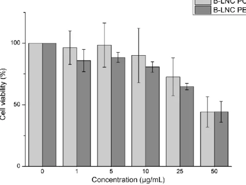
176
wt% and pH 5.
177
178
2.5. Atomic force microscopy (AFM)
179
Atomic force microscopy was performed on Nanoman (Bruker Instrument) and monitored by Nanoscope V
180
software. The LNC samples were diluted 100 times in MilliQ water and 5 µL of the solution was deposited on a
181
silicon wafer. The tapping mode and harmonix mode were used after drying of the drop at 37°C. For the tapping
182
mode, PPP NCL tips (Nanosensors) were used at the resonance frequency of 157 kHz. Various amplitude
183
setpoints were tested (from 500 to 250 mV) to modify the resulting force applied onto the sample. The harmonix
184
mode was carried out using HMX-S tips (Bruker Instruments) by applying a force of 10 to 20 nN with a vertical
185
resonance frequency of 48 kHz and horizontal of 791 kHz. No morphology modification of the LNC was
186
observed.
187
188
2.6. Encapsulation efficacy (EE) and drug loading capacity (DL)
189
Drug loading (DL) and encapsulation efficacy (EE) were respectively calculated using the following equations:
190
(Eq.1)
191
Where total mass represents the mass of POx, Labrafac and quercetin.
192
(Eq.2)
193
The quercetin loaded in Q-LNC POx was quantified by HPLC (section 2.7) after filtration of the solution
194
through a 0.2 µm filter (Whatman) to remove the residual unloaded quercetin.
195
196
2.7. HPLC analysis
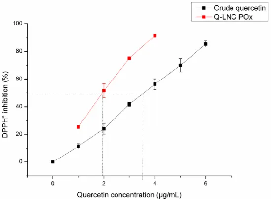
197
High Pressure Liquid Chromatography (HPLC) analysis of quercetin was performed on LC6-2012HT apparatus
198
(Shimadzu, Kyoto, Japan) using a Prontosil C18 column (120-5-C18 H5.0 µm, 250x4.0 mm). The detection was
199
achieved using a UV-vis detector (Shimadzu, Kyoto, Japan) at 368 nm (Yang et al., 2009).
8
Acetonitrile/phosphoric acid at 0.2 wt% and pH=1.9 (40/60 %v) was used as mobile phase. The calibration curve
201
was performed with solutions of quercetin in methanol from 1 to 100 µg/mL with a good linearity (r² = 0.9999).
202
The flow rate was 1 mL.min-1. To determine the concentration of quercetin released to the aqueous medium
203
receptor (see stability study of LNC, section 2.3) a calibration curve realized in PBS buffer at pH 7.4 and
204
Tween® 80 (2% wt) from 0.1 to 4 µg/mL (r² = 0.9996) was used.
205
206
2.8. In vitro antioxidant activity
207
The antioxidant activity of Q-LNC POx was determined from the reactivity of quercetin towards the free radical
208
2,2-diphenyl-1-picrylhydrazyl (DPPH°) (Blois, 1958). It was calculated from the decrease of the DPPH° radical
209
absorbance at 517 nm using the following equation (Eq.3):
210
211
(Eq.3)212
213
where Absorbance of control corresponds to the absorbance of a 100 µM DPPH° solution. Note that the
214
concentration of quercetin loaded in Q-LNC POx (2.5, 5, 7.5, 10, 12.5, 15 µM) was lower than that of DPPH°.
215
216
2.9. Cell culture
217
Mice fibroblasts cell lines (NIH3T3) were purchased from American Type Cell Culture organization (USA).
218
Cells were maintained in Dulbecco’s Modified Eagle Medium (DMEM) (Gibco) supplemented with 10% fetal
219
bovine serum (Life Technologies), 1% L-glutamine and 1% penicillin/streptomycin equivalent to a final
220
concentration of 2 nM for glutamine, 100 U.mL-1 for penicillin and 100 µg.mL-1 for streptomycin. They were
221
incubated at 37°C in humidified 5% CO2 atmosphere.
222
Normal Human Epidermal Keratinocytes (NHEK) were purchased from Promocell (C-123003) and cultured at
223
37°C in keratinocyte basal medium (C-20216 Promocell) supplemented with the supplement mix keratinocyte
224
from Promocell (C-39016) under a humidified atmosphere containing 5% CO2. Cells were seeded to reach
225
approximately 80% confluency at the time of treatment or irradiation. UVB irradiation was performed using a
226
BS-02 UV irradiation chamber (Dr. Gröbel UV-Elektronik GmbH, Ettlingen, Germany). The lamp emits UVB
227
irradiation with a peak at 311–312 nm and partially excludes shorter wavelengths, such as UVA.
228
2.10. Cell viability
229
NIH3T3 cells were seeded at 20,000 cells/cm² in 96 well plate and incubated for 24 hours at 37°C, 5% CO2 to
230
allow cell adhesion. Cells were treated or not (control) with quercetin, POx (B-LNC POx, Q-LNC POx) and
231
PEG (B-LNC PEG, Q-LNC PEG) preparation. After 24 hours of exposure, a CellTiter 96® Aqueous
Non-232
Radioactive Cell Proliferation Assay (Promega, USA) was used to evaluate the cell viability following the
233
manufacturer’s instructions. The absorbance was recorded at 490 nm using MultiskanTM GO Microplate
234
Spectrophotometer (Thermo ScientificTM, Waltham, Massachusetts, USA). Absorbance of the basal media (with
235
no cells and no treatment) was subtracted to the recorded absorbance for all conditions. Values of treated cells
236
were normalized to non-treated cells (100% intensity).
237
238
239
9
2.11. Antioxidant effect of quercetin on TBHP treated NIH3T3 cells240
NIH3T3 cells were seeded at 20,000 cells/cm² in a 96 well black plate with clear bottom (Corning®
241
Massachusetts, USA) and incubated 24 hours at 37°C, 5% CO2 to allow cell adhesion. Then B-LNC (POx and
242
PEG), crude quercetin, or Q-LNC (POx and PEG) possessing an equivalent quercetin concentration of 5 µg/mL
243
were added and incubated for another 24 hours in supplemented DMEM. Cells were washed twice with PBS
244
before addition of 200 µL of 2,7-dichlorofluorescein diacetate (DCFDA) reagent (20 µM in DMEM without
245
phenol red). The serum free medium used provides reliable data by avoiding deacetylation of the DCFDA into
246
non fluorescent compound that could later be oxidized by reactive oxygen species (ROS) into
247
dichlorofluorescein (DCF). After 30 minutes of incubation at 37°C, 5% CO2, cells were rinsed with PBS and
248
placed in 200 µL DMEM without phenol red. A solution of tert-butyl hydroperoxide (TBHP) (500 µM in PBS)
249
was then added and the cells were incubated for additional 30 minutes. The fluorescence signal of DCF produced
250
by the reaction of DCFDA reagent with ROS was then measured (λexc 485 nm, λem 535 nm) using a Tristar
251
LB941 Spectrofluorimeter (Berthold Technologies, Germany). Values of treated cells were normalized to
non-252
treated cells (100% intensity).
253
254
2.12. Antioxidant effect of quercetin on UVB irradiated NHEK cells
255
NHEK cells were seeded at 30,000cells per well in a 24-well plate. They were allowed to settle for 24 hours
256
before treatment for 2 more hours with 5 μg/ml of the crude quercetin, Q-LNC POx or Q-LNC PEG
257
preparations. Cells were then incubated with 20 µM DCFDA in a serum free medium for 30 minutes before a
258
100 mJ/cm2 UVB irradiation was triggered. The cells were harvested and cellular fluorescence was assessed by
259
flow cytometry (Canto, Becton Dickinson) directly after irradiation (Masaki et al., 2009). Data were processed
260
using FlowJo software. Values of treated cells were normalized to non-treated cells (representing the cells with
261
100% ROS intensity).262
263
2.13. Statistical analysis264
The statistical analysis of the data resulting from the cell viability and the antioxidant effect on cells was
265
conducted with Origin Pro software 8.1 (OriginLab, USA). A one-sample t-test with equal variance was
266
performed to compare cell viability and antioxidant effect with formulations to viability of untreated cells
267
(100%). A two-sample t-test with unequal variance was carried out to compare cell viability of formulations two
268
by two. The P value reflects the significance with * =P <0.05, ** =P <0.01 and *** =P < 0.001.
269
270
271
10
3.
Results and discussion
272
3.1. LNC formulation and characterization
273
The amphiphilic nonionic POx polymer (C16POx15) used in this study is highly soluble in water and has a
274
molecular weight of 1520 g/mol. It possesses a low viscosity (e.g. PEtOx 50 kDa [η] = 0.23 dL/g) and a high
275
stability towards degradation (Lorson et al., 2018). Its solubility in water remains constant with temperature (no
276
LCST characteristic for this poly(-2-methyl-2-oxazoline) POx derivative) (Glassner et al., 2018)), impeding the
277
use of the phase inversion process developed by Heurtault et al (Heurtault et al., 2002) to produce LNC. As a
278
consequence, the protocol to form POx based LNC was redefined as represented in Scheme 1. Heating and
279
cooling cycles were first performed to solubilize and homogenize the system by melting and mixing the solid
280
lipids (Lipoid® S75) with the liquid lipids (Labrafac®). Then, a short sonication step supplied the energy
281
necessary to disperse the preparation (Cohen et al., 2013) and reduce its size from 300 nm to 30 nm. Quercetin
282
was added to the mixture to produce antioxidant LNC after this stage, thus avoiding its degradation from
283
ultrasonic energy (Qiao et al., 2014). One of the advantages of this process is thus to preserve active compound
284
(AC) sensitive to many temperature cycles and ultrasound treatments by post-insertion into the LNC. One last
285
heating and cooling cycle was performed to favor quercetin loading. Then, the mixture was heated at 85°C and
286
cold water (4°C) was rapidly introduced to anchor the system and generate LNC stabilized by polyoxazoline
287
(LNC POx).
288
Using this process, a pre-formulation work was conducted to determine the suitable composition leading to the
289
most stable nanosized LNC, with the lowest amount of POx and resulting in the highest drug loading. The POx
290
concentration range explored varied from 10 to 20 mass percent, the Labrafac® oily phase from 10 to 25 mass
291
percent and water from 55 to 80 mass percent, whereas the phospholipids Lipoid® S75 and NaCl were
292
respectively fixed at 1.5 mass percent and 3 mass percent. The stability was evaluated from DLS measurement
293
(data not shown).
294
295
296
Scheme 1 : Process of lipid nanocapsules formulation
297
298
As a result, POx was introduced at 20 mass percent, Labrafac® oil at 15 mass percent and water 65 mass percent.
299
Other components such as NaCl and Lipoid® S75 were added at 3 mass percent and 1.5 mass percent. The
300
physicochemical and morphological properties of blank LNC (B-LNC POx) and LNC loaded with quercetin
(Q-301
LNC POx) were fully characterized; their main properties are gathered in Table 2. The hydrodynamic diameter
302
of B-LNC was measured by DLS at 30.5 ± 1.5 nm with a polydispersity index (PDI) of 0.16. Loading with
303
quercetin (Q-LNC POx) resulted in almost similar hydrodynamic diameter (26.4 ± 0.6 nm with a PDI of 0.18 ±
304
0.01, Table 2). The zeta potential for both LNC was close to neutral, indicating that POx chains at the surface of
305
the LNC stabilize the formulation by steric repulsions. LNC PEG, free and loaded with quercetin were prepared
306
using Hatahet et al. protocol (Hatahet et al., 2017) for comparative purposes. The hydrodynamic diameter of
B-307
LNC PEG and Q-LNC PEG were measured at 34.7 nm and 33.5 nm with a PDI of 0.03 and 0.05, respectively.
308
As for LNC POx, the presence of quercetin did not change the LNC size.
309
310
Table 2 : Characteristics of LNC POx
11
312
The morphology of LNC was observed by TEM after coloration with uranyl acetate solution. Well-defined
313
spherical particles of similar size (< 70 nm) were observed for both B-LNC POx and Q-LNC POx. A typical
314
image obtained with Q-LNC POx is presented on Figure S1. The spherical shape was confirmed by AFM
315
(Figure 1). It also allowed determination of the nanoparticle stiffness using the tapping mode to evaluate the
316
contact force and more especially the repulsive force of the nanoparticle by decreasing the amplitude (setpoint).
317
The topography of the LNC POx showed a positive phase indicating a repulsive force and mechanical properties.
318
The latter were quantified using the harmoniX mode for determination of the indentation modulus (DMT). The
319
DMT profile of Q-LNC POx was measured at 1-2 GPa (Figure S2) and the associated topography profiles
320
resulted in no permanent morphology deformation (Figures S3 and S4) up to a constraint of about 10-20 nN.
321
This evidences the mechanical properties of the LNC POx composed of a rigid capsule embedding on oily core
322
just like the ones designed by Heurtault et al. (Heurtault et al., 2002) for which a contact force > 10nN was
323
applied.
324
325
326
Figure 1 : LNC POx topography from AFM measurement
327
It is reasonable to assume that the stiffness of the POx-based nanoparticles results from the rigid capsule made of
328
phospholipids from the Lipoid® S75 and POx surrounding the oily core of Labrafac® as for the LNC produced
329
with polyethylene glycol (15)-hydroxystearate as a surfactant (Heurtault et al., 2002). For LNC POx, the
330
amphiphilic non ionic POx stabilized the hydrophobic part of the formulation into spherical nano-objects
331
covered with the POx chains as previously described for similar amphiphilic POx (Korchia et al., 2015). An
332
illustrative representation of the assembly is depicted in Scheme 2.
333
334
Scheme 2 : Inner assembly of LNC POx
335
336
LNC formulations are well known for their high stability to dilution, centrifugation and over the long term
337
(Huynh et al., 2009). These properties were also investigated for the newly developed POx based LNC. The
338
LNC POx stability to dilution by 1000 (data not shown) and to high speed centrifugation at 15 000 rpm for 15
339
minutes at 25°C was evaluated. The size and the PDI remained the same as reported in Table 2. Interestingly,
340
after 5 days at -22°C the LNC POx hydrodynamic diameter did not change. The long term stability of B-LNC
341
POx was assessed at 4, 25 and 37°C by measuring the hydrodynamic diameter and PDI over a period of one
342
month (Figure 2). At each temperature, the size of the particles slightly increased in the first 7 days (from 30 to
343
38 nm at 4°C, 42 nm at 25°C and 49 nm at 37°C), after 7 days the PDI remained almost identical at a low value
344
of ~ 0.1, indicating stable monodisperse preparations. In any case, after a month, the size of the B-LNC POx
345
remained well below 100 nm, which is fully compatible for topical delivery applications. It is noteworthy to
346
mention that after 2 months at 25°C, the size of the B-LNC POx had only marginally increased; it reached 57 nm
347
whereas it was 52 nm one month before (PDI < 0.1).
348
349
12
Figure 2 : Hydrodynamic diameter (column) and PDI (symbol) of B-LNC POx at 4, 25 and 37 °C over time
350
(n=3)
351
A cell viability test was first conducted on mice fibroblasts (NIH3T3) to ensure that the platform was non toxic
352
by itself. B-LNC POx and B-LNC PEG were both tested from 1 to 50 µg/mL to evaluate the IC50(cell viability)
353
(Figure S5). The IC50(cell viability) value was 43± 8 µg/mL for B-LNC PEG and 45± 8 µg/mL for B-LNC POx and
354
the statistical analysis did not reveal any difference in term of cell viability. Thus, the cell viability in presence of
355
B-LNC POx seems to be well-suited for topical delivery of AC as it is similar to that of B-LNC PEG.
356
357
3.2. Quercetin loaded LNC and scavenging capacity
358
The LNC POx was then loaded with an AC model to evaluate the encapsulation capacity and protection of
359
bioactivity. The natural antioxidant quercetin was chosen for its hydrophobic nature, poor solubility and
360
sensitivity to oxidation. The post-insertion method successfully led to a high encapsulation efficiency (EE) of 93
361
± 1% corresponding to a drug loading (DL) of 7.9 ± 0.1%. Addition of ethanol (from 100 µL to 500µL) had no
362
impact on DL and rather tended to destabilize the LNC by creating two phases, contrary to Q-LNC PEG
363
synthesized by Hatahet et al. adapted from Heurtault protocol (Hatahet et al., 2017). Compared to others LNC
364
(Barras et al., 2009) and NLC (Chen-yu et al., 2012; Pivetta et al., 2019) developed to encapsulate quercetin, it
365
has to be mentioned that Q-LNC POx exhibits the highest drug loading, which exceed that obtained with Q-LNC
366
PEG (Hatahet et al., 2017) using Cremophor and ethanol to reach a drug loading of 2.6 ± 0.1% with an
367
encapsulation efficiency of 96.4 ± 1.2%. Moreover, Q-LNC POx preparation also improved quercetin loading
368
compared to the POx stabilized mixed-micelles we recently developed (Simon et al., 2019) (from 3.6 to 7.9 %).
369
The stability of Q-LNC POx with time was investigated at 4 °C, a temperature relevant in the presence of
370
quercetin that is sensitive to thermal degradation (Wang and Zhao, 2016). As for the blank LNC POx, the size of
371
quercetin loaded nanocapsules slightly evolved over one month, from 26 nm to 40 nm with a PDI staying under
372
0.2 (Figure 3). As for B-LNC POx, Q-LNC POx was also stable to dilution and centrifugation (Table 2).
373
374
Figure 3 : Hydrodynamic diameter (column) and PDI (symbol) of Q-LNC POx at 4 °C with time (n=3)
375
376
The stability study of Q-LNC POx was completed by evaluating the quercetin leakage from the nanocapsules in
377
aqueous media (Figure S6). After 7 hours, only 1.5% of quercetin was released from Q-LNC POx and it reached
378
a low value of 5% after 48 hours. This result confirmed that quercetin is localized within the core of the LNC.
379
Interestingly, the LNC POx formulation reduced by four times the quercetin leakage in aqueous media compared
380
to the mixed-micelles formulation (Simon et al., 2019). The Q-LNC POx also seemed to better retain quercetin
381
than Q-LNC PEG that showed a leakage of quercetin of almost 15% after 24 hours using the same experimental
382
conditions (Hatahet et al., 2017). This result might be due to the rigid capsule surrounding the oily core acting as
383
a sealed reservoir and ensuring a high stability for the particles.
384
385
Antioxidant tests were performed in order to evaluate the ability of the LNC POx platform to protect and
386
maintain the bioactivity of the encapsulated compound. The radical scavenging capacity was assessed in vitro
13
using a chemical assay (DPPH). It was also evaluated on mice fibroblasts and human keratinocytes by generating
388
reactive oxygen species (ROS) respectively using organic peroxide and UVB irradiation.
389
The quercetin antioxidant is able to react with the 2,2-diphenyl-1-picrylhydrazyl (DPPH°) molecule by its free
390
radical on the hydrazine position leading to DPPHH. The radical scavenging capacity of quercetin encapsulated
391
in the Q-LNC POx was assessed by looking at its inhibitive interaction with DPPH°. The IC50 (DPPH° inhibition) value
392
deduced from the DPPH° decrease shows higher antioxidant activity for Q-LNC POx (IC50 (DPPH° inhibition) = 2
393
µg/mL) than for crude quercetin, which exhibited a twice higher IC50 (Figure S7). The higher antioxidant activity
394
of Q-LNC POx can be due to a better protection of the antioxidant to oxidation, a greater conformation or
395
organization in the nanocapsule compared to crude quercetin.
396
397
3.3. Quercetin loaded antioxidant activity
398
To go further in the evaluation of the antioxidant effect of Q-LNC POx with respect to classical PEG based
399
LNC, both POx and PEG based Q-LNC were loaded at the same quercetin concentration and tested on mice
400
fibroblasts and human keratinocytes. A cell viability test was performed on NIH3T3 to determine the maximum
401
non toxic quercetin concentration to be used for the antioxidant tests. The IC50(cell viability) values were determined
402
for crude quercetin (17.5 µg/mL), Q-LNC POx (23 µg/mL) and Q-LNC PEG (16 µg/mL) (Figure S8). The cell
403
viability of blank LNCs was also evaluated B-LNC POx (43 µg/mL) and B-LNC PEG (45 µg/mL).
404
As for the uncharged POx and PEG-based LNC, the IC50(cell viability) value is not affected by the nature of the
405
stabilizing polymer.
406
407
To perform the antioxidant study, the concentration of quercetin was selected such as to avoid formulation
408
toxicity from the cell viability results; it was set at 5 µg/mL. The radical scavenging capacity of Q-LNC POx
409
was first tested on NIH3T3 and compared to Q-LNC PEG and crude quercetin (Figure 4). Cells were treated
410
with the formulations for 24 hours, then an oxidative stress was induced by tert-butyl hydroperoxide (TBHP).
411
The quantity of ROS generated was determined by fluorescence spectroscopy using the DCFDA probe. The
412
results were normalized to untreated cells with TBHP exposure (non treated). The crude quercetin was able to
413
reduce excess ROS generation to 69 ± 6% (P < 0.01) and similarly Q-LNC POx decreased surplus ROS to 70 ±
414
6% (P < 0.01). Q-LNC PEG was less able to counter ROS generation with a reduction to 81 ± 9% (P < 0.01).
415
The antioxidant activity of quercetin once loaded in LNC POx was thus maintained and even slightly enhanced
416
compared to Q-LNC PEG. B-LNC POx was also tested and the cells resisted to the peroxides generated
417
reflecting the innocuousness of POx upon oxidation.
418
419
420
Figure 4 : ROS relative intensity for crude quercetin, Q-LNC POx and Q-LNC PEG at a quercetin concentration
421
of 5 µg/mL (n=3) on NIH3T3 cells. Fluorescence intensity was normalized to TBHP treated cells without
422
formulation. A one sample t-test relative to untreated cells was performed (*: P < 0.05, **: P < 0.01 and ***: P <
423
0.001)
424
425
14
Another antioxidant test was performed on human keratinocytes with UV irradiation as a source of ROS.
426
Therefore, the antioxidant effect observed on mice fibroblasts can be transposed to human cells with ROS
427
simulated from an environmental factor.
428
We first evaluated the scavenging activity of ROS naturally generated by the cells (Table 3, “No UV
429
irradiation”). The fluorescence intensity was normalized to untreated cells (no formulation and no UV
430
irradiation). UV irradiation was then carried out to evaluate the antioxidant capacity on overproduction of ROS
431
(Table 3, “UV irradiation” column). The results were normalized to untreated cells. As shown in Table 3, the
432
crude quercetin significantly reduced the ROS naturally produced by the cells to 59 ± 19% (P < 0.01) which is
433
not the case for Q-LNC POx and Q-LNC PEG maintaining a quantity of ROS of 91 ± 16% and 94 ± 13%
434
respectively. On the contrary, once the cells irradiated, the crude quercetin was the one reducing most of the
435
ROS over generated to 65 ± 6% (P < 0.01) whereas Q-LNC POx was able to reduce ROS at 83 ± 4% (P < 0.01)
436
and Q-LNC PEG at 91 ± 15%. The statistical analysis revealed that crude quercetin and Q-LNC POx
437
significantly decreased the ROS over generated. Therefore, Q-LNC POx was able to maintain the antioxidant
438
activity of quercetin alike Q-LNC PEG. It has to be noticed that, even if the crude quercetin reduced most of the
439
ROS, its too powerful activity could scavenge the ROS essential for cell survival.
440
441
Table 3 : ROS relative intensity for crude quercetin, Q-LNC POx and Q-LNC PEG at a quercetin concentration
442
of 5 µg/mL (n=3) on human keratinocytes. Fluorescence intensity was normalized to irradiated cells (not treated
443
with formulation). A one sample t-test relative to untreated cells was performed (*: P < 0.05, **: P < 0.01 and
444
***: P < 0.001).445
446
447
448
4.Conclusion
449
This work demonstrates the ability of amphiphilic non ionic polyoxazolines (POx) to act as a surfactant to form
450
lipid based nanoformulation suitable for topical delivery of quercetin. The optimized nanoformulation allows to
451
shape well-defined monodisperse spherical lipid nanocapsules (LNC) of 30 nm diameter size. They are
452
composed of a Labrafac® oily core stabilized by a solid phospholipid and POx surfactant shell, whose stiffness
453
ensured a high stability over time, centrifugation, freezing and dilution. Most importantly, LNC POx are free
454
from the PEG-dependant inversion process universally used to date to design LNC. The process developed here
455
associates a sonication step to heating/cooling cycles allowing loading the natural antioxidant quercetin at a high
456
drug loading of 7.9 ± 0.1% without inducing any morphological change in the particles nor altering their
457
stability. Interestingly, the LNC POx leads to a higher encapsulation and a slightly better radical scavenging
458
capacity on mice fibroblasts and human keratinocytes than LNC PEG. This evidences the crucial role
459
amphiphilic polyoxazolines might take in the future as PEG free topical delivery platform for topical and
460
intravenous administration for applications in nanomedecine, dermatology and cosmetics.
15
Acknowledgements462
The authors thank Michel Ramonda (CTM, Univ Montpellier, France) for help on AFM measurements,
463
Christophe Dorandeu (ICGM, Univ Montpellier, France) for assistance on HPLC analysis and Thomas
464
Cacciaguerra (ICGM, Univ Montpellier, France) for assistance on TEM experiments. We acknowledge the
465
imaging facility MRI, member of the national infrastructure France-BioImaging infrastructure supported by the
466
French National Research Agency (ANR-10-INBS-04, «Investments for the future»)" for Cytometry Analysis.
16
References
468
Abdel-Mottaleb, M.M.A., Neumann, D., Lamprecht, A., 2011. Lipid nanocapsules for dermal
469
application: A comparative study of lipid-based versus polymer-based nanocarriers. European Journal
470
of Pharmaceutics and Biopharmaceutics 79, 36-42.
471
Anton, N., Gayet, P., Benoit, J.-P., Saulnier, P., 2007. Nano-emulsions and nanocapsules by the PIT
472
method: An investigation on the role of the temperature cycling on the emulsion phase inversion.
473
International Journal of Pharmaceutics 344, 44-52.
474
Barras, A., Mezzetti, A., Richard, A., Lazzaroni, S., Roux, S., Melnyk, P., Betbeder, D.,
Monfilliette-475
Dupont, N., 2009. Formulation and characterization of polyphenol-loaded lipid nanocapsules.
476
International Journal of Pharmaceutics 379, 270-277.
477
Blois, M.S., 1958. Antioxidant Determinations by the Use of a Stable Free Radical. Nature 181,
1199-478
1200.
479
Chen-yu, G., Chun-fen, Y., Qi-lu, L., Qi, T., Yan-wei, X., Wei-na, L., Guang-xi, Z., 2012. Development of
480
a Quercetin-loaded nanostructured lipid carrier formulation for topical delivery. International Journal
481
of Pharmaceutics 430, 292-298.
482
Cohen, J., Deloid, G., Pyrgiotakis, G., Demokritou, P., 2013. Interactions of engineered nanomaterials
483
in physiological media and implications for in vitro dosimetry. Nanotoxicology 7, 417-431.
484
Dargaville, T.R., Park, J.-R., Hoogenboom, R., 2018. Poly(2-oxazoline) Hydrogels: State-of-the-Art and
485
Emerging Applications. Macromolecular Bioscience 18, 1800070.
486
Dragicevic, N., Atkinson, J.P., Maibach, H.I., 2015. Chemical Penetration Enhancers: Classification and
487
Mode of Action, in: Dragicevic, N., Maibach, H.I. (Eds.), Percutaneous Penetration Enhancers
488
Chemical Methods in Penetration Enhancement: Modification of the Stratum Corneum. Springer
489
Berlin Heidelberg, Berlin, Heidelberg, pp. 11-27.
490
Elias, P.M., 1983. Epidermal Lipids, Barrier Function, and Desquamation. Journal of Investigative
491
Dermatology 80, S44-S49.
492
Garcês, A., Amaral, M.H., Sousa Lobo, J.M., Silva, A.C., 2018. Formulations based on solid lipid
493
nanoparticles (SLN) and nanostructured lipid carriers (NLC) for cutaneous use: A review. European
494
Journal of Pharmaceutical Sciences 112, 159-167.
495
Glassner, M., Vergaelen, M., Hoogenboom, R., 2018. Poly(2-oxazoline)s: A comprehensive overview
496
of polymer structures and their physical properties. Polymer International 67, 32-45.
497
Guillerm, B., Monge, S., Lapinte, V., Robin, J.-J., 2012. How to Modulate the Chemical Structure of
498
Polyoxazolines by Appropriate Functionalization. Macromolecular Rapid Communications 33,
1600-499
1612.
500
Hatahet, T., Morille, M., Hommoss, A., Devoisselle, J.M., Müller, R.H., Bégu, S., 2016. Quercetin
501
topical application, from conventional dosage forms to nanodosage forms. European Journal of
502
Pharmaceutics and Biopharmaceutics 108, 41-53.
503
Hatahet, T., Morille, M., Hommoss, A., Devoisselle, J.M., Müller, R.H., Bégu, S., 2018. Liposomes, lipid
504
nanocapsules and smartCrystals®: A comparative study for an effective quercetin delivery to the skin.
505
International Journal of Pharmaceutics 542, 176-185.
506
Hatahet, T., Morille, M., Shamseddin, A., Aubert-Pouëssel, A., Devoisselle, J.M., Bégu, S., 2017.
507
Dermal quercetin lipid nanocapsules: Influence of the formulation on antioxidant activity and cellular
508
protection against hydrogen peroxide. International Journal of Pharmaceutics 518, 167-176.
509
Heurtault, B., Saulnier, P., Pech, B., Proust, J., Benoit, J.-P., 2002. A Novel Phase Inversion-Based
510
Process for the Preparation of Lipid Nanocarriers.
511
Huynh, N.T., Passirani, C., Saulnier, P., Benoit, J.P., 2009. Lipid nanocapsules: A new platform for
512
nanomedicine. International Journal of Pharmaceutics 379, 201-209.
513
Krutmann, J., Bouloc, A., Sore, G., Bernard, B.A., Passeron, T., 2017. The skin aging exposome. Journal
514
of Dermatological Science 85, 152-161.
515
Lorson, T., Lübtow, M.M., Wegener, E., Haider, M.S., Borova, S., Nahm, D., Jordan, R.,
Sokolski-516
Papkov, M., Kabanov, A.V., Luxenhofer, R., 2018. Poly(2-oxazoline)s based biomaterials: A
517
comprehensive and critical update. Biomaterials 178, 204-280.
518
17
Lubich, C., Allacher, P., de la Rosa, M., Bauer, A., Prenninger, T., Horling, F.M., Siekmann, J.,
519
Oldenburg, J., Scheiflinger, F., Reipert, B.M., 2016. The Mystery of Antibodies Against Polyethylene
520
Glycol (PEG) - What do we Know? Pharmaceutical Research 33, 2239-2249.
521
Masaki, H., Izutsu, Y., Yahagi, S., Okano, Y., 2009. Reactive Oxygen Species in HaCaT Keratinocytes
522
After UVB Irradiation Are Triggered by Intracellular Ca2+ Levels. Journal of Investigative Dermatology
523
Symposium Proceedings 14, 50-52.
524
Moreadith, R.W., Viegas, T.X., Bentley, M.D., Harris, J.M., Fang, Z., Yoon, K., Dizman, B., Weimer, R.,
525
Rae, B.P., Li, X., Rader, C., Standaert, D., Olanow, W., 2017. Clinical development of a
poly(2-526
oxazoline) (POZ) polymer therapeutic for the treatment of Parkinson’s disease – Proof of concept of
527
POZ as a versatile polymer platform for drug development in multiple therapeutic indications.
528
European Polymer Journal 88, 524-552.
529
Nagula, R.L., Wairkar, S., 2019. Recent advances in topical delivery of flavonoids: A review. Journal of
530
Controlled Release 296, 190-201.
531
Pivetta, T.P., Silva, L.B., Kawakami, C.M., Araújo, M.M., Del Lama, M.P.F.M., Naal, R.M.Z.G.,
Maria-532
Engler, S.S., Gaspar, L.R., Marcato, P.D., 2019. Topical formulation of quercetin encapsulated in
533
natural lipid nanocarriers: Evaluation of biological properties and phototoxic effect. Journal of Drug
534
Delivery Science and Technology 53, 101148.
535
Qiao, L., Sun, Y., Chen, R., Fu, Y., Zhang, W., Li, X., Chen, J., Shen, Y., Ye, X., 2014. Sonochemical
536
effects on 14 flavonoids common in citrus: relation to stability. PLoS One 9, e87766-e87766.
537
Roberts, M.S., Mohammed, Y., Pastore, M.N., Namjoshi, S., Yousef, S., Alinaghi, A., Haridass, I.N.,
538
Abd, E., Leite-Silva, V.R., Benson, H.A.E., Grice, J.E., 2017. Topical and cutaneous delivery using
539
nanosystems. Journal of Controlled Release 247, 86-105.
540
Rudmann, D.G., Alston, J.T., Hanson, J.C., Heidel, S., 2013. High Molecular Weight Polyethylene Glycol
541
Cellular Distribution and PEG-associated Cytoplasmic Vacuolation Is Molecular Weight Dependent
542
and Does Not Require Conjugation to Proteins. Toxicologic Pathology 41, 970-983.
543
Sala, M., Diab, R., Elaissari, A., Fessi, H., 2018. Lipid nanocarriers as skin drug delivery systems:
544
Properties, mechanisms of skin interactions and medical applications. International Journal of
545
Pharmaceutics 535, 1-17.
546
Schäfer-Korting, M., Mehnert, W., Korting, H.-C., 2007. Lipid nanoparticles for improved topical
547
application of drugs for skin diseases. Advanced Drug Delivery Reviews 59, 427-443.
548
Simon, L., Vincent, M., Le Saux, S., Lapinte, V., Marcotte, N., Morille, M., Dorandeu, C., Devoisselle,
549
J.M., Bégu, S., 2019. Polyoxazolines based mixed micelles as PEG free formulations for an effective
550
quercetin antioxidant topical delivery. International Journal of Pharmaceutics, 118516.
551
Viegas, T.X., Fang, Z., Yoon, K., Weimer, R., Dizman, B., 2018. 6 - Poly(oxazolines), in: Parambath, A.
552
(Ed.), Engineering of Biomaterials for Drug Delivery Systems. Woodhead Publishing, pp. 173-198.
553
Wang, J., Zhao, X.H., 2016. Degradation kinetics of fisetin and quercetin in solutions affected by
554
medium pH, temperature and co-existing proteins. Journal of the Serbian Chemical Society 81,
243-555
254.
556
Yang, L., Li, P., Gao, Y.-J., Li, H.-F., Wu, D.-C., Li, R.-X., 2009. Time Resolved UV-Vis Absorption Spectra
557
of Quercetin Reacting with Various Concentrations of Sodium Hydroxide.
558
Zalipsky, S., Hansen, C.B., Oaks, J.M., Allen, T.M., 1996. Evaluation of Blood Clearance Rates and
559
Biodistribution of Poly(2-oxazoline)-Grafted Liposomes§. Journal of Pharmaceutical Sciences 85,
133-560
137.
561
Zhai, Y., Yang, X., Zhao, L., Wang, Z., Zhai, G., 2014. Lipid nanocapsules for transdermal delivery of
562
ropivacaine: in vitro and in vivo evaluation. International Journal of Pharmaceutics 471, 103-111.
563
Zhang, P., Sun, F., Liu, S., Jiang, S., 2016. Anti-PEG antibodies in the clinic: Current issues and beyond
564
PEGylation. Journal of Controlled Release 244, 184-193.
565
Declaration of interests
x
The authors declare that they have no known competing financial interests or personal relationships
that could have appeared to influence the work reported in this paper.
☐The authors declare the following
financial interests/personal relationships
which may be considered
as potential competing interests:
Credit Author Statement:
-Laurianne Simon: Conceptualization, formal analysis, methodology, validation, investigation, writing original draft, writing review & editing, visualization
-Vincent Lapinte: Conceptualization, methodology, writing review & editing, visualization, supervision, project administration
-Loic Lionnard: methodology, investigation, writing original draft, writing review & editing
-Nathalie Marcotte: Conceptualization, methodology, writing review & editing, visualization, supervision, project administration
-Marie Morille: methodology, writing review & editing, resources, supervision -Abdel Aouacheria: methodology, resources, writing review & editing
-Karima Kissa: resources
-Jean-Marie Devoisselle: resources, project administration
-Sylvie Bégu: Conceptualization, methodology, writing review & editing, visualization, supervision, project administration
Highlights
- Innovative PEG free lipid nanocapsules made of an oily core surrounded by a phospholipid and
polyoxazoline shell.
- Stable nanoformulations loaded with quercetin, a natural antioxidant.
- Lipid nanocapsules stabilized by polyoxazoline successfully led to higher drug loading than the
original one with PEG.
- Powerful antioxidant activity of polyoxazoline based lipid nanocapsules on human keratinocytes
after UV exposure.
Type of lipid formulation Solid lipid nanoparticles (SLN) Nanostructured lipid carriers (NLC) Liposomes Lipid nanocaspules (LNC) Phospholipid Composition
Solid lipid core with polymer shell (Schäfer-Korting et al., 2007)
Mixture of solid and liquid lipid core and surfactant layer (Schäfer-Korting et al., 2007)
Lipid bilayer enclosing an aqueous core (Zhai and Zhai, 2014)
Liquid lipid core surrounded by a solid membrane from lecithin and surfactant (Huynh et al., 2009) Size 40- 1000 nm (Sala et al., 2018) 40- 1000 nm (Sala et al., 2018) 20-3000 nm (Sala et al., 2018) 20-100 nm (Huynh et al., 2009) Preparation method Hot or cold HPH (Schäfer-Korting et al., 2007) or microemulsion technique (Montenegro et al., 2016) Hot or cold HPH (Schäfer-Korting et al., 2007) or microemulsion technique (Montenegro et al., 2016) -Bangham method -Detergent depletion method -Injection method…(Mahera ni et al., 2011) PIT (Heurtault et al., 2002) Advantages Biocompatibility, drug protection against degradation (Sala et al., 2018); low toxicity and feasible scaling up (Zhai and Zhai, 2014)
Higher stability and DL than SLN (Montenegro et al., 2016) Flexibility and deformability; high DL of hydrophilic AC (Sala et al., 2018) Better encapsulation and greater stability (Huynh et al., 2009) than SLN and NLC; excellent tolerability for dermal application (Zhai and Zhai, 2014)
Disavantages
High degree of order leading to low DL (Zhai and Zhai, 2014)
Lower skin permeation than LNC (Zhai and Zhai, 2014) Low capacity to encapsulate lipophilic drug; presence of traces of organic solvent; unstable in biological fluid (Huynh et al., 2009) Low DL of hydrophilic AC (Huynh et al., 2009) Table 1
B-LNC POx Q-LNC POx
Hydrodynamic diameter (nm) 30.5±1.5 26.4±0.6
PDI 0.16±0.01 0.18±0.01
Hydrodynamic diameter after centrifugation (nm) 30.0±3 27.0±2
PDI after centrifugation 0.20±0.03 0.19±0.01
Hydrodynamic diameter after freezing (nm) 33.6 25.8
PDI after freezing 0.16 0.17
Zeta potentiel (mV) 5.1±18.4 7.9±12.5
Drug loading (%) 7.9 ± 0.1
Encapsulation efficiency (%) 93 ± 1
Conditions
No UV irradiation UV irradiation
ROS relative intensity (%) ROS relative intensity (%)
Untreated 100 100
Crude quercetin 59 ± 19 ** 65 ± 6 **
Q-LNC POx 91 ± 16 83 ± 4 **
Q-LNC PEG 94 ± 13 91 ± 15
Supplementary Material
Supplementary Figures
Figure S1: TEM image of Q-LNC POx
Figure S2: AFM modulus for Q-LNC POx
AFM microscopy was performed on the Q-LNC POx with topography on the left and the modulus on the right. Five LNC were measured to determine their topography and modulus profiles
Figure S3: Topography profile Harmonix mode of AFM image for Q-LNC POx
Figure S4: Modulus profile Harmonix mode of AFM image for Q-LNC POx
The shape of the LNC was observed with the topography profile (Figure S3), indicating that the nanoparticles remained spherical with no deformation. The modulus profile (Figure S4) showed that the constraint applied on the LNC was about 1-2 GPa.
Figure S5: Cell viability of mice fibroblasts (NIH3T3) co-incubated with B-LNC POx and B-LNC PEG for 24 h
(n=3)
Figure S7: DPPH° scavenging activity of crude quercetin and Q-LNC POx
Figure S8: Cell viability of mice fibroblasts (NIH3T3) co-incubated with crude quercetin, LNC POx and