Microbial life in the grapevine: what can we expect from the leaf microbiome?
Léo Vionnet1, Mout De Vrieze2,3, Agnès Dutartre1,3, Aurélie Gfeller1,2, Angelika Lüthi1,3, Floriane L’Haridon3and Laure Weisskopf1,2,31*
1Changins School of Viticulture and Oenology, Route de Duillier 50, 1269 Nyon, Switzerland 2Institute of Plant Production Sciences, Agroscope, Route de Duillier 50, 1260 Nyon, Switzerland 3Department of Biology, University of Fribourg, Route Albert-Gockel 3, 1700 Fribourg, Switzerland
Aim: Recent studies have shown that plants harbor complex bacterial communities, the so-called “microbiome”. We are only beginning to unravel the origin of these bacterial plant inhabitants, their community structure and their roles, which, in analogy to the gut microbiome, are likely to be of essential nature. The aim of this work was to analyze the abundance and diversity of the cultivable members of the bacterial microbiome living in and on the leaves of grapevine, and to identify microbiome members with putative plant-protective activities against phytopathogenic organisms.
Methods and results: Grapevine leaves were sampled three times during the growing season at one location and used to quantify the abundance and isolate representative members of the epiphytic and endophytic bacterial communities. Results were compared for three grapevine cultivars: Pinot noir, Chasselas and Solaris. Greater bacterial abundance and diversity was observed among epiphytes than among endophytes. Leaf imprints revealed a higher colonization density for the upper than for the lower surface of leaves. A high relative frequency of strains belonging to the genera Staphylococcus and Bacillus was observed, especially in the endophytic communities.
Conclusions: The first results of this ongoing study led to the conclusion that epiphytic bacteria of the cultivable grapevine microbiome were more abundant and diverse than endophytic bacteria. A tendency towards more abundant bacteria in the resistant variety Solaris than in the susceptible varieties Chasselas and Pinot noir contrasted with a higher diversity of epiphytic bacteria in the Pinot noir variety. A trend for higher frequency of strains showing antagonistic activity towards the grapevine pathogen Botrytis cinerea among isolates from the resistant variety Solaris could indicate a putative contribution of microbiome bacteria to this resistant phenotype, even though this remains to be confirmed.
Significance and impact of the study: This study constitutes a first step in characterizing the endo- and epiphytic cultivable bacterial communities of three grapevine varieties grown at the same location. Further, presently ongoing studies shall reveal i) the true complexity of these leaf-associated communities through cultivation-independent, next generation sequencing techniques, and ii) their potential as a source of biofungicidal agents. In this last point, the contribution of the emission of volatile organic compounds (VOCs) as a newly discovered type of bacterial weapon against fungal and oomycetal pathogens shall be quantified.
Keywords: microbiome, endophyte, epiphyte, Bacillus, Staphylococcus, Botrytis cinerea, grapevine Abstract
http://doc.rero.ch
Published in "OENO One 52(3): 219–224, 2018"
which should be cited to refer to this work.
Introduction
For a long time, agronomists have assumed that healthy plants were free of microbes, with the exception of well-characterized symbiotic partners such as nitrogen-fixing bacteria or mycorrhizal fungi. In those days, recovering bacteria or fungi from surface-sterilized plant tissues was interpreted as a sign of plant disease. Later studies dealing with the characterization of the root environment (i.e. the rhizosphere) revealed that a variety of microbes of both bacterial and fungal nature could colonize the root surface as well as the interior of plant roots (Philippot et al., 2013) and that these microbes could play a beneficial role in plant growth and health (Berendsen et al., 2012). Recent findings have demonstrated that similarly to the roots, the above-ground plant part, the so-called “phyllosphere”, hosts microbes as well (Vorholt, 2012; Müller et al., 2016). These bacteria and fungi can be found both on the surface and in the interior tissues of stems, leaves, flowers and fruits. Since the phyllosphere is a harsh environment for microbes given the poor nutrient availability, the rapidly changing climatic conditions and the solar irradiation, lesser diversity and abundance of microbes is observed in leaves compared with roots. Different plant species harbor different microbial communities, yet some consistency can be observed – at least at higher bacterial taxonomic level – between species as distantly related as rice, soybean, clover or the model plant Arabidopsis thaliana (Delmotte et al., 2009; Coleman-Derr et al., 2016). Among others, the genera Methylobacterium, Sphingomonas, or Pseudomonas are frequently retrieved from phyllosphere samples, as well as bacteria belonging to the phyla Actinobacteria and Bacteroidetes. This consistency suggests that the plant regulates the microbial colonization of its leaves in a relatively stringent manner. Other studies suggest that the plant microbiome can also be vertically transmitted, i.e. through the seeds (Truyens et al., 2015) and that it could contribute to confer abiotic stress tolerance to the host plant. Beyond abiotic stress, protecting functions against biotic stress such as diseases were also reported for the typical leaf colonizers Sphingomonas and Pseudomonas (Innerebner et al., 2011; Ritpitakphong et al., 2016).
How do phyllosphere bacteria protect plants against their fungal enemies? There are many ways by which bacteria can restrict disease progression, e.g. through induction of resistance or through direct inhibition of the disease-causing agents (Bulgarelli et al., 2013). Among the various metabolites inducing resistance or inhibiting growth and development of pathogenic
organisms, a particular class of compounds has recently attracted the attention of the scientific community: volatile organic compounds. While the importance of volatiles in the context of plant-insect interaction has long been recognized (Clavijo McCormick et al., 2012), the fact that microbes emit volatiles with plant-protecting activity is a relatively recent discovery. Bacterial volatiles have been shown both to induce systemic resistance (Ryu et al., 2004; Farag et al., 2013) and to directly inhibit phytopathogenic organisms (Effmert et al., 2012; Groenhagen et al., 2013; De Vrieze et al., 2015; Hunziker et al., 2015). The discovery of such highly bioactive metabolites highlights the potential of the microbiome of native plants as a source of new active substances, which might be implemented into new plant protection practices and therefore contribute to diminish the environmental footprint of crop production in general, and of viticulture in particular.
This manuscript reports first results of a study carried out to answer the following three main questions:
- Do grapevine varieties differ in their phyllosphere microbiome?
- What is the disease-inhibiting potential of the grapevine microbiome?
- Can we use grapevine’s native microbiome as a source of biocontrol agents and/or of bioactive metabolites?
Materials and methods 1. Leaf microbiome isolation
Grapevine leaves were collected at three different time points from an experimental vineyard located in Prangins, Switzerland. Three different varieties were selected: Pinot noir and Chasselas as disease-sensitive varieties and Solaris as disease-resistant variety. For each of three replicate blocks, six plants from two different rows were selected, from which leaves were harvested (at the first harvest, only one type of leaves was harvested, at the two later time points, both young and older leaves were collected). From these six leaves per treatment/block, two were used to perform so-called leaf imprints, i.e. to obtain an idea of the diversity and localization of bacteria on the upper and lower surface of the leaf. The other four were used to provide leaf discs, which were either frozen to allow later molecular analyses or ground to allow recovery of the cultivable fraction of bacteria. For bacterial isolation, two leaf compartments were compared: i) epiphytes, which were recovered after incubation of the leaf discs in sterile saline solution,
sonication and vortexing, and ii) endophytes, which were recovered following ethanol-mediated surface sterilization of the leaves, grinding in sterile saline solution and plating of the serially diluted leaf extracts. Leaf imprints were performed using a minimal medium containing glucose as the carbon source, while plates devoted to counting and isolating of bacteria contained plate count agar (PCA). Both media were supplemented with pimaricin to avoid proliferation of fungi. After growth, colony forming units (CFU) were counted and representative colonies were isolated. To avoid overly redundant isolation, each clearly distinguishable morphotype per treatment/block combination was isolated only once. Isolated strains were purified and kept at -80°C for long-term storage.
2. Taxonomic identification
Taxonomic identification to the genus level was carried out by amplifying the full length 16S rRNA gene, purifying the PCR product (QIAquick PCR Purification Kit, Qiagen) and sequencing at least 800 bp. 16S rRNA gene sequences were then aligned and blasted against the NCBI database. This sequencing procedure allowed identifying most isolates to the genus level.
3. Functional characterization
For functional characterization of the strains’ protective potential, two major disease-causing agents of grapevine were selected: i) Botrytis cinerea isolate BMM was provided by Prof. Brigitte Mauch-Mani (University of Neuchâtel, Switzerland), and ii) Plasmopara viticola sporangia were collected from naturally infected leaves of untreated grapevines. Sporangia were concentrated into a filter-containing micropipette tip through gentle suction driven by a peristaltic pump and thereafter stored at -80°C. The first screening of the isolates’ putative protective activity was carried out with B. cinerea as a target pathogen. To this end, a plug of B. cinerea culture (maintained on PDA medium) was inoculated in the center of a PCA plate, while three drops of liquid
culture of each bacterial strain were inoculated at the border. Inhibition zones around the bacterial colonies indicated an antagonistic activity of the bacteria towards the pathogenic fungus. To evaluate the volatile-mediated antagonistic activity of the isolates, two-compartment plates were used, in which the volatile-emitting bacteria were separated from the target fungi by a plastic border within the Petri dish. The most promising strains were finally tested for effects against Plasmopara infection using infected leaf discs treated – or not – with the respective strains.
Results and discussion
One first, striking observation from the leaf imprints was that independently of the variety used, the adaxial (upper) side of plant leaves seemed to harbor a greater abundance of bacterial epiphytes than the abaxial (lower) side (Figure 1). This was surprising considering that the abaxial leaf surface is likely to be better protected against solar irradiation or drastic humidity/temperature changes. In general, a high frequency of pigmented colonies was observed, indicating a widespread bacterial protection strategy against UV irradiation.
A young leaf from the Pinot noir grapevine variety was pressed on either the abaxial (A) or the adaxial (B) side onto a minimal medium containing glucose as the carbon source. Note the very high proportion of pigmented bacteria indicative of UV stress.
Comparing the abundance of cultivable bacteria between the different varieties, although strong variability was observed between the replicated blocks and the harvests, a tendency seemed to emerge that the resistant variety harbored a higher density of epiphytic bacteria than the sensitive ones (the average over two harvests revealed ca. 300 cells/leaf cm2for Solaris and ca 70 - 80 cells/leaf cm2 for Pinot noir and Chasselas). As expected, abundance of endophytic bacteria was on average lower than that of epiphytes, but no trend towards a difference between the varieties was observed.
Figure 1. Representative pictures of leaf imprints of a young leaf from Pinot noir
Larger scale studies with greater numbers of varieties sampled from different locations would be needed to confirm this tendency and investigate whether this seemingly denser microbiome colonization is involved in disease resistance.
Sequence analyses of 194 bacterial strains isolated from the leaves of Pinot noir, Chasselas and Solaris varieties identified 17 different genera (Figure 2). As expected, much greater diversity was observed in the epiphytic communities (12 genera) than in the endophytic communities (6 genera). A strong dominance of Staphylococcus and Bacillus isolates was observed in all varieties and especially in the endophytes. Bacteria belonging to these genera are frequently found in soils and on the human skin; their dominance in our samples could therefore either come from soil-borne inoculation of grapevine roots and subsequent migration to the leaves as suggested by Compant et al. (2008) and Zarraonaindia et al. (2015), or from sample contaminations by the experimen-tator’s own microflora.
Within each sample type (sampling date, variety, endo- vs. epiphytic compartment), non-redundant isolation of each different morphotype was carried out. A fragment spanning 800 bp of the 16S rRNA gene was amplified and sequenced. It was thereafter blasted against available databases to identify the genus of the respective isolates. Total number of sequenced isolates and cultivable diversity for each variety (Pinot noir, Chasselas and Solaris) are depicted for both endophytic and epiphytic communities.
The latter seems rather unlikely given the number of strains recovered and their phylogenetic diversity. Moreover, earlier studies using
cultivation-independent techniques also identified OTUs (operational taxonomic units) affiliated with the genera Bacillus and Staphylococcus in the endosphere of grapevine, although not to such a great extent as that observed in our study (Campisano et al., 2014; Yousaf et al., 2014). Interestingly, the vineyard from which we sampled our leaf material is an organically managed one and OTUs affiliated with Staphylococcus have been reported to be more abundant in organically managed fields than in conventional ones (Campisano et al., 2014). A planned microbiome survey on the same samples but using next-generation sequencing technologies rather than cultivation-dependent methods shall enable to determine whether this dominance is a true biological phenomenon or a laboratory artefact. Within the more diverse epiphytic communities, frequent phyllosphere inhabitants such as Microbacterium, Cupriavidus, Methylobacte-rium or Sphingomonas were retrieved. Among those, Microbacterium has been previously reported to be part of the cultivable endophytic grapevine microbiome (Baldan et al., 2014). Interestingly, although a tendency for higher abundance in epiphytic bacteria was observed for Solaris, the cultivable epiphytic communities appeared more diverse in the two disease-sensitive varieties, with a specifically high diversity in Pinot noir. Here again, the planned culture-independent characterization of the bacterial communities living in and on the leaves of these different grapevine varieties shall enable us to confirm whether this apparent higher diversity is maintained when considering the entire community and not only the cultivable fraction.
From very first and preliminary results on the antagonistic activity of the isolates towards B. cinerea, the observed tendency was towards higher
Figure 2. Diversity of cultivable bacteria isolated from leaves of three grapevine varieties
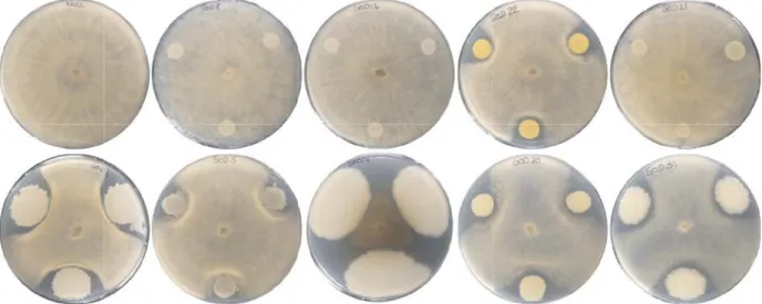
proportion of antagonists within bacteria isolated from the Solaris than from the Pinot noir and Chasselas varieties. Interestingly, the ratio between Bacillus and Staphylococcus was higher in Solaris than in the two other varieties (Figure 2) and preliminary results of the antagonistic assay against Botrytis revealed that the frequency of active strains was higher in the Bacillus genus than in the Staphylococcus genus (see Figure 3 for few representative examples). Further and more detailed studies will allow determining the extent of disease protection conferred by the microbial component of the grapevine holobiont.
PCA plates were inoculated with three 10 µl liquid bacterial culture (OD600= 1) at the border of the plate. After one day, a plug of Botrytis cinerea mycelium was placed in the middle of the plates. Pictures were taken after 6 days. The first row contains a negative control (left) and four phylogenetically distinct Staphylococcus strains, while the second row contains five phylogenetically distinct Bacillus strains. All strains were isolated from the endophytic fraction of the Solaris microbiome.
Conclusion
The next steps in this ongoing work will be i) to use the sequences obtained for the isolates to compile a non-redundant grapevine strain collection for further, more in-depth functional characterizations, and to exclude putative opportunistic animal, human or plant pathogens from further studies, ii) to analyze the strains’ potential biocontrol activity using in vivo experiments on leaf discs, pot-grown plants and ultimately small field experiments, and iii) to identify
the volatile or non-volatile metabolites leading to disease inhibition.
Acknowledgements :The authors are thankful for
financial support provided by the Swiss National Science Foundation (grant 31003A_149271) and by Agroscope (MicBioDiv research program). Scientific and logistic support from Jean-Philippe Burdet, Dr. Anne-Claire Silvestri and Serge Hautier is gratefully acknowledged.
References
Baldan E., Nigris S., Populin F., Zottini M., Squartin A. and Baldan B., 2014. Identification of culturable bacterial endophyte community isolated from tissues of Vitis vinifera “ Glera ”. Plant Biosyst. 148: 508–516. doi:10.1080/11263504.2014.916364 Berendsen R.L., Pieterse C.M.J. and Bakker P.A.H.M.,
2012. The rhizosphere microbiome and plant health. Trends Plant Sci. 17: 478–486. doi:10.1016/ j.tplants.2012.04.001
Bulgarelli D., Schlaeppi K., Spaepen S., Ver Loren van Themaat E. and Schulze-Lefert P., 2013. Structure and functions of the bacterial microbiota of plants. Annu. Rev. Plant Biol. 64: 807–838. doi:10.1146/ annurev-arplant-050312-120106
Campisano A., Antonielli L., Pancher M., Yousaf S., Pindo M. and Pertot I., 2014. Bacterial endophytic communities in the grapevine depend on pest management. PLoS One 9: e112763. doi:10.1371/ journal.pone.0112763
Clavijo McCormick A., Unsicker S.B. and Gershenzon J., 2012. The specificity of herbivore-induced plant volatiles in attracting herbivore enemies. Trends Plant Sci. 17: 303–310. doi:10.1016/j.tplants. 2012.03.012
Figure 3. Representative pictures of dual assay experiments of anti-Botrytis activity.
Coleman-Derr D., Desgarennes D., Fonseca-Garcia C., Gross S., Clingenpeel S., Woyke T., North G., Visel A., Partida-Martinez L.P. and Tringe S.G., 2016. Plant compartment and biogeography affect microbiome composition in cultivated and native Agave species. New Phytol. 209: 798–811. doi:10.1111/nph.13697
Compant S., Nowak J., Coenye T., Clément C. and Ait Barka E., 2008. Diversity and occurrence of Burkholderia spp. in the natural environment. FEMS Microbiol. Rev. 32: 607–626. doi:10.1111/j.1574-6976.2008.00113.x
Delmotte N., Knief C., Chaffron S., Innerebner G., Roschitzki B., Schlapbach R., von Mering C. and Vorholt J.A., 2009. Community proteogenomics reveals insights into the physiology of phyllosphere bacteria. Proc. Natl. Acad. Sci. U. S. A. 106: 16428–16433. doi:10.1073/pnas.0905240106 De Vrieze M., Pandey P., Bucheli T.D., Varadarajan A.R.,
Ahrens C.H., Weisskopf L. and Bailly A., 2015. Volatile organic compounds from native potato-associated Pseudomonas as potential anti-oomycete agents. Front. Microbiol. 6: 1295. doi:10.3389/fmicb.2015.01295
Effmert U., Kalderas J., Warnke R. and Piechulla B., 2012. Volatile mediated interactions between bacteria and fungi in the soil. J. Chem. Ecol. 38: 665–703. doi:10.1007/s10886-012-0135-5
Farag M.A., Zhang H. and Ryu C.M., 2013. Dynamic chemical communication between plants and bacteria through airborne signals: induced resistance by bacterial volatiles. J. Chem. Ecol. 39: 1007–1018. doi:10.1007/s10886-013-0317-9
Groenhagen U., Baumgartner R., Bailly A., Gardiner A., Eberl L., Schulz S. and Weisskopf L., 2013. Production of bioactive volatiles by different Burkholderia ambifaria strains. J. Chem. Ecol. 39: 892–906. doi:10.1007/s10886-013-0315-y
Hunziker L., Bönisch D., Groenhagen U., Bailly A., Schulz S. and Weisskopf L., 2015. Pseudomonas strains naturally associated with potato plants produce volatiles with high potential for inhibition of Phytophthora infestans. Appl. Environ. Microbiol. 81: 821–830. doi:10.1128/AEM.02999-14
Innerebner G., Knief C. and Vorholt J.A., 2011. Protection of Arabidopsis thaliana against leaf-pathogenic Pseudomonas syringae by Sphingomonas strains in a controlled model system. Appl. Environ. Microbiol. 77: 3202–3210. doi:10.1128/AEM. 00133-11 Müller D.B., Vogel C., Bai Y. and Vorholt J.A., 2016. The
plant microbiota: systems-level insights and perspectives. Annu. Rev. Genet. 50: 211–234. doi:10.1146/annurev-genet-120215-034952
Philippot L., Raaijmakers J.M., Lemanceau P. and van der Putten W.H., 2013. Going back to the roots: the microbial ecology of the rhizosphere. Nat. Rev. Microbiol. 11: 789–799. doi:10.1038/nrmicro3109 Ritpitakphong U., Falquet L., Vimoltust A., Berger A.,
Métraux J.P. and L’Haridon F., 2016. The microbiome of the leaf surface of Arabidopsis protects against a fungal pathogen. New Phytol. 210: 1033–1043. doi:10.1111/nph.13808
Ryu C.-M., Farag M.A., Hu C.-H., Reddy M.S., Kloepper J.W. and Paré P.W., 2004. Bacterial volatiles induce systemic resistance in Arabidopsis. Plant Physiol. 134: 1017–1026. doi:10.1104/pp.103. 026583 Truyens S., Weyens N., Cuypers A. and Vangronsveld J.,
2015. Bacterial seed endophytes: genera, vertical transmission and interaction with plants. Environ. Microbiol. Rep. 7: 40–50. doi:10.1111/1758-2229.12181
Vorholt J.A., 2012. Microbial life in the phyllosphere. Nat. Rev. Microbiol. 10: 828–840. doi:10.1038/ nrmicro2910
Yousaf S., Bulgari D., Bergna A., Pancher M., Quaglino F., Casati P. and Campisano A., 2014. Pyrosequencing detects human and animal pathogenic taxa in the grapevine endosphere. Front. Microbiol. 5: 327. doi:10.3389/fmicb.2014.00327 Zarraonaindia I., Owens S.M., Weisenhorn P., West K.,
Hampton-Marcell J., Lax S., Bokulich N.A., Mills D.A., Martin G., Taghavi S., van der Lelie D. and Gilbert J.A., 2015. The soil microbiome influences grapevine-associated microbiota. MBio 6: e02527-14. doi:10.1128/mBio.02527-14