published: 05 November 2018 doi: 10.3389/fnmol.2018.00406
Edited by: Ralf J. Braun, University of Bayreuth, Germany Reviewed by: Md. Golam Sharoar, University of Connecticut Health Center, United States Markus Babst, The University of Utah, United States *Correspondence: Joris Winderickx joris.winderickx@kuleuven.be Christophe Cullin christophe.cullin@u-bordeaux.fr
†These authors have contributed
equally to this work
‡Co-first authorship Received: 13 April 2018 Accepted: 16 October 2018 Published: 05 November 2018 Citation: Fruhmann G, Marchal C, Vignaud H, Verduyckt M, Talarek N, De Virgilio C, Winderickx J and Cullin C (2018) The Impact of ESCRT on Aβ1−42Induced Membrane
Lesions in a Yeast Model for Alzheimer’s Disease. Front. Mol. Neurosci. 11:406. doi: 10.3389/fnmol.2018.00406
The Impact of ESCRT on A
β
1−42
Induced Membrane Lesions in a
Yeast Model for Alzheimer’s Disease
Gernot Fruhmann1†, Christelle Marchal2†‡, Hélène Vignaud2, Mathias Verduyckt1, Nicolas Talarek3, Claudio De Virgilio4, Joris Winderickx1* and Christophe Cullin2*1Functional Biology, KU Leuven, Leuven, Belgium,2Institut de Chimie et Biologie des Membranes et des Nano-objets,
University of Bordeaux, CNRS UMR 5248, Pessac, France,3Institut de Génétique Moléculaire de Montpellier, University of
Montpellier, CNRS, Montpellier, France,4Department of Biology, Université de Fribourg, Fribourg, Switzerland
Aβ metabolism plays a pivotal role in Alzheimer’s disease. Here, we used a yeast model to monitor Aβ42 toxicity when entering the secretory pathway and demonstrate that
processing in, and exit from the endoplasmic reticulum (ER) is required to unleash the full Aβ42 toxic potential. Consistent with previously reported data, our data suggests
that Aβ42 interacts with mitochondria, thereby enhancing formation of reactive oxygen
species and eventually leading to cell demise. We used our model to search for genes that modulate this deleterious effect, either by reducing or enhancing Aβ42 toxicity,
based on screening of the yeast knockout collection. This revealed a reduced Aβ42
toxicity not only in strains hampered in ER-Golgi traffic and mitochondrial functioning but also in strains lacking genes connected to the cell cycle and the DNA replication stress response. On the other hand, increased Aβ42 toxicity was observed in strains
affected in the actin cytoskeleton organization, endocytosis and the formation of multivesicular bodies, including key factors of the ESCRT machinery. Since the latter was shown to be required for the repair of membrane lesions in mammalian systems, we studied this aspect in more detail in our yeast model. Our data demonstrated that Aβ42 heavily disturbed the plasma membrane integrity in a strain lacking the
ESCRT-III accessory factor Bro1, a phenotype that came along with a severe growth defect and enhanced loading of lipid droplets. Thus, it appears that also in yeast ESCRT is required for membrane repair, thereby counteracting one of the deleterious effects induced by the expression of Aβ42. Combined, our studies once more validated the use
of yeast as a model to investigate fundamental mechanisms underlying the etiology of neurodegenerative disorders.
Keywords: Aβ42, amyloid beta, Alzheimer’s disease, ESCRT, membrane repair, Saccharomyces cerevisiae, yeast
INTRODUCTION
Amyloidβ1−42(referred to as Aβ42) is an intensively investigated peptide involved in Alzheimer’s
disease (AD) but beyond its role as a possibly malignant factor, the physiological functions of Aβ42 are still speculative. It has been shown that it has a role in activity dependent synaptic
vesicle release and there is evidence that it might act as an antimicrobial agent in the brain, but besides that, not much is known about its physiological function (Pearson and Peers, 2006;
Abramov et al., 2009; Kagan et al., 2012; Kumar et al., 2016). Thus, although several efforts have been put in investigating its role in AD, still most of its functions remain elusive. In humans, the membrane bound amyloid precursor protein, APP, is processed in two possible ways: it can enter the non-amyloidogenic pathway or it might be cleaved by β- and γ-secretases in the amyloidogenic pathway thus producing Aβ peptides of lengths between 37 and 43 amino acids (LaFerla et al., 2007). In AD, these peptides are then secreted and form inert extracellular plaques, but monomers and especially small oligomers are transported to several intracellular organelles where they reveal their toxic potential. For instance, strong evidence was found for that Aβ42 perturbs proper function
of mitochondria through blocking respiration at complex IV, resulting in cells producing more reactive oxygen species (ROS) and eventually apoptotic cell death (Wang et al., 2010;Ittner and Gotz, 2011;Benilova et al., 2012;Jackrel et al., 2014;Alexandrov et al., 2016;Vicente Miranda et al., 2016).
In the past decade, yeast was established as a model organism for studying fundamental aspects related to neurodegenerative disorders such as Huntington’s, Parkinson’s, or AD (Winderickx et al., 2008;Franssens et al., 2010;Swinnen et al., 2011;Porzoor and Macreadie, 2013;Jiang et al., 2016; Verduyckt et al., 2016; Fruhmann et al., 2017). Since most of the overall cellular architecture and most basic biochemical processes are conserved between mammalian cells and yeast, and given the fact that about 20–30 per cent of all human genes have orthologs in Saccharomyces cerevisiae (Foury, 1997;Hamza et al., 2015; Liu et al., 2017), it is obvious that certain disease mechanisms can be studied in this easy to handle model organism. In connection to AD and Aβ42, we and others reported that
expression of this peptide triggers toxicity in yeast when targeted to the secretory pathway as to mimic its multi-compartment trafficking observed in mammalian systems. This led to the observation that Aβ42 expression in yeast alters endocytosis
of plasma membrane resident proteins (Treusch et al., 2011; D’Angelo et al., 2013), induces ER-stress and the unfolded protein response (Chen et al., 2017) and triggers mitochondrial dysfunction (D’Angelo et al., 2013;Chen and Petranovic, 2015; Chen et al., 2017). In the studies presented in this paper, we used previously reported constructs (D’Angelo et al., 2013; Vignaud et al., 2013) where the yeast mating type α-prepro factor directs Aβ42 into the Golgi. Here, theα-prepro factor is
cleaved off, followed by transport of the peptide to the plasma membrane. In addition, the constructs contain a C-terminal linker-GFP tag in order to ensure stable expression and easy localization of Aβ42in the yeast cells. Besides the wild-type Aβ42
and the clinical arctic mutant, we also expressed two synthetic mutants generated by random mutagenesis and previously shown to be either more toxic (Aβ42G37C) to, or to be moderately
toxic (Aβ42L34T) compared to Aβ42wt (D’Angelo et al., 2013; Vignaud et al., 2013). Using these constructs, we performed genome-wide screenings as to identify Aβ42toxicity modulators.
We confirm the previously reported Aβ42 toxicity phenotypes
and in addition demonstrate that Aβ42 introduces membrane
lesions that require the ESCRT system in order to become repaired.
MATERIALS AND METHODS
Yeast Strains, Plasmids, and Media
We used the haploid Saccharomyces cerevisiae strain BY4741 MATa his311 leu210 met1510 ura310 and BY4742 MATα his311 leu210 lys210 ura310 for all specified experiments. All deletion strains were obtained from the commercial EUROSCARF knock-out library (Y.K.O. collection). For a full list of strains used in this study see Table 1.
The pYe plasmids with the galactose inducible, episomal αAβ42-linker-GFP isoforms, the similar Aβ42-GFP construct
without the α-prepro sequence and the control constructs for expressing of either GFP (ev-GFP) or a α-prepro-GFP fusion (ev-αGFP) were described previously (Tong et al., 2001). The p426-GAL vector (Addgene) was used as additional empty vector (ev) control. For a full list of plasmids used in this study see
Table 2.
Standard yeast techniques were applied. We used minimal medium containing Yeast Nitrogen Base (YNB) without ammonium sulfate (FORMEDIUM). Supplements were ammonium sulfate (5 g/L; VWR), histidine (100 mg/L; MP Biomedicals), methionine (20 mg/L; Acros Organics), leucine (30 mg/L; FORMEDIUM) and lysine (30 mg/L; FORMEDIUM). Synthetic Drop-Out (SD) medium was used for microscopy and contained YNB with ammonium sulfate and was depleted for either uracil or uracil and leucine (FORMEDIUM). For solid media, 1.5% Difco-agar (BD) was added. Pre-cultures were grown on medium supplemented with 4% glucose and gene expression was induced by washing the cells with medium without sugar followed by transfer to medium supplemented with 2% galactose.
Synthetic Genetic Array and Suppressor
screening
The synthetic genetic array (SGA) screening was essentially performed as previously described in Tong et al. (2001). The query strain (MATα can11::STEpr-HIS5sp lyp11 his311 leu210 met1510 ura310 LYS2) expressing αAβ42wt-linker-GFP
(further designated as αAβ42wt), the αAβ42G37C-linker-GFP
(designated αAβ42G37C) or the ev-αGFP was mated with the
non-essential deletion mutant array (MATatarget_gene::kanMX4 his311 leu210 met1510 ura310) on SD plates lacking uracil. Diploids were selected on SD medium lacking uracil but containing G418 (geneticin). Next, sporulation was induced by plating diploids onto sporulation medium containing G418. Then, haploids were selected in two steps. First, spores were plated onto YNB lacking arginine, lysine, and histidine but containing canavanine and thialysine, which ensures uptake of canavanine. This allowed growth of MATa haploids only. In a second step, the selected haploids were grown on YNB lacking uracil, arginine, lysine, and histidine but containing canavanine, thialysine and G418 to select for haploid knock-out mutants still carrying the αAβ42wt,
αAβ42G37C, or ev-αGFP plasmids. Growth analysis was
performed with the ScreenMill software (Dittmar et al., 2010).
TABLE 1 | Yeast strains used in this study.
Name Genotype Source
Query strain (SGA) MATα can11::STEpr-HIS5sp lyp11 his311 leu210 met1510 ura310 LYS2 Tong et al., 2001
Deletion mutant strains (SGA) MATa Target_gene::kanMX4 his311 leu210 met1510 ura310 Tong et al., 2001
BY4741 MATa his311 leu210 met1510 ura310 Openbiosystems
BY4742 MATα his311 leu210 lys210 ura310 Openbiosystems
JW 12 918 MATa his311 leu210 met1510 ura310 erv29::kanMX Y.K.O. collection
JW 23 168 MATα his311 leu210 lys210 ura310 hse11::kanMX Y.K.O. collection
JW 23 771 MATα his311 leu210 lys210 ura310 vps27::kanMX Y.K.O. collection
JW 20 178 MATα his311 leu210 lys210 ura310 srn2::kanMX Y.K.O. collection
JW 21 184 MATα his311 leu210 lys210 ura310 mvb12::kanMX Y.K.O. collection
JW 21 335 MATα his311 leu210 lys210 ura310 stp22::kanMX Y.K.O. collection
JW 22 115 MATα his311 leu210 lys210 ura310 vps28::kanMX Y.K.O. collection
JW 23 142 MATα his311 leu210 lys210 ura310 vps25::kanMX Y.K.O. collection
JW 21 849 MATα his311 leu210 lys210 ura310 vps36::kanMX Y.K.O. collection
JW 22 164 MATα his311 leu210 lys210 ura310 snf8::kanMX Y.K.O. collection
JW 20 891 MATα his311 leu210 lys210 ura310 doa4::kanMX Y.K.O. collection
JW 22 100 MATα his311 leu210 lys210 ura310 bro1::kanMX Y.K.O. collection
JW 22 220 MATα his311 leu210 lys210 ura310 vps4::kanMX Y.K.O. collection
JW 22 777 MATα his311 leu210 lys210 ura310 did2::kanMX Y.K.O. collection
JW 24 370 MATα his311 leu210 lys210 ura310 vps60::kanMX Y.K.O. collection
JW 21 424 MATα his311 leu210 lys210 ura310 vta1::kanMX Y.K.O. collection
JW 23 123 MATα his311 leu210 lys210 ura310 chm7::kanMX Y.K.O. collection
JW 20 444 MATα his311 leu210 lys210 ura310 ist1::kanMX Y.K.O. collection
JW 20 124 MATα his311 leu210 lys210 ura310 snf7::kanMX Y.K.O. collection
JW 21 479 MATα his311 leu210 lys210 ura310 vps24::kanMX Y.K.O. collection
JW 22 806 MATα his311 leu210 lys210 ura310 vps20::kanMX Y.K.O. collection
JW 11 560 MATα his311 leu210 lys210 ura310 did4::kanMX Y.K.O. collection
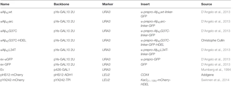
TABLE 2 | Plasmids used in this study.
Name Backbone Marker Insert Source
αAβ42wt pYe-GAL10 2U URA3 α-prepro-Aβ42
wt-linker-GFP
D’Angelo et al., 2013
αAβ42arc pYe-GAL10 2U URA3 α-prepro-Aβ42
arc-linker-GFP
D’Angelo et al., 2013
αAβ42G37C pYe-GAL10 2U URA3 α-prepro-Aβ42
G37C-linker-GFP
D’Angelo et al., 2013
αAβ42G37C-HDEL pYe-GAL10 2U URA3 α-prepro-Aβ42
G37C-linker-GFP-HDEL
Christophe Cullin
αAβ42L34T pYe-GAL10 2U URA3 α prepro-Aβ42
L34T-linker-GFP
D’Angelo et al., 2013
ev-αGFP pYe-GAL10 2U URA3 α-prepro-GFP D’Angelo et al., 2013
ev-GFP pYe-GAL10 2U URA3 GFP D’Angelo et al., 2013
Ev p426-GAL1 URA3 Mumberg et al., 1994
pHS12-mCherry pHS12-ADH1 LEU2 COX4 Addgene
pYX242-mCherry pYX242-TPI LEU2 Kar2(1−135)
-mCherry-HDEL
Swinnen et al., 2014
In a second so-called suppressor screening, the Euroscarf collection of deletion strains was pooled transformed with αAβ42G37C. Transformants were plated on minimal medium lacking uracil. After incubation, transformants were selected that grew similar as the isogenic wild-type carrying the empty vector control, also when replica plated on SD medium supplemented with casamino acids. Transformants were then used for bar-code
PCR sequencing as to identify their corresponding ORF deletion.
Electron Microscopy
Electron microscopic analysis of yeast cells was done similar as previously described (Lefebvre-Legendre et al., 2005). Briefly, pellets of yeast cells were placed on the surface of a copper
EM grid (400 mesh) coated with formvar. Grids were immersed in liquid propane held at −180◦
C by liquid nitrogen and then transferred in a 4% osmium tetroxide solution in dry acetone at −82◦
C for 72 h. They were warmed progressively to RT, and washed three times with dry acetone and stained with 1% uranyl acetate. After washing in dry acetone, the grids were infiltrated with araldite (Fluka). Ultra-thin sections were contrasted with lead citrate and observed in an electron microscope (80 kV; 7650; Hitachi) at the EM facility of the Bordeaux Imaging Center.
Growth Profile Analysis and Spot Assays
Cells were grown under non-inducing conditions in 96-well plates shaking at 30◦
C in a Multiskan GO or Multiskan FC microplate spectrophotometer (Thermo Scientific) to an OD595 nm or OD600 nm, respectively, of 0.5 to 0.9. Cells were
washed with minimal medium containing galactose and diluted to an OD595 nm or OD600 nm, respectively, of 0.5 after which
growth was monitored every 2 h by OD measurement. Four different transformants were taken per experiment and at least three independent experiments were performed. Growth curves were analyzed with GraphPad Prism v7.03, error bars represent standard deviations.
For spot assays, serial dilutions of precultures were spotted on solid medium containing either glucose or galactose and cells were grown at 30◦
C. The plates were scanned at days 3 to 6.
Cytometry and Fluorescence Microscopy
To monitor plasma membrane disruption with propidium iodide (PI) staining and the formation of ROS by dihydroethidium (DHE) staining, cells were grown in SD medium lacking uracil and containing 4% glucose. Once in exponential phase, the cells were washed with and re-suspended in SD medium without uracil but containing 2% galactose as to induce expression of αAβ42-linker-GFP and control constructs. Flow
cytometric analysis was performed on at least four independent transformants with a Guava easyCyte 8HT benchtop flow cytometer (Millipore) after staining with 5µM PI for 30 min at 30◦
C and subsequent washing. Data were analyzed with FlowJo v10 and GraphPad Prism 7.03 software packages. Gates were set in FlowJo v10 with single stained ev-αGFP samples. Further statistical analysis was done in GraphPad Prism v7.03. Error bars represent standard deviations and asterisks the significance calculated with an ordinary Two-way ANOVA.
For the FM4-64/CMAC (Thermo Fisher) and Nile Red (Acros) stainings, the cells were grown as described above. After pre-incubating the cells with CAMAC at 30◦
C for 30 min, FM4-64 stainings were performed as described before (Zabrocki et al., 2005). For the Nile Red staining, the cells were fixed with formaldehyde (4% final concentration) and stained with 2% Nile Red (60µg/mL stock) for 30 min with shaking at 30◦
C. Then, the cells were washed twice with PBS and either stored at 4◦
C or taken immediately for fluorescence microscopy.
For epifluorescence pictures, cells were pre-grown in selective glucose (4%) containing SD medium to exponential phase. After transfer to SD medium containing 2% galactose to induce expression of αAβ42-linker-GFP, the cells were grown at 30◦C
and pictures were taken after different time intervals using a
Leica DM4000B or a DMi8 microscope. For Hoechst stainings, the cells were incubated with 20 nM Hoechst 33342 (Sigma-Aldrich) for 10 min. Pictures were deconvolved with Huygens Essential software (v18.04.0p4 64, Scientific Volume Imaging B.V.) and further processed with the standard FIJI software package (v1.51n) (Schindelin et al., 2012).
Immunological Techniques
Cultures were grown to exponential phase in 4% glucose containing SD medium, transferred to 2% galactose containing medium and grown overnight. Then, three OD units were harvested by centrifugation and protein extracts were prepared by using an alkaline lysis method. The cells were permeabilized with 0.185 M NaOH plus 2%β-mercaptoethanol. After 10 min incubation on ice, trichloroacetic acid (TCA) was added to a final concentration of 5%, followed by an additional 10 min incubation step on ice. Precipitates were collected by centrifugation for 5 min at 13000 g and pellets were resuspended in 50µL of sample buffer (4% sodium dodecyl sulfate, 0.1 M Tris-HCl pH 6.8, 4 mM EDTA, 20% glycerol, 2% β-mercaptoethanol, and 0.02% bromophenol blue) plus 25µL of 1 M Tris-Base. Samples were separated by standard SDS-PAGE on 12% polyacrylamide gels and further analyzed using standard Western blotting techniques. An anti-GFP primary antibody (Sigma-Aldrich) and anti-Mouse (GAM)-HRP conjugated secondary antibody (Biorad) were used. The ECL method (SuperSignal West Pico or Femto, Thermo Scientific) was used for detection and visualization of the blots was performed with a UVP Biospectrum R
Multispectral Imaging System.
RESULTS
αAβ
42-Linker-GFP Is Toxic in an AD Yeast
Model
To date, the exact molecular basis of how Aβ42 impacts cell
functions remains largely elusive. To address this question, we used a yeast model transformed with plasmids carrying the galactose-inducible GAL10 promoter to control expression of wild-type or mutant Aβ42 that is N-terminally fused to
the α-prepro sequence and C-terminally to a linker and GFP (in this paper referred to as αAβ42; Figure 1A) (D’Angelo et al., 2013; Vignaud et al., 2013). Besides the wild-type αAβ42 (αAβ42wt), we additionally expressed the clinical E22G
arctic mutant (αAβ42arc) that is associated to a familial
form of AD as well as the previously described synthetic mutants αAβ42G37C and αAβ42L34T (Figure 1A) (D’Angelo et al., 2013; Vignaud et al., 2013). We used three control vectors, i.e., an empty vector allowing for the expression of an α-prepro fused GFP (ev-αGFP), GFP alone (ev-GFP) or an empty vector just carrying the galactose promotor (ev). To disperse concerns about the processing efficacy of the α-prepro factor in the Golgi system, we tested both the BY4741 MATa and BY4742 MATα strains with all control vectors and the aforementioned wild-type and mutant αAβ42-linker-GFP
constructs (Figures 1B–D and Supplementary Figure S1). Albeit the strains transformed with the ev-αGFP control grew somewhat
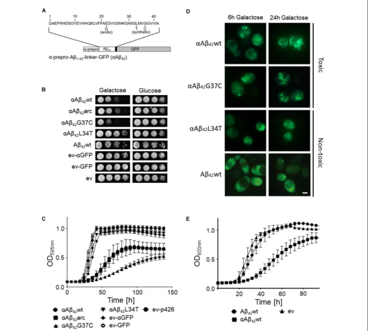
FIGURE 1 | Toxicity profiles of wild-type or mutantαAβ42in the haploid Saccharomyces cerevisiae strains BY4741. (A) Schematic representation of the
α-prepro-Aβ42-linker-GFP construct with indication of the mutants used in this study. (B) Spot assays under inducing (galactose) and repressing (glucose) conditions
and (C) growth profiles under inducing conditions of the BY4741 wild-type strain transformed with constructs allowing for expression ofαAβ42wt,αAβ42arc,
αAβ42G37C,αAβ42L34T, Aβ42wt and control vectors (ev-αGFP, ev-GFP, ev) as indicated. Error bars in the growth profiles represent the standard deviation of at least
four independent transformants. (D) Fluorescence microscopy pictures show localization ofα-prepro fused Aβ42mutants to the ER 6 and 24 h after induction on
galactose-containing medium. 24 h after induction of gene expression the toxicαAβ42isoforms show a more “patchy” pattern while the non-toxicαAβ42L34T still
localizes at the ER. The Aβ42wt construct lacking theα-prepro sequence is seen in the cytoplasm. The scale bar represents 2 µm. (E) Growth profiles of the BY4741
wild-type strain transformed with an empty vector (ev) or constructs allowing for expression ofαAβ42wt and Aβ42wt.
slower than those transformed with ev-GFP or ev (Figures 1B,C and Supplementary Figure S1A), both spot assays and growth analysis in liquid medium confirmed that wild-type or mutant αAβ42 instigated a significantly higher level of toxicity that was
similar in the BY4741 and the BY4742 strains (Figures 1B,C and
Supplementary Figure S1). The synthetic αAβ42G37C mutant
was the most toxic followed by αAβ42wt and αAβ42arc. The
syntheticαAβ42L34T mutant, on the other hand, did not yield
a toxic phenotype and these transformants grew similar as those expressing ev-αGFP, thereby confirming previously reported data (Vignaud et al., 2013).
Fluorescence microscopy showed that all αAβ42 constructs
clearly stained the perinuclear ER and to a lesser extent the cortical ER and that particularly the cells expressing the toxic
αAβ42forms often displayed ER-associated foci and filamentous
structures (Figure 1D). These were previously believed to be vesicles (D’Angelo et al., 2013), but recent studies suggests that these may as well-represent ER-aggregates or clustering of ER membranes, both indicative for ER stress (Varadarajan et al., 2012, 2013;Vevea et al., 2015). In contrast, when the Aβ42wt was
expressed from a construct that lacks theα-prepro sequence, the GFP fusion was mainly found to be distributed in the cytoplasm though some cells presented foci after prolonged induction (Figure 1D). Despite of these foci, the expression of the Aβ42wt
construct without theα-prepro sequence only triggered a small growth retardation (Figures 1B,E). This nicely demonstrates that the processing in the ER/Golgi system is required to unleash the full toxic capacity ofαAβ42.
Retention of
αAβ
42in the ER Diminishes
Its Toxicity
Previously, we reported that the processing of theαAβ42
-linker-GFP fusion constructs in the ER/Golgi system yields three distinct isoforms when performing Western blot analysis, i.e., the α-prepro precursor (41 kDa), the glycosylated precursor (50 kDa), and the matured Aβ42-linker-GFP form (34 kDa)
(D’Angelo et al., 2013; Vignaud et al., 2013). Given that the latter is shuttled into the secretory pathway, we wondered if retention of the αAβ42-linker-GFP fusion in the endoplasmic
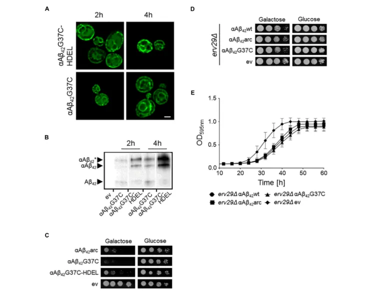
reticulum (ER) would affect its toxic capacity. To this end, we introduced a HDEL retention signal at the C-terminal end of the construct. The yeast HDEL sequence is equivalent to the mammalian KDEL retention signal that shuttles the KDEL containing proteins back to the ER lumen (Dean and Pelham, 1990; Lewis and Pelham, 1990; Ruan et al., 2017). Consistently, microscopic analysis demonstrated that while both αAβ42G37C andαAβ42G37C-HDEL are equally present at the
perinuclear ER, the latter accumulated more in the peripheral cortical ER (Figure 2A). Also, Western blot analysis showed that in case of expression ofαAβ42G37C-HDEL, both the
non-processed αAβ42G37C precursor as well as the glycosylated
version accumulated, while the fully processed Aβ42G37C was
significantly reduced (Figure 2B). Next, a growth analysis was performed. This revealed that the strain expressingαAβ42
G37C-HDEL construct grew much better than that withαAβ42G37C,
thereby displaying a level of toxicity comparable to a strain with theαAβ42arc mutant (Figure 2C).
In order to confirm these results with a different approach, we expressed αAβ42wt, αAβ42arc, as well as the super-toxic
αAβ42G37C mutant in a BY4741 strain deleted forERV29, which
encodes a transmembrane factor involved in COP-II dependent vesicle formation and in trafficking of theα-prepro factor from the ER to the Golgi apparatus (Belden and Barlowe, 2001). Upon deletion ofERV29,αAβ42gets stuck in the ER and cannot transit
to the Golgi. Strikingly, cytotoxicity of all tested mutants was indeed significantly diminished in thiserv291 strain and even the super-toxicαAβ42G37C mutant almost completely lost its toxic
power (Figures 2D,E). Thus, the data above confirm thatαAβ42
needs to be fully processed and exit the ER in order to gain its full toxic potential.
αAβ
42Affects Mitochondrial Functioning
SinceαAβ42 confers toxicity by entering the secretory pathway,we wanted to know more about the targets and processes being affected. Therefore, we first focused on mitochondria given that previous studies have associated Aβ42 to mitochondrial
dysfunction in yeast (Chen and Petranovic, 2015; Chen et al., 2017;Hu et al., 2018). To this end, we co-expressed αAβ42wt,
αAβ42arc,αAβ42G37C,αAβ42L34T or the ev-αGFP together with
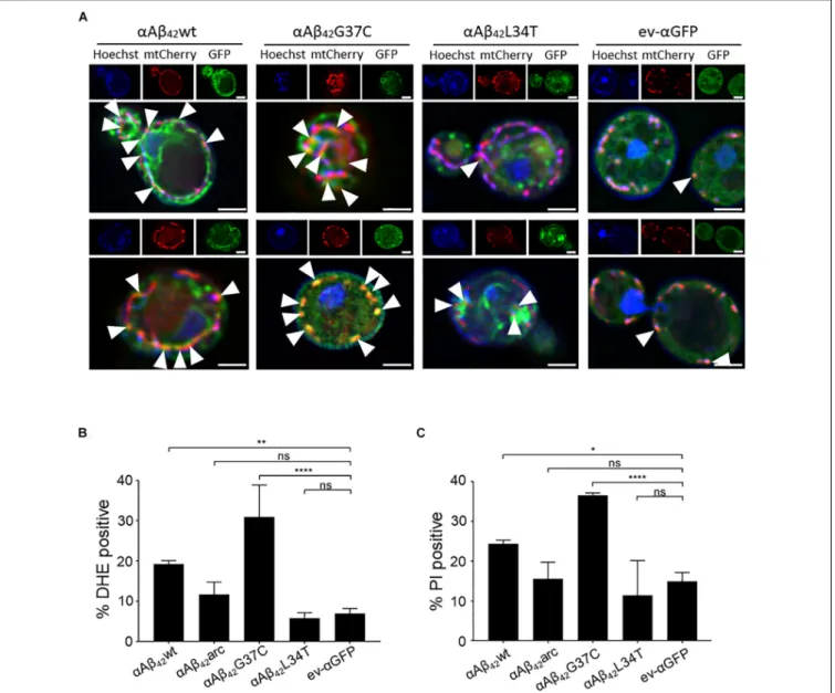
a mCherry labeled mitochondrial marker Cox4 in wild-type cells. As illustrated in Figure 3A, this suggests a toxicity dependent co-localization sinceαAβ42wt and even moreαAβ42G37C seem
to partially co-localize with mitochondria. We then performed a DHE staining to estimate the ROS levels as marker for mitochondrial dysfunction and a PI staining to monitor the amount of cells with disrupted plasma membranes as marker for cell demise (Figures 3B,C and Supplementary Figure S2). As expected, and consistent with previously reported data (Chen and Petranovic, 2015;Chen et al., 2017), also these aspects correlated to the observed αAβ42 instigated toxicity. Although our data
on co-localization suggest that αAβ42 may directly interfere
with mitochondrial functioning, we cannot exclude that these effects on mitochondrial function or morphology are indirect, for instance due alterations in ER-mitochondrial communication via ERMES contact sites.
Identification of Additional Processes
Underlying
αAβ
42Toxicity
To decipher which additional processes sustain the αAβ42
toxicity, we performed two unbiased genetic screens. In a first screening setup, we aimed to identify alleviators of Aβ42G37C
toxicity. Therefore, we transformed a pooled Euroscarf knock-out (KO) library with theαAβ42G37C-linker-GFP construct and
looked for transformants that grew similar as the empty vector control. Out of 90,000 transformants obtained when plated on repressive glucose containing medium, only 465 were able to form visible colonies after 72 h on galactose-containing medium where the expression of the super-toxic Aβ42mutant is induced.
Each colony was then plated on SD medium supplemented with casamino acids to manually confirm the suppressive effect on αAβ42 toxicity. Of the 465 initial clones, 268 were still able to
grow. Finally, we characterized each KO strain by sequencing the PCR amplicon of its bar-code region. This led us to identify 113 different KO strains. In the second screening setup, we performed an unbiased SGA analysis with the full gene knock-out library using either αAβ42wt, the super-toxic αAβ42G37C
mutant and the ev-GFP control. The additional use of the empty vector control and αAβ42wt, which displays a more moderate
toxicity phenotype, allowed for more complete results since not only suppressors but also toxicity aggravators could be scored. With this SGA screen, we identified 87 additional KO strains that modulatedαAβ42 toxicity, one of which contains a deletion in
the overlapping genesINP52/RRT16. Finally, the 200 KO strains that were identified by either one of the screening procedures (Table 3 and Supplementary Table S1) were also transformed individually with a construct allowing for the expression of αAβ42arc, which has an intermediate toxicity similar to that
FIGURE 2 | Retention ofαAβ42in the ER and the consequence for toxicity. (A) Fluorescence microscopy pictures showingαAβ42G37C andαAβ42G37C-HDEL
localization at the perinuclear and cortical ER. Scale bar represents 2µm. (B) Western blot analysis of total protein extracts obtained from cells expressing either αAβ42G37C andαAβ42G37C-HDEL. The different processing isoforms are indicated, i.e.,αAβ∗42indicates glycosylatedαAβ42,αAβ42indicates un-glycosylated
αAβ42and Aβ42indicates fully processed form where theα-prepro sequence is cleaved off. Spot assays under inductive (galactose) and repressive (glucose)
conditions of wild-type (C) or erv291 (D) cells transformed with the empty vector (ev) or constructs allowing for expression of αAβ42wt,αAβ42arc,αAβ42G37C, or
αAβ42G37C-HDEL as indicated. (E) Growth profiles of erv291 transformed with the empty vector (ev) or constructs allowing for expression of αAβ42wt,αAβ42arc, or
αAβ42G37C when grown on galactose-containing medium.
of αAβ42wt and thus is again ideal to provide confirmation of
both aggravators and suppressors of toxicity (D’Angelo et al., 2013). This provided an independent confirmation that all the KO strains selected in one of the two genetic screens are indeed involved in modulating αAβ42arc toxicity. For each of the KO
strains, the cell density under galactose inducing conditions was monitored allowing to rank KO strains from 0 to 5 depending on the growth capacity in comparison to the wild-type strain transformedαAβ42arc. For 29 KO strains, growth was improved
strongly (scored as 5), for 18 KO strains growth was clearly improved (scored as 4) and for 129 KO strains growth was only slightly better (scored as 3). On the other hand, we also identified 2 KO strains in whichαAβ42arc toxicity was strongly enhanced
(scored as “0”) and 17 KO strains that enhancedαAβ42arc toxicity
moderately (scored as 1) or weakly (scored as 2) (Table 3 and
Supplementary Table S1). Next, a gene ontology (GO) analysis
using the SGD Gene Ontology Slim Mapper1 allowed to sort
the KO strains into functional categories depending on the gene deleted (Table 3). These included, amongst others, cytoskeleton organization and endocytosis, protein sorting and trafficking, protein ubiquitination, plasma membrane transport, cell cycle, translation, and transcription.
Interestingly, among the many KO strains that alleviated αAβ42arc toxicity we found not only the strain deleted for
ERV29, which is in line with the data described above, but also strains lacking other genes that impact on ER/Golgi functioning
FIGURE 3 |αAβ42induces mitochondrial dysfunction and cell demise. (A) Fluorescence microscopy pictures suggesting partial co-localization of toxicαAβ42but not
of the non-toxicαAβ42L34T nor of theαGFP control (green) with mitochondria (red) in wild-type cells. Hoechst staining (blue) shows mitochondrial as well as nuclear
DNA. The white arrowheads indicate sites of co-localization betweenαAβ42and mitochondria. Two single cells shown per strain. Scale bars represent 2µm.
Percentage of wild-type yeast cells expressing wild-type or mutantαAβ42and a control stained with DHE as a marker for ROS-formation (B) or PI as a marker for
plasma membrane integrity (C).∗P ≤ 0.05;∗∗P ≤ 0.01;∗∗∗∗P ≤ 0.0001;nsP> 0.05.
and traffic, such as theSPC2 encoded subunit of the peptidase complex, which cleaves the signal sequence from proteins targeted to the ER, EMP24, which encodes a component of the p24 complex that mediates ER-to-Golgi transport of GPI anchored proteins, or ARF2 and YPT31, which both encode GTPases required for intra-Golgi traffic. Also found were several strains lacking genes encoding mitochondrial functions and this included AIF1 that codes for the mitochondrial cell death effector, indicative thatαAβ42actively induces programmed cell
death pathways. Furthermore, the fact that we retrieved the KO strains for ATG1, ATG2, and ATG20 suggests thatαAβ42
may overstimulate the autophagy and the cytoplasm-to-vacuole targeting pathways (Kim and Klionsky, 2000;Nice et al., 2002).
Finally, it was recently shown by transcriptome analysis that Aβ42
impacts on lipid metabolism withINO1 being most significantly upregulated (Chen et al., 2017). We found the KO strain lacking ITC1 to reduceαAβ42 toxicity.ITC1 encodes a subunit of
ATP-dependent Isw2p-Itc1p chromatin remodeling complex that is required for repression of INO1. In addition, also KO strains lacking genes involved sphingolipid and ceramide metabolism were retrieved, i.e., FAA1, SUR1, YDC1. Overall, these data confirm that the noxious effect ofαAβ42is associated to changes
in lipid metabolism.
The KO strains that aggravated αAβ42 toxicity were often
missing functions associated to maintenance and organization of the actin cytoskeleton, endocytosis and the multivesicular
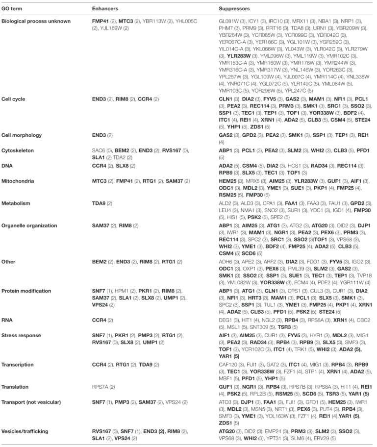
TABLE 3 |αAβ42toxicity modifiers.
GO term Enhancers Suppressors
Biological process unknown FMP41 (2), MTC3 (2), YBR113W (2), YHL005C (2), YJL169W (2)
GL081W (3), ICY1 (3), IRC10 (3), MRX11 (3), NBA1 (3), NRP1 (3), PHM7 (3), PRM9 (3), RRT16 (3), TDA8 (3), URN1 (3), YBR209W (3), YBR284W (3), YCR085W (3), YCR099C (3), YDR042C (3), YER067C-A (3), YER186C (3), YGL101W (3), YGR259C (3), YIL014C-A (3), YKL066W (3), YL043W (3), YLR042C (3), YLR279W (3), YLR283W (3), YML096W (3), YML119W (3), YMR102C (3), YMR153C-A (3), YMR160W (3), YMR178W (3), YMR244W (3), YMR316C-A (3), YMR317W (3), YNL146W (3), YOR263C (3), YPL257W (3), YGL109W (4), YJL007C (4), YMR114C (4), YNL338W (4), YNR071C (4), YGL072C (5), YLR149C (5), YML084W (5), YMR103C (5), YOR296W (5), YPL247C (5)
Cell cycle END3 (2), RIM8 (2), CCR4 (2) CLN1 (3), DIA2 (3), FYV5 (3), GAS2 (3), MAM1 (3), NFI1 (3), PCL1 (3), PEA2 (3), REC114 (3), PRM3 (3), SMK1 (3), SRC1 (3), SSO2 (3), SSP1 (3), TEC1 (3), TEP1 (3), TOF1 (3), YOR338W (3), BDF2 (4), ITC1 (4), REI1 (4), XRN1 (4), ADA2 (5), CLB3 (5), CSM4 (5), STE24 (5), YHP1 (5), ZDS1 (5)
Cell morphology END3 (2) GAS2 (3), GPD2 (3), PEA2 (3), SMK1 (3), SSP1 (3), TEP1 (3), REI1
(4) Cytoskeleton SAC6 (0), BEM2 (2), END3 (2), RVS167 (0),
SLA1 (2) TDA2 (2)
ABP1 (3), PCL1 (3), PEA2 (3), SLM2 (3), WHI2 (3), CLB3 (5), PFD1 (5)
DNA CCR4 (2), SLX8 (2) ADA2 (5), CSM4 (5), DIA2 (3), HCS1 (3), RAD34 (3), REC114 (3),
RPB9 (3), SLX5 (3), TEC1 (3), TOF1 (3)
Mitochondria MTC3 (2), FMP41 (2), RTG1 (2), SAM37 (2) HEM25 (3), MRX5 (3), AIM25 (3), YLR283W (3), GUF1 (3), AIF1 (3), ODC1 (3), MDL2 (3), YME1 (3), SUE1 (3), PKP1 (4), FMP25 (4), RSM25 (5), FMP30 (5)
Metabolism TDA9 (2) ALD2 (3), ALD3 (3), CPA1 (3), FAA1 (3), FAA3 (3), FAU1 (3), GPD2 (3),
LEU4 (3), NMA1 (3), SNO2 (3), SUR1 (3), YDC1 (3), IGD1 (4), FMP30 (5), HIS1 (5), PSK2 (5), SPE2 (5)
Organelle organization SAM37 (2), RIM8 (2) ABP1 (3), AIM25 (3), ATG1 (3), ATG2 (3), ATG20 (3), DID2 (3), DJP1 (3), IWR1 (3), MAM1 (3), NGR1 (3), PEA2 (3), PEX6 (3), PRM3 (3), REC114 (3), SPC2 (3), SRC1 (3), SSO2 (3)TOF1 (3), VPS68 (3), WHI2 (3), YME1 (3), BDF2 (4), FMP25 (4), ADA2 (5), CLB3 (5), CSM4 (5) SCD6 (5)
Other BEM2 (2), END3 (2), RIM8 (2), RTG1 (2) ADH6 (3), APE2 (3), ARF2 (3), DIA2 (3), FDO1 (3), FYV5 (3), IGO2 (3), ODC1 (3), OXP1 (3), PEX6 (3), PML39 (3), SLM2 (3), GAS2 (3), SMK1 (3), SSO2 (3), SSP1 (3), SUE1 (3), TEC1 (3), TEP1 (3), TVP18 (3), YML082W (3), YOR338W (3), ECM4 (4), PDE2 (4), YGR111W (4) Protein modification SNF7 (1), HPM1 (2), PKR1 (2), RIM8 (2),
SAM37 (2), SLA1 (2), SLX8 (2), UMP1 (2), VPS24 (2)
ABP1 (3), ATG1 (3), CLN1 (3), CPS1 (3), CUL3 (3), CUR1 (3), DIA2 (3), NFI1 (3), HRT3 (3), MAM1 (3), PCL1 (3), SLX5 (3), SMK1 (3), SPC2 (3), SSP1 (3), TUL1 (3), YME1 (3), FMP25 (4), PKP1 (4), XRN1 (4), ADA2 (5), CLB3 (5), PFD1 (5), PSK2 (5), STE24 (5)
RNA CCR4 (2) DEG1 (3), HIT1 (4), NGL2 (3), RPB4 (3), RPS8A (3), XRN1 (4), CBC2
(5), MSL1 (5), SNT309 (5), TSR3 (5) Stress response SNF7 (1), PKR1 (2), PMP3 (2), RTG1 (2),
RVS167 (0), SLX8 (2), UMP1 (2)
AIF1 (3), AIM25 (3), CUR1 (3), FYV5 (3), HYR1 (3), MDL2 (3), MIG1 (3), PEA2 (3), RAD34 (3), RPB4 (3), RPB9 (3), SLX5 (3), SMF3 (3), TOF1 (3), YCR102C (3), ITC1 (4), TRK1 (5), WHI2 (3), ADA2 (5), YAR1 (5)
Transcription CCR4 (2), RTG1 (2), TDA9 (2) CAF120 (3), FUI1 (3), GAT2 (3), ITC1 (4), MIG1 (3), RPB4 (3), RPB9 (3), TEC1 (3), YOR338W (3), FZF1 (4), STP1 (4), XRN1 (4), ADA2 (5), MBF1 (5), PFD1 (5), YHP1 (5)
Translation RPS7A (2) GUF1 (3), NGR1 (3), RPB4 (3), RPS7B (3), RPS8A (3), HIT1 (4), REI1
(4), PSK2 (5), RPL2B (5), RSM25 (5), SCD6 (5), TSR3 (5), YAR1 (5) Transport (not vesicular) SNF7 (1), PMP3 (2), SAM37 (2), VPS24 (2) ATO3 (3), DJP1 (3), FAA1 (3), FUI1 (3), GFD1 (5), HEM25 (3), IWR1
(3), MDL2 (3), MSN5 (3), NRT1 (3), PEX6 (3), PUT4 (3), RPB4 (3), SMF3 (3), YME1 (3), YOL163W (3), FZF1 (4), REI1 (4),YAR1 (5), ZDS1 (5)
Vesicles/trafficking RVS167 (0), SNF7 (1), END3 (2), RIM8 (2), SLA1 (2), VPS24 (2)
ATG20 (3), DID2 (3), EMP24 (3), PRM3 (3), SLM2 (3), SSO2 (3), VPS68 (3), WHI2 (3), YPT31 (3), SLM6 (4), ERV29 (5)
Enhancers and suppressors of theαAβ42instigated toxicity are shown. Genes are sorted according to the Gene Ontology (GO) terms. Category “Mitochondria” was
manually added according to GO terms list. Genes that occur in more than 1 GO-term category are in bold. The numbers between brackets refer to the growth scores ranging from 0 (worst growth) to 5 (best growth). See main text for details.
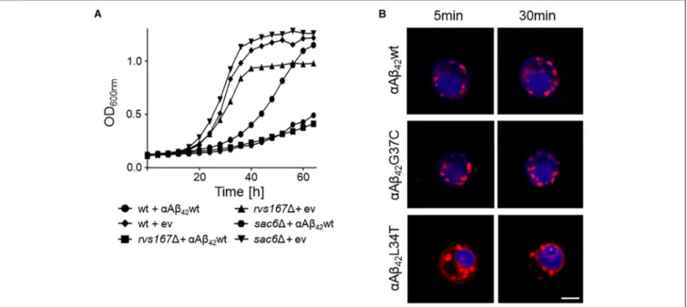
FIGURE 4 | Interference ofαAβ42with endocytosis. (A) Growth profiles of wild-type cells and rvs1671 or sac61 cells transformed with the empty vector (ev) or a
construct allowing for expression ofαAβ42wt when grown on galactose-containing medium. (B) Fluorescence microscopy pictures of strains expressing the toxic
αAβ42wt andαAβ42G37C or the non-toxicαAβ42L34T stained with the endocytosis tracker FM4-64 (red) and CMAC (blue), a dye to stain the vacuolar lumen. The
scale bar represents 2µm.
body (MVB) pathway. The two strains with the strongest enhanced toxicity were those carrying a deletion of SAC6 or RVS167. Sac6 is an actin-bundling protein that is required for endocytosis (Penalver et al., 1997;Gheorghe et al., 2008). Rvs167 is a homolog of mammalian amphiphysin that interacts with actin as well and that functions in the internalization step of endocytosis (Lombardi and Riezman, 2001). As illustrated for the expression ofαAβ42wt, we indeed observed a similar severe
growth phenotype in both the sac61 and rvs1671 mutants as compared to the isogenic wild-type strain (Figure 4A). To further confirm the link between endocytosis andαAβ42toxicity, we also
monitored the uptake of the endocytosis tracker FM4-64 by wild-type cells expressing either the toxicαAβ42wt andαAβ42G37C
and non-toxic αAβ42L34T isoforms after 4 h induction on
galactose-containing medium. As shown, FM4-64 already stained the vacuolar membrane within 30 min in cells expressing the non-toxic construct while no, or only a minimal staining of the vacuolar membrane was observed in cells expressing the toxic αAβ42 species, even not after 60 min of incubation (Figure 4B).
This demonstrates that the latter have a direct impact on the endocytic process.
αAβ
42Enhances the Occurrence of
Plasma Membrane Lesions and
Formation of Lipid Droplets
Closely connected to endocytosis, we noticed that our screening retrieved some ESCRT components, which function in the MVB pathway that is required for the turnover of plasma membrane proteins and lipids. The first step in MVB formation is the binding of ESCRT-0 (Hse1 and Vps27) and ESCRT-I (involving
Vps28, Mvb12, Srn2, and Stp22) to ubiquitinated MVB cargoes. Next, the ESCRT-II complex (involving Vps25, Vps36, and Snf8) mediates the recruitment of ESCRT-III accessory factors (Bro1 and Doa4), which in turn loads general ESCRT-III factors (Vps20, Snf7, Did4, Chm7, Ist1, and Vps24) to direct the continued sorting of cargoes into invaginating vesicles during MVB formation. ESCRT-III dissociation factors (Vps4, Vps60, Did2, and Vta1) mediate the release and recycling of all involved factors (Hurley, 2010; Babst, 2011). All the 21 corresponding genes are not essential and could therefore be tested for their implications in αAβ42 toxicity. After transformation of the
corresponding KO strains with either αAβ42wt, αAβ42G37C
or the ev-αGFP control construct, we evaluated the impact of the deletions on αAβ42 toxicity through spot assays. This
revealed that for several components of the MVB pathway their deletion significantly aggravated αAβ42 toxicity. This included
the ESCRTIII components Did4, the ESCRTIII accessory components Bro1 and Doa4 as well as the ESCRTIII dissociation mediator Vps4 (Supplementary Figures S3, S4). Recent studies in mammalian cells have demonstrated that besides its role in the MVB pathway, ESCRT plays key roles in a variety of other processes, including membrane lesion repair (Jimenez et al., 2014; Sundquist and Ullman, 2015; Campsteijn et al., 2016; Denais et al., 2016; Raab et al., 2016). It is well-established that Aβ42 can introduce membrane lesions and different
non-excluding mechanisms were proposed, including membrane lipid interaction, alterations in membrane fluidity, pore formation, or lipid oxidation (Eckert et al., 2000; Muller et al., 2001; Ambroggio et al., 2005; Rauk, 2008; Axelsen et al., 2011;Viola and Klein, 2015). We speculated that αAβ42 would also trigger
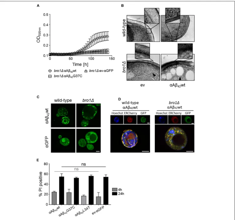
FIGURE 5 | The role of Bro1 for membrane lesion repair. (A) Growth profile on galactose-containing medium of a strain deleted for the ESCRT-III accessory factor BRO1 transformed with an empty vector (ev-αGFP) or constructs allowing for expression of αAβ42wt orαAβ42G37C. Cryo-EM pictures (B) and fluorescence
microscopy pictures (C) of wild-type and bro11 cells transformed with an empty vector (ev) or expressing αAβ42wt and grown for 6 h on galactose-containing
medium. The indents in panel (B) zoom in on the plasma membrane and cell wall. The black arrowhead in (B) indicates a lipid droplet. Scale bars for cryo-EM pictures represent 200 nm. (D) BY4742 wild-type and a bro11 strains transformed with a plasmids carrying αAβ42wt and additionally a plasmid allowing the
expression of Kar2(1−135)-mCherry-HDEL (ERCherry), a marker for the ER. DNA was stained with Hoechst. Cells were grown in medium allowing for gene expression for 6 h prior to microscopy. Scale bars for fluorescence pictures represent 2µm. (E) PI staining of cells deleted for BRO1 transformed with constructs allowing for expression ofαAβ42wt,αAβ42L34T,αAβ42G37C, orαGFP after 4 or 24 h growth on galactose-containing medium. Error bars represent standard
deviations of at least four independent transformants.
the plasma membrane integrity of wild-type and bro11 cells by cryo-EM. We chose thebro11 strain because here αAβ42wt
and αAβ42G37C was almost lethal (Figure 5A) and because
Bro1 is the yeast ortholog of human Alix, a proposed biomarker for AD (Sun et al., 2015). Consistent with our hypothesis, the cryo-EM study showed that the plasma membrane of bro11
cells expressing αAβ42wt seemed heavily corrugated while the
bro11 cells transformed with empty vector displayed a more modest phenotype. In the wild-type strain, the plasma membrane remained ostensibly smooth even upon expression ofαAβ42wt
(Figure 5B). However, although we did not quantify, the observed membrane corrugating effects of specifically thebro11
strain expressing αAβ42wt are strikingly obvious. Moreover,
when compared to the wild-type cells, the ER morphology in thebro11 mutant was completely different and appeared to be deteriorated. Indeed, only a minimal perinuclear and cortical ER was detected and the cells displayed theαAβ42-linker-GFP
fusion mostly in filamentous structures and foci (Figures 5C,D). Thus, expression of αAβ42wt seems to dramatically affect all
membranous structures evidencing that Bro1, and by extension the ESCRT system, is absolutely required for the repair of membrane lesions induced byαAβ42. In line with this essential
requirement of ESCRT, we observed that while still seeing a tendency of increased PI uptake after 4 h induction for those bro11 cells expressing toxic αAβ42, a maximal PI uptake in all
bro11 transformants is seen after 24 h, even for those strains expressing the non-toxic αAβ42L34T mutant or the ev-αGFP
control (Figure 5E).
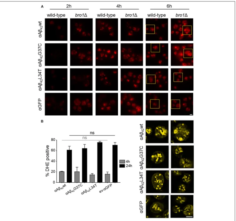
The cryo-EM study also pointed to the formation of cortical vesicle-like structures, which we believe may correspond to lipid droplets. Since previous studies have shown that the formation of lipid droplets denotes an adaptive response to a chronic lipid imbalance (Vevea et al., 2015; Garcia et al., 2018), which is likely to occur in bro11 strain because of hampered MVB formation and lipid turnover, we decided to perform a Nile Red staining to visualize the droplets. While during the first hours of induction on galactose-containing medium we observed an overall enhanced lipid droplet biogenesis in allbro11 strains as compared to the respective wild-type strains, the increase was persistent and especially more dense droplets were seen with bro11 cells expressing αAβ42G37C orαAβ42wt (Figure 6A). This
observation confirms previously reported data on an increased lipid droplet load in an Aβ42wild-type strain (Chen et al., 2017).
The perturbation of lipid homeostasis has also been linked to defects in the ER and mitochondria (Vevea et al., 2015), and the latter incited us to monitor the level of ROS in thebro11 strains. As shown, and comparable to the PI uptake, we found similar high levels of ROS in all strains tested indicative that this is a characteristic mainly associated to the deletion ofBRO1 itself (Figure 6B).
DISCUSSION
During the past decades, several studies validated the use of yeast to decipher the pathobiology underlying a variety of human disorders. Especially for degenerative protein folding diseases, like Huntington’s, Parkinson’s, or Alzheimer’s disease, this led to the discovery of processes and molecular pathways contributing to cell demise (Winderickx et al., 2008; Franssens et al., 2010; Swinnen et al., 2011; Porzoor and Macreadie, 2013; Tenreiro et al., 2013, 2017;Chen and Petranovic, 2016;Jiang et al., 2016; Verduyckt et al., 2016; Fruhmann et al., 2017). The insight gained from these studies were not only relevant in the context of disease, but they also clarified fundamental aspects on how a cell manages to maintain proteostasis and the consequences in case this system fails. In this paper, we used a previously reported model to study the repercussions when the APP-derived peptide Aβ42is expressed in yeast (D’Angelo et al., 2013;Vignaud
et al., 2013). Though this model makes use of a GFP fusion, the use of the super-toxic Aβ42G37C and the non-toxic Aβ42L34T
mutants clearly demonstrated that the properties of the GFP fusion are dictated by the Aβ42peptide moiety. Furthermore, by
comparing constructs with or without an N-terminal fusion with the α-prepro sequence and conditions that retain the α-prepro in the ER, it became obvious that the processing in the ER/Golgi system and the subsequent shuttling into the secretory pathway is essential to unleash the full toxic capacity of the Aβ42
-linker-GFP fusion. Our data also contradict the argument that theαAβ42
instigated toxicity would simply be due to an overload of the ER/Golgi processing system because both wild-type and mutant αAβ42 are processed in the same manner (D’Angelo et al., 2013; Vignaud et al., 2013) and no toxicity is seen in case of expression of mutantαAβ42L34T.
The use of our yeast model allowed us to confirm some data previously reported. This included the impact of αAβ42
on endocytosis (Treusch et al., 2011; D’Angelo et al., 2013), where we now show that strains with a deletion of SAC6 or RVS167 display an increased αAβ42 toxicity, as well as
the impact on mitochondrial functioning (D’Angelo et al., 2013; Chen and Petranovic, 2015; Chen et al., 2017), which we illustrated by co-localization studies and the observation that αAβ42 enhances ROS formation. These data are relevant
because, indeed, changes in endocytic capacity and mitochondrial dysfunction are typically seen in the pathogenesis of AD and have been observed in other AD models as well (Thomas et al., 2011;Wang et al., 2011;Avetisyan et al., 2016;Schreij et al., 2016; Dixit et al., 2017;Shoshan-Barmatz et al., 2018;Xu et al., 2018). However, the observed partial co-localization of toxic αAβ42
with mitochondria may either indicate a direct interaction of the peptide with this organelle or it may simply be a reflection of the interaction between the ER and mitochondria through the membrane contact sites known as ERMES (Kornmann et al., 2009). This aspect needs to be analyzed in more detail. Interestingly, mitochondria associated membranes (MAMs), the mammalian counterpart of ERMES, have already been implicated in AD (Area-Gomez and Schon, 2016;Paillusson et al., 2016). Closely related to the observed mitochondrial dysfunction is our observation that deletion of AIF1, encoding a cell death effector, has a protective effect on the αAβ42 expressing yeast
cells. This suggests that αAβ42 may induce an apoptotic-like
program in yeast, which fits the finding that neuronal cells die through apoptosis in AD (Ohyagi et al., 2005; Shoshan-Barmatz et al., 2018). Our data also show that particularly the expression of the toxic αAβ42 isoforms is linked to the
formation of ER-associated foci and filamentous structures. Though we did not study these structures in detail and previously believed these to represent vesicles (D’Angelo et al., 2013), it is well-possible that they may in fact be ER aggregates or clustering of ER membranes, which are both indicative for ER stress (Varadarajan et al., 2012, 2013; Vevea et al., 2015). Also ER stress is associated to AD (Gerakis and Hetz, 2018) and several links between ER stress and mitochondrial dysfunction have been proposed in this neurodegenerative disorder (Costa et al., 2012; Barbero-Camps et al., 2014; Erpapazoglou et al., 2017).
FIGURE 6 |αAβ42instigated lipid droplet biogenesis. (A) Nile Red stainings (top panel), a marker for lipid droplets, of wild-type and the bro11 cells expressing
αAβ42wt,αAβ42G37C, orαAβ42L34T orαGFP after 2, 4, or 6 h growth on galactose-containing medium. The bottom panel (yellow) shows magnifications and
deconvolved parts of the top-panel pictures at time point of 6 h. Note that due to deconvolution the picture intensities are enhanced. Scale bars represent 2µm. (B) DHE staining of cells deleted for BRO1 transformed with constructs allowing for expression ofαAβ42wt,αAβ42L34T,αAβ42G37C, orαGFP after 4 or 24 h growth
on galactose-containing medium. Error bars represent standard deviations of at least four independent transformants.
One of the most striking observations made in our studies is the role of ESCRT in modulating theαAβ42toxicity. The ESCRT
system functions in the MVB pathway and several studies have linked this role of ESCRT to AD. Neurons of AD transgenic mice were shown to display enlarged MVBs as compared to the neurons of wild-type mice, and ESCRT was demonstrated to modulate intracellular Aβ42 accumulation by directing APP
to lysosomal degradation and by enhancing Aβ42 secretion.
In addition, ESCRT components were found associated with
amyloid plaques in transgenic mice and to granular structures hippocampal neurons of AD diseased human brain (Yamazaki et al., 2010; Edgar et al., 2015; Willen et al., 2017). However, apart from its function in the MVB pathway, ESCRT is also required for the repair of membrane lesions. Here, both cryo-EM and PI-staining give a strong impression of the presence of such lesions at the plasma membrane of cells lacking the ESCRT component Bro1, probably explaining in part the sick phenotype of the bro11 mutant. As such, our data strongly
suggest that the role of ESCRT for plasma membrane repair, which so far was only demonstrated in mammalian cells, is evolutionary well-conserved. Interestingly, we found that the disruption of the plasma membrane integrity in the bro11 strain is dramatically exacerbated upon expression ofαAβ42and
that this came along with the deterioration of the ER and an almost lethal phenotype. This is intriguing for several reasons. It demonstrates that when the fully processed Aβ42-linker-GFP
arrives at the plasma membrane, the peptide further aggravates plasma membrane disruption, which given the observed effect on the ER, might well be involving fusion of secretory vesicles that contain disordered membranes. The fact that our screens retrieved the KO strain lacking SSO2 as suppressor ofαAβ42
toxicity favors the last possibility. Indeed,SSO2 encodes a plasma membrane t-SNARE that is required for fusion secretory vesicles (Grote et al., 2000). Moreover, the data recapitulate observations made for AD where, as mentioned, Aβ42was shown to introduce
membrane lesions via different non-excluding mechanisms, including membrane lipid interaction, alterations in membrane fluidity, pore formation, or lipid oxidation (Eckert et al., 2000; Muller et al., 2001;Ambroggio et al., 2005;Rauk, 2008;Axelsen et al., 2011;Viola and Klein, 2015).
Given the effect of Aβ42 on plasma membrane integrity, the
presence of this peptide also impacts on the overall cellular lipid homeostasis. In fact, extensive lipid alterations are implicated in the AD disease pathology but it is still a matter of debate whether such alterations are the cause or the consequence of AD (Grosgen et al., 2010;Xiang et al., 2015;El Gaamouch et al., 2016). In yeast, the expression of Aβ42has been linked to a transcriptional upshift
of key regulators of lipid metabolism as well as an enhanced formation of lipid droplets (Chen et al., 2017). Our screens with the yeast deletion collection and our Nile Red stainings support the link between Aβ42 and lipid metabolism. Intriguingly, a
recent study demonstrated lipid droplet formation to be an adaptive response to an acute lipid imbalance in yeast cells. The same study then also showed that the biogenesis of these droplets occurs at ER aggregates (Vevea et al., 2015), which we
believe to correspond to the ER-associated foci and filamentous structures seen when yeast cells express toxic forms of αAβ42,
as mentioned. Notably, also in transgenic mouse models of AD an enhanced lipid droplet formation is observed (Hamilton et al., 2010;Yang et al., 2014), again underscoring the relevance of the data obtained in yeast.
AUTHOR CONTRIBUTIONS
GF, CM, HV, MV, and NT performed experiments and analyzed data. GF and JW wrote the draft. CDV, CC, and JW contributed conception and design of the studies. CC and JW corrected and edited the draft.
FUNDING
This work was financially supported by the KU Leuven and the FWO Vlaanderen (Project No. G0A6315N). CDV was supported by the Swiss National Science Foundation and the Canton of Fribourg.
ACKNOWLEDGMENTS
We want to thank Joëlle Rosseels, Dorien Vliegen, and Elodie Cougouille for technical and experimental support. We also want to thank Dr. B. Salin and to the Bordeaux Imaging Center (BIC) for performing TEM experiments.
SUPPLEMENTARY MATERIAL
The Supplementary Material for this article can be found online at: https://www.frontiersin.org/articles/10.3389/fnmol. 2018.00406/full#supplementary-material
REFERENCES
Abramov, E., Dolev, I., Fogel, H., Ciccotosto, G. D., Ruff, E. (2009). Amyloid-β as a positive endogenous regulator of release probability at hippocampal synapses. Nat. Neurosci. 12, 1567–1576. doi: 10.1038/nn.2433
Alexandrov, A. I., Serpionov, G. V., Kushnirov, V. V., and Ter-Avanesyan, M. D. (2016). Wild type huntingtin toxicity in yeast: Implications for the role of amyloid cross-seeding in polyQ diseases. Prion 10, 221–227. doi: 10.1080/ 19336896.2016.1176659
Ambroggio, E. E., Kim, D. H., Separovic, F., Barrow, C. J., Barnham, K. J., Bagatolli, L. A., et al. (2005). Surface behavior and lipid interaction of Alzheimer β-amyloid peptide 1-42: a membrane-disrupting peptide. Biophys. J. 88, 2706– 2713. doi: 10.1529/biophysj.104.055582
Area-Gomez, E., and Schon, E. A. (2016). Mitochondria-associated ER membranes and Alzheimer disease.Curr. Opin. Genet. Dev. 38, 90–96. doi: 10.1016/j.gde. 2016.04.006
Avetisyan, A. V., Samokhin, A. N., Alexandrova, I. Y., Zinovkin, R. A., Simonyan, R. A., and Bobkova, N. V. (2016). Mitochondrial dysfunction in neocortex and hippocampus of olfactory bulbectomized mice, a model of Alzheimer’s disease. Biochemistry (Mosc) 81, 615–623. doi: 10.1134/S00062979160 60080
Axelsen, P. H., Komatsu, H., and Murray, I. V. (2011). Oxidative stress and cell membranes in the pathogenesis of Alzheimer’s disease.Physiology (Bethesda) 26, 54–69. doi: 10.1152/physiol.00024.2010
Babst, M. (2011). MVB vesicle formation: ESCRT-dependent, ESCRT-independent and everything in between.Curr. Opin. Cell Biol. 23, 452–457. doi: 10.1016/j. ceb.2011.04.008
Barbero-Camps, E., Fernandez, A., Baulies, A., Martinez, L., Fernandez-Checa, J. C., and Colell, A. (2014). Endoplasmic reticulum stress mediates amyloid beta neurotoxicity via mitochondrial cholesterol trafficking.Am. J. Pathol. 184, 2066–2081. doi: 10.1016/j.ajpath.2014.03.014
Belden, W. J., and Barlowe, C. (2001). Role of Erv29p in collecting soluble secretory proteins into ER-derived transport vesicles.Science 294, 1528–1531. doi: 10. 1126/science.1065224
Benilova, I., Karran, E., and De Strooper, B. (2012). The toxic Aβ oligomer and Alzheimer’s disease: an emperor in need of clothes.Nat. Neurosci. 15, 349–357. doi: 10.1038/nn.3028
Campsteijn, C., Vietri, M., and Stenmark, H. (2016). Novel ESCRT functions in cell biology: spiraling out of control?Curr. Opin. Cell Biol. 41, 1–8. doi: 10.1016/j. ceb.2016.03.008
Chen, X., Bisschops, M. M. M., Agarwal, N. R., Ji, B., Shanmugavel, K. P., and Petranovic, D. (2017). Interplay of energetics and ER stress exacerbates
Alzheimer’s amyloid-beta (Abeta) toxicity in yeast.Front. Mol. Neurosci. 10:232. doi: 10.3389/fnmol.2017.00232
Chen, X., and Petranovic, D. (2015). Amyloid-β peptide-induced cytotoxicity and mitochondrial dysfunction in yeast.FEMS Yeast Res. 15, 1–21. doi: 10.1093/ femsyr/fov061
Chen, X., and Petranovic, D. (2016). Role of frameshift ubiquitin B protein in Alzheimer’s disease. Wiley Interdiscip. Rev. Syst. Biol. Med. 8, 300–313. doi: 10.1002/wsbm.1340
Costa, R. O., Ferreiro, E., Martins, I., Santana, I., Cardoso, S. M., Oliveira, C. R., et al. (2012). Amyloid beta-induced ER stress is enhanced under mitochondrial dysfunction conditions.Neurobiol. Aging 33, 824.e825-e816. doi: 10.1016/j. neurobiolaging.2011.04.011
D’Angelo, F., Vignaud, H., Di Martino, J., Salin, B., Devin, A., Cullin, C., et al. (2013). A yeast model for amyloid-beta aggregation exemplifies the role of membrane trafficking and PICALM in cytotoxicity.Dis. Model Mech. 6, 206–216. doi: 10.1242/dmm.010108
Dean, N., and Pelham, H. R. (1990). Recycling of proteins from the golgi compartment to the ER in yeast.J. Cell Biol. 111, 369–377. doi: 10.1083/jcb. 111.2.369
Denais, C. M., Gilbert, R. M., Isermann, P., McGregor, A. L., te Lindert, M., Weigelin, B., et al. (2016). Nuclear envelope rupture and repair during cancer cell migration.Science (New York, N.Y.) 352, 353–358. doi: 10.1126/science. aad7297
Dittmar, J. C., Reid, R. J., and Rothstein, R. (2010). ScreenMill: a freely available software suite for growth measurement, analysis and visualization of high-throughput screen data.BMC Bioinformatics 11:353. doi: 10.1186/1471-2105-11-353
Dixit, S., Fessel, J. P., and Harrison, F. E. (2017). Mitochondrial dysfunction in the APP/PSEN1 mouse model of Alzheimer’s disease and a novel protective role for ascorbate.Free Radic. Biol. Med. 112, 515–523. doi: 10.1016/j.freeradbiomed. 2017.08.021
Eckert, G. P., Cairns, N. J., Maras, A., Gattaz, W. F., and Muller, W. E. (2000). Cholesterol modulates the membrane-disordering effects of beta-amyloid peptides in the hippocampus: specific changes in Alzheimer’s disease.Dement. Geriatr. Cogn. Disord. 11, 181–186. doi: 10.1159/000017234
Edgar, J. R., Willen, K., Gouras, G. K., and Futter, C. E. (2015). ESCRTs regulate amyloid precursor protein sorting in multivesicular bodies and intracellular amyloid-beta accumulation.J. Cell Sci. 128, 2520–2528. doi: 10.1242/jcs.170233 El Gaamouch, F., Jing, P., Xia, J., and Cai, D. (2016). Alzheimer’s disease risk genes
and lipid regulators.J. Alzheimers Dis. 53, 15–29. doi: 10.3233/JAD-160169 Erpapazoglou, Z., Mouton-Liger, F., and Corti, O. (2017). From dysfunctional
endoplasmic reticulum-mitochondria coupling to neurodegeneration. Neurochem. Int. 109, 171–183. doi: 10.1016/j.neuint.2017.03.021
Foury, F. (1997). Human genetic diseases: a cross-talk between man and yeast.Gene 195, 1–10. doi: 10.1016/S0378-1119(97)00140-6
Franssens, V., Boelen, E., Anandhakumar, J., Vanhelmont, T., Buttner, S., and Winderickx, J. (2010). Yeast unfolds the road map toward alpha-synuclein-induced cell death.Cell Death Differ. 17, 746–753. doi: 10.1038/cdd.2009.203 Fruhmann, G., Seynnaeve, D., Zheng, J., Ven, K., Molenberghs, S., Wilms, T., et al.
(2017). Yeast buddies helping to unravel the complexity of neurodegenerative disorders.Mech. Ageing Dev. 161(Pt B), 288–305. doi: 10.1016/j.mad.2016.05. 002
Garcia, E. J., Vevea, J. D., and Pon, L. A. (2018). Lipid droplet autophagy during energy mobilization, lipid homeostasis and protein quality control. Front. Biosci. (Landmark Ed) 23, 1552–1563. doi: 10.2741/4660
Gerakis, Y., and Hetz, C. (2018). Emerging roles of ER stress in the etiology and pathogenesis of Alzheimer’s disease.FEBS J. 285, 995–1011. doi: 10.1111/febs. 14332
Gheorghe, D. M., Aghamohammadzadeh, S., Smaczynska-de, R. II, Allwood, E. G., Winder, S. J., and Ayscough, K. R. (2008). Interactions between the yeast SM22 homologue Scp1 and actin demonstrate the importance of actin bundling in endocytosis. J. Biol. Chem. 283, 15037–15046. doi: 10.1074/jbc.M71033 2200
Grosgen, S., Grimm, M. O., Friess, P., and Hartmann, T. (2010). Role of amyloid beta in lipid homeostasis.Biochim. Biophys. Acta 1801, 966–974. doi: 10.1016/j. bbalip.2010.05.002
Grote, E., Carr, C. M., and Novick, P. J. (2000). Ordering the final events in yeast exocytosis.J. Cell Biol. 151, 439–452. doi: 10.1083/jcb.151.2.439
Hamilton, L. K., Aumont, A., Julien, C., Vadnais, A., Calon, F., and Fernandes, K. J. (2010). Widespread deficits in adult neurogenesis precede plaque and tangle formation in the 3xTg mouse model of Alzheimer’s disease.Eur. J. Neurosci. 32, 905–920. doi: 10.1111/j.1460-9568.2010.07379.x
Hamza, A., Tammpere, E., Kofoed, M., Keong, C., Chiang, J., Giaever, G., et al. (2015). Complementation of yeast genes with human genes as an experimental platform for functional testing of human genetic variants.Genetics 201, 1263– 1274. doi: 10.1534/genetics.115.181099
Hu, W., Wang, Z., and Zheng, H. (2018). Mitochondrial accumulation of amyloid beta (Abeta) peptides requires TOMM22 as a main Abeta receptor in yeast. J. Biol. Chem. 293, 12681–12689. doi: 10.1074/jbc.RA118.002713
Hurley, J. H. (2010). The ESCRT complexes.Crit. Rev. Biochem. Mol. Biol. 45, 463–487. doi: 10.3109/10409238.2010.502516
Ittner, L. M., and Gotz, J. (2011). Amyloid-beta and tau–a toxic pas de deux in Alzheimer’s disease.Nat. Rev. Neurosci. 12, 65–72. doi: 10.1038/nrn2967 Jackrel, M. E., DeSantis, M. E., Martinez, B. A., Castellano, L. M., Stewart, R. M.,
Caldwell, K. A., et al. (2014). Potentiated Hsp104 variants antagonize diverse proteotoxic misfolding events.Cell 156, 170–182. doi: 10.1016/j.cell.2013.11.047 Jiang, Y., Chadwick, S. R., and Lajoie, P. (2016). Endoplasmic reticulum stress: The cause and solution to Huntington’s disease?Brain Res. 1648, 650–657. doi: 10.1016/j.brainres.2016.03.034
Jimenez, A. J., Maiuri, P., Lafaurie-Janvore, J., Divoux, S., Piel, M., and Perez, F. (2014). ESCRT machinery is required for plasma membrane repair.Science 343:1247136. doi: 10.1126/science.1247136
Kagan, B. L., Jang, H., Capone, R., Teran Arce, F., Ramachandran, S., Lal, R., et al. (2012). Antimicrobial properties of amyloid peptides.Mol. Pharm. 9, 708–717. doi: 10.1021/mp200419b
Kim, J., and Klionsky, D. J. (2000). Autophagy, cytoplasm-to-vacuole targeting pathway, and pexophagy in yeast and mammalian cells.Annu. Rev. Biochem. 69, 303–342. doi: 10.1146/annurev.biochem.69.1.303
Kornmann, B., Currie, E., Collins, S. R., Schuldiner, M., Nunnari, J., Weissman, J. S., et al. (2009). An ER-mitochondria tethering complex revealed by a synthetic biology screen. Science 325, 477–481. doi: 10.1126/science.117 5088
Kumar, D. K., Choi, S. H., Washicosky, K. J., Eimer, W. A., Tucker, S., Ghofrani, J., et al. (2016). Amyloid-beta peptide protects against microbial infection in mouse and worm models of Alzheimer’s disease.Sci. Transl. Med. 8:340ra372. doi: 10.1126/scitranslmed.aaf1059
LaFerla, F. M., Green, K. N., and Oddo, S. (2007). Intracellular amyloid-β in Alzheimer’s disease.Nat. Rev. Neurosci. 8, 499–509. doi: 10.1038/nrn2168 Lefebvre-Legendre, L., Salin, B., Schaeffer, J., Brethes, D., Dautant, A., Ackerman,
S. H., et al. (2005). Failure to assemble the alpha 3 beta 3 subcomplex of the ATP synthase leads to accumulation of the alpha and beta subunits within inclusion bodies and the loss of mitochondrial cristae in Saccharomyces cerevisiae. J. Biol. Chem. 280, 18386–18392. doi: 10.1074/jbc.M4107 89200
Lewis, M. J., and Pelham, H. R. B. (1990). A human homologue of the yeast HDEL receptor.Nature 348, 162–163. doi: 10.1038/348162a0
Liu, W., Li, L., Ye, H., Chen, H., Shen, W., Zhong, Y., et al. (2017). From Saccharomyces cerevisiae to human: the important gene co-expression modules. Biomed. Rep. 7, 153–158. doi: 10.3892/br.2017.941
Lombardi, R., and Riezman, H. (2001). Rvs161p and Rvs167p, the two yeast amphiphysin homologs, function together in vivo. J. Biol. Chem. 276, 6016–6022. doi: 10.1074/jbc.M008735200
Muller, W. E., Kirsch, C., and Eckert, G. P. (2001). Membrane-disordering effects of beta-amyloid peptides.Biochem. Soc. Trans. 29(Pt 4), 617–623. doi: 10.1042/ bst0290617
Mumberg, D., Müller, R., and Funk, M. (1994). Regulatable promoters of Saccharomyces cerevisiae: comparison of transcriptional activity and their use for heterologous expression.Nucleic Acids Res. 22, 5767–5768. doi: 10.1093/nar/ 22.25.5767
Nice, D. C., Sato, T. K., Stromhaug, P. E., Emr, S. D., and Klionsky, D. J. (2002). Cooperative binding of the cytoplasm to vacuole targeting pathway proteins, Cvt13 and Cvt20, to phosphatidylinositol 3-phosphate at the pre-autophagosomal structure is required for selective autophagy.J. Biol. Chem. 277, 30198–30207. doi: 10.1074/jbc.M204736200
Ohyagi, Y., Asahara, H., Chui, D. H., Tsuruta, Y., Sakae, N., Miyoshi, K., et al. (2005). Intracellular Abeta42 activates p53 promoter: a pathway to