NBS1 promotes the endonuclease of the
MRE11-RAD50 complex by sensing CtIP phosphorylation
Postprint (Author accepted manuscript)
Anand, Roopesh; Institute for Research in Biomedicine (IRB), Faculty of
Biomedical Sciences, Università della Svizzera italiana, Switzerland
Jasrotia, Arti; Department of Gynecology, University of Zurich, Switzerland
Bundschuh, Diana; Department of Gynecology, University of Zurich,
Switzerland
Howard, Sean Michael; Institute for Research in Biomedicine (IRB), Faculty of
Biomedical Sciences, Università della Svizzera italiana, Switzerland
Ranjha, Lepakshi; Institute for Research in Biomedicine (IRB), Faculty of
Biomedical Sciences, Università della Svizzera italiana, Switzerland
Stucki, Manuel; Department of Gynecology, University of Zurich, Switzerland
Cejka, Petr; Institute for Research in Biomedicine (IRB), Faculty of
Biomedical Sciences, Università della Svizzera italiana, Switzerland - Institute
of Biochemistry, Department of Biology, ETH Zürich, Switzerland
The EMBO Journal
2019 / Vol. 38 / 7 / e101005
Published version:
https://doi.org/10.15252/embj.2018101005
Online publication: 2019-02-20
Abstract DNA end resection initiates DNA break repair by homologous recombination. MRE11- RAD50-NBS1 and phosphorylated CtIP perform the first resection step by MRE11-catalyzed endonucleolytic DNA cleavage. Human NBS1, more than its Xrs2 homologue from Saccharomyces cerevisiae, is crucial for this process, highlighting complex mecha- nisms that regulate the MRE11 nuclease in high eukaryotes. Using a reconstituted sys-tem, we show here that NBS1, through its FHA and BRCT domains, functions as a sensor of CtIP phosphorylation. NBS1 then activates the MRE11-RAD50 nuclease through direct physical interactions with MRE11. In absence of NBS1, MRE11-RAD50 exhibits a weaker nuclease activity, which requires CtIP but not strictly its phosphorylation. This identifies at least two mechanisms by which CtIP promotes MRE11: a phosphorylation-dependent mode through NBS1, and a phosphorylation-independent mode without NBS1. In sup-port, we show that limited DNA end resection in absence of the FHA and BRCT domains of NBS1 occurs in vivo. Collectively, our data suggest that NBS1 restricts the MRE11-RAD50 nuclease to S-G2 phase when CtIP is extensively phosphorylated. This defines mechanisms that regulate the MRE11 nuclease in DNA metabolism.
Introduction The maintenance of genomic integrity is critically important as cells are constantly ex- posed to various genotoxic agents. DNA double-strand breaks (DSBs) are difficult to re-pair due to the loss of genetic information in both DNA strands. While unrepaired DSBs may result in cell death, their inaccurate repair can lead to mutagenesis and inappropri-ate chromosomal translocations (Ranjha, Howard et al., 2018). Cells possess two main mechanisms for DSB repair. This includes end-joining pathways including Ku-dependent non-homologous end-joining (NHEJ) and Ku-independent microhomology-mediated end-joining (MMEJ), which are functional in any phase of the cell cycle and do not re- quire a DNA template (Ranjha et al., 2018). The second mechanism is template-dependent homologous recombination (HR), which is generally restricted to the S-G2 phase of the cell cycle. The sister chromatid serves as an HR template in most cases in vegetative cells, explaining why recombination can only function in the cell cycle phases when sister chromatids are available (Ranjha et al., 2018). This is achieved by a regula-tory mechanism involving cyclin-dependent kinases (CDKs), which phosphorylate key factors that function in the first step of the HR pathway termed DNA end resection (Huertas, Cortes-Ledesma et al., 2008, Huertas & Jackson, 2009). Resection involves nu-cleolytic degradation of the 5'-terminated DNA strands at DSBs, leading to 3' overhangs that are essential for the downstream steps in the recombination pathway. While lim- ited DNA end resection may be involved in MMEJ, DNA that underwent extended resec- tion is no longer ligatable and therefore unsuitable for end-joining. The decision wheth-er and to which extent to resect DNA ends therefore regulates the pathway choice in DSB repair (Ranjha et al., 2018). The MRE11-RAD50-NBS1 (MRN, in human cells) or Mre11-Rad50-Xrs2 (MRX, in bud-ding yeast) complex has multiple key evolutionarily conserved functions to initiate and coordinate the repair of DSBs (Paull, 2010, Stracker & Petrini, 2011). This includes roles in both end-joining and HR pathways (Carney, Maser et al., 1998, de Jager, van Noort et al., 2001, Moore & Haber, 1996, Paull & Gellert, 1998, Paull & Gellert, 1999, Rass, Grabarz et al., 2009). Additionally, MRN promotes DNA end tethering and is required to signal the presence of DSBs via the ATM kinase (Carney et al., 1998, de Jager et al., 2001, Lee & Paull, 2005, Stewart, Maser et al., 1999, Usui, Ogawa et al., 2001, Williams, Moncalian et al., 2008). This is achieved by a conserved interaction between the MRN subunit NBS1 (Xrs2 in S. cerevisiae) and ATM (Tel1 in yeast). ATM in turn phosphory-lates hundreds of protein targets that regulate the response to DSBs. Beyond ATM, NBS1 also interacts with MDC1, which binds phosphorylated proteins, including H2AX
(γH2AX), to amplify the DNA damage signaling beyond the vicinity of the DSB (Goldberg, Stucki et al., 2003). In recombination, the MRN complex has a direct role in DNA end re-section mediated by the MRE11 nuclease in the S-G2 phase of the cell cycle. MRE11 is likely the first nuclease to resect DSBs, which is particularly important for the pro-cessing of breaks with secondary DNA structures or protein adducts such as stalled topoisomerases or Ku (Anand, Ranjha et al., 2016, Deshpande, Lee et al., 2016, Reginato, Cannavo et al., 2018, Shibata, Moiani et al., 2014, Wang, Daley et al., 2017). The current models posit that resection by MRE11 is initiated by endonucleolytic DNA cleavage in-ternal to the DSB past the protein block, followed by 3'→5' exonucleolytic degradation back toward the DNA end (Cannavo & Cejka, 2014, Garcia, Phelps et al., 2011, Keeney & Kleckner, 1995, Neale, Pan et al., 2005, Shibata et al., 2014). The endonucleolytic cleav-age sites also represent entry sites for processive DNA end resection nucleases that function downstream of MRN in the 5'→3' direction, including EXO1 and DNA2 (Gravel, Chapman et al., 2008, Mimitou & Symington, 2008, Nimonkar, Genschel et al., 2011, Zhu, Chung et al., 2008). The MRN nuclease furthermore likely functions to promote MMEJ independently of the cell cycle, which to date remains very poorly defined (Deng, Gibb et al., 2014, Ma, Kim et al., 2003, Rahal, Henricksen et al., 2010, Sharma, Javadekar et al., 2015, Taylor, Cecillon et al., 2010, Truong, Li et al., 2013). The processing of protein-blocked DSBs is an evolutionarily conserved capacity of MRE11 homologues and their co-factors. In bacteria, the MRE11-RAD50-like SbcC-SbcD complex endonucleotically cleaves DNA near blocked DNA ends (Connelly, de Leau et al., 2003). Eukaryotic cells possess a third member of the MRE11-RAD50 complex, NBS1/Xrs2, as well as CtIP/Sae2, which function as co-factors of the MRE11 nuclease in resection (Carney et al., 1998, Sartori, Lukas et al., 2007). In S. cerevisiae, Sae2 phos- phorylated by CDK and other kinases has a critical function to promote the Mre11-Rad50 endonuclease near protein blocks (Cannavo & Cejka, 2014, Huertas et al., 2008, Reginato et al., 2018, Wang et al., 2017), while Xrs2 per se is not essential for the DNA clipping reaction in vitro (Oh, Al-Zain et al., 2016), but may have a stimulatory function (Wang et al., 2017). It has been demonstrated that Xrs2 is responsible for the nuclear import of the MRX complex (Carney et al., 1998, Tsukamoto, Mitsuoka et al., 2005). When this function was bypassed by placing the nuclear localization signal on Mre11, Xrs2 became partially dispensable for the Mre11-Rad50-dependent functions in resec-tion also in vivo (Oh et al., 2016). In contrast, Xrs2 per se was strictly required for the DNA-damage signaling function of the MRX complex (Oh et al., 2016). In humans, both CtIP, when phosphorylated by CDK, and NBS1 promote the MRE11-RAD50 endonucle-
ase (Anand et al., 2016, Deshpande et al., 2016). The comparatively stronger require-ment for NBS1 in human cells with respect to budding yeast likely reflects the need for more complex regulatory mechanisms to control the MRE11 nuclease in high eukary-otes. How NBS1 performs this function however remains poorly characterized. Beyond DSB processing, unscheduled DNA degradation by MRE11 at stalled DNA replication forks may be responsible for the toxicity associated with defects in BRCA1 or BRCA2, further highlighting the importance to understand how MRE11 nuclease is regulated in human cells (Feng & Jasin, 2017, Mijic, Zellweger et al., 2017, Ray Chaudhuri, Callen et al., 2016, Schlacher, Christ et al., 2011). Human NBS1 consists of 754 amino acids. The N-terminus contains a forkhead-associated domain (FHA) and tandem BRCA1 C-terminal (BRCT) motifs (BRCT1 and BRCT2), which bind phosphorylated proteins, including CtIP and MDC1 (Spycher, Miller et al., 2008, Wang, Shi et al., 2013, Williams, Dodson et al., 2009). At its C-terminal part, NBS1 contains ATM and MRE11 interaction sites, as well as three potential nuclear lo-calization signals that promote the nuclear import of MRN (Carney et al., 1998, Falck, Coates et al., 2005, Nakada, Matsumoto et al., 2003, Tsukamoto et al., 2005, You, Chahwan et al., 2005). The FHA domain of S. cerevisiae Xrs2 binds phosphorylated Sae2, although this capacity appears to be partially dispensable for DNA end resection in vitro and in vivo (Liang, Suhandynata et al., 2015, Oh et al., 2016). Structural and biochemical characterization of MRN in Schizosaccharomyces pombe revealed that it also binds phos-phorylated Ctp1, an ortholog of CtIP/Sae2 in S. pombe, through the FHA motif of Nbs1 (Lloyd, Chapman et al., 2009, Williams et al., 2009). A point mutation that affects this interaction resulted in hypersensitivity to ionizing radiation and camptothecin, as well as impaired Ctp1 enrichment at DSBs (Williams et al., 2009). Similarly in human cells, CtIP phosphorylated by CDK at multiple sites in the center of the protein binds the FHA-BRCT domains of NBS1, which is important for DNA end resection (Wang et al., 2013). However, this CDK-dependent phosphorylation was also important for the subsequent modification of T859 by ATM that is required for resection in vivo. Phosphomimetic T859E could partially bypass the requirement for the phosphorylation of CtIP at the CDK sites at the center of the protein that mediate its interaction with the FHA-BRCT domains of NBS1. This raised questions whether modification of these central CDK sites is required for resection per se, or only serves as a platform to help phosphorylate CtIP at T859 (Wang et al., 2013). Furthermore, mutations in FHA-BRCT abrogate the interac-tion of NBS1 with MDC1, which may have an indirect effect on resection as a result of disrupted signaling or MRN recruitment (Hari, Spycher et al., 2010). Because of the mul-tiple functions of the MRN complex and potential pleiotropic phenotypes associated
with NBS1 defects, the interpretation of cell-based resection assays with NBS1 variants is challenging. Here, we primarily employ in vitro reconstituted reactions to define the function of NBS1 in DNA end resection by MRN-CtIP. We show that both FHA and BRCT domains of NBS1 promote resection by MRE11 through interactions with phosphorylated CtIP. When NBS1 senses that CtIP is phosphorylated, it promotes resection by a mechanism that is dependent on its interaction with MRE11. This is in agreement with a recent study showing that an NBS1 fragment containing the MRE11 binding site but not the FHA-BRCT domains rescues the inviability of NBS1-deficient mouse embryonic fibro-blasts (Kim, Grosbart et al., 2017). Importantly, we identify an NBS1-independent DNA cleavage activity of MRE11-RAD50 and CtIP. Although less efficient than the resection capacity of the MRN-CtIP holocomplex, the NBS1-independent activity is promoted by CtIP but surprisingly does not strictly require its phosphorylation. Accordingly, we find limited CtIP- and MRE11-dependent but NBS1-independent DSB resection activity in vivo. These results suggest that a mechanism that interferes with the function of NBS1 might allow limited resection in the absence of CDK-dependent modification of CtIP, which might be relevant for understanding MRE11 nuclease functions in G1.
Results: NBS1 in trans promotes endonucleolytic cleavage by MRE11-RAD50 and phos-phorylated CtIP Previously, we showed that phosphorylated CtIP (pCtIP) promotes the clipping of the 5'-terminated DNA strand near protein blocked DSBs by the MRE11 nuclease within the MRE11-RAD50-NBS1 (MRN) complex (Anand et al., 2016). This reaction is believed to initiate DNA end resection by MRN. The dsDNA clipping efficiency of MRE11-RAD50 (MR) was strongly reduced compared to MRN, showing that NBS1 has an important function to stimulate this activity (Anand et al., 2016, Deshpande et al., 2016). To define the function of NBS1 in the regulation of the MRE11 nuclease, we purified NBS1, as well as MR, MRN and pCtIP from baculovirus-infected Spodoptera frugiperda 9 (Sf9) insect cells (Fig EV1A-D). NBS1 was purified by affinity chromatography by utilizing maltose binding protein (MBP) and 10x histidine (his) tags, located at the N- and C-termini, respectively. As the removal of the MBP tag by PreScission protease cleavage was not very efficient (Fig EV1E), the MBP tag was retained during the final purification (Fig EV1A). MBP-NBS1-his added in trans promoted dsDNA clipping by MR and pCtIP (Fig 1A and B). This was al-most as efficient as DNA cleavage by pCtIP and MRN purified as a complex, where NBS1 was untagged (Figs 1A, B and EV1F). Therefore, the affinity tags did not notably impair the stimulatory function of NBS1 on dsDNA clipping by MR and pCtIP in vitro. To further confirm this, we note that treatment of MBP-NBS1 with PreScission protease, which cleaves ~50% of the MBP tag off NBS1, did not affect dsDNA clipping efficiency (Fig EV1E and G). The MBP tag, expressed separately, did not affect the cleavage capacity of MR and pCtIP (Fig EV1H-J). Finally, FLAG-NBS1-his construct promoted DNA cleavage similarly as MBP-NBS1-his (Fig EV1K and L). In summary, we conclude that MBP-NBS1-his (hereafter NBS1 for brevity) added in trans promotes the capacity of MR and pCtIP ensemble to clip 5'-terminated DNA in the vicinity of protein blocks. FHA and BRCT domains of NBS1 are important for the processing of protein blocked DSBs, while its interaction with MRE11 is essential NBS1 mediates physical interactions of the MRN complex with ATM, BRCA1, BLM, MDC1, CtIP and possibly other factors (Carney et al., 1998, Chen, Nievera et al., 2008, Goldberg et al., 2003, Hari et al., 2010, Spycher et al., 2008, Stewart et al., 1999, Wang et
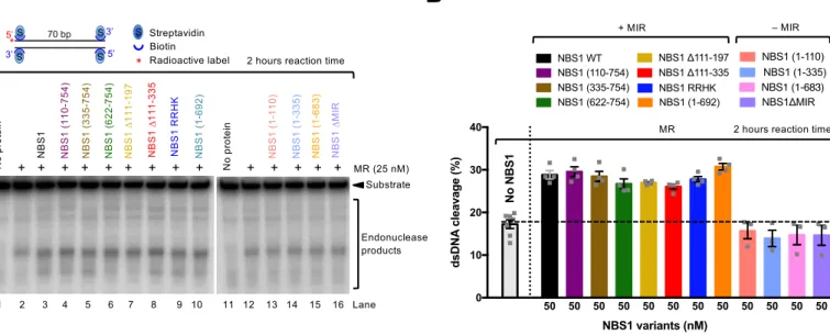
al., 2013, Wang, Cortez et al., 2000, Williams et al., 2008). Out of these, the interactions with CtIP and MDC1 are at least in part phosphorylation-dependent. To specifically de-fine the functional interaction of NBS1 with MR and pCtIP in resection, we employed our in vitro reconstituted system (Anand et al., 2016). To this point, we prepared NBS1 vari- ants lacking particular domains or carrying mutations specifically affecting known in-teractions with the MR and pCtIP ensemble (Figs 2A and EV2A), and assayed them in nuclease assays to determine their effect on the MRE11 endonuclease. We first varied the concentrations of wild type NBS1 in reactions with MR and pCtIP. While weak NBS1-independent DNA cleavage was observed (Fig EV2B and C), we found that less than equimolar concentrations of NBS1, compared to the MR complex, were sufficient for maximal DNA clipping activity (Fig EV2B and C). We next tested various NBS1 fragments lacking FHA, BRCT or both domains (Figs 2B and EV2D). As phosphory-lation of CtIP is required for resection (Huertas & Jackson, 2009), and the MRN complex primarily binds phosphorylated CtIP via the FHA and BRCT domains of NBS1 (Fig 2C and D)(Wang et al., 2013, Williams et al., 2009), a strong defect in resection was antici-pated. As shown in Fig 2B, a deletion of either FHA or BRCT domain in NBS1 had only a minor effect on resection in vitro, while elimination of both domains significantly re-duced, but did not eliminate resection. Similarly to the elimination of both FHA and BRCT domains, a stronger inhibitory effect was observed with the NBS1 RRHK variant, carrying point mutations in FHA-BRCT (R28A, R43A, H45A in FHA and K160M in BRCT1, Fig 2A and B)(Wang et al., 2013). The RRHK mutations decreased the phosphor-ylation-dependent interaction of NBS1 with phosphorylated MDC1 (Hari et al., 2010), as well as with pCtIP (Fig 2C), in agreement with a dramatic DNA end resection defect in vivo, as scored by the single-strand annealing reporter assay (Wang et al., 2013). Despite the strong physical interaction between the FHA and BRCT domains of NBS1 and pCtIP (Fig 2C), which is dependent on CtIP phosphorylation (Fig 2D), we conclude that both domains of NBS1 are important, but not essential for the DNA clipping activity in vitro together with MR and pCtIP. Furthermore, NBS1 (335-754), which lacks FHA-BRCT but contains a central linker region, exhibited similar stimulatory activity to NBS1 (622-754) lacking the central region (Fig 2B and E). This result revealed that the central NBS1 region (residues 335-621) is largely dispensable for MR and pCtIP-mediated resection, despite this region mediated residual interaction with pCtIP (Fig 2C). It has been demonstrated that the MRE11-RAD50 complex directly interacts with NBS1 via the MRE11-interaction region (MIR) within the C-terminal part of NBS1, with the most important motif located between residues 684-690 of NBS1 (Desai-Mehta,
Cerosaletti et al., 2001, Kim et al., 2017, Schiller, Lammens et al., 2012). This interaction facilitates MRN entry into the nucleus as only NBS1 contains the nuclear localization se- quence. Therefore, in vivo experiments with mutated MIR of NBS1 cannot easily distin-guish effects related to impaired nuclear entry from direct effects on the biochemical activities of the MR complex. Using our in vitro system, where any effects on nuclear im-port are irrelevant, we observed that in contrast to the FHA and BRCT domains, the MRE11 interaction region in NBS1 was absolutely essential for the stimulatory function of NBS1 on the MRE11-RAD50 endonuclease in conjunction with pCtIP (Figs 2E and EV2E). Specifically, the NBS1 (1-692) fragment lacking the very C-terminal region of NBS1, but possessing MIR, exhibited similar activity as full-length NBS1 (Figs 2E and EV2D). In contrast, the NBS1 (1-683) mutant lacking 9 residues comprising MIR (resi-dues 684-692) completely lost its stimulatory activity (Figs 2E and EV2E). Likewise, the internal deletion of MIR (residues 684-690) totally abolished NBS1 function in DNA clipping, even at high concentrations (Figs 2E and EV2E, F). In accord with previous studies (Desai-Mehta et al., 2001, Schiller et al., 2012), we observed a dramatic reduc-tion of the MRE11 and NBS1 physical interaction when NBS1 ∆MIR variant was used in pulldown experiments instead of wild type NBS1 (Fig EV2G and H). None of the NBS1 variants used here had any DNA cleavage activity, as expected (Fig EV2I). In summary, the NBS1-dependent interaction with MRE11-RAD50, more than that with pCtIP, is im- portant for the endonuclease activity of the MRE11-RAD50 and pCtIP nuclease ensem-ble. In the absence of pCtIP, NBS1 promotes MR cleavage independently of its FHA and BRCT domains Recently, it has been shown that NBS1 alone is capable of stimulating the endonucleolyt-ic activity of MR on protein-blocked dsDNA, independently of CtIP (or pCtIP)(Deshpande et al., 2016). Although CtIP-independent DNA end resection does not likely occur in vivo (Sartori et al., 2007), we used the in vitro assay to learn more about the function of NBS1. To this point, we employed our NBS1 mutants in an MR-dependent nuclease as-say with streptavidin-blocked dsDNA. In contrast to the assays that included pCtIP, the reactions were incubated for 2 h instead of 30 min to compensate for the lower cleavage efficacy in the absence of pCtIP. We observed that all NBS1 fragments containing MIR stimulated the clipping activity of MR almost indistinguishably, irrespectively of FHA and BRCT domains (Fig 3A and B). Notably, one of these constructs, the short C-terminal
NBS1 fragment containing 133 residues (NBS1 622-754), stimulated the endonucleolyt-ic cleavage to the same extent as full-length wild type NBS1. Conversely, NBS1 fragments lacking MIR did not at all promote MR (Fig 3A and B). Very similar results were obtained using circular ssDNA as a substrate, which forms a variety of secondary DNA structures (Fig EV3A and B). However, as noted previously, we believe that the cleavage of dsDNA near protein blocks is a better model for resection taking place in cells (Fig EV3C-G) (Anand et al., 2016, Chen, Trujillo et al., 2005, Deshpande, Williams et al., 2014). In summary, we show that in the absence of pCtIP, the phosphoprotein binding domains of NBS1 become dispensable for resection in vitro. Our data support a model for resection in wild type cells, where the FHA-BRCT domains of NBS1 promote resection by first binding phosphorylated CtIP, and NBS1 in turn activates the MR complex by interacting with MRE11. BRCT and FHA domains of NBS1 are important but not absolutely essential for re-section in vivo To verify the physiological significance of our data, we first designed an NBS1 comple-mentation system in U2OS cells to define the requirement of the FHA and BRCT domains of NBS1 for DNA end resection in vivo. The expression of endogenous wild type NBS1 was suppressed with siRNA, and siRNA resistant myc-tagged NBS1 variants were ex-pressed from a construct containing doxycycline-inducible promoter stably integrated into chromosomal DNA (Fig 4A). Downregulation of endogenous NBS1 led to cellular sensitivity to ionizing radiation, retention of MRE11 in the cytoplasm and a DNA end resection deficiency, as anticipated (Figs 4B-E and EV4A-D). Expression of wild type myc-tagged NBS1 overcame these defects, verifying the functionality of the complemen-tation system (Figs 4B-E, and EV4A-D). To test for the involvement of the FHA and BRCT domains of NBS1 in resection, we gen-erated cell lines expressing wild type NBS1, NBS1 R28A (a point mutation in the FHA domain), NBS1 K160M (a point mutation in the BRCT1 domain), or the NBS1 R28A K160M double mutant. The respective point mutations in the FHA and BRCT domains affect their phosphorylation-dependent protein binding function (Cerosaletti & Concannon, 2003, Hari et al., 2010, Wang et al., 2013), and disrupted the interaction with CtIP in cell extracts (Fig EV4E). The cell lines were treated with siNBS1 for 72 h to knock down endogenous NBS1 (Fig 4A). For the last 24 h, the expression of the comple- menting NBS1 variant was induced by the addition of doxycycline. Cells were then irra-diated (5 Gy) and DNA end resection was scored by automated counting of BrdU foci in
ssDNA under non-denaturing conditions or by counting of RPA2 foci (Fig 4B-E). Muta-tions in either FHA or BRCT domains led to an intermediate defect in DNA end resection in both resection assays. The NBS1 R28A K160M double mutant led to a more severe, but not complete abrogation of resection, comparable to NBS1 depletion (Fig 4B-E). Both FHA and BRCT domains thus individually promote, but are not absolutely essential, for the DNA end resection function of NBS1, which is in full agreement with our bio-chemical analysis. Depletion of CtIP brought about a more severe defect than that of NBS1, and nearly completely eliminated BrdU or RPA2 foci upon IR (Fig 4B-E). This result confirmed that CtIP is essential for almost all DNA end resection activity, as observed previously by others (Sartori et al., 2007). Furthermore, the depletion of NBS1 brought about lesser defects compared to CtIP depletion in homologous recombination as scored by a DR- GFP reporter assay (Fig EV4F and G). Finally, depletion of NBS1 did not completely elim-inate single-strand annealing (SSA)(Fig 4F), which is a good readout for extensive DNA end resection (Stark, Pierce et al., 2004). We also note that the residual SSA activity ob-served in the reporter assay without NBS1 was MRE11 dependent (Fig 4F). The depletion of NBS1 by siRNA may not be complete and may thus explain the residual resection we observed. To this point, using CRISPR/Cas9, we constructed a U2OS hypo-morphic NBS1ΔN cell line that expresses low levels of a C-terminal fragment of NBS1, which completely lacks the FHA and BRCT domains (Fig EV4H). Similarly as in mouse embryonic fibroblasts, where a C-terminal fragment of NBS1 rescued viability and ATM activation (Kim et al., 2017), the NBS1ΔN cells were viable and grew normally in the ab-sence of exogenous DNA damage. MRE11 was partially retained in the cytoplasm in this cell line, most likely owing to the low expression level of the hypomorphic NBS1 allele that may not be sufficient to fully localize MRE11 in the nucleus (Fig EV4I). However, stable expression of recombinant wild type or NBS1 R28A K160M (C-terminally tagged with the monomeric GFP analogue mNeonGreen), fully restored MRE11 nuclear localiza-tion (Fig EV4I). DNA end resection in the NBS1DN cell line was similarly compromised as in the cells depleted from NBS1 by siRNA (Fig EV4J). Consistent with our siRNA-based complementation system, stable expression of wild type NBS1 in the NBS1DN cell line efficiently restored DNA end resection activity, whereas expression of the NBS1 R28A K160M variant did not, indicating that even though MRE11 nuclear localization was re-stored by NBS1 R28A K160M, full resection activity was not (Fig 4G). Interestingly, both in wild type and NBS1 R28A K160M expressing cells, the residual resection activity was dependent on CtIP (Fig 4G), thus suggesting that CtIP can stimulate resection inde-pendently of NBS1 also in vivo. Together, we demonstrated that limited CtIP-dependent
DNA end resection in vivo can occur without the involvement of FHA-BRCT domains of NBS1. Phosphorylation of CtIP is partially dispensable for stimulation of MR in the ab-sence of NBS1 NBS1 specifically binds phosphorylated CtIP (Fig 2D), and phosphorylation of CtIP by CDKs is key for stimulating the MRN endonuclease activity (Anand et al., 2016). There- fore, the observation that limited CtIP-dependent resection can occur without the FHA-BRCT domains of NBS1 raised the question whether phosphorylation of CtIP is required under these conditions. In Figs 2B and EV2B, C, we observed weak NBS1-independent resection in vitro. To better define the mechanism of this reaction, we modified our bio-chemical assay conditions and employed the extended reaction incubation time (2 h) to improve the detection limit. As shown in Fig 5A and B, we observed that pCtIP stimulat-ed dsDNA cleavage by MR in the absence of NBS1 in a concentration-dependent manner. Our previous experiments established NBS1 as a reader of CtIP phosphorylation, and the NBS1-independent reaction thus identifies a separate mechanism for CtIP-facilitated stimulation of MR. Next, we set out to test whether phosphorylation of CtIP was re-quired. We treated or mock-treated pCtIP with λ phosphatase. As shown previously (Anand et al., 2016), we observed that only pCtIP (i.e. mock-treated), but not λCtIP, stimulated DNA cleavage by MRN in a concentration-dependent manner (Fig 5C-E). In contrast, we observed a robust stimulation of MR-dependent DNA clipping by both phosphorylated and λ phosphatase-treated CtIP (Fig 5C-E). Very similar results were obtained in kinetic assays: with MRN, pCtIP was ~7-fold more efficient in promoting the DNA cleavage compared to λCtIP; with MR, the difference was only ~1.5-fold (Figs 5F and G, and EV5A). Therefore, NBS1 appears to restrict DNA cleavage by MR to condi- tions when CtIP is phosphorylated. Without NBS1, although less efficient, the MR- dependent DNA cleavage is more promiscuous and can occur without CtIP phosphoryla-tion. One of the key residues in CtIP that needs to be phosphorylated by CDKs to allow DNA end resection during S-phase is T847, which maps to a conserved region that is equiva- lent to S267 of S. cerevisiae Sae2 (Huertas et al., 2008, Huertas & Jackson, 2009). Non- phosphorylatable substitution of T847 with alanine, resulting in T847A mutation, ren-ders pCtIP incapable to promote DNA end resection (Anand et al., 2016, Huertas & Jackson, 2009). Unexpectedly, we observed that the pCtIP T847A mutant was incapable
to promote both MR and MRN (Fig 5H and I). This result suggested that the T847A vari-ant is likely structurally impaired, beyond being non-phosphorylatable at the key CDK consensus site. Alternatively, the failure of pCtIP T847A to promote MR, in contrast to λCtIP, may stem from the presence of other phosphorylated residues in pCtIP T847A that have an inhibitory function together with MR. To test for this scenario, we treated pCtIP T847A with λ-phosphatase, and observed a minor, yet statistically significant, res-cue of DNA clipping by MR (Fig EV5B and C). As wild type, dephosphorylated λCtIP was still more efficient in promoting the MR endonuclease than dephosphorylated λCtIP T847A, the T847A substitution in pCtIP likely brings about structural changes within the conserved CtIP region that affect CtIP activity beyond mimicking a non-phosphorylated threonine. Together, our results demonstrate that NBS1, as CtIP, is a co-factor that stimulates the DNA cleavage efficacy of the MR complex. Reactions with MRN require CtIP phosphory-lation, and lead to extensive DNA end resection activity that is required for HR and SSA. In contrast, in the absence of NBS1, the MR-dependent cleavage is more restricted, and CtIP phosphorylation is partially dispensable for the DNA cleavage activity. The physical interaction of MRE11 and RAD50 with CtIP does not require CtIP phosphorylation Our observation that CtIP was able to stimulate the DNA cleavage capacity of the MR complex prompted us to define physical interactions between MRE11, RAD50 and phos-phorylated or non-phosphorylated CtIP. CtIP is known to interact with all subunits of the MRN complex (Chen et al., 2008, Sartori et al., 2007, Williams et al., 2009, Yuan & Chen, 2009); in accord, we detected direct interactions between pCtIP/CtIP and both MRE11 and RAD50 (Fig 6A and B). While phosphorylation of CtIP was absolutely re-quired for its interaction with NBS1 (Fig 2D), it was not the case for MRE11 and RAD50. In contrast, λ phosphatase-treated CtIP interacted with both MRE11 and RAD50 even better than pCtIP, suggesting that CtIP phosphorylation may interfere with the interac-tion with the isolated MRE11 and RAD50 subunits (Fig 6A and B). We cannot exclude that the apparent increase in the protein-protein interactions was due to aggregation of non-phosphorylated CtIP, or that our preparation of pCtIP contained phosphorylated residues that are not normally modified in vivo, which could inhibit the interaction with MRE11 and RAD50. Nevertheless, NBS1 was mostly responsible for the interaction be-tween the MRN complex and pCtIP, and phosphorylation of CtIP was indispensable for this interaction (Fig 6C). The interaction between MRN and pCtIP was not affected by
the presence of ATP and/or DNA (Fig EV6A). Finally, NBS1 largely mediated the interac- tion of the MRN complex even when DNA bound (Fig EV6B). In summary, the interac-tions of MRE11 and RAD50 with CtIP did not require CtIP phosphorylation. These data are in agreement with the results from the nuclease assays, which indicated that CtIP phosphorylation was not strictly required to promote the nuclease of the MR complex. NBS1 is dispensable for DNA cleavage opposite to a strand discontinuity To learn more about the involvement of NBS1 in the DNA clipping reaction, we em- ployed a substrate that contains a preexisting nick in the strand opposite to the antici-pated cleavage site, which was shown to increase the efficacy of DNA cleavage (Deshpande et al., 2016). To this point, we performed nuclease analyses with both nicked and non-nicked DNA substrates, which had phosphorothioate bonds at the 3' ends to prevent exonucleolytic DNA degradation by MRE11 (or MR/MRN). We observed that MRE11 alone had no endonuclease activity on any of the substrates tested, with or without pCtIP, as expected (Fig 7A and B, lanes 3 and 6). NBS1 promoted the cleavage of the non-nicked DNA substrate by MR, and pCtIP was essential (Fig 7A). In contrast, NBS1 was dispensable when nicked DNA substrate was used (Fig 7B). Under these con-ditions, pCtIP provided a moderate stimulatory effect, with or without NBS1 (Fig 7B). The same results were obtained in kinetic assays (Fig 7C and D). The endonuclease ac-tivity was promoted by a nick or a gap in the top strand, as a substrate lacking the top left oligonucleotide was not endonucleolytically cleaved by MR-pCtIP or MRN-pCtIP (Fig EV7). Together, our results suggested that NBS1, and to a lesser extent pCtIP, is dispen-sable for the endonucleolytic DNA cleavage in case of a pre-existing nick opposite to the cleavage site. Discussion The MRE11 nuclease plays critical functions at DSBs as well as at stalled, collapsed or reversed replication forks (Mijic et al., 2017, Schlacher et al., 2011, Stracker & Petrini, 2011). Scheduled activation of the MRE11 nuclease is important for both homologous recombination and microhomology-mediated end-joining DSB repair pathways. In con-trast, inappropriate MRE11 nuclease may result in illegitimate recombination leading to genome rearrangements or excessive DNA degradation at challenged replication forks, leading to cell lethality in certain genetic backgrounds (Schlacher et al., 2011). There- fore, the activity of the MRE11 nuclease must be finely controlled and restricted to situa-
tions when it is appropriate for genome stability maintenance. MRE11 is a 3'→5' exonu-clease on its own (Paull & Gellert, 1998). In complex with RAD50 and other co-factors, MRE11 can cleave DNA endonucleolytically (Paull & Gellert, 1998, Paull & Gellert, 1999). It is believed that the processing of DSBs or reversed DNA replication forks is initiated by the endonucleolytic cleavage of the 5'-terminated DNA strand by MRE11 and co-factors (Cannavo & Cejka, 2014, Garcia et al., 2011, Lemacon, Jackson et al., 2017, Neale et al., 2005). Eukaryotic cells possess two key regulators of the MR nuclease complex: CtIP/Sae2 and NBS1/Xrs2. In yeast, phosphorylated Sae2 was required for the endonu-cleolytic DNA cleavage by MR, while the lack of Xrs2 had a more moderate effect (Oh et al., 2016). In humans, both NBS1 and phosphorylated CtIP promote the activity of the MR endonuclease, revealing that NBS1 is a regulator of MRE11 that appears more im-portant in high eukaryotes (Anand et al., 2016, Deshpande et al., 2016). In this study, we define the mechanism by which NBS1 regulates the MRE11 endonuclease using primari-ly a reconstituted in vitro system. We show that NBS1 functions as a sensor of CtIP phosphorylation, which allows MRE11 nuclease activation only when CtIP is phosphorylated (Fig 8A-C). The FHA and BRCT are highly conserved phosphoprotein binding domains, which mediate the physical interac-tion of NBS1 with phosphorylated CtIP and both these domains promote resection in vitro as well as in vivo. Once NBS1 senses that CtIP is phosphorylated, it activates the MR endonuclease, which is dependent on a direct physical interaction between NBS1 (resi-dues 684-692) and MRE11 (Fig 8A). We found that the FHA and BRCT domains of NBS1 are important, but not absolutely essential for the MR and pCtIP-dependent cleavage of dsDNA near protein blocks. More than the FHA-BRCT domains, the short region within the C-terminus of NBS1 that interacts with MRE11 is absolutely required for NBS1 to promote the MR complex in conjunction with pCtIP. In support of this model, we ob-served residual NBS1-dependent, but pCtIP-independent nuclease activity of the MR complex in vitro (Fig 8C). Without CtIP, NBS1 still promotes DNA cleavage by MR. In this case, the FHA-BRCT domains of NBS1 were entirely dispensable, while the MRE11 inter-action region was required. Nevertheless, while this in vitro reaction informs us about the underlying mechanism, we believe that CtIP-independent resection does not occur in vivo, as depletion of CtIP entirely eliminated resection in cells in all assays tested (Fig 4)(Sartori et al., 2007). Next, we observed that in the absence of NBS1, CtIP still promotes the endonuclease of MR, albeit to a lesser extent. This functional interaction was observed in vitro (Fig 5), as well as in cells upon NBS1 depletion or gene editing that eliminated the FHA and BRCT domains of NBS1 (Fig 4). Strikingly, CtIP phosphorylation was not strictly required in
the absence of NBS1 (Figs 5C-G and 8B). While these data are in agreement with the function of NBS1 as a sensor of CtIP phosphorylation, the result was nevertheless sur-prising, as it revealed fundamental differences between the budding yeast and human systems. In yeast, Sae2 phosphorylation regulates its physical interaction with Rad50 (Cannavo, Johnson et al., 2018), in addition to Xrs2 (Liang et al., 2015). Even in the ab-sence of Xrs2, Sae2 phosphorylation is still fully required for the stimulation of the Mre11-Rad50 endonuclease. Using human proteins instead, we found that the phos-phorylation of CtIP was largely but not completely dispensable for the activation of MR in the absence of NBS1. This result might help explain how CtIP promotes limited resec-tion in G1. It is well established that CDK and ATM-dependent phosphorylation of CtIP is necessary for the activation of extended resection and hence homologous recombination in S-G2 (Huertas et al., 2008, Huertas & Jackson, 2009, Wang et al., 2013). Nevertheless, CtIP likely activates MRE11 in MMEJ, which is not restricted to S-G2 and requires only minimal resection (Averbeck, Ringel et al., 2014, Dutta, Eckelmann et al., 2017, Sharma et al., 2015, Truong et al., 2013). In G1, CtIP is phosphorylated at S327, which mediates its interaction with BRCA1, but the G1 form of CtIP will likely lack other phosphorylation modifications that are present in S-G2 (Biehs, Steinlage et al., 2017, Yu & Chen, 2004). We speculate that this form of CtIP could promote the MR complex in case the NBS1 function is inhibited. However, we were unable to test this hypothesis directly, as a CtIP variant that is non-phosphorylatable at the key CDK site at T847 appears to be impaired beyond mimicking a non-phosphorylated residue (Fig 5H and I). While MRE11 and RAD50 form a constitutively interacting dimer of dimers complex, there are conflicting reports about the stoichiometry of NBS1 within the MRN complex (Paull & Gellert, 1999, Schiller et al., 2012, Trujillo, Yuan et al., 1998). Furthermore, it has been reported that the physical interaction of NBS1 with MR may be differentially regulated in G1 vs. S-G2, upon DNA damage or at various subcellular locations, such as at telomeres (Limbo, Yamada et al., 2018, Zhou, Kawamura et al., 2017, Zhu, Kuster et al., 2000). Specifically, the MR complex was shown to localize to DSBs in the absence of NBS1 (Limbo et al., 2018), and NBS1 co-localized with MR to telomeres in S but not dur-ing interphase (Zhu et al., 2000). NBS1 also shows slower kinetics of recruitment to DSBs compared to MRE11 (Haince, McDonald et al., 2008). Finally, we show here that DNA end resection was strongly reduced but not completely eliminated in the absence of NBS1 in cells, despite the reduced nuclear import of MR, showing that NBS1 may not be an obligate component of the resection machinery under all conditions. Our data sug-gest that a reduction of the MR interaction with NBS1 may allow the complex to respond to non-phosphorylated or differently phosphorylated CtIP. It is additionally possible
that the function of NBS1 as a sensor of CtIP phosphorylation might be regulated by posttranslational modifications of NBS1 (which is heavily phosphorylated) and/or by other proteins that could regulate NBS1 function. More research is needed to fully un- derstand the mechanisms of resection in G1, and the involvement of MRN and CtIP pro- teins in this context. Together, our results demonstrate that the MRE11-RAD50 endonu-clease is on its own restricted and becomes fully functional in the presence of NBS1 and phosphorylated CtIP. By regulating these co-factors through multiple mechanisms, cells can fine-tune the function of the MR endonuclease to optimally promote genome stabil-ity in various physiological contexts. Material and Methods Cloning, expression and purification of recombinant proteins Recombinant MRN, MR and phosphorylated CtIP were expressed in insect Spodoptera frugiperda 9 (Sf9) cells and purified by affinity and ion exchange chromatography as de-scribed previously (Anand, Pinto et al., 2018, Anand et al., 2016). MBP-NBS1-his and its variants, as well as MRE11-his and FLAG-NBS1-his were expressed in Sf9 cells and puri- fied as described in detail in Supplementary information. To prepare the pFB-MBP-NBS1-his expression construct, NBS1 was amplified from pTP36 (a kind gift from Tanya Paull, University of Texas) by PCR using primers NBS1_F and NBS1_R (Tables S1 and S2). The PCR product was digested with NheI and XmaI (New England Biolabs) and cloned into corresponding sites of pFB-MBP-MLH3-his (Ranjha, Anand et al., 2014). The preparation of other NBS1 variant expression constructs is described in Supplementary information. To prepare FLAG-NBS1-his, a NBS1 sequence that was codon-optimized for expression in Sf9 cells (NBS1co) was purchased from Synbio Technologies and cloned into BamHI-XhoI sites of pFB-MBP-NBS1-his to create pFB-FLAG-NBS1co-his. To pre-pare pFB-MBP-MRE11-his, the MRE11 gene was amplified by PCR from pTP17 (a kind gift from Tanya Paull, University of Texas) using primers HMRE11-FO and HMRE11-RE (Tables S1 and S2). The amplified product was digested with NheI and XmaI, and cloned into the same sites in pFB-MBP-Sgs1-his plasmid (Cejka & Kowalczykowski, 2010); the sequence of Sgs1 was removed during this step. The scheme and sequences of oligonu-cleotides used for cloning of the NBS1 fragments and all other constructs in this study are listed in Tables S1 and S2. Where indicated, 1.2 μg pCtIP was dephosphorylated in 20 μl reactions with 200 units of λ phosphatase (λ-PP, New England Biolabs) in 1x PMP buffer (New England Biolabs) supplemented with 1 mM magnesium chloride for 15 min
at 30°C. For "mock" control, λ-PP was excluded from the reaction. Nuclease assays Unless indicated otherwise, nuclease assays (15 μl volume) were carried out in nuclease buffer containing 25 mM Tris-HCl pH 7.5, 5 mM magnesium acetate, 1 mM manganese acetate, 1 mM dithiothreitol (DTT), 1 mM ATP, 0.25 mg/ml BSA (New England Biolabs), 1 mM phosphoenolpyruvate (Sigma), 80 U/ml pyruvate kinase (Sigma), and 1 nM oligo-nucleotide-based DNA substrate (in molecules). Unless specified otherwise, the final NaCl concentration in the reactions was adjusted to 15 mM, accounting for salt brought to the reactions with protein storage or dilution buffer. The reactions were supplement-ed with 15 nM streptavidin (Sigma), and incubated for 5 min at room temperature to block the biotinylated end(s) of the DNA substrates. The recombinant proteins were then added to the reactions on ice and samples were incubated at 37°C for 30 min, un-less indicated otherwise. Reactions were stopped by adding 0.5 μl of 0.5 M ethylenedia-minetetraacetic (EDTA) and 1 μl Proteinase K (19 mg/ml, Roche), and incubated at 50°C for 30 min. Finally, 15 μl loading buffer (5% formamide, 20 mM EDTA, bromophenol blue) was added to all samples and the products were separated on 15% polyacryla-mide denaturing urea gels (19:1 acrylamide-bisacrylamide, Bio-Rad), as described (Pinto, Anand et al., 2018). The gels were fixed in fixing solution (40% methanol, 10% acetic acid, 5% glycerol) for 30 min at room temperature and dried on a 3MM Chr paper (Whatman). The dried gels were exposed to storage phosphor screen (GE Healthcare) and scanned by a Typhoon Phosphor Imager (FLA 9500, GE Healthcare). Nuclease as-says with circular ssDNA substrate (M13, 100 ng per reaction, New England Biolabs) were carried out similarly except the reaction buffer contained 60 mM NaCl and incuba-tion time was extended to 2 h. The reaction products were separated on 1.1% agarose gels, which were stained with GelRed (1:20000, Biotium) after completion of electro-phoresis for 45 min. The gels were imaged with gel imager (Ingenius3, Syngene). Preparation of oligonucleotide-based DNA substrates All oligonucleotides were purified by polyacrylamide gel electrophoresis and purchased from Eurogentec. The labeling of oligonucleotides at the 5'-end was carried out by T4 polynucleotide kinase (New England Biolabs) and [γ-32 P] ATP (Perkin Elmer). The label- ing of oligonucleotides at the 3'-end was carried out by terminal deoxynucleotidyl trans-ferase (New England Biolabs) and [α-32P] cordycepin 5' triphosphate (Perkin Elmer), as described (Pinto et al., 2018). To prepare quadruple blocked 70-bp long DNA substrate, PC210 and PC211 oligonucleotides were used, as described previously (Cannavo &
Cejka, 2014). To prepare single-blocked non-nicked substrate (Fig 7A), PC216C_5XSS (GATGCATGAGGTGGAGTACGCGCCCGGGGAGCCCAAGGGCACGCCCTGGCACCCGCACCGCG GCA*C*T*T*A*C) was labeled at 5'-end and annealed with PC216_5XSS (GTAAGTGCCGCGGTGCGGGTGCCAGGGCGTGCCCTTGGGCTCCCCGGGCGCGTACTCCACCTC AT*G*C*A*T*C), the bold T represents the biotin modification and * represent phos- phorothioate linkages between nucleotides. To prepare the single blocked nicked sub-strate (Fig 7B), PC216C_5XSS labeled at 5'-end was annealed with PC216_50nt (GTAAGTGCCGCGGTGCGGGTGCCAGGGCGTGCCCTTGGGCTCCCCGGGCG) and PC216_20nt_5XSS (CGTACTCCACCTCAT*G*C*A*T*C). To prepare substrate used in Fig EV7, PC216C_5XSS labeled at 5'-end was annealed to PC216_20nt_5XSS. Protein interaction assays To test for interactions between recombinant proteins, the "bait" proteins were first immobilized on a resin (either M2 anti FLAG affinity gel [Sigma] or Protein G agarose [Invitrogen] coupled to specific antibodies). The resin was then incubated with "prey" proteins in binding buffer, washed, and bound proteins were analyzed by western blot- ting. As a negative control, prey proteins were incubated with resin without bait pro-teins. Specifically, to detect the interaction between pCtIP and NBS1 variants (Fig 2C and D), 1.5 μg anti CtIP antibody (Active Motif, 61141) was captured on 10 μl protein G beads upon incubation in 150 μl PBS-T pH 7.4 (1.37 mM NaCl, 2.7 mM KCl, 10 mM Na2HPO4, 1.8 mM KH2PO4 , 0.1% Tween 20) for 40 min at room temperature with continuous rota-tion. Next, 1 μg recombinant CtIP (mock-treated pCtIP or dephosphorylated λCtIP) was added and incubated in 60 μl binding buffer (25 mM Tris-HCl pH 7.5, 1 mM DTT, 3 mM EDTA, 50 mM NaCl, 0.2 mg/ml BSA) for 1 h at 4°C with continuous rotation. The resin with the immobilized antibody was washed 2 times with 150 μl wash buffer (25 mM Tris-HCl pH 7.5, 1 mM DTT, 3 mM EDTA, 80 mM NaCl, 0.05% Triton-X), and incubated with 320 nM NBS1 variants in 60 μl binding buffer for 1 h at 4° C with continuous rota-tion. The resin was then washed 3 times with 150 μl wash buffer. The proteins were eluted by incubation of the washed resin for 3 min at 95°C in 60 μl SDS buffer (50 mM Tris-HCl pH 6.8, 1.6% sodium dodecyl sulphate, 100 mM DTT, 10% Glycerol, 0.01% bromophenol blue). CtIP and NBS1 variants were detected by western blot using anti CtIP (Active Motif, 61141, 1:1000) and anti NBS1 (Novus Biologicals, NB100-143, 1:1000) antibodies. In assays shown in Figs 6C and EV6A, 90 nM MRE11, MR or MRN
was incubated with immobilized λCtIP or pCtIP as described above. MRE11 was detect-ed with anti-MRE11 antibody (Abcam, ab214, 1:1000). To detect the interaction between MRE11-his and MBP-NBS1-his/MBP-NBS1 ∆MIR-his, or between MRE11-his and pCtIP/λCtIP as shown in Figs EV2H and 6A, respectively, 1.4 μg MRE11-his was immobilized on beads and coupled to anti MRE11 antibody (Abcam, ab214). After washing, 200 nM MBP-NBS1-his/MBP-NBS1∆MIR-his, or 330 nM pCtIP/λCtIP were incubated with the MRE11-coated resin in a 60 μl reaction in binding buffer as described above. For the assay shown in Fig EV2H, both MRE11-his and MBP-NBS1-his/MBP-NBS1 ∆MIR-his were detected by anti his antibody (MBL, M091-3, 1:1000) by western blotting. For the interaction assay shown in Fig 6B, 50 ml Sf9 cells were infected with RAD50-FLAG baculovirus. Cells were lysed and RAD50-FLAG (~300 ng) was immobilized on 30 μl anti FLAG M2 affinity gel. Next, in a 150 μl reaction, 330 nM pCtIP/λCtIP was added and incubated in binding buffer for 1 h at 4°C with continuous rotation. Beads were washed 4 times with 300 μl wash buffer and proteins were eluted with wash buffer sup-plemented with 3X FLAG peptide (150 ng/μl, Sigma). Bound RAD50-FLAG was detected by western blotting using anti FLAG antibodies (Sigma, F3165, 1:2000). For FLAG co-IP experiments (Fig EV4E), 293-T cells were co-transfected with FLAG-CtIP expression constructs (Sartori et al., 2007) and NBS1-myc expression constructs (Hari et al., 2010). Immunoprecipitations were done using Anti FLAG M2 affinity beads (Sig-ma, A2220). Western blots were probed with rabbit anti FLAG (Sigma, F7425) and mouse anti myc (GeneTex, 9E10, GTX80249) antibodies. DR-GFP and SSA reporter assays For DR-GFP HR repair reporter assays, DR-GFP U2OS cells (a gift from J. Stark) were plated at 20’000 cells/cm2 in 6-well plates. After 24 h, cells were transfected with con-trol siRNA or siRNA against CtIP (5’-GCUAAACAGGAACGAAUCTTdTdT-3’) and NBS1 (5’-GGAGGAAGAUGUCAAUGUUTTdTdT-3’), respectively. 48 h after siRNA transfection, cells were transfected with I-SceI expression plasmid (pCBASce). 4 h after transfection, the medium was replaced and a second transfection with siRNA oligonu- cleotides was carried out. 48 h after I-SceI transfection, cells were analyzed for GFP ex- pression by flow cytometry on a LSRII Fortessa flow cytometer (BD). For the SSA re-porter assays, 0.4 x10⁵ SA-GFP U2OS cells were seeded in 0.5 ml antibiotic free media onto 24 well plates with 5 pmol siRNA (siCTRL, 5'-TGGTTTACATGTCGACTAA; siCtIP, 5'-GCTAAAACAGGAACGAATC; siNBS1, 5'-GGAGGAAGATGTCAATGTT) and 1.8 μl RNAiMAX
(Invitrogen) in 100 μl Opti-MEM (Gibco) and cultured overnight (20 h). Following RNAi treatment, the cells were transfected with 0.4 μg I-SceI (pCBASce) and 0.2 μg empty vec-tor (pcDNA3.1) with Lipofectamine 2000 (Invitrogen) in 100 μl Opti-MEM and 0.5 ml antibiotic free media for 3 h. After the I-SceI transfection, cells were washed and treated with MRE11 inhibitors 50 μM PFM03 and 50 μM PFM39 (a kind gift from Davide Moiani and John Tainer (MD Anderson Cancer Center) (Shibata, Moiani et al., 2014) or mock treated with DMSO and cultured for 3 days. GFP positive cells were quantified on an LSRFortessa flow cytometer (BD). Generation of siRNA-based NBS1 complementation system in U2OS cells To generate the siRNA-based complementation system for NBS1, the Flp-InTM T-RExTM system (Thermo Fisher Scientific) was used according to the manufacturer’s protocols. Briefly, myc-tagged full-length NBS1 cDNAs (wild type and mutants) were sub-cloned from retroviral expression constructs (Hari et al., 2010) and ligated into the pcDNA5/FRT/TO vector. Silent mutations were introduced in the siRNA target sequence (see below) by site-directed mutagenesis using the following mutagenesis primer (sense): 5’-GTTCAAAAACAGGAGGAAGACGTGAACGTTAGAAAAAGGCCAAGG-3’. A Flp-In T-REx U2OS cell line (gift from D. Durocher) was cultured in Dulbecco’s modified Eagle medium (DMEM), supplemented with 10% fetal calf serum (FCS), 2 mM L-glutamine and penicillin-streptomycin antibiotics under standard cell culture conditions in a CO2 incu-bator (37°C; 5% CO2 ). NBS1-myc harboring vectors and the Flp-In recombinase expres-sion plasmid pOG44 were mixed in a 1:9 ratio and transfected into the Flp-In T-REx U2OS cells. Stable clones were established by Hygromycin B (250 µg/ml) and Blasticidin (10 µg/ml) selection and were characterized for inducible NBS1-myc expression by im-munofluorescence and western blotting. Generation of the NBS1DN cell line The NBS1DN cell line was generated according to a protocol published by the Zhang la- boratory (Ran, Hsu et al., 2013). The sgRNA target sequence (5’- GCGTTGAGTACGTTGTTGGA-3’) was cloned into the PX330-U6-hSpCas9 vector and veri-fied by sequencing. The targeting vector was then transfected into U2OS cells and clonal cell lines were isolated by dilution in 96-well plates. Single clones were analyzed for NBS1 expression by immunofluorescence. Several clones without detectable NBS1 ex- pression were isolated and one of them was further characterized (data will be pub-lished elsewhere). Frame-shift inducing insertion/deletion (indel) mutations in all of the
three NBS1 alleles present in U2OS cells were verified by Sanger sequencing of a PCR- amplified genomic fragment that was cloned in the pCR II Blunt TOPO plasmid (se- quences will be published elsewhere), indicating that the FHA BRCT domain coding re- gion was eliminated. Western blotting however revealed the presence of weakly ex-pressed ~40 kDa protein that is recognized by a monoclonal antibody raised against full-length NBS1 (Genetex, clone 1D7, GTX70224), which was sensitive to downregula- tion of the NBS1 mRNA by siRNA transfection, thus suggesting that this cell line ex- presses low levels of hypomorphic C-terminal fragment of NBS1 that is produced by in- ternal translation initiation within the NBS1 mRNA using an open reading frame gener-ated by one of the indel mutations, similar to the NBS1 hypomorphic alleles expressed in cell lines derived from NBS patients (Maser, Zinkel et al., 2001). In vivo resection assay U2OS cell lines were grown on coverslips for 24 h prior to treatment. When indicated, cells were transfected with siRNAs against NBS1 or CtIP (siRNA sequence indicated in the previous section where the generation of siRNA-based NBS1 complementation sys-tem was explained) and incubated for 72 h. For heterologous expression of full-length recombinant myc-tagged NBS1, doxycyclin (1 µg/ml) was added 24 h prior to the exper-iment. For BrdU staining under native conditions, BrdU (10 µg/ml) was also added 24 h prior to the experiment. Cells were exposed to 5 Gy of IR and then incubated for 3 h at 37°C. The medium was removed, and cells were washed with PBS, followed by a brief pre-extraction on ice in CSK buffer (100 mM PIPES, pH 7.0; 100 mM NaCl; 300 mM Su-crose; 3 mM MgCl2; 0.4% Triton X-100). Cells were then fixed in 4% paraformaldehyde, washed and prepared for immunofluorescence analysis (see below). Immunofluorescence Cells were grown on glass coverslips and fixed with either ice-cold methanol for 10 min (for staining with NBS1 and MRE11 antibodies), or with 4% buffered formaldehyde (for staining with all other antibodies) for 15 min at room temperature, and subsequently permeabilized for 5 min in PBS containing 0.2% Triton X-100. Following 1 h of blocking in blocking buffer (10% FBS, 3% BSA in PBS), primary antibody incubations were per-formed at room temperature for 2 h. Coverslips were washed three times with PBS and secondary antibody incubations were performed for 1 h at room temperature in the dark. After washing with PBS for three times, coverslips were mounted on glass micros- copy slides with Vectrashield mounting medium containing 0.5 μg/ml 4’,6-diamidino-2-phenylindole dihydrochloride (DAPI). Widefield image acquisition was done on either a
Leica DMI6000B inverted microscope equipped with a 63x Apochromat oil immersion objective (NA 1.3) using Leica’s LAS-AF software or on a Zeiss AxioObserver Z1 wide-field microscope, equipped with a Lumencor SpectraX illumination system and a 63x, 1.4-NA, i-plan apochromat oil-immersion objective, using Zeiss’ ZEN (blue edition) soft-ware. Images were analyzed using Fiji software (Schindelin, Arganda-Carreras et al., 2012). Counting of BrdU foci or RPA2 foci was done with a Fiji macro written for auto-matic image analysis. The following antibodies were used at the indicated dilutions: rabbit anti NBS1 (Novus Biologicals, NB100-143, 1:500), affinity purified sheep anti MRE11 (Goldberg et al., 2003, 1:1000), mouse anti Myc (GeneTex, 9E10, GTX80249, 1:250), mouse anti BrdU (Amersham/GE healthcare, BU-1, RPN202, 1:100), mouse anti RPA2 (Abcam, 9H8, ab2175, 1:250). Western blotting of total cell extracts SDS-PAGE and western blotting were performed using total extracts prepared from cul-tured cells following standard procedures. The following antibodies were used at the indicated dilutions: rabbit anti ATMpS1981 (Eptimics, YE070901r, 1:5000), rabbit anti ATM (Calbiochem, PC-116, 1:250), mouse anti CtIP (Santa Cruz, sc-271339, 1:250), mouse anti NBS1 (GeneTex, 1D7, GTX70224, 1:500), mouse anti RPA2 (Abcam, 9H8, ab2175, 1:500), rabbit anti SMC1 (Abcam, ab9262, 1:500), mouse anti RAD50 (GeneTex, 13B3, GTX70228, 1:1000), mouse anti MRE11 (Abcam, 12D7, ab214, 1:500), mouse anti Myc (GeneTex, 9E10, GTX80249, 1:250), mouse anti Tubulin (Sigma, DM1A, T6199, 1:2000). For western blot shown in Fig 4F, the following antibodies were used: rabbit anti NBS1 (Novus Biologicals, NB100 143, 1:1000), mouse anti CtIP (Active Motif, 61141, 1:1000), mouse anti β actin (Abcam, ab8227, 1:1000) Clonogenic survival assay U2OS FlpIn T-REx cell lines were transfected with siNBS1 for 48 h and then plated at low density in medium supplemented with 0.5 µg/ml doxycycline. 24 h later, the plates were exposed to the indicated IR doses, followed by incubation for 12-14 days. Colonies were fixed and stained with Coomassie brilliant blue and the number of surviving colo-nies for each cell was counted. The percentage of surviving colonies for each cell line was calculated using the plating efficiencies of the non-irradiated cells as a reference. Survival experiments were carried out in biological triplicates. Acknowledgements
We thank Dr. Jeremy Stark (City of Hope), Dr. Xiohua Wu (Scripps), Dr. Davide Moiani and Dr. John Tainer (MD Anderson Cancer Center), Dr. Tanya Paull (UT Austin), A. Sarto-ri (University of Zurich) and Dan Durocher (Lunenfeld-Tanenbaum Research Insitute) for cell lines, plasmids and reagents. We thank members of the Cejka (L. Ranjha, S. Hal-der, G. Reginato, E. Cannavo, I. Ceppi, A. Acharya) and Stucki laboratories for comments on the manuscript. This work was supported by the Swiss National Science Foundation grants 31003A_144284 to MS, 31003A_175444 to PC and European Research council grant 681630 to PC. Author contributions RA performed all biochemical experiments. LR performed the experiment shown in Fig EV6B. SMH, AJ and DB performed cell biology experiments. All authors designed the ex-periments and analyzed the data. RA, PC and MS wrote the paper. Conflict of interest The authors declare that they have no conflict of interest. References