Eur Arch Otorhinolaryngol (2008) 265:327–330 DOI 10.1007/s00405-007-0459-x
123
L A R Y N G O L O G YLaryngeal involvement in patients with active pulmonary
tuberculosis
Murat Topak · Cagatay Oysu · Kursat Yelken · Asli Sahin-Yilmaz · Mehmet Kulekci
Received: 14 March 2006 / Accepted: 30 August 2007 / Published online: 6 October 2007 © Springer-Verlag 2007
Abstract The aim of this study was to determine the incidence of laryngeal tuberculosis (LT) among patients with active pulmonary tuberculosis. A total of 319 patients under treatment for pulmonary tuberculosis were sub-jected to laryngoscopy. Five patients (1.5%) with LT were identiWed. Odynophagia was the most common complaint, followed by alteration in voice. The larynx returned to its normal appearance in 3–8 months (average 18 weeks) by antituberculous medication. Physicians dealing with pul-monary tuberculosis should keep in mind that symptoms of laryngeal involvement may be minor, and laryngoscopy should always be performed when laryngeal involvement is suspected in order to isolate highly infectious patients. Response to antituberculous medication is usually late in LT and diagnosis by “wait and watch” policy will cause a signiWcant delay in the diagnosis of a possible larynx carcinoma.
Keywords Laryngeal tuberculosis · Pulmonary tuberculosis
Introduction
Pulmonary tuberculosis remains a signiWcant health prob-lem in developing countries. Since 1990, tuberculosis has shown a rise in incidence worldwide and was declared a global emergency by the World Health Organization (WHO) in 1993 [1]. WHO reported 373,497 new tuberculo-sis cases in Europe in 2002, compared to 231,608 new cases in 1991[1]. Pulmonary tuberculosis is an endemic disease in Turkey. The incidence of pulmonary tuberculosis was 28/100,000 in 1999 [2].
Laryngeal tuberculosis (LT) is one of the most common extra-pulmonary manifestations of this disease [3, 4]. The incidence of LT has signiWcantly declined to the rate of 1.8, 1.5, and <1% in the last Wfty years [3, 5–7]. The aim of this study is to determine the current incidence of laryngeal involvement in a relatively large group of patients with active pulmonary tuberculosis.
Materials and methods
This study was conducted from January through September of the year 2003. 319 unselected patients with active pul-monary tuberculosis admitted in two major institutions of chest diseases in Istanbul were enrolled in the study. Active pulmonary tuberculosis was deWned as positive sputum smear microscopy and/or culture [8]. In smears, detection of acid fast bacilli with the carbolfuchsin method (Ziehl– Neelsen stain) was considered positive. In cultures, myco-bacterial growth in specimens inoculated onto egg-based culture media (Lowenstein–Jensen) was considered posi-tive. The sputum specimens were subject to drug sensitivity testing on Lowenstein-Jensen medium if: (a) the patient had a previous history of tuberculosis infection, (b) if there was M. Topak · K. Yelken · A. Sahin-Yilmaz · M. Kulekci
Department of Otolaryngology,
Taksim Education and Research Hospital, Istanbul, Turkey
C. Oysu
Department of Otolaryngology,
Haydarpasa Numune Education and Research Hospital, Istanbul, Turkey
C. Oysu (&)
Haci Hakki Bey sk. Basel Ap. no.: 4/7, Erenkoy, 34728 Istanbul, Turkey e-mail: [email protected]
328 Eur Arch Otorhinolaryngol (2008) 265:327–330
123
a clinical suspicion of multidrug resistant tuberculosis. If the smear or sputum culture was negative, the diagnosis was conWrmed by positive tuberculin test results and/or radiological evidence of tuberculosis [8]. All the subjects had been receiving antituberculous medication including isoniazid, rifampicin and pyrazinamide with a duration varying from 2 weeks to 3 months at the time of inclusion.
All patients were subject to bedside indirect laryngos-copy by mirror or by endoscopic examination. The diagno-sis of LT was established by biopsy and histological examination or rapid regression of the laryngeal lesions by antituberculous medication. Following the diagnosis of LT, patients were subject to query in detail in order to discover any neglected symptoms of LT. Taksim Education and Research Hospital ethical committee approved the study and we obtained informed consent from all subjects.
Results
A total of 319 patients with pulmonary tuberculosis were examined. All of the patients were from lower socio-economic classes. A total of 212 were living in rural areas. Totally, 273 were men, and 46 were women (male:female = 6:1). The age range was 14–80 years, with an average of 31.8 in men and 33.2 in women. Three hundred patients (94%) had a history of tobacco use and Wve (1.5%) were alcoholic. Totally, 249 of the patients were recently diag-nosed with tuberculosis, and 70 (22%) patients were admit-ted due to reactivation of the disease.
Laryngeal involvement was identiWed in 5 out of 319 (1.5%). All the patients with laryngeal involvement were male and age-range was 25–46 years (mean 37.2). All patients were heavy smokers (mean: 22 packs/year). Only one patient had a history of heavy alcohol consumption. All patients had been under medical treatment for tuberculosis for 2 weeks to 3 months (mean: 7 weeks) prior to inclusion. Commonly observed constitutional symptoms included weight loss in all Wve patients and fatigue in four of the patients with laryngeal involvement. None of the patients reported fever. In four of the cases, a biopsy was performed and histological examination revealed chronic granuloma-tous infection. In the remaining one case with diVuse laryn-geal inXammation and edema, diagnosis was made by rapid recovery of laryngeal lesions with medical therapy.
Laryngoscopic examination revealed lesions in the supraglottic area in all patients and involvement of the true vocal cords in case 2 (Table1). Cases 1, 3 and 4 had an involvement of the epiglottis with an ulcerovegetative appearance (Fig.1). The appearance of the lesion in case 2 was irregularity and edema. Case 5 had a granular lesion. In all cases except case 3, the larynx returned to its near nor-mal appearance in 3–8 months (average 18 weeks); in case
Table 1 The summary of clinical Wndings of Wve cases with laryngeal
tuberculosis
VC vocal cord, FVC false vocal cord, TVC true vocal cord
a The time interval between the date of initial pulmonary tuberculosis
diagnosis and the date of laryngeal involvement diagnosis Case Age/sex Durationa Site of lesion
1 45/male 2 weeks Ulcerovegetative lesion of epiglottis 2 28/male 8 weeks Irregularity of right TVC and edema
of the right FVC
3 25/male 3 weeks Destructive ulcerovegetative lesion 4 42/male 7 years Ulcerovegetative lesion of the epiglottis 5 46/male 15 years Granular lesion of the right FVC
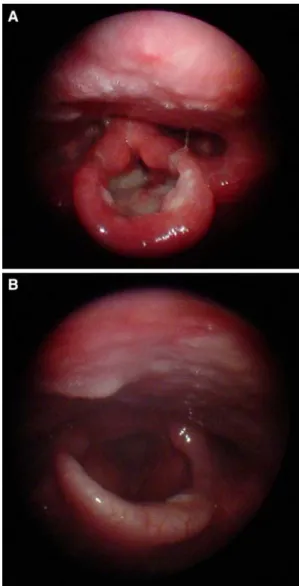
Fig. 1 This patient is a 48-year-old male with complaints of weight
loss, fatigue, odynophagia, hemoptysis and foreign body sensation in the throat for the last 15 days. Diagnosis of laryngeal tuberculosis was conWrmed by laryngeal biopsy. Endoscopic view at 2nd week of anti-tuberculous medical therapy was as shown (a). Endoscopic view at 6 months of antituberculous medical therapy was as shown (b)
Eur Arch Otorhinolaryngol (2008) 265:327–330 329
123
3, the tuberculous laryngitis healed in 8 months withsequel, but resulted in partial loss and scarring of the epiglottis.
The query of symptoms revealed that odynophagia was reported by all the patients. Mild to moderate changes in voice existed in three of the cases. Hemoptysis and foreign body sensation in the throat was reported by one patient each.
Discussion
According to recent reports from the WHO, the situation of tuberculosis in the countries of central and eastern Europe is alarming [1]. As the incidence of tuberculosis increases, LT is encountered by an ever-increasing number of otolar-yngologists and chest physicians [4]. The number of LT cases reported from industrialized countries remained low in the late twentieth century. Several studies reported an incidence of laryngeal involvement varying from <1% to 1.8% [5–7]. In developing countries such as Pakistan and Tanzania, the incidence of laryngeal involvement was reported to be higher and equal to 37% and 27%, respec-tively [4, 9]. In autopsies of patients who died of pulmonary tuberculosis, Fetterolf [10] showed laryngeal lesions in up to 83% in 1914. This Wnding suggests that it is diYcult to estimate the exact current prevalence of LT among patients with pulmonary tuberculosis. It is likely that LT is more common than is clinically recognized. Our study revealed an incidence of asymptomatic LT among hospitalized patients with pulmonary tuberculosis of 1.5%. However, the patients included in this study had already received anti-tuberculous medication, and it is possible that the incidence of LT would have been much higher if the patients were examined prior to therapy.
Our study supports Wndings that the male gender pre-dominates among tuberculosis patients, a Wnding that has been reported both in pulmonary and extrapulmonary tuberculosis [11–13]. Lower socio-economic and cultural conditions and higher level of social activity have been pos-tulated as factors that cause male predominance in tubercu-losis [14]. Besides these factors, one cannot exclude the possibility of higher exposure to cigarette smoke in the male population of Turkey [15].
The most commonly reported symptom of LT is hoarse-ness, followed by sore throat and odynophagia, whereas most of our patients complained of odynophagia followed by alteration in voice [16]. Because our study was designed to scan unselected, asymptomatic cases, instead of hoarse-ness, minor symptoms such as odynophagia and mild to moderate changes in voice were predominant complaints. This situation may also be attributed to the fact that the supraglottic larynx was the predominant site of
involve-ment in our patients. Furthermore, the fact that all of our cases were under antituberculous medication might have aVected the symptoms. Since all cases were heavy smokers, these symptoms may have been ignored by the patients and attributed to chronic laryngitis by the physicians. Because LT is the most infectious form of the disease, even minor symptoms in patients at risk should be taken into account by the physicians [17, 18].
Moreover, laryngeal carcinoma that may easily mimic LT or laryngeal carcinoma may concurrently exist with laryngeal or pulmonary tuberculosis [19]. Biopsy or rapid response to antituberculous treatment are two methods in the diagnosis of LT [20, 21]. Although biopsy is crucial for deWnitive diagnosis, the hazard of close proximity to the “highly infectious” patient during indirect laryngos-copy and biopsy may sometimes lead the physician to fol-low a “wait and watch” policy [21]. Contrary to our experience, there are reports that most lesions resolve over a 2-month period [6, 19, 20]. To our experience, a rapid response to the medication is possible only in a minority of the cases. In our study, only one of Wve cases showed a rapid response. It took our patients several months to get back to a near normal laryngeal appearance. For this reason, a “wait and watch” strategy will not be appropriate in patients with LT as it may cause a delay in diagnosis of laryngeal carcinoma.
Conclusion
• The incidence of unrecognized LT among patients with active pulmonary tuberculosis is 1.5%.
• Physicians dealing with pulmonary tuberculosis should keep in mind that symptoms of laryngeal involvement may be minor, such as odynophagia and minor alteration in voice. In these cases, an otolaryngological examina-tion should be performed. Early diagnosis of LT may protect the health care professionals from exposure to “highly infectious” patients.
• Laryngeal carcinoma may mimic LT. Response to antitu-berculous medication is usually late. Diagnosis by “wait and watch” policy will cause a signiWcant delay in the diagnosis of larynx carcinoma. Therefore, early biopsy should always be considered.
References
1. World Health Organization (2001). Global tuberculosis control, WHO report 2001. Geneva, Switzerland, WHO/CDS/TB/ 2001.287
2. Mert A (2003) Eriokinlerde yüzeysel tüberküloz lenfadenopati. Proceedings of tuberculosis and its diagnosis in 21st century sym-posium, Samsun, Turkey, pp 93–105
330 Eur Arch Otorhinolaryngol (2008) 265:327–330
123
3. Munck K, Mandpe AH (2003) Mycobacterial infections of the head and neck. Otolaryngol Clin North Am 36(4):569–76 4. Beg MH, Marfani S (1985) The larynx in pulmonary tuberculosis.
J Laryngol Otol 99(2):201–203
5. Rohwedder JJ (1974) Upper respiratory tract tuberculosis. Sixteen cases in a general hospital. Ann Intern Med 80(6):708–713 6. Brodovsky DM (1975) Laryngeal tuberculosis in an age of
chemo-therapy. Can J Otolaryngol 4(1):168–176
7. Williams RG, Douglas-Jones T (1995) Mycobacterium marches back. J Laryngol Otol 109(1):5–13
8. Diagnostic standards and classiWcation of tuberculosis in adults and children (2000) This oYcial statement of the American Tho-racic Society and the Centers for Disease Control and Prevention was adopted by the ATS Board of Directors, July 1999. This state-ment was endorsed by the Council of the Infectious Disease Soci-ety of America, September 1999. Am J Respir Crit Care Med 161(4 Pt 1):1376–1395
9. Manni H (1983) Laryngeal tuberculosis in Tanzania. J Laryngol Otol 97(6):565–570
10. Fetterhof G (1914) Cited by Du Plessis A, Hussey G (1987). La-ryngeal tuberculosis in childhood. Pediatr Infect Dis J 6:678–681 11. BorgdorV MW, Nagelkerke NJ, Dye C, Nunn P (2000) Gender and
tuberculosis: a comparison of prevalence surveys with notiWcation data to explore sex diVerences in case detection. Int J Tuberc Lung Dis 4(2):123–132
12. Holmes CB, Hausler H, Nunn P (1998) A review of sex diVerences in the epidemiology of tuberculosis. Int J Tuberc Lung Dis 2(2):96–104 13. Tu HY, Li HY, Huang TS (1997) Laryngeal tuberculosis: a series
of 46 patients. Changgeng Yi Xue Za Zhi 20(2):94–99
14. Martinez AN, Rhee JT, Small PM, Behr MA (2000) Sex diVer-ences in the epidemiology of tuberculosis in San Francisco. Int J Tuberc Lung Dis 4(1):26–31
15. Watkins RE, Plant AJ (2006) Does smoking explain sex diVer-ences in the global tuberculosis epidemic? Epidemiol Infect 134(2):333–339
16. Shin JE, Nam SY, Yoo SJ, Kim SY (2000) Changing trends in clinical manifestations of laryngeal tuberculosis. Laryngoscope 110:1950–1953
17. Ramadan HH, Tarazi AE, Baroudy FM (1993) Laryngeal tubercu-losis: presentation of 16 cases and review of the literature. J Oto-laryngol 22(1):39–41
18. Riley EC, Amundson DE (1992) Laryngeal tuberculosis revised. Am Fam Physician 49:759–762
19. Bailey CM, Windle-Taylor PC (1981) Tuberculous laryngitis: a series of 37 patients. Laryngoscope 91(1):93–100
20. Thaller SR, Gross JR, Pilch BZ, Goodman ML (1987) Laryngeal tuberculosis as manifested in the decades 1963–1983. Laryngo-scope 97(7 Pt 1):848–850
21. Harney M, Hone S, Timon C, Donnelly M (2000) Laryngeal tuber-culosis: an important diagnosis. J Laryngol Otol 114(11):878–880