Using Gene Expression Arrays to Elucidate Transcriptional
Profiles Underlying Prolactin Function
Sandra Gass,
1Jessica Harris,
2Chris Ormandy,
2and Cathrin Brisken
1,3Prolactin is an ancient hormone, with different functions in many species. The binding of prolactin to its receptor, a member of the cytokine receptor superfamily, results in the acti-vation of different intracellular signaling pathways, such as JAK2/STAT5, MAP kinase, and PI3K/AKT. How prolactin elicits so many different biological responses remains unclear. Re-cently, microarray technology has been applied to identify prolactin target genes in different systems. Here, we attempt to summarize and compare the available data. Our comparison of the genes reported to be transcriptionally regulated by prolactin indicates that there are few genes in common between the different tissues. Among the organs studied, mammary and prostate glands displayed the largest number of overlaps in putative prolactin target genes. Some of the candidates have been implicated in tumorigenesis. The relevance and validation of microarray data, as well as comparison of the results obtained by different groups, will be discussed.
KEY WORDS: prolactin target genes; expression arrays; mammary gland; prostate; ovary; uterus; Nb2 cells.
Prolactin is a peptide hormone, the bulk of which is secreted by the anterior pituitary gland. In females, prolactin serum levels are elevated during the luteal phase of the menstrual cycle (1), they increase dur-ing late pregnancy and lactation, and peak follow-ing bouts of nursfollow-ing (2). It is an ancient hormone, which plays a role in very different processes in verte-brates, such as freshwater osmoregulation in fish (3), nesting behavior in birds, and growth and metamor-phosis in amphibians (4). Prolactin acts pleiotropi-cally on a variety of target tissues in mammals (5), affecting, directly and indirectly, the development of the mammary gland, the ovaries, the prostate, and lacrimal glands, the intestine, and the skin. The hormone binds to the prolactin receptor (PrlR), a
1Swiss Institute for Experimental Cancer Research, National Cen-ter of Competence in Research (NCCR) Molecular Oncology, Epalinges s/Lausanne, Switzerland.
2Garvan Institute of Medical Research, St. Vincents Hospital, Sydney, Australia.
3To whom correspondence should be addressed at ISREC, 155 Chemin des Boveresses, CH-1066 Epalinges s/Lausanne, Switzerland; e-mail: [email protected].
member of the cytokine receptor family (6), thereby activating different intracellular signaling pathways
Abbreviations used: 20α-HSD, 20 alpha-hydroxysteroid
dehydro-genase; 3β-HSD, 3β-hydroxysteroid dehydrogenase; bHLH, ba-sic Helix–Loop–Helix; BRCA1, breast cancer 1; C/EBP, CCAAT enhancer-binding protein; cDNA, complementary deoxyribonu-cleic acid; CL, corpus luteum; DNA, deoxyribonudeoxyribonu-cleic acid; ECM, extracellular matrix; EGF, epidermal growth factor; EST, expressed sequence tag; GATA3, GATA binding protein 3; GlyCAM 1, glycosylation-dependent cell adhesion molecule 1; HB-EGF, heparin-binding epidermal growth factor; HE5, human epididymal protein 5; IGF-2, insulin-like growth factor 2; IRF-1, interferon-regulated factor 1; JAK, janus kinase; MAP, mitogen activated protein; MAS, MicroArray Suite; MEC, mammary ep-ithelial cell; MIAME, minimal information on microarray ex-periment; mRNA, messenger ribonucleic acid; NCCR, national center of competence in research; PCR, polymerase chain re-action; PI3K, phosphoinositol 3-phosphate kinase; PPARγ, per-oxisome proliferator-activated receptorγ ; PR, progesterone re-ceptor; PrlR, prolactin rere-ceptor; QRT-PCR, quantitative reverse transcriptase polymerase chain reaction; RANKL, receptor acti-vator of nuclear factor-κB ligand; RDA, representational differ-ence analysis; SSH, subtractive suppressive hybridization; STAT, signal transducer and activator of transcription; SVP, seminal vesi-cle protein; TIGR, the institute for genome research; WAP, whey acidic protein; Wt, wild type.
269
such as the JAK2/Stat5 (7–9), the MAP kinase (10), and PI3K/AKT pathways (11).
In the mammary gland, prolactin signaling in the epithelium controls the formation of alve-oli and the differentiation of mammary epithe-lial cells (MECs) into milk-secreting cells (12). Moreover, prolactin indirectly affects ductal side branching through the stimulation of progesterone secretion by the corpora lutea (13). Prolactin is also synthesized locally in the breast epithe-lium (14); this production may be important for cell proliferation during late pregnancy (15). Lo-cal prolactin synthesis as well as expression of the PrlR is upregulated in breast carcinomas (16), suggesting that localized deregulation of pro-lactin signaling may contribute to breast carcino-genesis. Consistent with this notion are observa-tions that blocking prolactin signaling interferes with the growth of various breast cancer cell lines (17,18) and that mice lacking the prolactin gene show a delay in polyoma middle-T antigen-induced breast tumorigenesis (19). Furthermore, high serum prolactin levels have been related to an in-creased relative risk of developing breast cancer (20).
How can one hormone ensure milk production, control uterus, and ovaries, and affect the physiology of T cells and hair follicles?
A number of mechanisms may be responsible for these contrasting, tissue-specific effects. Different forms of prolactin have been described (21) and dif-ferent receptor forms exist with distinct cytoplasmic lengths, which may differentially activate distinct in-tracellular signaling pathways (22). The expression of receptor and ligand isoforms in cell-type-specific con-stellations may contribute to the tissue-specificity of prolactin action. Alternatively, a very similar stimu-lus may elicit different biological responses in differ-ent target cells, the signaling networks of which are wired in distinct ways. This latter hypothesis is sup-ported by our recent finding that a chimeric recep-tor consisting of the signaling domain of the erythro-poietin receptor could, when coupled to the ligand binding domain of PrlR, rescue the defective alve-ologenesis and differentiation of PrlR−/−MECs (23). Recent studies exploiting new techniques for tran-scription profiling lend further support to this hypoth-esis. Our analysis of transcription profiling studies, albeit preliminary, suggest that the tissue/cell-type-specific responses at the transcriptional level differ substantially.
KNOWN TRANSCRIPTIONAL TARGETS OF PROLACTIN
In an early differential screening experiment, interferon-regulatory factor 1 (IRF-1) was identified as an immediate-early gene in prolactin-stimulated T cells (24). In hepatocytes, prolactin regulates tran-scription of the Na+/taurocholate cotransporter (25). Furthermore, prolactin induces estrogen receptor ex-pression (26) and progesterone secretion in the cor-pus luteum (CL) during pregnancy. It also represses transcription of 20-α-hydroxysteroid dehydrogenase, which plays a role in pregnancy termination in the rat (27). Another molecule known to be involved in progesterone synthesis, 3β-hydroxysteroid dehy-drogenase (3β-HSD), is also regulated by prolactin in the CL (28). In NIH-3T3 cells, prolactin stim-ulates the transcription of the transcription factors CCAAT enhancer-binding protein (C/EBP) and per-oxisome proliferator-activated receptorγ (PPARγ ) (29); these effects may explain its adipogenic function in these cells.
In the mammary gland, prolactin regulates tran-scription of milk proteins, such as casein (30), whey acidic protein (WAP) (31) andβ-lactoglobulin (32). The hormone regulates the transcription of a number of other genes in various human breast cancer cell lines. BRCA1 transcription increases in MCF-7 and T47-D cells following treatment with prolactin (33), and cyclin D1 is induced in T47D cells (34). Additional prolactin target genes were discovered using mouse MEC lines, such as the p100 coactivator in HC11 cells (35) and the CBP/p300 transactivator Cited4 in SCp2 cells (36).
DISCOVERING NOVEL MEDIATORS OF PROLACTIN ACTION THROUGH TRANSCRIPT PROFILING
Considering the variety of biological functions, the number of target genes identified is limited. How does prolactin trigger alveolar morphogenesis? What is the role of prolactin signaling in breast carcinogen-esis? How does it affect other organs? One way of addressing these questions is to examine changes in gene expression induced by activation of the prolactin signaling cascade in different contexts. With the ad-vent of microarrays, the research community has a powerful tool for surveying at once all known mRNA transcripts in a broad, unbiased fashion. Several
studies have now been published using arrays, some of them with more limited transcript sets, to learn more about genes transcribed following prolactin action in different biological processes.
PROSTATE
Using a mouse model in which prolactin is overexpressed from the metallothionine promoter, Dillner and colleagues (37) looked for genes that may be important for the prostate hyperplasias seen in these transgenic animals. cDNA from dorsolateral and ventral lobes of 4 transgenic and 5 control mice, at 4–6 months of age, was compared. Representational difference analysis (RDA) identified 152 unique se-quences, 69 of which had been annotated previously while 83 sequences were novel. These 152 sequences were printed on microarrays to which total RNA from hyperplastic and control prostate tissue was co-hybridized. Thirty percent of the sequences, i.e. 48 dif-ferent transcripts, were detected in the starting pool of total RNA. Thirty-one percent of these, i.e. 15, were identified as significantly modulated, 10 of them were annotated. Muscle creatine kinase, vimentin, and cy-tochrome C oxidase polypeptide III were upregulated and cytokeratin 8, aldose reductase and RIL protein downmodulated in the transgenic prostates.
Robertson et al. (38) studied the role of prolactin in the growth of the normal and cancerous prostate of PrlR−/−mice by transcript profiling the dorsal and ventral lobes of prostates from 50-week-old animals. RNA was collected from five to eight lobes for each homozygous mutant and wild-type (wt) mouse. Mor-phological analysis of the prostates from these ani-mals demonstrated that prolactin plays a subtle role in the morphogenesis of the prostate, since PrlR−/− animals displayed a 20% increase in prostate weight and a reduced epithelium content, but no alteration in branching morphology. Three experiments using Affymetrix microarrays were performed on the ven-tral prostate. As triplicates were not substantially more informative than duplicates, only two experi-ments were performed on the dorsal prostate. Genes identified as differentially expressed between PrlR−/− and PrlR+/+prostates are shown in Table I.
The majority of differentially expressed genes were decreased in the PrlR−/−tissue, suggesting that prolactin has overall stimulatory effects on transcrip-tion in both lobes of the prostate. Surprisingly, the genes most differentially regulated were found to be
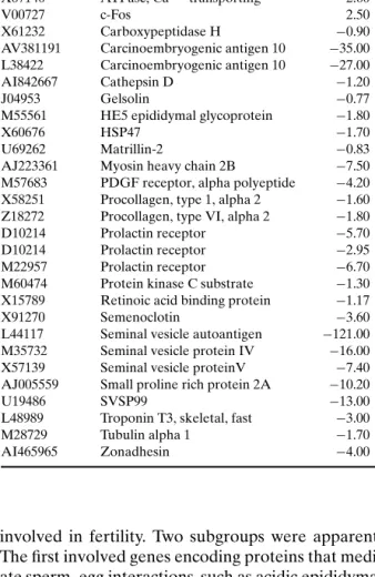
Table I. Genes Differentially Expressed Between PrlR−/−and PrlR+/+Prostates (38)
Accession # Title Fold change
M92849 Acidic epididymal glycoprotein 1 −4.30 M88127 Adenemoatosis polyposis coli −0.80
X04673 Adipsin −1.80
U03419 Alpha-1 type I procollagen −0.80 X67140 ATPase, Ca++transporting −2.60
V00727 c-Fos 2.50
X61232 Carboxypeptidase H −0.90
AV381191 Carcinoembryogenic antigen 10 −35.00 L38422 Carcinoembryogenic antigen 10 −27.00
AI842667 Cathepsin D −1.20
J04953 Gelsolin −0.77
M55561 HE5 epididymal glycoprotein −1.80
X60676 HSP47 −1.70
U69262 Matrillin-2 −0.83
AJ223361 Myosin heavy chain 2B −7.50 M57683 PDGF receptor, alpha polyeptide −4.20 X58251 Procollagen, type 1, alpha 2 −1.60 Z18272 Procollagen, type VI, alpha 2 −1.80
D10214 Prolactin receptor −5.70
D10214 Prolactin receptor −2.95
M22957 Prolactin receptor −6.70
M60474 Protein kinase C substrate −1.30 X15789 Retinoic acid binding protein −1.17
X91270 Semenoclotin −3.60
L44117 Seminal vesicle autoantigen −121.00 M35732 Seminal vesicle protein IV −16.00 X57139 Seminal vesicle proteinV −7.40 AJ005559 Small proline rich protein 2A −10.20
U19486 SVSP99 −13.00
L48989 Troponin T3, skeletal, fast −3.00
M28729 Tubulin alpha 1 −1.70
AI465965 Zonadhesin −4.00
involved in fertility. Two subgroups were apparent. The first involved genes encoding proteins that medi-ate sperm–egg interactions, such as acidic epididymal glycoprotein HE5 and zonadhesin. Acidic epididymal glycoprotein is an androgen-regulated gene secreted by the epithelium of the epididymis that is thought to be involved in sperm–oocyte plasma membrane fu-sion (39–41). It is found on maturing spermatozoa and shows variable surface presentation during sperm ca-pacitation (42). Zonadhesin is a sperm-zona pellucida binding protein that confers species specificity (43). The second subgroup contains the anti-inflammatory and procoagulant seminal vesicle proteins, which play a role in copulatory plug formation and protection of sperm from the female immune response: namely SVP2/SV-IV, the semenoclotin and seminal vesicle F protein/SVP5 (44–46). The changes in the expression
of these fertility-related genes prompted the authors to examine the fertility of PrlR−/−males. They found overall reduced fertility, a prolonged latency to first pregnancy and a 40% probability to produce a first pregnancy compared to PrlR+/+animals. This study demonstrates that transcript profiling can assist in identifying phenotypes in mutant mouse strains.
OVARY
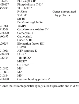
Stocco and colleagues asked the question whether prolactin and prostaglandin F2α (PGF2α), which have opposing effects on the corpus luteum, control the expression of the same genes in an antag-onistic fashion (47). Pregnant rats were hypophysec-tomized on day 4 of pregnancy and injected with pro-lactin or vehicle. Both groups were sacrificed 7 days later, and RNA was isolated from CL. Out of the 1176 distinct rat genes on the cDNA microarrays, 13 were controlled by both prolactin and PGF2α in opposite fashion. The 27 genes identified as differentially reg-ulated by prolactin are listed in Table II.
Table II. Genes Regulated by Prolactin in the Corpus Luteum (47)
Accession # Title Annotation
L12382 ADP-ribosylation factor 3 Genes downregulated Annexin V by prolactin X17163 c-jun 20a-HSDa U47287 PGF2 alpha-Ra M20637 Phospholipase C d1a X52498 TGF beta1a P450scc Genes upregulated 3b-HSD by prolactin SR-B1 Beta2-microglobulin L31884 TIMP2 X14209 Cytochrome c oxidase IV M36320 Cathepsin H Y00697 Cathepsin L Cu/Zn SOD
L29259 Elongation factor SIII HSP60 D10021 ATP synthase D M26199 LH-Ra U22424 11b-HSD2a MGSTa GST m2a D10862 Id1a D10863 Id2a D10864 Id3a
M86870 Calcium binding protein 2a
aGenes that are antagonistically regulated by prolactin and PGF2α.
Nb2 RAT LYMPHOMA CELLS
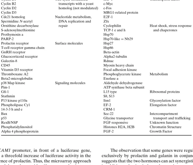
Bole-Feysot et al. (48) used the rat Nb2-11c lym-phoma cell line, which depends on prolactin for pro-liferation, to identify Prl target genes. By means of mRNA differential display, RDA, subtractive sup-pressive hybridization (SSH), analysis of weakly expressed genes and screening of an organized li-brary, they identified 70 genes that were differen-tially transcribed by Nb2-11c cells within 2–24 h of prolactin treatment. Twenty of these were un-known; the other 50 genes were classified to 10 functional categories, such as cell cycle regulators, nucleotide metabolism/DNA replication and repair proteins, cell-surface antigens/adhesion proteins, sig-naling moleciles involved in apoptosis or prolifera-tion, or transcription factors (see Table III).
UTERUS
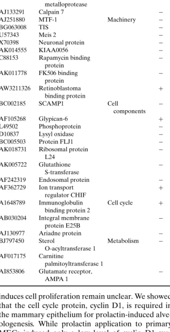
Deletion of the PrlR in mice causes female in-fertility and failure of implantation (49). Applying mRNA differential display to PrlR−/− and wt uteri implantation sites, Baran et al. isolated 45 transcripts, out of which 29 corresponded to known mouse ESTs (see Table IV) (50).
MAMMARY GLAND
To identify new prolactin target genes in the mammary gland, Hou et al. made use of mice that are homozygous for a disrupted prolactin allele (Prl−/−) (51). Prl−/−females are infertile but their mammary glands develop a normal ductal system. The authors
compare RNA from Prl−/− and Prl+/− mammary
glands from 10-week-old females, pooled from five mice of each genotype. One hundred seventy-nine out of 588 transcripts represented on the Clontech membrane were detected (30%), 33 (18%) were el-evated in the Prl+/− mammary glands, and 7 (4%) were elevated in Prl−/− mammary glands. The au-thors focused on one particular gene, the expression of which was decreased in the Prl−/−mammary glands, the glycosylation-dependent cell adhesion molecule 1 (GlyCAM1). Immunohistochemistry confirmed that GlyCAM1 protein is expressed in the mammary ep-ithelium and the ductal lumen of Prl+/−virgin mice. In the mouse mammary epithelial cell line HC11, which can be induced to express milk proteins in vitro, Gly-CAM1 mRNA is increased after 48 h of prolactin and progesterone treatment. An 800 bp fragment of the
Table III. Transcripts Modulated by Prolactin in the Rat Nb2-11c Lymphoma Cell Line (48)
Title Annotation Title Annotation
Cyclin E1 Myosin heavy chain Cytoskeleton
EGR-1 Transcripts that have a Tubulin, a, b Cdc5-like protein cell-cycle modulated Beta-actin
Cdk2, Cdk5 homolog in yeast FAKp125
Cyclin B1 Cell-cycle-modulated IRF-1 Transcription factor
Cyclin B2 transcripts with a yeast c-Myc Cyclin D2 homolog (not modulated) c-Fos
Cyclin D3 MRG1-related protein
Cdc21 homolog Nucleotide metabolism, E2F-1 Spermidine N-acetyl DNA replication and Zfx
Ornithine decarboxylase repair Cyclophilin Heat shock, stress response
S-adenosylmethionine TCP-1 e and h and chaperones
Prothymosin a Hsp70
PARP-2 Hsp70-like= Nb29
Prolactin receptor Surface molecules Hsp27
T-cell receptor gamma chain Hsp86
GnRH receptor Beta-actin
Glucocorticoid receptor Alpha2-tubulin
Galectin-8 Rdnuc
CD45 Myosin heavy chain
Vitamin D3 receptor Focal adhesion kinase
Thromboxane A2 Phosphoglycerate kinase Metabolism
Beta2-microglobulin Enolase a
p38 Map kinase Signaling molecules Aldehyde dehydrogenase
Pim-1 ATP synthase beta subunit
Gfi-1 L15 type Ribosomal proteins
Stathmin S8, S13
P13 kinase p110a Itm1 Glycosylation factor
Phospholipase Cy1 EF-2 Elongation factor
14-3-3 h and e CRM-1
Bax Sec-22 Intercompartment
p53 Glycine transporter transport and trafficking
RexB/NSP FGF-responsive Unknown function
Phosphatidylinositol Histones H2A, H2B Chromatin Structure
Alpha 4 phosphoprotein FGF-2 Growth Factor
GlyCAM1 promoter, in front of a luciferase gene, gives a threefold increase of luciferase activity in the presence of prolactin. Thus, the microarray approach allowed the authors to identify a new prolactin target gene in the mammary epithelium. The biological im-portance of GlyCAM1 in mediating the response to prolactin remains to be evaluated.
Using organ cultures, Naylor et al. demonstrated that the neuropeptide galanin acts on the mammary gland to ensure final stage differentiation and milk se-cretion (52). Transcript profiling of explants treated with either galanin, prolactin, or both factors was per-formed. The genes identified as transcriptionally reg-ulated by either factor or both are listed in Table V. Among them were milk protein genes, markers of MEC differentiation, and several known JAK/STAT target genes.
The observation that some genes were regulated exclusively by prolactin and galanin in conjunction suggests that the two hormones can act synergistically in addition to having independent actions.
The overall number of genes regulated by pro-lactin was larger than the number of genes regulated by galanin. This result suggests that prolactin has a much stronger effect on transcription in the mam-mary gland than galanin. Interestingly, prolactin and galanin have opposite effects on the transcription lev-els of some of the genes which are regulated by both of them, reflecting the complexity of the interactions occurring in the mammary gland organ cultures fol-lowing galanin and prolactin treatments.
As discussed above, prolactin acts as a potent mitogen and morphogen on the mammary epithe-lium in vivo (53), but the mechanisms by which it
Table IV. Differentially Expressed Transcripts in the PrlR−/− Uterus at Day 5.5 of Pregnancy (50)
Accession # Title Annotation Change
AK011085 HSPC 307 Immunity +
AF039584 Decay accelerating −
factor precursor
AE000664 T cell receptor beta −
AJ242912 Disintegrin + metalloprotease AJ133291 Calpain 7 − AJ251880 MTF-1 Machinery − BG063008 TIS − U57343 Meis 2 − X70398 Neuronal protein − AK014555 KIAA0056 − C88153 Rapamycin binding − protein AK011778 FK506 binding − protein AW3211326 Retinoblastoma + binding protein BC002185 SCAMP1 Cell − components AF105268 Glypican-6 + L49502 Phosphoprotein − D10837 Lysyl oxidase − BC005503 Protein FLJ1 −
AK018731 Ribosomal protein −
L24
AK005722 Glutathione −
S-transferase
AF242319 Endosomal protein −
AF362729 Ion transport +
regulator CHIF
A1648789 Immunoglobulin Cell cycle + binding protein 2
AB030204 Integral membrane −
protein E25B
AJ130977 Ariadne protein −
BJ797450 Sterol Metabolism −
O-acyltransferase 1 AF017175 Carnitine
palmitoyltransferase 1
AI853806 Glutamate receptor, −
AMPA 1
induces cell proliferation remain unclear. We showed that the cell cycle protein, cyclin D1, is required in the mammary epithelium for prolactin-induced alve-ologenesis. While prolactin application to primary MECs induced only a low level of cyclin D1 syn-thesis, longer exposures—18 h—led to a significantly higher cyclin D1 protein expression (54). These ob-servations suggested that prolactin might be induc-ing the expression of an intermediate growth factor, which in turn induced cyclin D1 and proliferation. To identify such a growth factor, we conducted a screen that selects specifically for genes that function
down-stream of the PrlR and updown-stream of cyclin D1 (54). We compared the transcription profiles of pairs of con-tralateral cleared inguinal mammary fat pads, which had been reconstituted with PrlR−/−or cyclin D1−/− MECs. The host mice were wt. At 3 weeks of age, their inguinal fat pads were surgically cleared of en-dogenous epithelium and engrafted with MECs from PrlR−/−and cyclin D1−/−females. As first shown by DeOme (55), such epithelial grafts can reconstitute the mammary stroma, forming a new ductal system. Four weeks later, when the grafts had fully penetrated the fat pad, the hosts were mated. Since the morpho-genetic block shown by MECs of these two genotypes are similar, these two types of mammary glands, as-sayed at an identical day of pregnancy, are presumed to carry comparable numbers of cells in comparable states of proliferation. The only differences in gene ex-pression pattern between them should involve those genes that lie downstream of PrlR signaling and up-stream of cyclin D1 expression.
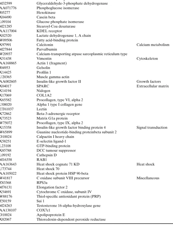
Out of the 6500 transcripts surveyed, 319 were expressed at more than threefold higher levels in the
cyclin D1−/−than in the PrlR−/−grafts, whereas 430
transcripts were downregulated more than threefold. Previous observations had indicated that, in contrast to the PrlR−/− epithelium, the cyclin D1−/− epithe-lium retains its ability to undergo differentiation, as manifested by its ability to synthesize milk proteins late in pregnancy (56). Accordingly, many genes en-coding proteins secreted with the milk or involved in milk production, such as metabolic enzymes, cal-cium transport genes, and intracellular vesicular traf-ficking genes, were differentially expressed, with their transcripts being found at far higher levels in the
cy-clin D1−/− grafts than in the fat pads engrafted with
PrlR−/− MECs. In addition, genes involved in signal transduction and in the construction of the cytoskele-ton and the extracellular matrix (ECM) were prefer-entially expressed in the cyclin D1−/−recombinants.
An overview of the genes whose expression lev-els differed by more than 10-fold is given in Ta-ble VI. We concentrated our further analysis on one of the secreted factors that were particularly highly expressed in the cyclin D1−/− grafts, namely insulin-like growth factor-2 (IGF-2), which was found at a level 13.2-fold higher in the glands engrafted with cyclin D1−/− epithelia than in their PrlR−/− counterparts. QRT-PCR showed a 30-fold differ-ence. Consistent with a model in which prolactin relies on a secreted factor IGF-2 to induce a pro-liferative response, we found that prolactin induces IGF-2 mRNA expression in primary MECs and
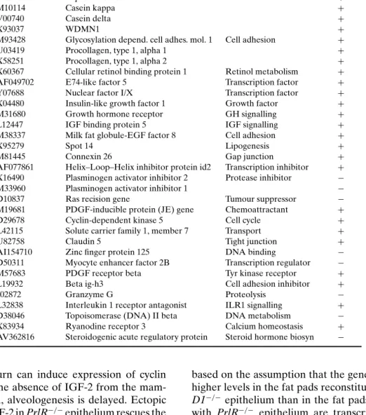
Table V. Genes Found to be Regulated by Prolactin in Mammary Gland Organ Cultures (52)
Accession # Title Annotation Change
V00856 Whey acidic protein Milk protein +
M36780 Casein alpha + D10215 Casein gamma + X04490 Casein beta + M87863 Alpha lactalbumin + M10114 Casein kappa + V00740 Casein delta + X93037 WDMN1 +
M93428 Glycosylation depend. cell adhes. mol. 1 Cell adhesion +
U03419 Procollagen, type 1, alpha 1 +
X58251 Procollagen, type 1, alpha 2 +
X60367 Cellular retinol binding protein 1 Retinol metabolism + AF049702 E74-like factor 5 Transcription factor +
Y07688 Nuclear factor I/X Transcription factor +
X04480 Insulin-like growth factor 1 Growth factor +
M31680 Growth hormone receptor GH signalling +
L12447 IGF binding protein 5 IGF signalling +
M38337 Milk fat globule-EGF factor 8 Cell adhesion +
X95279 Spot 14 Lipogenesis +
M81445 Connexin 26 Gap junction +
AF077861 Helix–Loop–Helix inhibitor protein id2 Transcription inhibitor + X16490 Plasminogen activator inhibitor 2 Protease inhibitor −
M33960 Plasminogen activator inhibitor 1 −
D10837 Ras recision gene Tumour suppressor −
M19681 PDGF-inducible protein (JE) gene Chemoattractant +
D29678 Cyclin-dependent kinase 5 Cell cycle +
L42115 Solute carrier family 1, member 7 Transport +
U82758 Claudin 5 Tight junction +
AI154710 Zinc finger protein 125 DNA binding −
D50311 Myocyte enhancer factor 2B Transcription regulator −
M57683 PDGF receptor beta Tyr kinase receptor +
L19932 Beta ig-h3 Cell adhesion inhibitor +
J02872 Granzyme G Proteolysis −
L32838 Interleukin 1 receptor antagonist ILR1 signalling + D38046 Topoisomerase (DNA) II beta DNA metabolism − X83934 Ryanodine receptor 3 Calcium homeostasis + AV362816 Steroidogenic acute regulatory protein Steroid hormone biosyn −
that IGF-2 in turn can induce expression of cyclin D1 protein. In the absence of IGF-2 from the mam-mary epithelium, alveologenesis is delayed. Ectopic expression of IGF-2 in PrlR−/−epithelium rescues the proliferative defect, arguing that IGF-2 is a physiolog-ically important mediator of prolactin-induced pro-liferation/morphogenesis. Hovey et al. independently drew the same conclusion based on transcript quan-tification in PrlR−/−mouse mammary glands (57).
Certainly, more can be learned from the data obtained with this screen. Another growth factor found to be differentially expressed is HB-EGF (Gass and Brisken, unpublished observations), and its role in alveologenesis remains to be studied. In further exploring the data, we need to be aware of several potential limitations of our approach. This screen is
based on the assumption that the genes expressed at higher levels in the fat pads reconstituted with cyclin D1−/− epithelium than in the fat pads reconstituted with PrlR−/− epithelium are transcriptionally acti-vated by prolactin signaling. However, it is formally possible, that differences in gene expression levels are independent of prolactin signaling and result instead from a lack of cyclin D1-dependent repression. This concern can be addressed by comparing expression levels in cyclin D1−/−versus cyclin D1 wt transplants, as done for IGF-2. More general limitations, such as the choice of genes arrayed and the sensitivity, are discussed below.
We used a novel approach to elucidate the tran-scriptional response to prolactin specifically in the mammary gland during early pregnancy. By transcript
Table VI. Genes Expressed at a 10-Fold Higher Level in PrlR−/−Mammary Epithelial Transplants than Cyclin D1 Transplanted Glands (54)
Accession # Title Annotation
X93037 WDNM1 Milk secretion
W18308 Ferritin heavy chain gene
X04673 Adipsin
X61431 ACYL-COA-binding protein Y00516 Fructose-bisphosphate aldolase A
M32599 Glyceraldehyde-3-phosphate dehydrogenase AA071776 Phosphoglucose isomerase
J05277 Hexokinase
X04490 Casein beta
L09104 Glucose phosphate isomerase M21285 Stearoyl-Coa desaturase AA117004 KDEL receptor
X02520 Lactate dehydrogenase 1, A chain W09506 Fatty acid-binding protein
X97991 Calcitonin Calcium metabolism
M27844 Parvalbumin
W20937 Calcium-transporting atpase sarcoplasmic reticulum type
X51438 Vimentin Cytoskeleton
AA168865 Actin 1 (fragment)
J04953 Gelsolin
X14425 Profilin 1
U20365 Muscle gamma actin
AA002605 Insulin-like growth factor II Growth factors
X04017 SPARC Extracellular matrix
X14194 Nidogen
X17069 COL1A2
X65582 Procollagen, type VI, alpha 2 U08020 Alpha 1 type I collagen gene
ET61037 Lectin
X72862 Beta-3-adrenergic receptor X73523 Matrix G1a protein W75072 Procollagen, type IX, alpha 2
X15358 Insulin-like growth factor binding protein 4 Signal transduction W65899 Guanine nucleotide-binding proteinbeta subunit 2
D10024 Calpactin I heavy chain X58251 E-selectin ligand-1 L23108 GTP-binding protein X85788 DCC tumour suppressor
L09192 Cathepsin D
M16358 RAB1
AA163643 Heat shock cognate 71 KD Heat shock
U73744 Heat shock 70
AA105022 Heat shock protein HSP 90-beta
W41817 C oxidase subunit VIII precursor Miscellaneous
Z83368 RPS3a
M76131 Elongation factor 2
X54691 Cytochrome C oxidase, subunit IV W88176 Thiol-specific antioxidant protein (PRP)
Z50159 Sui 1
M24263 Testosterone 16-alpha-hydroxylase gene
AA138107 COX7c1
D10024 Apolipoprotein E
profiling PrlR−/− and PrlR+/+ epithelial transplants during early pregnancy, we reduced the effect of dif-ferences in epithelial cell number seen during late pregnancy, when PrlR−/− epithelium fails to form alveoli. Additionally, we examined transcript profiles of PrlR+/+mammary fat pads cleared of epithelium to distinguish transcripts present in the epithelium from those in the fat pad. The profiling was performed with pools of RNA from four to six animals on high-density oligonucleotide arrays (Affymetrix MGU74A GeneChips). Data were analyzed using MicroArray Suite 4.0 (MAS 4.0 Affymetrix) and sorted using Ex-cel (Microsoft). Fold changes, calculated by MAS 4.0 for a number of genes were confirmed by quantitative PCR using the LightCycler (Roche). Overall, there was a dramatic loss of expression in the PrlR−/− ep-ithelial transplants at all 3 days of pregnancy profiled (days 2, 4, and 6), including epithelial markers such as keratins. Nevertheless, merely 15% of “epithelial transcripts,” as defined by their presence in PrlR+/+ transplants and concomitant absence from PrlR+/+ cleared fat pads, displayed decreased expression lev-els in the PrlR−/−transplants. These results suggested that the changes in gene expression were due to a lack of prolactin action and not due to differences in ep-ithelial cell number.
The genes identified by MAS 4.0 were sorted into groups, depending on their gene ontology as annotated in NetAffx (Affymetrix). This abbreviated list (to be published in full elsewhere; Harris et al., submitted) does not include cDNAs of unknown function or genes associated with expressed se-quence tags (ESTs), which are the focus of ongoing investigations. Extensive literature searches suggest that many of the genes whose expression decreased in PrlR−/− transplants are usually upregulated during pregnancy and localize predominantly to the epithelium.
Thus, four milk protein genes (caseinα, β, κ, and WDNM1) were decreased in the glands reconstituted with PrlR−/− epithelium. The isolation of ß-casein, a known prolactin-regulated gene, confirms that our model can identify prolactin-regulated genes involved in MEC differentiation.
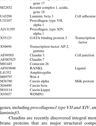
The development of the mammary gland is not only influenced by systemic hormones but also de-termined by the cell’s microenvironment. One com-ponent of this environment which helps to deter-mine tissue-specific gene expression is the ECM (58). As shown in Table VII, we identified several ECM components, involved in cell adhesion and alveolar development as potential prolactin target
Table VII. Genes Reduced in Expression During Early Pregnancy in the PrlR−/−Mammary Epithelial Grafts (59)
Accession # Title Annotation
X15662 Keratin complex 2, basic, Cytoskeleton gene 8
M36120 Keratin complex 1, acidic, gene 19
M13805 Keratin complex 1, acidic, gene 17
M22832 Keratin complex 1, acidic, gene 18
U43298 Laminin, beta 3 Cell adhesion U32107 Procollagen, type VII,
alpha 1
AJ131395 Procollagen, type XIV, alpha 1
X55123 GATA binding protein 3 Transcription factor X94694 Transcription factor AP-2,
gamma
AF09505 Claudin 3 Cell junction
AF087825 Claudin 7 M81445 Connexin 26
AF019048 RANKL Ligand
L41352 Amphiregulin
M89797 Wnt-4
M36780 Casein alpha Milk protein X04490 Casein beta
M10114 Casein kappa
X93037 WDMN1
genes, including procollagenα1 type VII and XIV, and lamininβ3.
Claudins are recently discovered integral mem-brane proteins that are major structural compo-nents of tight junction strands. Tight junction clo-sure increases during lactation to prevent diffusion of molecules across the mammary epithelium. This process is mediated by progesterone withdrawal fol-lowing parturition and requires activation of the PrlR (59). Our finding that claudin 3 and 7 expression is decreased in PrlR−/−mammary glands suggests that prolactin may further influence these junctions by reg-ulating the transcription of their components.
Connexin-26 is a member of a large family of proteins that form gap junctions and allow exchange of small ions between epithelial cells. Connexin-26 mRNA and protein expression are significantly up-regulated during pregnancy and remain elevated dur-ing lactation (60); expression is confined to the alveo-lar epithelium and the protein localizes to sites of cell contact (61).
Our screen also identified several secreted lig-ands of importance for cell–cell communication and cell differentiation. One such gene, Wnt-4, is a
member of the Wnt family of secreted glycoproteins implicated in cell–cell signaling. We have shown that Wnt-4 acts downstream of progesterone to induce ductal side-branching during pregnancy (62). Over-expression of Wnt-4 in the mammary gland by retro-viral delivery resulted in increased ductal side branch-ing and alveolar-like structures in virgin animals (63). Another gene of interest, amphiregulin, is a member of the epidermal growth factor (EGF) family, whose members bind to the EGF receptor family. In mice lacking both amphiregulin alleles, the ductal tree fails to fully penetrate the fat pad. Ectopic expression of amphiregulin in the mammary epithelium can result in hyperplastic ducts and lobules (64). Finally, tumor necrosis factor (ligand) superfamily member 11, also known as receptor activator of nuclear factor-κB lig-and (RANKL) lig-and osteoprotegrin liglig-and, was found to be decreased in the PrlR−/−epithelium at all three
time points. The mammary glands of RANKL−/−
mice, similar to PrlR−/−mammary glands, fail to de-velop alveoli, resulting in lactational failure in the mutant animals. Our preliminary studies have shown that prolactin modulates RANKL expression during early pregnancy, suggesting that prolactin is the mas-ter regulator of signaling events necessary for alveolar development.
Interestingly, all three, Wnt-4 (62), amphiregulin (65), and RANKL (54), are known targets of proges-terone signaling. It remains to be seen whether they are also direct prolactin targets or whether their ex-pression is indirectly controlled by prolactin. Grimm
et al. (66) demonstrated that PrlR−/− mammary
ep-ithelial grafts showed increased progesterone recep-tor expression compared to their wt counterparts and a decrease in proliferation after combined estrogen and progesterone treatments. These findings suggest that progesterone response of the mammary gland is altered in the absence of prolactin signaling. Further-more, studies by Hovey et al. (79) demonstrate that the PrlR and progesterone receptor (mRNA) show simi-lar expression patterns, and synergistic effects of both hormones arguing further for cross talk of these two signaling pathways.
Eventually, a number of transcription factors were discovered to be important for the prolactin-stimulated development of alveolar cells, among them the GATA binding protein 3, a member of a family of transcription factors that bind to DNA through a highly conserved zinc finger domain.
Thus, these transcript profiling experiments show that, in addition to the induction of milk proteins at the late stage of differentiation, prolactin also induces
the transcription of genes important for intracellular and extracellular structure, cell permeability, cell–cell communication, and differentiation.
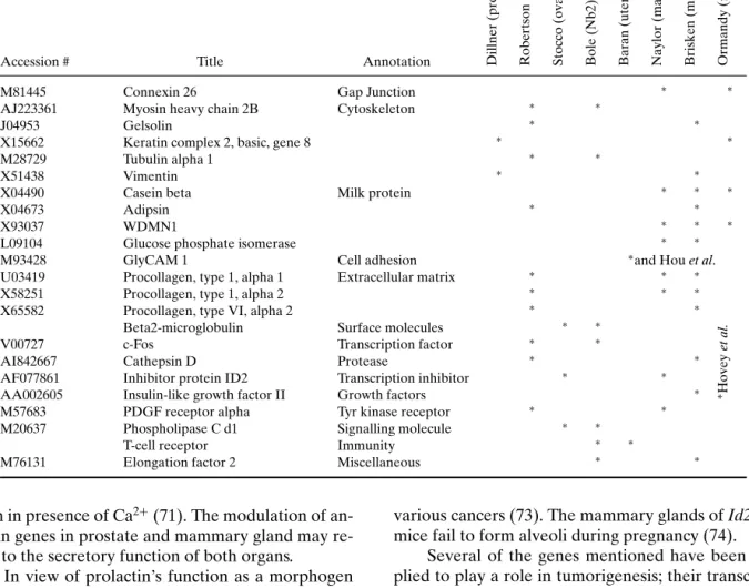
COMPARISON OF THE DIFFERENT SCREENS The recent studies presented above have applied expression profiling techniques to investigate the in-fluence of prolactin on transcription in different tis-sues or cell lines and have generated a list of puta-tive prolactin targets. Our comparison of the genes identified indicates that few of them are common to different tissues (see Table VIII and Table IX).
As illustrated by the Venn diagram in Fig. 1, among the different organs studied, mammary and prostate glands displayed the largest overlap in puta-tive prolactin target genes. Three of the genes shared between these two were equally modified in the ovary; these code for proteins of the cathepsin, cytochrome c oxidase, and annexin families. On the other hand, transcription of genes coding for elongation factors of the E2F family was affected in the ovary, Nb2 cells and the mammary gland, whereas phospholipase genes were differentially transcribed in the prostate, the ovary, and the lymphoma cell line Nb2. The mean-ing of these findmean-ings remains open for speculation.
Cathepsins are lysosomal cysteine proteases that are optimally active in the slightly acidic-reducing mi-lieu of lysosomes and that participate in the degra-dation of lysosomal proteins (67). Several members of the cathepsin family, such as cathepsin B and D, were found to be involved in the processes of apop-tosis, tumor invasion, or metastasis (68), and their in-creased expression is correlated with poor prognosis in colorectal carcinoma (69). Interestingly, cathepsin L was also reported to play an active role in mouse mammary gland involution (70). It is conceivable that cathepsins play a role in tissue remodeling in the prostate and the mammary gland, both of which are organs that undergo extensive branching morphogen-esis during their hormonally controlled development. Annexins are Ca2+ and phospholipid-binding
proteins, members of a conserved multigene family which are expressed throughout animal and plant kingdoms. They are known to participate in the reg-ulation of membrane organization, membrane traffic, Ca2+ion currents across membranes, and intracellular Ca2+concentrations. Annexin II and V were shown to play roles in heart diseases, coagulation, cell growth, and transformation. Moreover, annexins promote in-sulin secretion and induce secretion in neutrophils,
Table VIII. Genes Common to Two or More Genomic Screens Outlined in This Paper
Accession # Title Annotation Dillner
(prostate) Robertson (prostate) Stocco (ovary) Bole (Nb2) Baran (uterus) Naylor (mammary gland) Brisken (mammary gland) Ormandy (mammary gland)
M81445 Connexin 26 Gap Junction ∗ ∗
AJ223361 Myosin heavy chain 2B Cytoskeleton ∗ ∗
J04953 Gelsolin ∗ ∗
X15662 Keratin complex 2, basic, gene 8 ∗ ∗
M28729 Tubulin alpha 1 ∗ ∗
X51438 Vimentin ∗ ∗
X04490 Casein beta Milk protein ∗ ∗ ∗
X04673 Adipsin ∗ ∗
X93037 WDMN1 ∗ ∗ ∗
L09104 Glucose phosphate isomerase ∗ ∗
M93428 GlyCAM 1 Cell adhesion ∗and Hou et al.
U03419 Procollagen, type 1, alpha 1 Extracellular matrix ∗ ∗ ∗
X58251 Procollagen, type 1, alpha 2 ∗ ∗ ∗
X65582 Procollagen, type VI, alpha 2 ∗ ∗
Beta2-microglobulin Surface molecules ∗ ∗
V00727 c-Fos Transcription factor ∗ ∗
AI842667 Cathepsin D Protease ∗ ∗
AF077861 Inhibitor protein ID2 Transcription inhibitor ∗ ∗
AA002605 Insulin-like growth factor II Growth factors ∗ ∗Hovey
et
al.
M57683 PDGF receptor alpha Tyr kinase receptor ∗ ∗
M20637 Phospholipase C d1 Signalling molecule ∗ ∗
T-cell receptor Immunity ∗ ∗
M76131 Elongation factor 2 Miscellaneous ∗ ∗
both in presence of Ca2+(71). The modulation of
an-nexin genes in prostate and mammary gland may re-late to the secretory function of both organs.
In view of prolactin’s function as a morphogen and differentiation inducing factor, it is easy to ratio-nalize that several genes involved in protein synthesis, such as elongation factor 2 and different ribosomal proteins, are upregulated by the hormone in several target tissues. The prominent induction of cytochrome C oxidase subunits and heat shock proteins, however, remains puzzling to us.
Id proteins (Inhibitor of differentiation or In-hibitor of DNA-binding), differentially transcribed in ovary and mammary glands, are proteins acting as dominant negative inhibitors of differentiation-specific basic Helix–Loop–Helix transcription factors. They inhibit cellular differentiation and induce prolif-eration by modulating different cell cycle regulators, by both direct and indirect mechanisms (72). Recent reports show that Id proteins are overexpressed in
various cancers (73). The mammary glands of Id2−/− mice fail to form alveoli during pregnancy (74).
Several of the genes mentioned have been im-plied to play a role in tumorigenesis; their transcrip-tional modulation by prolactin is suggestive of a rela-tionship between this hormone and cancer.
In addition to the above-mentioned candidates, genes encoding cytoskeletal proteins were widely rep-resented in prostate and mammary gland studies. This observation can be explained by the nature of the molecules. Indeed, transcription factors and signal-ing molecules tend to be expressed at low levels, so expression per se or changes in expression may not be detected. On the contrary, cytoskeletal proteins and corresponding mRNAs are much more abun-dant, simplifying their detection and making changes in their expression levels easy to reveal.
While few genes were common targets of pro-lactin, a large number of genes were specifically modulated in any given organ, arguing either for a
Fig. 1. Venn diagram presenting the total numbers of putative prolactin target genes in different systems. Note. Phospolipase genes, and ribosomal proteins, identified as differentially expressed in ovary-uterus, prostate and NB2 lymphoma cells, are not represented here.
tissue-specific expression pattern or for discrepancies between the experimental conditions.
LIMITATIONS AND CAVEATS OF THE GENE EXPRESSION ARRAY APPROACH
The data summarized above have to be consid-ered carefully for various reasons. First of all, different systems were used to undertake the expression profil-ing. Bole-Feysot et al., for instance, used a lymphoma cell line while the other studies were based on in vivo models. Cell lines may not conserve the expression profiles displayed by their in vivo counterparts. In-deed, recent investigations by Mackay et al. (75) on expression profiles in different models where ErbB2 was overexpressed, namely tumor biopsies, BT474 cell lines, and ErbB2-transfected cell lines, showed that there was only one gene, apart from ErbB2 itself, that was transcribed in an ErbB2-dependent fashion in all of the models. This result clearly argues for sub-stantial variations in transcription profiles between in vivo and in vitro systems.
Moreover, the absence of a gene in a given tissue may simply be explained by the fact that the corre-sponding probe was not present on the array used for the screen. Indeed, different arrays were used; while Dillner et al. selected 3500 clones out of the TIGR rat index, Naylor and colleagues screened 36000 full length mouse genes and ESTs retrieved from the Uni-gene databank. Obviously, which Uni-genes can be identi-fied depends upon the design of the chip or library that
was screened, the number of genes arrayed, and the spectrum of functional gene categories. The 91 probes used by Bole-Feysot et al. in their differential display experiment were specifically chosen among signaling molecules and transcription factors, with the assump-tion that expression of these genes was likely to be modulated in response to prolactin action. Hence, the results obtained do not necessarily reflect the overall actual expression profile, but are biased by this arbi-trary selection step.
Further discrepancies observed between the genes listed for each experiment may be due to the way the data were analyzed. Arbitrarily, different fold changes were chosen as the thresholds between back-ground signals and signals that were considered rele-vant for further analysis (1.5-fold for Dillner and col-leagues, 10-fold for Brisken et al.) (Table IX).
PERSPECTIVES
The present review allowed us to highlight genes modulated in various tissues upon prolactin treat-ment. Some of these genes may be involved in the potential cancer-promoting role of prolactin. It will be interesting to test whether they can be used as mark-ers in breast, prostate, or ovarian cancmark-ers.
Our review also raises the issue of the relevance of microarray data, their validation, and comparison of the results obtained by different groups. Few sur-veys have been done to assess the accuracy of ar-ray data, comparing the results to “laboratory-based
Table IX. Families of Genes Common to the Genomic Screens Outlined in This Paper
Accession # Title Annotation Dillner
(prostate) Robertson (prostate) Stocco (ovary) Bole (Nb2) Baran (uterus) Naylor (mammary gland) Brisken (mammary gland) Ormandy (mammary gland)
AJ131395 Procollagen, type XIV alpha 1 Cell Adhesion ∗
M93428 Glycosylation depend. cell adhes. ∗
U03419 Procollagen, type 1, alpha 1 ∗ ∗ ∗
U32107 Procollagen, type VII alpha 1 ∗
W75072 Procollagen, type IX, alpha 2 ∗
X58251 Procollagen, type 1, alpha 2 ∗ ∗ ∗
X65582 Procollagen, type VI, alpha 2 ∗ ∗
M81445 Connexin 26 Cell junction ∗ ∗
AF09505 Claudin 3 ∗
AF087825 Claudin 7 ∗
U82758 Claudin 5 ∗
M20637 Phospholipase C Signaling ∗ ∗
J04953 Gelsolin Cytoskeleton ∗ ∗
M13805 Keratin complex 1, acidic, gene 17 ∗
M22832 Keratin complex 1, acidic, gene 18 ∗
M36120 Keratin complex 1, acidic, gene 19 ∗
X15662 Keratin complex 2, basic, gene 8 ∗ ∗
X04673 Adipsin ∗
X51438 Vimentin ∗ ∗
Myosin heavy chain ∗ ∗
M76131 Elongation factor 2 Elongation factor ∗ ∗
L29259 Elongation factor SIII ∗
X04480 Insulin-like growth factor I IGF signalling ∗
AA002605 Insulin-like growth factor II ∗Hovey
et
al.
L12447 IGF binding protein 5 ∗
X81584 IGF binding protein 6 ∗
X15358 Insulin-like growth factor binding protein 4 ∗
U73744 Heat shock 70 Heat shock ∗
AA163643 Heat shock cognate 71 KD ∗
AA105022 Heat shock protein HSP 90-beta ∗
Hsp27 ∗ Hsp70 ∗ Hsp86 ∗ HSP60 ∗ X60676 HSP47 ∗ AK011085 HSPC 307
ATP synthase Metabolism ∗ ∗
D10215 Casein gamma Milk protein ∗
L09104 Glucose phosphate isomerase ∗
M10114 Casein kappa ∗ ∗ M36780 Casein alpha ∗ ∗ V00740 Casein delta ∗ X04490 Casein beta ∗ ∗ ∗ X04673 Adipsin ∗ X93037 WDMN1 ∗ ∗ ∗
X54691 Cytochrome C oxidase, subunit IV Cytochrome C ∗ ∗
J01420 Cytochrome c oxidase polypeptide ∗
L09192 Cathepsin D Cathepsin ∗ ∗
Table IX. (Continued)
Accession # Title Annotation Dillner
(prostate) Robertson (prostate) Stocco (ovary) Bole (Nb2) Baran (uterus) Naylor (mammary gland) Brisken (mammary gland) Ormandy (mammary gland) Y00697 Cathepsin L ∗ b2-microglobulin Steroidogenesis ∗ ∗ D10862 Id1 Transcription ∗
AF077861 Helix–Loop–helix inhibitor protein id2 Inhibitor ∗ ∗
D10864 Id3 ∗
V00727 c-Fos Transcription factor ∗ ∗
M57683 PDGF receptor alpha Tyr Kinase ∗ ∗
X04367 PDGF receptor beta Receptor ∗
M19681 PDGF-inducible protein (JE) gene ∗
results” using, for instance, Northern Blot or Real-Time PCR approaches. Taniguchi et al. (76) showed that microarrays, obtained by designing their own ar-rays with selected mouse ESTs, and Northern Blot analyses gave globally consistent results, except for 4 out of 46 genes, their fold change being too low to be detected by the array. Similarly, Rajeevan and colleagues (77) demonstrated that genes identified by DNA array with a two- to fourfold difference in ex-pression levels could not be accepted as true or false positive without confirmation by Real-Time PCR ap-proaches. Microarray analysis may therefore over-look genes of significant interest because they display deceivingly small differences in expression levels.
Indeed, as mentioned above, for many signaling molecules such as transcription factors, small changes in expression can have significant biological impact. Furthermore, there is the general problem of microar-rays, that all the regulatory changes at protein lev-els are ignored. It is not clear to date whether pro-tein expression correlates with the transcription level, as measured on arrays. In fact, preliminary surveys by the National Cancer Institute to evaluate expres-sion levels of the corresponding protein products in-dicate that they correlate in less than 50% of the cases (see http://www.cancer.gov/tarp). This argues that proteomics will be more helpful for understand-ing of the molecular mechanisms involved in differen-tiation, carcinogenesis, and other biological processes. Overall these new technologies, such as comparative expression profiling using polysome-bound mRNA,
yield a better representation of the proteome than ex-pression profiling employing total mRNA (see issue of Journal of Mammary Gland Biology and Neoplasia, Vol. 7, October 2002, for more detailed information). The development of widely accepted and appli-cable standards, such as the MIAME (Minimal infor-mation on Microarray Experiment), that will enable meaningful comparison of array data between differ-ent research groups, the developmdiffer-ent of uniform val-idation methods, and a more complete understanding of how to compare and contrast results derived by dif-ferent techniques, are therefore of great interest (78). ACKNOWLEDGMENTS
We thank Ayyakannu Ayyannan and Laura Ciarloni for critical advice and Patrick Urban for help-ful discussions on microarray data analysis. This work was supported by the National Center of Competence in Research (NCCR) Molecular Oncology, a research instrument of the Swiss National Science Foundation and U. Gaspard.
REFERENCES
1. P. Franchimont, C. Dourcy, J. J. Legros, A. Reuter, Y. Vrindts-Gevaert, and J. R. Van Cauwenberge (1976). Prolactin levels during the menstrual cycle. Clin. Endocrinol. (Oxf). 5:643–650. 2. N. D. Horseman (2001). Prolactin. In L. J. DeGroot and J. L. Jameson (eds.), Endocrinology, 4th ed. Vol. 1, Philadelphia. Saunders, pp. 209–220.
3. L. A. Manzon (2002). The role of prolactin in fish osmoregula-tion: A review. Gen. Comp. Endocrinol. 125:291–310. 4. DeVlaming. (1979). Actions of prolactin among the
verte-brates. In E. J. W. Barrington (ed.), Hormones and evolution, Academic Press, New York, pp. 561–642.
5. C. Bole-Feysot, V. Goffin, M. Edery, N. Binart, and P. A. Kelly (1998). Prolactin (PRL) and its receptor: Actions, signal trans-duction pathways and phenotypes observed in PRL receptor knockout mice. Endocr. Rev. 19:225–68.
6. J. F. Bazan (1989). A novel family of growth factor recep-tors: A common binding domain in the growth hormone, pro-lactin, erythropoietin and IL-6 receptors, and the p75 IL-2 re-ceptor beta-chain. Biochem. Biophys. Res. Commun. 164:788– 95.
7. G. S. Campbell, L. S. Argetsinger, J. N. Ihle, P. A. Kelly, J. A. Rillema, and C. Carter-Su (1994). Activation of JAK2 tyrosine kinase by prolactin receptors in Nb2 cells and mouse mammary gland explants. Proc. Natl. Acad. Sci. U.S.A. 91:5232–5236. 8. J. J. Lebrun, S. Ali, L. Sofer, A. Ullrich, and P. A. Kelly (1994).
Prolactin-induced proliferation of Nb2 cells involves tyrosine phosphorylation of the prolactin receptor and its associated tyrosine kinase JAK2. J. Biol. Chem. 269:14021–14026. 9. H. Rui, R. A. Kirken, and W. L. Farrar (1994). Activation of
receptor-associated tyrosine kinase JAK2 by prolactin. J. Biol.
Chem. 269:5364–5368.
10. R. Piccoletti, P. Maroni, P. Bendinelli, and A. Bernelli-Zazzera (1994). Rapid stimulation of mitogen-activated protein ki-nase of rat liver by prolactin. Biochem. J. 303(Pt. 2):429– 433.
11. K. A. al-Sakkaf, P. R. Dobson, and B. L. Brown (1996). Activa-tion of phosphatidylinositol 3-kinase by prolactin in Nb2 cells.
Biochem. Biophys. Res. Commun. 221:779–784.
12. C. Brisken, S. Kaur, T. Chavarria, N. Binart, R. Sutherland, R. Weinberg, P. A. Kelly, and C. J. Ormandy (1999). Prolactin controls mammary gland development via direct and indirect mechanisms. Dev. Biol. 210:96–106.
13. N. Binart, C. Helloco, C. J. Ormandy, J. Barra, P. Clement-Lacroix, N. Baran, P. A. Kelly (2000). Rescue of preimplan-tatory egg development and embryo implantation in prolactin receptor-deficient mice after progesterone administration.
En-docrinology 141:2691–2697.
14. C. Reynolds, K. Montone, C. Powell, J. Tomaszewski, and C. Clevenger (1997). Expression of prolactin and its receptor in human breast carcinoma. Endocrinology 138:5555–5560. 15. M. J. Naylor, J. A. Lockefeer, N. D. Horseman, and C. J.
Ormandy (2003). Prolactin regulates mammary epithelial cell proliferation via autocrine/paracrine mechanism. Endocrine 20:111–114.
16. C. Clevenger, W. Chang, W. Ngo, T. Pasha, K. Montone, and J. Tomaszewski (1995). Expression of prolactin and prolactin receptor in human breast carcinoma. Evidence for an au-tocrine/paracrine loop. Am. J. Pathol. 146:695–705.
17. G. Fuh and J. Wells (1995). Prolactin receptor antagonists that inhibit the growth of breast cancer cell lines. J. Biol. Chem. 270:13133–13137.
18. E. Ginsburg and B. Vonderhaar (1995). Prolactin synthesis and secretion by human breast cancer cells. Cancer Res. 55:2591– 2595.
19. A. J. Vomachka, S. L. Pratt, J. A. Lockefeer, and N. D. Horseman (2000). Prolactin gene-disruption arrests mam-mary gland development and retards T-antigen-induced tumor growth. Oncogene 19:1077–1084.
20. C. V. Clevenger, P. A. Furth, S. E. Hankinson, and L. A. Schuler (2003). The role of prolactin in mammary carcinoma. Endocr.
Rev. 24:1–27.
21. Y. N. Sinha (1995). Structural variants of prolactin: Occurrence and physiological significance. Endocr. Rev. 16:354–369. 22. M. Shirota, D. Banville, S. Ali, C. Jolicoeur, J. M. Boutin, M.
Edery, J. Djiane, and P. A. Kelly (1990). Expression of two forms of prolactin receptor in rat ovary and liver. Mol. Endocrinol. 4:1136–1143.
23. C. Brisken, M. Socolovsky, H. F. Lodish, and R. A. Weinberg (2002). The signaling domain of the erythropoeitin receptor rescues prolactin receptor-mutant epithelium. Proc. Natl. Acad.
Sci. U.S.A. 99:14241–14245.
24. L. Y. Yu-Lee, J. A. Hrachovy, A. M. Stevens, and L. A. Schwarz (1990). Interferon-regulatory factor 1 is an immediate-early gene under transcriptional regulation by prolactin in Nb2 T cells. Mol. Cell Biol. 10:3087–3094.
25. T. Ganguly, J. F. Hyde, and M. Vore (1993). Prolactin in-creases Na+/taurocholate cotransport in isolated hepatocytes from postpartum rats and ovariectomized rats. J. Pharmacol.
Exp. Ther. 267:82–87.
26. J. Frasor and G. Gibori (2003). Prolactin regulation of estrogen receptor expression. Trends Endocrinol. Metab. 14:118–123. 27. C. T. Albarracin, T. G. Parmer, W. R. Duan, S. E. Nelson,
G. Gibori (1994). Identification of a major prolactin-regulated protein as 20 alpha-hydroxysteroid dehydrogenase: Coordinate regulation of its activity, protein content, and messenger ri-bonucleic acid expression. Endocrinology 134:2453–2460. 28. F. A. Feltus, B. Groner, and M. H. Melner (1999).
Stat5-mediated regulation of the human type II 3beta-hydroxysteroid dehydrogenase/delta5-delta4 isomerase gene: Activation by prolactin. Mol. Endocrinol. 13:1084–1093.
29. R. Nanbu-Wakao, Y. Fujitani, Y. Masuho, M. Muramatu, and H. Wakao (2000). Prolactin enhances CCAAT enhancer-binding protein-beta (C/EBP beta) and peroxisome proliferator-activated receptor gamma (PPAR gamma) messenger RNA expression and stimulates adipogenic conversion of NIH-3T3 cells. Mol. Endocrinol. 14:307–316.
30. W. A. Guyette, R. J. Matusik, and J. M. Rosen (1979). Prolactin-mediated transcriptional and post-transcriptional control of ca-sein gene expression. Cell 17:1013–1023.
31. L. Hennighausen, C. Westphal, L. Sankaran, and C. W. Pittius (1991). Regulation of expression of genes for milk proteins.
Biotechnology 6:65–74.
32. T. G. Burdon, K. A. Maitland, A. J. Clark, R. Wallace, and C. J. Watson (1994). Regulation of the sheep beta-lactoglobulin gene by lactogenic hormones is mediated by a transcription factor that binds an interferon-gamma activation site-related element. Mol. Endocrinol. 8:1528–1536.
33. D. A. Favy, P. Rio, J. C. Maurizis, C. Hizel, Y. J. Bignon, and D. J. Bernard-Gallon (1999). Prolactin-dependent up-regulation of BRCA1 expression in human breast cancer cell lines. Biochem.
Biophys. Res. Commun. 258:284–291.
34. J. L. Brockman, M. D. Schroeder, and L. A. Schuler (2002). PRL activates the cyclin D1 promoter via the Jak2/Stat pathway. Mol.
Endocrinol. 16:774–784.
35. M. K. Broadhurst and T. T. Wheeler (2001). The p100 coactiva-tor is present in the nuclei of mammary epithelial cells and its abundance is increased in response to prolactin in culture and in mammary tissue during lactation. J. Endocrinol. 171:329–337. 36. T. Yahata, H. Takedatsu, S. L. Dunwoodie, J. Braganca, T.
S. Bhattacharya, and T. Shioda (2002). Cloning of mouse Cited4, a member of the CITED family p300/CBP-binding tran-scriptional coactivators: Induced expression in mammary ep-ithelial cells. Genomics 80:601–613.
37. K. Dillner, J. Kindblom, A. Flores-Morales, S. T. Pang, J. Tornell, H. Wennbo, and G. Norstedt (2002). Molecular charac-terization of prostate hyperplasia in prolactin-transgenic mice by using cDNA representational difference analysis. Prostate 52:139–149.
38. F. G. Robertson, J. Harris, M. J. Naylor, S. R. Oakes, J. Kindblom, K. Dillner, H. Wennbo, J. Tornell, P. A. Kelly, J. Green, and C. J. Ormandy (2003). Prostate development and carcinogenesis in prolactin receptor knockout mice.
En-docrinology 144:3196–3205.
39. M. Hayashi, S. Fujimoto, H. Takano, T. Ushiki, K. Abe, H. Ishikura, M. C. Yoshida, C. Kirchhoff, T. Ishibashi, and M. Kasahara (1996). Characterization of a human glycoprotein with a potential role in sperm–egg fusion: cDNA cloning, im-munohistochemical localization, and chromosomal assignment of the gene (AEGL1). Genomics 32:367–374.
40. L. M. Klemme, K. P. Roberts, L. B. Hoffman, K. M. Ensrud, J. E. Siiteri, and D. W. Hamilton (1999). Cloning and character-ization of the rat Crisp-1 gene. Gene 240:279–288.
41. K. P. Roberts, L. B. Hoffman, K. M. Ensrud, and D. W. Hamilton (2001). Expression of crisp-1 mRNA splice variants in the rat epididymis, and comparative analysis of the rat and mouse crisp-1 gene regulatory regions. J. Androl. 22:157–163. 42. C. H. Yeung, F. Perez-Sanchez, S. Schroter, C. Kirchhoff, and
T. G. Cooper (2001). Changes of the major sperm maturation-associated epididymal protein HE5 (CD52) on human ejac-ulated spermatozoa during incubation. Mol. Hum. Reprod. 7:617–624.
43. I. A. Lea, P. Sivashanmugam, and M. G. O’Rand (2001). Zonad-hesin: Characterization, localization, and zona pellucida bind-ing. Biol. Reprod. 65:1691–1700.
44. A. Lundwall, A. Peter, J. Lovgren, H. Lilja, and J. Malm (1997). Chemical characterization of the predominant proteins se-creted by mouse seminal vesicles. Eur. J. Biochem. 249:39–344. 45. C. Romano-Carratelli, C. Bentivoglio, I. Nuzzo, N. Benedetto, E. Buommino, A. Cozzolino, M. Carteni, F. Morelli, M. R. Costanza, B. Metafora, V. Metafora, and S. Metafora (2002). Effect of protein SV-IV on experimental Salmonella enterica serovar Typhimurium infection in mice. Clin. Diagn. Lab
Im-munol. 9:115–125.
46. L. Williams, C. McDonald, and S. Higgins (1985). Sequence organisation of rat seminal vesicle F gene: Location of tran-scriptional start point and sequence comparison with six other androgen-regulated genes. Nucleic Acids Res. 13:659–672. 47. C. Stocco, E. Callegari, and G. Gibori (2001). Opposite effect
of prolactin and prostaglandin F(2 alpha) on the expression of luteal genes as revealed by rat cDNA expression array.
En-docrinology 142:4158–4161.
48. C. Bole-Feysot, E. Perret, P. Roustan, B. Bouchard, and P. A. Kelly (2000). Analysis of prolactin-modulated gene expression profiles during the NB2 cell cycle using differential screening techniques. Genome Biol. 4: RESEARCH0008.Epub 2000 Oct. 16.
49. C. J. Ormandy, A. Camus, J. Barra, D. Damotte, B. Lucas, H. Buteau, M. Edery, N. Brousse, C. Babinet, N. Binart, and P. A. Kelly (1997). Null mutation of the prolactin receptor gene produces multiple reproductive defects in the mouse. Genes
Dev. 11:167–178.
50. N. Baran, P. A. Kelly, and N. Binart (2002). Characterization of a prolactin-regulated gene in reproductive tissues using the pro-lactin receptor knockout mouse model. Biol. Reprod. 66:1210– 1218.
51. Z. Hou, J. P. Bailey, A. J. Vomachka, M. Matsuda, J. A. Lockefeer, and N. D. Horseman (2000). Glycosylation-dependent cell adhesion molecule 1 (GlyCAM 1) is induced by prolactin and suppressed by progesterone in mammary ep-ithelium. Endocrinology 141:4278–4283.
52. M. J. Naylor, E. Ginsburg, T. P. Iismaa, B. K. Vonderhaar, D. Wynick, and C. J. Ormandy (2003). The neuropeptide galanin augments lobuloalveolar development. J. Biol. Chem. 278:29145–29152.
53. S. Nandi (1958). Endocrine control of mammary-gland de-velopment in the C3H/He Crgl mouse. J. Natl. Cancer Inst. 21:1039–1063.
54. C. Brisken, A. Ayyannan, C. Nguyen, A. Heineman, F. Reinhardt, J. Tan, S. K. Dey, G. P. Dotto, R. A. Weinberg, and T. Jan (2002). IGF-2 is a mediator of prolactin-induced mor-phogenesis in the breast. Dev. Cell. 3:877–887.
55. K. B. DeOme, L. J. Faulkin Jr., H. A. Bern, and P. B. Blair (1959). Development of mammary tumors from hyperplastic alveolar nodules transplanted into gland-free mammary fat pads of fe-male C3H Mice. Cancer Res. 19:511–520.
56. V. Fantl, P. Edwards, J. Steel, B. Vonderhaar, and C. Dickson (1999). Impaired mammary gland development in Cyl-1(−/−) mice during pregnancy and lactation is epithelial cell au-tonomous. Dev. Biol. 212:1–11.
57. R. C. Hovey, J. Harris, D. L. Hadsell, A. V. Lee, C. J. Ormandy, and B. K. Vonderhaar (2003). Local insulin-like growth factor-II mediates prolactin-induced mammary gland development.
Mol Endocrinol. 17:460–471.
58. A. Howlett and M. Bissell (1993). The influence of tissue mi-croenvironment (stroma and extracellular matrix) on the devel-opment and function of mammary epithelium. Epithelial Cell.
Biol. 2:79–89.
59. C. J. Ormandy, M. Naylor, J. Harris, F. Robertson, N. D. Horseman, G. J. Lindeman, J. Visvader, and P. A. Kelly (2003). Investigation of the transcriptional changes underlycing functional defects in the mammary glands of prolactin re-ceptor Knockout mice. Recent. Prog. Horm. Res. 58: 297– 323.
60. Z. J. Tu, R. Kollander, and D. T. Kiang (1998). Differential up-regulation of gap junction connexin 26 gene in mammary and uterine tissues: The role of Sp transcription factors. Mol.
Endocrinol. 12:1931–1938.
61. D. Locke, N. Perusinghe, T. Newman, H. Jayatilake, W. H. Evans, and P. Monaghan (2000). Developmental expression and assembly of connexins into homomeric and heteromeric gap junction hemichannels in the mouse mammary gland. J.
Cell. Physiol. 183:228–237.
62. C. Brisken, A. Heineman, T. Chavarria, B. Elenbaas, J. Tan, S. K. Dey, J. A. McMahon, A. P. McMahon, and R. A. Weinberg (2000). Essential function of Wnt-4 in mammary gland devel-opment downstream of progesterone signaling. Genes. Dev. 14:650–654.
63. J. M. Bradbury, P. A. Edwards, C. C. Niemeyer, and T. C. Dale (1995). Wnt-4 expression induces a pregnancy-like growth pat-tern in reconstituted mammary glands in virgin mice. Dev. Biol. 170:553–563.
64. N. J. Kenney, G. H. Smith, K. Rosenberg, M. L. Cutler, and R. B. Dickson (1996). Induction of ductal morphogenesis and
lobular hyperplasia by amphiregulin in the mouse mammary gland. Cell Growth Differ 7:1769–1781.
65. S. K. Das, I. Chakraborty, B. C. Paria, X. N. Wang, G. Plowman, and S. K. Dey (1995). Amphiregulin is an implantation-specific and progesterone-regulated gene in the mouse uterus. Mol.
En-docrinol. 9:691–705.
66. S. L. Grimm, T. N. Seagroves, E. B. Kabotyanski, R. C. Hovey, B. K. Vonderhaar, J. P. Lydon, K. Miyoshi, L. Hennighausen, C. J. Ormandy, A. V. Lee, M. A. Stull, T. L. Wood, and J. M. Rosen (2002). Disruption of steroid and prolactin receptor pat-terning in the mammary gland correlates with a block in lobu-loalveolar development. Mol. Endocrinol. 16:2675–2691. 67. V. Turk, B. Turk, and D. Turk (2001). Lysosomal cysteine
pro-teases: Facts and opportunities. EMBO J. 20:4629–4633. 68. A. Adenis, G. Huet, F. Zerimech, B. Hecquet, M. Balduyck, and
J. P. Peyrat (1995). Cathepsin B, L, and D activities in colorectal carcinomas: Relationship with clinico-pathological parameters.
Cancer Lett. 96:267–275.
69. Y. Kanber, N. R. Demirbag, A. D. Sam, and N. Aydin (2002). Cathepsin D expression in colorectal adenocarcinomas and adenomas. Int. J. Biol. Markers 17:165–168.
70. M. A. Burke, D. Hutter, R. P. Reshamwala, and J. E. Knepper (2003). Cathepsin L plays an active role in involution of the mouse mammary gland. Dev. Dyn. 227:315–322.
71. V. Gerke, and S. E. Moss (2002). Annexins: From structure to function. Physiol. Rev. 82:331–371.
72. H. A. Sikder, M. K. Devlin, S. Dunlap, B. Ryu, and R. M. Alani (2003). Id proteins in cell growth and tumorigenesis. Cancer
Cell 3:525–530.
73. J. Hasskarl and K. Munger (2002). Id proteins—tumor markers or oncogenes? Cancer Biol. Ther. 1:91–96.
74. K. Miyoshi, B. Meyer, P. Gruss, Y. Cui, J. P. Renou, F. V. Morgan, G. H. Smith, M. Reichenstein, M. Shani, L. Hennighausen, and G. W. Robinson (2002). Mammary epithelial cells are not able to undergo pregnancy-dependent differentiation in the absence of the Helix–Loop-Helix inhibitor Id2. Mol. Endocrinol. 16:2892– 2901.
75. A. Mackay, C. Jones, T. Dexter, R. L. Silva, K. Bulmer, A. Jones, P. Simpson, P. A. Harris, P. S. Jat, A. M. Neville, L. F. Reis, S. R. Lakhani, and M. J. O’Hare (2003). cDNA microarray analysis of genes associated with ERBB2 (HER2/neu) overexpression in human mammary luminal epithelial cells. Oncogene 22:2680– 2688.
76. M. Taniguchi, K. Miura, H. Iwao, and S. Yamanaka (2001). Quantitativ assessment of DNA microarrays— omparison with Northern blot analyses. Genomics 71:34– 39.
77. M. S. Rajeevan, D. G. Ranamukhaarachchi, S. D. Vernon, and E. R. Unger (2001). Use of real-time quantitative PCR to val-idate the results of cDNA array and differential display PCR technologies. Methods 25:443–451.
78. R. F. Chuaqui, R. F. Bonner, C. J. Best, J. W. Gillespie, M. J. Flaig, S. M. Hewitt, J. L. Phillips, D. B. Krizman, M. A. Tangrea, M. Ahram, and W. M. Linehan (2002). Post-analysis follow-up and validation microarray experiments. Nat. Genet. 32(Suppl):509– 514.
79. R. C. Hovey, J. F. Trott, E. Ginsburg, A. Godhar, M. M. Sasaki, S. J. Fountain, K. Sundarajan, B. K. Vonderhaar (2001). Tran-scriptional and spatiotemporal regulation of prolactin receptor mRNA and cooperativity with progesterone receptor function during ductal branch growth in the mammary gland. Dev. Dyn. 222:192–205.