CASE REPORT
Neurofibromatosis type 1 (NF1) with an unusually severe
phenotype due to digeny for NF1 and ryanodine receptor 1
associated myopathy
Florence Martin&Veronika Kana&Andrea Capone Mori&
Dirk Fischer&Nicolas Parkin&Eugen Boltshauser&
Elisabeth Jane Rushing&Andrea Klein
Received: 30 December 2013 / Revised: 21 February 2014 / Accepted: 26 March 2014 / Published online: 8 April 2014 # Springer-Verlag Berlin Heidelberg 2014
Abstract We describe a 5-year-old girl with marked hypoto-nia, poor feeding and reduced facial expression since birth. Congenital myopathy was suspected; muscle biopsy showed unspecific type 1 fibre predominance. The possibility of a ryanodine receptor 1 gene (RYR1)-associated myopathy was considered, but not further investigated. At the age of 2 years, she presented with exophthalmos. Brain MRI revealed optic pathway glioma. On clinical examination, she had six café-au-lait spots, thus fulfilling the diagnostic criteria for neurofi-bromatosis type 1 (NF1). The hypotonia was then attributed to NF1. At the age of 3 years, she developed scoliosis and had an unusually severe motor delay for NF1, as she was not able to
walk independently. Dual pathology was suspected, and muscle MRI showed the typical pattern for RYR1-related myopathy. This was genetically confirmed with the discovery of two heterozygous mutations. Conclusion: NF1 is one of the most frequent genetic diseases in children. RYR1-related myopathy is one of the most frequent causes of congenital myopathy. The combination of these two pathologies has not yet been de-scribed. In cases of unusual presentations or clinical course, the possibility of genetic“double trouble” should be considered. Keywords Neurofibromatosis type 1 . Ryanodine receptor 1 . Myopathy . Dual pathology . Central core disease
Communicated by Beat Steinmann F. Martin
Department of Paediatric Neurology, Kantonsspital Winterthur, Winterthur, Switzerland
e-mail: Florence.martin@ksw.ch V. Kana
:
E. J. RushingDepartment of Neuropathology, University Hospital Zürich, Zürich, Switzerland V. Kana e-mail: Veronika.kana@usz.ch E. J. Rushing e-mail: Elisabethjane.rhushing@usz.ch A. C. Mori
Department of Pediatric Neurology, Kantonsspital Aarau, Aarau, Switzerland
e-mail: andrea.capone@ksa.ch D. Fischer
Department of Neurology, University Hospital Basel, Basel, Switzerland
e-mail: dirk.fischer@ukbb.ch
D. Fischer
Division of Neuropaediatrics, University Children’s Hospital Basel, Basel, Switzerland
N. Parkin
DNA Laboratory, Guy’s Hospital, London, UK e-mail: Nicolas.parkin@gsts.com
E. Boltshauser
:
A. Klein (*)Department of Paediatric Neurology, University Children’s Hospital Zürich, 8032 Zürich, Switzerland
e-mail: andrea.klein@kispi.uzh.ch
E. Boltshauser
e-mail: eugen.boltshauser@bluewin.ch Eur J Pediatr (2014) 173:1691–1694
Abbreviation
NF1 Neurofibromatosis type 1 RYR1 Ryanodine receptor 1
Introduction
Neurofibromatosis type 1 (NF1) (OMIM 613113, cytogenetic location on chromosome 17q11.2) is one of the most frequent inherited diseases in childhood with a birth prevalence of 1:3,000. The diagnosis is based on family history and clinical findings [2, 14]. In infancy, hypotonia and developmental delay are often among the first symptoms [7]. Ryanodine receptor 1 gene (RYR1; OMIM 180901, cytogenetic location 19q13.2)-related myopathies are probably the most frequent forms of congenital myopathies with an estimated prevalence of 1:90,000 [8]. Typical central core disease and malignant hyperthermia are usually dominantly inherited, but in recent years, recessively inherited RYR1-related myopathies have been increasingly recognised [6]. The typical clinical mani-festation in childhood consists of relatively stable proximal, axial and often facial weakness. More generalised weakness and involvement of extraocular muscles have been described in patients with recessive mutations. Orthopaedic complica-tions such as dislocation of the hip, scoliosis and joint laxity are frequent.
The combination of NF1 and RYR1-related myopathy has not been described before.
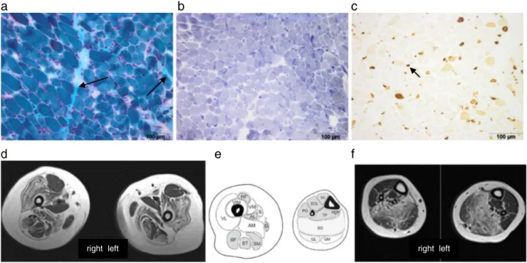
Case report This 5-year-old girl was born at term by caesar-ean section due to transversal position after an uneventful pregnancy. Her mother and older brother are healthy; the father is affected by ankylosing spondylitis. The girl adapted with an APGAR 1/5/5. Because of respiratory insufficiency and bradycardia, she was resuscitated and ventilated for 2 days. After 5 days, breathing was sufficient, except for rare obstructive apnoeic episodes due to secretion. She had re-duced spontaneous movements and facial expression, absent tendon reflexes and reduced muscle tone. Anti-gravity move-ments were not full. Prader-Willi syndrome, spinal muscular atrophy and myotonic dystrophy were excluded by genetic investigation. Brain magnetic resonance imaging (MRI), cre-atine kinase, metabolic screening tests, electroencephalogra-phy and nerve conduction studies of the tibial nerve were normal. Because of persistent poor feeding at the age of 3 months, a gastrostomy was placed and a muscle biopsy of the quadriceps femoris was performed, which showed vari-ability of fibre size and type 1 fibre predominance and atrophy (Fig1a, b, c). She was discharged at the age of 4 months. At age of 12 months, she was able to sit independently and started to mouth feed. Cognitive development was mildly delayed. At the age of 21 months, she presented with strabismus, exoph-thalmos and 6 café-au-lait spots. Brain MRI revealed bilateral
a b
c
d
e
f
right left right left
Fig. 1 Biopsy from the quadriceps muscles (a Gomori trichrome) show-ing variation in fibre size, mild endomysial fibrosis (arrow) and (b NADH) fibre-type uniformity, but no cores and c numerous fibres ex-pressing neonatal myosin (small dark fibres, see arrow). d Muscle MRI of the upper leg: selected involvement of adductor magnus (AD of schematic
image), sartorius (S), vastus lateralis (VL), vastus intermedius (VIM), vastus medialis (VM), spared rectus femoris (RF), adductor longus (AD) and gracilis (G). e Schematic axial image of left upper and lower leg. f Muscle MRI of lower leg: most affected muscle is soleus (SO), followed by the lateral head of the gastrocnemius (GM)
optic pathway glioma. The diagnostic criteria for NF1 were thus fulfilled. Because of good visual function, the optic pathway glioma was left untreated. The marked motor delay with hypotonia was interpreted as within the limits of a severe NF1 phenotype. She learned to sit up by herself, started to shuffle, but was still not able to stand or crawl. Oral feeding was sufficient to remove gastrostomy. At age of 3 years, she developed freckling as an additional criterion for NF1 and a rapidly progressive thoracic-lumbar scoliosis. Spinal MRI failed to reveal vertebral dysplasia or spinal neurofibroma. The possibility of an additional neuromuscular disorder was re-evaluated. On neurological examination, proximal and fa-cial weaknesses were noted, next to the known hypotonia. Muscle MRI showed symmetrical signal change of vasti, adductor magnus, gastrocnemius and soleus muscles. Rectus femoris, adductor longus, gracilis and the peroneal group muscles were relatively spared (Fig.1d, e). This pattern is suggestive for RYR1-related myopathy. Full gene sequencing revealed two novel variants (c.11360-1_11374del16, predict-ed to remove the splice acceptor site at the start of exon 80; c.14928C>G; p.Phe4976Leu, a point mutation at a position that is highly conserved). Pathogenicity was assessed using bioinformatics software Alamut v2.0. Both unaffected parents carried one mutation (c.11360-1_11374del16 was inherited from the father, c.14928C>G from the mother); therefore, recessive inheritance is suggested.
Discussion
Our case demonstrates the difficulty of discerning hypotonia due to a central problem as in NF1 from weakness caused by a neuromuscular disease in the first few years of life. Even though a myopathy was suspected initially, this differential consideration was rejected at the time when the diagnosis of NF1 was confirmed. The marked motor difficulties and hypo-tonia were then attributed to a severe NF1 phenotype. Of note, hypotonia, facial dysmorphic features, scoliosis, joint laxity and cognitive impairment have been described in patients with large deletions in the NF1 region [9]. We did not perform genetic testing of NF1 because our patient fulfilled the clinical diagnostic criteria and developed freckling in addition. The differential diagnosis of Legius syndrome (SPRED1; OMIM 611431) was not considered a possibility, because patients with Legius syndrome do not develop optic pathway gliomas [10]. On follow up, the progress in motor development was poor and could not be attributed to hypotonia alone, but was caused by additional proximal weakness. Therefore, the clin-ical suspicion of an additional underlying congenital myopa-thy was re-evaluated.
Congenital myopathies are group of neuromuscular dis-eases with early onset, defined by the predominant
histopathological features and structural abnormalities. With recent advances in molecular genetics, it has become clear that different genetic congenital myopathies can share clinical and pathological findings and that many individuals with a genet-ically confirmed congenital myopathy have only non-specific histopathological features. Cores as the classical histopatho-logical finding of RYR1-related myopathies may be absent, and unspecific findings such as fibre-type uniformity or type 1 fibre predominance are frequent, as in our patient. Muscle MRI has been shown to be a helpful tool in the evaluation of congenital myopathies [1,5]. The typical selective pattern of muscle involvement in RYR1-related myopathies led to the genetic investigation in our patient. Two novel mutations were detected; one is a splice site mutation and therefore predicted to be pathogenic. The second variant is a point mutation at a highly conserved position, but the physicochemical difference between the amino acids is low. This mutation has not been reported as a benign polymorphism so far, suggesting a dis-ease causing role in this patient. Since the introduction of diagnostic sequencing of the whole RYR1 gene, many novel mutations have been detected but assigning pathogenicity is often difficult [6].
The combination of two different genetic diseases has been reported in a few case reports of patients with different neu-romuscular disorders, where the combination led to a more severe or unusual phenotype [3,4,11–13].
Conclusion
In an unusually severe or atypical presentation,“double trou-ble” should be suspected. In our patient, careful clinical re-evaluation led to further investigations that ultimately led to the two diagnoses. NF1 is one of the most common genetic diseases in child neurology; the combination with another disorder is therefore a possibility. The diagnosis of the RYR1-related myopathy has important implications for genetic counselling and clinical management regarding the risk of malignant hyperthermia for the patient and the family.
Conflict of interest None of the authors declare a conflict of interest.
References
1. Carlier PG, Mercuri E, Straub V (2012) Applications of MRI in muscle diseases. Neuromuscul Disord 22(Suppl 2):S41
2. Ferner RE, Huson SM, Thomas N, Moss C, Willshaw H, Evans DG, Upadhyaya M, Towers R, Gleeson M, Steiger C, Kirby A (2007) Guidelines for the diagnosis and management of individuals with neurofibromatosis 1. J Med Genet 44:81–88
3. Filosto M, Tonin P, Scarpelli M, Savio C, Greco F, Mancuso M, Vattemi G, Govoni V, Rizzuto N, Tupler R, Tomelleri G (2008)
Novel mitochondrial tRNA Leu(CUN) transition and D4Z4 partial deletion in a patient with a facioscapulohumeral phenotype. Neuromuscul Disord 18:204–209
4. Jokela M, Udd B, Paivarinta M (2012) Double trouble: spinal mus-cular atrophy type II and seropositive myasthenia gravis in the same patient. Neuromuscul Disord 22:129–130
5. Klein A, Jungbluth H, Clement E, Lillis S, Abbs S, Munot P, Pane M, Wraige E, Schara U, Straub V, Mercuri E, Muntoni F (2011) Muscle magnetic resonance imaging in congenital myopathies due to ryanodine receptor type 1 gene mutations. Arch Neurol 68:1171– 1179
6. Klein A, Lillis S, Munteanu I, Scoto M, Zhou H, Quinlivan R, Straub V, Manzur AY, Roper H, Jeannet PY, Rakowicz W, Jones DH, Jensen UB, Wraige E, Trump N, Schara U, Lochmuller H, Sarkozy A, Kingston H, Norwood F, Damian M, Kirschner J, Longman C, Roberts M, Auer-Grumbach M, Hughes I, Bushby K, Sewry C, Robb S, Abbs S, Jungbluth H, Muntoni F (2012) Clinical and genetic findings in a large cohort of patients with ryanodine receptor 1 gene-associated myopathies. Hum Mutat 33:981–988
7. Lorenzo J, Barton B, Acosta MT, North K (2011) Mental, motor, and language development of toddlers with neurofibromatosis type 1. J Pediatr 158:660–665
8. Maggi L, Scoto M, Cirak S, Feng L, Lillis S, Cullup T, Robb S, Manzur A, Sewry CA, Abbs S, Jungbluth H, Muntoni F (2011) Congenital myopathies—clinical features and frequency of individual
subtypes diagnosed in a five-year period: the UK experience. Neuromuscul Disord 21:691–691
9. Mautner VF, Kluwe L, Friedrich RE, Roehl AC, Bammert S, Hogel J, Spori H, Cooper DN, Kehrer-Sawatzki H (2010) Clinical character-isation of 29 neurofibromatosis type-1 patients with molecularly ascertained 1.4 Mb type-1 NF1 deletions. J Med Genet 47:623–630 10. Muram TM, Stevenson DA, Watts-Justice S, Viskochil DH, Carey JC, Mao R, Jackson B (2013) A cost savings approach to SPRED1 mutational analysis in individuals at risk for neurofibromatosis type 1. Am J Med Genet A 161:467–472
11. Pandey R, Chandratre S, Roberts A, Dwyer JS, Sewry C, Quinlivan R (2011) Central core myopathy with RYR1 mutation masks 5q spinal muscular atrophy. Eur J Paediatr Neurol 15:70–73
12. Ricci G, Scionti I, Ali G, Volpi L, Zampa V, Fanin M, Angelini C, Politano L, Tupler R, Siciliano G (2012) Rippling muscle disease and facioscapulohumeral dystrophy-like phenotype in a patient carrying a heterozygous CAV3 T78M mutation and a D4Z4 partial deletion: further evidence for“double trouble” overlapping syndromes. Neuromuscul Disord 22:534–540
13. Rudnik-Schoneborn S, Weis J, Kress W, Hausler M, Zerres K (2008) Becker’s muscular dystrophy aggravating facioscapulohumeral mus-cular dystrophy–double trouble as an explanation for an atypical phenotype. Neuromuscul Disord 18:881–885
14. Neurofibromatosis. Conference statement (1988) National Institutes of Health Consensus Development Conference. Arch Neurol 45:575–578