HAL Id: inserm-00678288
https://www.hal.inserm.fr/inserm-00678288
Submitted on 8 Feb 2013HAL is a multi-disciplinary open access archive for the deposit and dissemination of sci-entific research documents, whether they are pub-lished or not. The documents may come from teaching and research institutions in France or abroad, or from public or private research centers.
L’archive ouverte pluridisciplinaire HAL, est destinée au dépôt et à la diffusion de documents scientifiques de niveau recherche, publiés ou non, émanant des établissements d’enseignement et de recherche français ou étrangers, des laboratoires publics ou privés.
integrin aIIbβ3 signalling without affecting P2Y
receptors desensitisation and function.
Mathieu Schaff, Nicolas Receveur, Catherine Bourdon, Philippe Ohlmann,
François Lanza, Christian Gachet, Pierre Henri Mangin
To cite this version:
Mathieu Schaff, Nicolas Receveur, Catherine Bourdon, Philippe Ohlmann, François Lanza, et al.. β-arrestin-1 participates in thrombosis and regulates integrin aIIbβ3 signalling without affecting P2Y receptors desensitisation and function.: Role of β-arrestins in platelet function. Thrombosis and Haemostasis, Schattauer, 2012, 107 (4), pp.735-48. �10.1160/TH11-06-0430�. �inserm-00678288�
β-arrestin-1 participates in thrombosis and regulates integrin α
IIbβ
3signalling without affecting P2Y receptors desensitisation and function
Mathieu Schaff1-3; Nicolas Receveur1-3; Catherine Bourdon1-3; Philippe Ohlmann1-3; François Lanza1-3; Christian Gachet1-3;
Pierre Henri Mangin1-3
1INSERM U949, Strasbourg, France; 2UMR-S949, Université de Strasbourg, Strasbourg, France; 3Etablissement Français du Sang-Alsace (EFS-Alsace), Strasbourg, France
Summary
β-arrestin-1 (β-arr1) and β-arrestin-2 (β-arr2) are cytosolic proteins well-known to participate in G protein-coupled receptor desensiti-sation and signalling. We used genetically-inactivated mice to evaluate the role of β-arr1 or β-arr2 in platelet function, P2Yreceptor desensiti-sation, haemostasis and thrombosis. Platelet aggregation, soluble fibri-nogen binding and P-selectin exposure induced by various agonists were near normal in β-arr1-/- and β-arr2-/- platelets. In addition,
defi-ciency in β-arr1 or β-arr2 was not critical for P2Y receptors desensiti-sation. A functional redundancy between β-arr1 and β-arr2 may ex-plain these unchanged platelet responses. Interestingly, β-arr1-/- but
not β-arr2-/- mice were protected against laser- and FeCl
3-induced
thrombosis. The tail bleeding times, number of rebleeds and volume of
Correspondence to: Pierre Henri Mangin
INSERM UMR-S949, Etablissement Français du Sang-Alsace (EFS-Alsace) 10, rue Spielmann, BP 36, F-67065 Strasbourg Cedex, France Tel.: +33 3 88 21 25 25, Fax: +33 3 88 21 25 21
E-mail: pierre.mangin@efs-alsace.fr
blood loss were unchanged in β-arr1-/- and β-arr2-/- mice, suggesting no
defect in haemostasis. β-arr1-/- platelet activation upon adhesion to
im-mobilised fibrinogen was inhibited, as attested by a 37 ± 5% (n = 3, p<0.0001) decrease in filopodia extension, suggesting defective sig-nalling through integrin αIIbβ3. β-arr1 appeared to be located down-stream of Src family kinases and to regulate αIIbβ3 signalling by increas-ing Akt phosphorylation. Overall, this study supports a role for β-arr1 in promoting thrombus formation, in part through its participation in αIIbβ3 signalling, and no role of β-arr1 and β-arr2 in agonist-induced
platelet activation and P2Yreceptors desensitisation.
Keywords
β-arrestins, platelets, thrombosis, αIIbβ3, Akt
Received: June 27, 2011
Accepted after major revision: January 5, 2012 Prepublished online : February 8, 2012 doi :10.1160/TH11-06-0430 Thromb Haemost 2012; 107: ■■■
Introduction
β-arrestin-1 (β-arr1) and β-arrestin-2 (β-arr2), also called arres-tin-2 and arrestin-3, respectively, are ubiquitously-expressed cyto-solic proteins found in mammals, and form the non-visual arrestin family (1). They bind to activated G protein-coupled receptors (GPCRs) and were originally described as serving to desensitise ac-tivation of G proteins and to target the receptors to clathrin-coated pits for endocytosis, thereby terminating the agonist-induced sig-nalling. More recently, it has been established that both β-arr1 and β-arr2 also transduce signals of agonist-occupied GPCRs by acting as adaptors, recruiting a variety of proteins including GTPases and diverse kinases such as phosphoinositide 3-kinase (PI3K), Akt, Src kinases and mitogen-activated protein kinases (1). The resulting multiprotein scaffolding promotes G protein-independent alter-native signallings which regulate many cellular responses such as adhesion, proliferation, differentiation, migration, and apoptosis.
P2Y1 and P2Y12 are G protein-coupled adenosine 5'-diphos-phate (ADP) receptors playing a key role in platelet activation and thrombosis (2). Platelets activated by ADP become refractory to restimulation, a phenomenon which has been attributed to P2Y1 desensitisation and internalisation (3–5). Although a substantial fraction of P2Y12 is rapidly internalised upon stimulation (4), whether this process is accompanied by a functional desensiti-sation remains unclear. We previously published that
ADP-pre-treated platelets retained P2Y12 activity (3, 4). For unidentified rea-sons, this was not the case in another study by Hardy et al. (5), sug-gesting that P2Y12 underwent desensitisation. Both β-arr1 and β-arr2 were shown to associate with stimulated P2Y1 andP2Y12 in transfected cell systems (6–8). However, whether β-arrestins me-diate P2Y receptor desensitisation in platelets remains to be estab-lished.
Integrin αΙIbβ3 plays a pivotal role in haemostasis and arterial thrombosis by supporting platelet adhesion and aggregation at sites of vascular injury (9). A key attribute of αΙIbβ3 is its capacity to shift from a resting low to an activated high affinity conformation for its main ligand, fibrinogen. This transformation is mediated by platelet-activating inside-out signals derived from tyrosine kinase-linked and G protein-coupled receptors. Upon fibrinogen binding, αΙIbβ3 initiates its own signalling cascade involving several proteins notably Src kinases, phospholipase Cγ2, PI3K and Akt (9–11). The so-called outside-in αΙIbβ3 signalling promotes intracellular Ca2+ mobilisation and cytoskeletal reorganisation leading to filopodia emission, and is important to platelet function in haemostasis and arterial thrombosis (12–15). While β-arrestins also regulate a var-iety of non GPCRs, their importance in αΙIbβ3 functionis unknown (1).
Recently, it has been described that β-arr1 deficiency resulted in a thrombosis defect in a FeCl3 carotid injury model (16). In vitro studies showed reduced soluble fibrinogen binding after
stimu-lation of the platelet protease-activated receptor (PAR) 4. Whether the decreased thrombosis observed in β-arr1-/- mice is due to the defective response through PAR4 or arises from additional signal-ling pathways remains to be established.
The objectives of this study were to evaluate the role of β-arr1 and β-arr2 in (i) agonist-induced platelet activation, (ii) P2Y1 and P2Y12 desensitisation, and (iii) haemostasis and experimental thrombosis. Mouse platelets with deficiency in β-arr1 or β-arr2 re-sponded normally to a series of agonists, and displayed similar de-sensitisation to ADP as wild-type (WT) platelets, suggesting that these proteins do not play a key role in these processes. We ob-served a contribution of β-arr1, but not β-arr2, to arterial throm-bosis after laser or FeCl3 injury. The tail bleeding times, rebleeding occurrences and volume of blood loss were unchanged in β-arr1 -/-and β-arr2-/- mice, suggesting no defect in haemostasis. Upon ad-hesion to immobilised fibrinogen, β-arr1-knock-out platelets ex-hibited defective integrin αΙIbβ3-mediated shape change. The role of β-arr1 in αΙIbβ3 outside-in signals appeared to be dependent on Src family kinases and linked to the upregulation of Akt phos-phorylation.
Materials and methods
Materials
Alexa Fluor 488-labelled fibrinogen, DIOC6 (3,3’-dihexyloxacar-bocyanine iodide) and the indicators Oregon Green 488 BAPTA/ AM-1 and Calcein red-orange/AM were from Molecular Probes (Paisley, UK). FITC-coupled anti-P-selectin antibody (RB40.34) was purchased from BD Pharmingen (Le Pont-De-Claix, France) and FeCl3 solution from VWR Prolabo (Fontenay-sous-Bois, France). Fatty acid-freehuman serum albumin (HSA), fura-2/AM, TRITC (tetramethylrhodamine isothiocyanate)-phalloidin, bov-ine thrombin, U46619, ADP, ADPβS, indomethacin, PP1 and PP2 were provided by Sigma-Aldrich (Lyon, France). AR-C69931MX (Cangrelor), a P2Y12 antagonist, was from the Medicines Company (Parsippany, NJ, USA) and MRS2179, a P2Y1 blocker, from Tocris Bioscience (Bristol, UK). Cross-linked collagen-related peptide (CRP-XL) was obtained from Dr. R.W. Farndale (University of Cambridge, Cambridge, UK), PAR4-selective agonist AYPGKF peptide was synthesised by PolyPeptide Group (Strasbourg, France) and Horm fibrillar type I collagen from equine tendon was from Nycomed (Zurich, Switzerland). Human fibrinogen was pro-vided by Kabi (Bad Homburg, Germany) and 1-paraformaldehyde (PFA) by Electron Microscopy Sciences (Euromedex, Souffel-weyersheim, France). Recombinant hirudin was from Transgene (Illkirch-Graffenstaden, France) and acid citrate dextrose (ACD) solution from Bioluz (St-Jean-de-Luz, France). Apyrase was puri-fied from potatoes as previously described (17). Monoclonal anti-bodies to total Akt and phosphorylated Akt at Ser473 (D9E) or Thr308 (244F9) were from Cell Signaling Technology (Ozyme, St-Quentin-en-Yvelines, France).
Mice
β-arr1- and β-arr2-deficient mice (18, 19) were provided by Pr. R.J. Lefkowitz (Duke University, Durham, NC, USA) and backcrossed for six generations on C57BL/6 background. WT mice were pur-chased from Charles River (L’Arbresle, France). Mice were main-tained in the animal facilities of the Etablissement Français du Sang-Alsace. We used 8– to 10-week-old mice, anesthetised intra-peritoneally with a mixture of xylazine (20 mg per kg body weight, Rompun®, Bayer, Leverkusen, Germany) and ketamine (100 mg per kg body weight, Imalgene 1,000®, Merial, Lyon, France). All ex-periments conformed to the French legislation for animal experi-mentation and followed the recommendations of the Guide for the Care and Use of Laboratory Animals.
Washed platelet preparation
Blood was drawn from the abdominal aorta into ACD anticoagu-lant, pooled (4–6 animals), and platelets were washed by sequential centrifugations and adjusted to 3 x 10⁸ platelets/ml in Tyrode’s al-bumin buffer containing 0.02 U/ml apyrase (17, 20)
Aggregation studies
Washed platelets aggregation was measured turbidimetrically using a four-channel CARAT TX4 aggregometer (Entec, Ilmenau, Germany) as previously described (17).
Measurement of fibrinogen binding and P-selectin exposure
Washed platelets (5 x 10⁷/ml) were stimulated with the indicated agonists in the presence of Alexa Fluor 488-fibrinogen (20 μg/ml) or FITC-anti-P-selectin antibody (25 μg/ml), mixed and at prede-termined time points fixed with PBS-20 mg/ml PFA for 20 minutes (min). Platelets were pelleted by centrifugation at 1,000 x g for 2 min and resuspended in 500 μl phosphate-buffered saline (PBS). The fluorescence intensity of each platelet sample was measured in the range 525–50 nm after excitation with a 488-nm argon-ion laser using a Gallios flow cytometer (Beckman Coulter, Villepinte, France). The extents of integrin αΙIbβ3 activation and P-selectin ex-posure were represented by the levels of Alexa Fluor 488-fibri-nogen and FITC-anti-P-selectin binding, respectively, expressed as the geometric mean of the relative fluorescence intensity of 10,000 platelets (in arbitrary units) (Kaluza software, Beckman Coulter). Basal fluorescence, as measured in resting platelets, was subtracted from all data points.
ELISA-based VASP whole blood assay
Vasodilator-stimulated phosphoprotein (VASP) phosphorylation state was determined using a standardised ELISA (CY-QUANT VASP/P2Y12®, Biocytex, Marseille, France) (21, 22). Briefly, hi-rudinated (100 U/ml) whole blood drawn from the abdominal aorta was pretreated for 20 min with Tyrode’s albumin buffer or ADP (10 μM). Each sample was then incubated for 10 min at room temperature (RT) with either prostaglandin E1 (PGE1) or PGE1 plus ADP in two separate wells of a 96-well plate. After cell lysis, VASP was captured by a selective anti-VASP antibody coated on the wells. A peroxidase-conjugated anti-phosphorylated VASP antibody was then added, and the plates were incubated with a chromogenic substrate solution and read at 450 nm. The platelet reactivity index (PRI), which reflects the ability of P2Y12 to inhibit PGE1-induced VASP phosphorylation, was calculated from blank-corrected optical densities (OD) as follows: PRI (%) = [(ODPGE1 – ODPGE1 + ADP) / ODPGE1] x 100.
Static adhesion assay
Glass coverslips were coated with 100 μg/ml fibrinogen for 2 hours (h) at RT and blocked with PBS-10 mg/ml HSA for 1 h. Washed platelets (3 x 10⁷/ml) were pretreated for 15 min with AR-C69931MX (10 μM), MRS2179 (100 μM) and indomethacin (100 μM) alone or in the presence of either vehicle (1:1,000 DMSO), PP1 or PP2 (10 μM), and allowed to adhere to the coated surfaces at 37°C. After 20 min, non-adherent platelets were washed away and adherent cells were fixed with PBS-40 mg/ml PFA, stained with TRITC-phalloidin (2 μg/ml) and imaged using a Leica DMI 4000 B epifluorescence microscope with a 63x, 1.4 numerical aperture oil objective (Leica Microsystems, Wetzlar, Germany) (23).
Scanning electron microscopy (SEM)
Adherent platelets were fixed with 25 mg/ml glutaraldhehyde in 0.1 M cacodylate buffer (pH 7.3) containing 20 mg/ml sucrose. SEM was performed as described elsewhere (23).
Analysis of cytosolic Ca2+ fluxes
ADP-induced Ca2+ elevations were measured as previously re-ported (24). Briefly, washed platelets resuspended in Ca2+-free Ty-rode’s buffer (7.5 x 10⁸ cells/ml) were loaded with fura-2/AM (15 μM) for 45 min at 37°C. The dyed platelets were pelleted at 1,900 x g for 3 min, resuspended to 2 x 10⁸/ml in Tyrode's buffer contain-ing apyrase and 1 mg/ml HSA, and challenged with P2Y receptor agonists using a PTI Deltascan spectrofluorimeter (Photon Tech-nology International, Ford, UK). Fluorescence measurements were
performed at 510 nm after exciting alternatively at 340 and 380 nm, and converted to intracellular Ca2+ levels. Cytosolic Ca2+ changes upon platelet adhesion to fibrinogen were measured using a previously published method (20). Briefly, washed platelets (5 x 10⁸/ml) were simultaneously loaded with Oregon Green 488 BAPTA/AM-1 (5 μM),Calcein red-orange/AM (10 μM) and the membrane transporter inhibitor probenecid (2.5 μM) for 45 min at 37°C. The dyed platelets were pelleted at 1,900 x g for 3 min, re-suspended at 1 x 10⁸/ml in Tyrode’s albumin buffer, and allowed to adhere to fibrinogen-coated (100 μg/ml) coverslips for 20 min. The increases in platelet and Ca2+-dependent fluorescence inten-sity upon adhesion were measured in the ranges 572–700 nm and 495–535 nm, respectively, by confocal laser scanning microscopy, and converted to intracellular Ca2+ levels (Leica TCS SP5).
Clot retraction
Blood drawn into sodium citrate (3.15 mg/ml) from the abdomi-nal aorta of 2–3 mice was pooled, centrifuged at 250 x g for 1 min and platelet-rich plasma (PRP) was adjusted to 8 x 10⁸ platelets/ml with autologous platelet-poor plasma. PRP (500 μl) was added to quartz cuvettes maintained at 37°C in the presence of 20 mM CaCl2. The clots were allowed to retract for up to 80 min and photographed every 5 min. The extent of retraction was then quantified using ImageJ (National Institutes of Health, Bethesda, MD, USA).
Measurement of Akt phosphorylation
Platelets adhering to fibrinogen were incubated in lysis buffer (1:50 Triton X-100, 40 mM Tris (tris(hydroxymethyl)aminomethane), 10 mM Na2VO3, 100 mM NaF, 2 mM EDTA, 2 mM EGTA, 300 mM NaCl, pH 7.4) for 10 min at 4°C and centrifuged at 7,000 x g for 7 min. The supernatants were resolved by SDS-PAGE and trans-ferred to PVDF membranes. The levels of total and phosphory-lated Akt were determined by Western blotting using monoclonal antibodies as previously described (25).
Laser-induced mesenteric artery thrombosis model
Thrombosis was performed as previously described (26). Briefly, platelets were labelled by injection of DIOC6 (5 μl of a 100 μM sol-ution per g body weight), a membrane fluorescent dye, and a loca-lised deep injury of a mesenteric arteriole was induced with a high intensity 440-nm-pulsed nitrogen dye laser applied for 30–40 sec-onds (s) (300–400 hits) with a Micropoint system (Photonic In-struments, Andor Technology, Belfast, UK). Thrombus formation was monitored (1 image/s) by fluorescence microscopy using a Sensicam charge-coupled device (CCD) camera (The Cooke
Cor-poration, Romulus, MI, USA), and images were analysed with the SlideBook software (Intelligent Imaging Innovations, Göttingen, Germany).
FeCl3-induced carotid artery thrombosis model
DIOC6 was injected into the jugular vein (5 μl of a 100 μM solution per g body weight) to label platelets and assist thrombus visual-isation. The left common carotid artery was exposed and vascular injury was induced by topically applying over the adventitia for 2 min a Whatman #1 filter paper (1 x 0.5 mm) saturated with 0.2 μl of a 75 mg/ml FeCl3 saline solution (27). The artery was then rinsed with saline and thrombus formation was monitored in real time under a fluorescent macroscope (Macrofluo, Leica Microsystems) using a 5x, 0.5 numerical aperture objective. Images (1 image/s) were acquired using a CCD camera (CoolSNAP HQ2, Photomet-rics, Roper Scientific, Evry, France) and analysed with Metamorph software version 7.6 (Molecular Devices, Roper Scientific).
Figure 1: Role of
β-arr1 and β-arr2 in
platelet aggregation, soluble fibrinogen binding and P-selectin exposure. A) Washed
platelets (2 x 10⁸/ml)
from WT, β-arr1-/- and
β-arr2-/- mice were
ag-gregated by a range of agonists in the presence (ADP, U46619, AYPGKF, type I collagen and CRP-XL) or absence (throm-bin) of fibrinogen (64 μg/ ml). Arrows indicate the point of agonist addition. Aggregation profiles are representative of four separate experiments.
Table 1: Platelet count and surface expression of different glycopro-teins in WT, β-arr1-/- and β-arr2-/- mice. Platelet counts are the mean ±
SEM for groups of at least 12 mice. Surface expression of the indicated gly-coproteins was determined in whole blood by staining with selective anti-bodies followed by flow cytometry analysis. Platelets were gated by FSC/SSC characteristics (10,000 platelets per sample). Results are expressed as the geometric mean ± SEM of the relative fluorescence intensity, in arbitrary units (three mice per group).
WT β-arr1-/- β-arr2-/- GPIIb/IIIa (αIIbβ3) 3.9 ± 0.1 4.2 ± 0.04 4.5 ± 0.1 GPIIa (β1) 0.43 ± 0.02 0.56 ± 0.09 0.46 ± 0.06 GPIa (α2) 0.73 ± 0.03 0.68 ± 0.07 0.69 ± 0.01 GPVI 0.20 ± 0.01 0.19 ± 0.01 0.21 ± 0.01 GPV 0.65 ± 0.01 0.63 ± 0.03 0.60 ± 0.02 GPIX 1.47 ± 0.06 1.44 ± 0.01 1.49 ± 0.03 GPIbα 1.74 ± 0.07 1.65 ± 0.06 1.64 ± 0.01 Platelet count (x106/ml) 1,038 ± 47 969 ± 53 1,151 ± 35
Bleeding time
The bleeding time was measured by transversally severing a 3-mm segment from the distal tail of 8– to 10-week-old isoflurane-anes-thetised mice. The amputated tail was immediately immersed in 9 mg/ml isotonic saline at 37°C during 30 min and the time required for arrest of bleeding was recorded. The tube containing saline with blood was then homogenised and centrifuged at 550 x g dur-ing 5 min. The supernatant was removed and 2 ml of lysis buffer (NH4Cl 150 mM, KHCO3 1 mM, EDTA 0.1 mM, pH 7.2) was added to the pellet. After homogenisation, the optical density was read at 540 nm and compared to a standard curve to determine the volume of blood loss.
Statistical analyses
All values are reported as mean ± standard error of the mean
(SEM) unless otherwise indicated. Data were compared by two-tailed Student’s t-tests using Prism software (GraphPad, La Jolla, CA, USA) and differences were considered significant at p<0.05.
Results
Platelet aggregation, soluble fibrinogen binding and
P-selectin exposure in β-arr1- and β-arr2-deficient
platelets
Deletion of β-arr1 or β-arr2 did not affect the platelet count and expression of the major surface glycoproteins (씰Table 1). We examined the ability of β-arr1-/- and β-arr2-/- platelets to become activated after stimulation with a range of agonists. As shown in
씰Figure 1A and 씰Suppl. Figure 1 (available online at www.throm bosis-online.com), WT and knock-out platelets displayed equival-ent aggregation profiles in response to various concequival-entrations of
Figure 1: B-E) WT,
β-arr1-/- (B, D) and
β-arr2-/- (C, E) washed
platelets (5 x 10⁷/ml)
were stimulated with various agonists in the presence of Alexa Fluor 488-fibrinogen (B, C) or a FITC-P-selectin anti-body (D, E). After 20 or 10 min, the samples were fixed and the extent of fibrinogen binding and P-selectin exposure was measured by flow cyto-metry. Results represent the geometric mean ± SEM of the relative fluor-escence intensity in arbit-rary units, obtained in four separate experi-ments performed in du-plicate (10,000 platelets analysed per experi-ment).
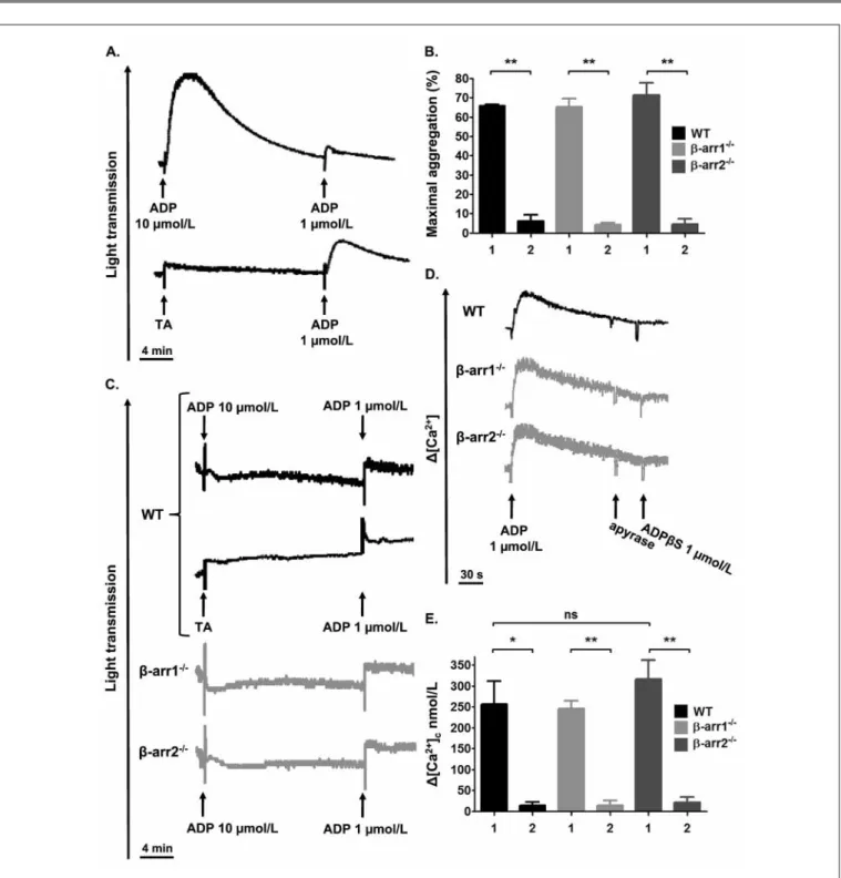
Figure 2: Role of β-arr1 and β-arr2 in P2Y receptors desensitisation. A) Top: WT platelets (2 x 10⁸/ml) were aggregated by 10 μM ADP in the
pres-ence of 64 μg/ml fibrinogen, followed by a second challenge with ADP (1 μM) after 20 min. Bottom: WT platelets were incubated with vehicle (Tyrode’s albumin buffer, [TA]) and stimulated with 1 μM ADP after 20 min. B) Washed platelets (2 x 10⁸/ml) from WT, β-arr1-/- and β-arr2-/- mice were aggregated
as in A. Bars represent the maximal amplitude of aggregation after addition of 10 μM ADP (1) and restimulation with 1 μM ADP (2). Data are the mean ± SEM from three separate experiments. **p<0.01 C) Aggregation: WT, β-arr1-/- and β-arr2-/- platelets (2 x 10⁸/ml) pretreated with AR-C69931MX (10
μM) for 15 min were incubated with 10 μM ADP or TA in the presence of 64 μg/ml fibrinogen, followed by a second challenge with ADP (1 μM) after 20 min. D, E) Variations in Ca2+ levels were measured by spectrofluorimetry in fura-2/AM-loaded (15 μM) WT, β-arr1-/- and β-arr2-/- platelets (2 x 10⁸/ml)
stimulated with 1 μM ADP. After 2 min, apyrase (0.2 U/ml) was added for 30 s and the cells were restimulated with 1 μM ADPβS. Representative profiles are shown (D). Bars represent the maximal increase in cytosolic Ca2+ after addition of ADP (1) and restimulation with ADPβS (2). Data are the mean ±
Figure 2: F) Black
curves: aggregation of WT, β-arr1-/- and β-arr2
-/-platelets (2 x 10⁸/ml) in
response to 0.05 U/ml thrombin, 13 min after a first addition of either 10 μM ADP or TA in the presence of 64 μg/ml fi-brinogen. Grey curves: WT, β-arr1-/- and β-arr2
-/-platelets were aggre-gated or not by ADP as above, followed by addi-tion of AR-C69931MX (10 μM), 3 min before stimulation with throm-bin. The profiles are rep-resentative of at least three independent ex-periments. G) Hirudi-nated (100 U/ml) whole blood from WT, β-arr1
-/-and β-arr2-/- mice was
pretreated or not with 10 μM ADP for 20 min, and the ability of P2Y12 to
in-hibit PGE1-induced VASP
phosphorylation was de-termined by ELISA. Dis-tribution of the platelet reactivity index in three different mice is shown. The mean value is indi-cated with horizontal bars.
ADP, thrombin, type I fibrillar collagen and CRP-XL, a glycopro-tein VI-selective ligand. In addition, serotonin-induced shape change and the ability of either adrenaline or serotonin to potenti-ate aggregation in response to 1 μM ADP were unchanged (data not shown). By contrast, β-arr2-/- but not β-arr1-/- platelet aggre-gation was delayed and exhibited a biphasic appearance restricted to the lowest concentration (1 μM) of the thromboxane A2 (TxA2) analog U46619, indicating minor participation of β-arr2 in TP re-ceptor signalling (씰Fig. 1A). A similar defect was observed in β-arr1-/- platelets using subthreshold concentrations (300 μM) of AYPGKF, a PAR4 agonist peptide (씰Fig. 1A). In parallel, we ob-served that soluble fibrinogen binding induced by ADP (2 μM), thrombin (0.25 U/ml), AYPGKF (1 mM) and CRP-XL (1 μg/ml) was normal in β-arr1- and β-arr2-null compared to control pla-telets (씰Fig. 1B, C). Finally, no defective P-selectin exposure, a marker of granule secretion, was found following thrombin (0.25 U/ml) and AYPGKF (1 mM) stimulation (씰Fig. 1D, E). Alto-gether, these results suggested that β-arr1 and β-arr2 play no major role in the signalling machinery leading to activation of platelets by a wide range of agonists.
β-arr1 and β-arr2 are not critical for P2Y1 andP2Y12 receptors desensitisation
WT platelets activated by ADP become refractory to restimulation, a phenomenon attributed to P2Yreceptordesensitisation (3–5), and illustrated by absence of shape change and reduced aggre-gation in response to a second ADP challenge (씰Fig. 2A and
씰Suppl. Fig. 2A and C, available online at www.thrombosis-on line.com). A similar refractoriness was observed in the knock-out platelets, suggesting that neither β-arr1 nor β-arr2 play a critical role in P2Y receptor desensitisation (씰Fig. 2B and 씰Suppl. Fig. 2B and D, available online at www.thrombosis-online.com). To in-vestigate this in more details, we studied P2Y1 desensitisation in platelets pretreated with 10 μM of the P2Y12 blocker AR-C69931MX. As expected, addition of ADP (10 μM) to WT platelets caused a shape change without aggregation (씰Fig. 2C) (2, 28). Upon restimulation with 1 μM ADP, no response was observed, re-flecting desensitisation of P2Y1 (씰Fig. 2C). Platelets lacking either β-arr1 or β-arr2 exhibited similar responses, indicating that these proteins do not contribute to P2Y1 desensitisation (씰Fig. 2C). These results were confirmed by looking at ADP-induced Ca2+ mo-bilisation, which is mediated by P2Y1 (3, 24). Similarly as in the WT, β-arr1-/- andβ-arr2-/- platelets stimulated with ADP followed by its removal with apyrase, were unable to exhibit Ca2+ rises in sponse to a second challenge with the metabolically stable P2Y re-ceptor agonist ADPβS (씰Fig. 2D, E). Although a substantial frac-tion of P2Y12 is rapidly internalised upon stimulation (4), whether this phenomenon is accompanied by a functional desensitisation is still a matter of debate (3–5). Therefore, we re-examined P2Y12 de-sensitisation and the potential role of β-arrestins in this process. We used the capacity of P2Y12 to potentiate thrombin-induced pla-telet aggregation (씰Fig. 2F, grey curves) as a marker of its
sensiti-sation state (2, 4). As shown in 씰Figure 2F, the rate and amplitude of aggregation in response to 0.05 U/ml thrombin were similar in WT platelets pre-stimulated or not with 10 μM ADP, suggesting no functional desensitisation of P2Y12. Equivalent aggregation pro-files were obtained when these experiments were performed in β-arr1-/- andβ-arr2-/- platelets (씰Fig. 2F). To get a more direct measurement of P2Y12 signalling, we analysed intraplatelet VASP phosphorylation, a method used to monitor the efficacy of anti-P2Y12 therapy (21, 22). ELISA showed that in WT platelets pre-treated with 10 μM ADP, P2Y12 was still able to profoundly inhibit PGE1-induced VASP phosphorylation, reflecting once again ab-sence of desensitisation (씰Fig. 2G). Similar results were found in β-arr1-/- andβ-arr2-/- platelets, confirming that these proteins are unlikely to contribute to P2Y12 desensitisation (씰Fig. 2G).
β-arr1 participates in integrin αΙIbβ3-mediated filopodia extension
We next evaluated the impact of a β-arrestin deficiency on platelet activation mediated by adhesion receptors. As compared to the WT, the ability of β-arr1-/- and β-arr2-/- platelets to adhere to von Willebrand factor or collagen and undergo shape change, which reflects activation of the cells, was unchanged (data not shown). In contrast, following adhesion to fibrinogen, the proportion of β-arr1-/- but not β-arr2-/- platelets extending filopodia was reduced by 37 ± 5% (n = 3, p<0.0001) (씰Fig. 3A, B), while the number of adherent cells remained unaltered (data not shown). As these ex-periments were performed in the presence of ADP and TxA2 blockers, this would suggest abnormal cytoskeletal reorganisation through integrin αΙIbβ3, the main platelet receptor for fibrinogen.
β-arr1 functions downstream of Src family kinases and regulates Akt phosphorylation mediated by αΙIbβ3
Integrin αΙIbβ3 signalling has been reported to be initiated by Src family kinases such as c-Src and Fyn (9). To explore whether β-arr1 is a target of Src kinases, washed platelets were preincubated with PP1 and PP2, two pan-inhibitors of this family of proteins, prior to adhesion to fibrinogen. As shown in 씰Figure 3B, PP1 reduced the proportion of WT (31 ± 6%) and β-arr1-/- (25 ± 5%) platelets ex-tending filopodia to a similar extent (n = 3, p = 0.44). Similar re-sults were obtained with PP2 (30 ± 4% of WT platelets with filopo-dia versus 33 ± 3% in mutants, n = 4, p = 0.50), suggesting that β-arr1 is located downstream of Src kinases in αΙIbβ3 signalling. Src kinases have been proposed to modulate various signalling events including Ca2+ mobilisation (9). Upon adhesion to fibrinogen, equivalent Ca2+ increases were observed in WT and mutant pla-telets, indicating that β-arr1 does not participate in this process (씰Fig. 3C). In addition, β-arr1-/- platelets exhibited normal fibrin clot retraction, another event in which αΙIbβ3 and Src kinases play a
Figure 3: Role of β-arr1 in αIIbβ3 signalling. A, B) WT and β-arr1-/- washed
platelets were pretreated for 15 min with AR-C69931MX (10 μM), MRS2179 (100 μM) and indomethacin (100 μM) alone or in the presence of PP1 or PP2 (10 μM), and applied for 20 min to fibrinogen-coated (100 μg/ml) coverslips. Adherent platelets were fixed and examined by SEM. Representative images of WT and β-arr1-/- platelets are shown. Scale bars, 10 μm (A). The adherent
platelets were also stained with TRITC-phalloidin and the mean percentage of cells with filopodial extensions was determined by epifluorescence micro-scopy in eight random fields in at least three independent experiments (B). C) WT and β-arr1-/- washed platelets loaded with Ca2+ and morphological dyes
were allowed to adhere to fibrinogen-coated (100 μg/ml) coverslips as in A, B. Changes in fluorescence were monitored for 20 min by confocal micro-scopy, and cytosolic Ca2+ concentrations were determined. The dot plot
dis-tribution of the relative maximal increase in individual adherent platelets is shown. D) Citrated platelet-rich plasma from WT and β-arr1-/- mice was
added to quartz cuvettes maintained at 37°C in the presence of 20 mM CaCl2
and clot retraction, visible as a consolidated morphology and transparent clot liquor, was photographed every 5 min for 80 min. Bars represent the mean percentage reduction of the initial clot surface area at different time points, determined by two-dimensional image analysis. E, WT and β-arr1
-/-washed platelets were allowed to adhere to fibrinogen as in A, B, lysed, cen-trifuged and the supernatants were resolved by SDS-PAGE followed by West-ern blotting. Bars represent the relative ratio of phosphorylated to total Akt, calculated by densitometry analysis. Data represent the mean ± SEM of three or four independent experiments. *p<0.05, **p<0.01, and ***p<0.0001.
Figure 4: β-arr1-/- but
not β-arr2-/- mice
ex-hibit a decreased thrombotic response after laser injury of the mesenteric artery. A
lo-calised deep injury of an exposed mesenteric arte-riole of WT, β-arr1-/- (A,
B) and β-arr2-/- (C, D)
mice was generated with a high intensity 440-nm-pulsed nitrogen dye laser applied for 30 s, and thrombus formation was monitored by fluor-escence microscopy (26). A, C) Time-course of the thrombus growth, repre-sented by its mean sur-face area. Dotted curves represent the SEM. B, D) Bar graphs represent the area under the curves shown in A and C, re-spectively. Data are from 12 vessels in five mice (A, B), three vessels in three β-arr2-/- mice or
five vessels in three WT mice (C, D). *p<0.05.
critical role (씰Fig. 3D). The downstream effectors of Src kinases also include Akt (10, 11), which has been proposed to be targeted by β-arr1 in various cellular systems (1, 16, 29). To determine whether β-arr1 participates in αΙIbβ3 signalling by regulating Akt, WT and β-arr1-/- washed platelets were allowed to adhere to fibri-nogen for 20 min and then lysed, and the level of Akt phosphory-lation was measured by Western blotting. Interestingly, β-arr1 -/-platelets presented a decrease of 52 ± 9% (n = 4, p<0.05) and 56 ± 12% (n = 4, p<0.01) in the phosphorylation of Ser473 and Thr308, respectively (씰Fig. 3E). Overall, these results suggested that upon binding of fibrinogen to αΙIbβ3, β-arr1 regulates filopodia emission through a process involving Src kinases and increased Akt phos-phorylation.
β-arr1-/- but not β-arr2-/- mice are protected in two models of arterial thrombosis
The role of β-arr1 and β-arr2 in arterial thrombosis was investi-gated using two in vivo models. When mesenteric arterioles were injured with a laser beam, a deficiency in β-arr1 led to a 28 ± 10% (n = 12 vessels in five mice, p<0.05) reduction in total thrombus surface area as compared to the WT (씰Fig. 4A, B). This result is in agreement with a recent study revealing that β-arr1-/- mice are less
sensitive to FeCl3-induced carotid thrombosis (16). Using a similar model, we observed a delay in thrombus growth with a thrombus area which was significantly reduced at early (350 s: 32 ± 9% reduc-tion, n = 6, p<0.05) but not later times (>480 s) (씰Fig. 5A-C). In contrast, thrombus formation was comparable between WT and β-arr2 knock-outs (씰Figs. 4C and D, 5D and E), pointing to a modest role of β-arr1 and no role of β-arr2 in this process.
β-arr1-deficient mice exhibit normal tail bleeding
To establish whether the decreased thrombosis observed in β-arr1-deficient mice is mirrored by defective haemostasis, stan-dardised tail-bleeding experiments were performed. The bleeding time was equivalent in β-arr1-null (149 ± 33 s, n = 12) and WT (137 ± 26 s, n = 12) mice (씰Fig. 6A), as were the number of reb-leeds (씰Fig. 6B) and total bleeding time (씰Fig. 6C) over 30 min. Despite an increased blood loss in a few mutant mice, the mean volume was not significantly different in WT (씰Fig. 6D). The bleeding phenotype was also unaltered in β-arr2-/- mice (씰Fig. 6A-D). Overall, these results suggested a minor contribution of β-arr1 and β-arr2 to primary haemostasis.
Figure 5: β-arr1-/- but
not β-arr2-/- mice
ex-hibit a delayed throm-botic response after FeCl3 injury of the
ca-rotid artery. A 75 mg/ml
FeCl3 solution was
ap-plied for 2 min to the ex-posed carotid artery of WT, β-arr1-/- (A-C) and
β-arr2-/- (D, E) mice, and
thrombus growth was recorded by video micro-scopy (27). A, D) Time-course of the thrombus growth, represented by its mean surface area. Dotted curves represent the SEM. B) Mean throm-bus area after 350 s in WT and β-arr1-/- mice. C,
E) Bar graphs represent the area under the curves shown in A and D, re-spectively. Data are from six (A-C) or seven (D, E) mice. *p<0.05.
Discussion
In the present study, we addressed the question of the role of β-ar-restins in platelet functions. Neither β-arr1 nor β-arr2 appeared to critically regulate inside-out signals induced by a wide range of agonists. We also provide evidence that P2Y1 receptor desensiti-sation and platelet refractoriness to ADP still occur in β-arr1-/- and β-arr2-/- platelets. Two in vivo models supported a modest role of β-arr1 and no role of β-arr2 in arterial thrombosis. The lack of β-arr1 or β-arr2 expression was not critical for haemostasis since the tail bleeding phenotype was unchanged. Our results identified a novel function for β-arr1 through the regulation of integrin αΙIbβ3 signalling. Upon fibrinogen binding to αΙIbβ3,β-arr1 partici-pates in cytoskeletal reorganisation leading to filopodia extension by a process involving Src kinases and probably upregulation of
Akt activity.
In agreement with our previous studies (3, 4), we observed that mouse platelets activated by ADP were unable to fully re-aggregate in response to a second challenge, as a result of P2Y1 but not P2Y12 receptor desensitisation. Most of GPCRs desensitise through phos-phorylation by GPCR kinases (GRKs), which promotes β-arrestin recruitment thereby precluding G protein coupling and ultimately leading to receptor internalisation (1). Although β-arr1 and β-arr2 were reported to interact with activated P2Y1 in transfected HEK-293 cells (6, 7), our study shows that the deficiency in either of these proteins did not prevent platelet P2Y1 desensitisation, as judged by the absence of re-aggregation and Ca2+ elevation in re-sponse to a second ADP challenge. However, one cannot exclude a compensatory mechanism between the two isoforms. This hy-pothesis is supported by the fact that in mouse embryonic
fibrob-Figure 6: β-arr1-/- and
β-arr2-/- mice present
normal tail bleeding. A
3-mm segment from the distal tail of 8– to 10-week-old WT, β-arr1-/-
and β-arr2-/- mice was
amputated and the time required for initial arrest of bleeding was measured (A). The reb-leeding occurrences (B), total bleeding time (C) and volume of blood lost (D) were then recorded over a 30-min period. Symbols represent indi-vidual mice (n = 12 or 13) and the mean ± SEM is indicated with horizon-tal bars.
lasts ablation of both β-arr1 and β-arr2 was required to inhibit de-sensitisation of the angiotensin II type 1A receptor (30). Since double β-arr1/β-arr2 deficiency is embryonically lethal, tissue-specific knock-outs would be needed to definitely resolve this question. Alternatively, P2Y1 phosphorylation on its own may be sufficient to induce desensitisation without any need for a β-arres-tin. Such a mechanism has been proposed in transfected 1321N1 cells in which P2Y1 phosphorylation and internalisation did not
require GRKs but protein kinase C (5, 8). Concerning P2Y12, we previously demonstrated that even though it was rapidly and tran-siently internalised upon stimulation, no functional desensiti-sation was objectivated (3, 4). In agreement with these observa-tions, the present study did not highlight a functional desensiti-sation of P2Y12 in mouse platelets. These results suggested that β-arrestins are not key players of P2Y12 desensitisation. However, recent identification of a patient carrying a mutation in P2Y12 gene
suggested that β-arrestins may bind a 4-amino acid motif at the ex-treme C-terminus of P2Y12, thereby promoting its internalisation and subsequent recycling (31).
Arrestins also act as adaptors, allowing for the assembly of multi-protein complexes which transduce the signals of a variety of receptors in many cell types (1). We provided evidence that β-arr1 and β-arr2 were not critical for the activation of platelets by a large series of agonists including ADP. This is in accordance with a recent report by Li et al. (16) showing normal ADP-induced Akt phosphorylation in β-arr1-/- platelets. These results appear sur-prising since β-arr1 and β-arr2 were proposed to bind and poten-tially regulate both P2Y1 and P2Y12 (6–8),which are indispensable for normal platelet response to ADP (2). As suggested above, a functional redundancy between β-arr1 and β-arr2 may help main-tain normal P2Y receptor function in the knock-out platelets. Ac-cording to Li et al. (16), β-arr1 modulates PAR4 signalling, as evi-denced by a decrease in Alexa Fluor 488-labelled fibrinogen bind-ing upon stimulation of β-arr1-/- platelets with 50 to 100 μM of the selective agonist AYPGKF. Using three different assays, we ob-served no defect in β-arr1-/- platelet activation in response to the lowest concentrations of AYPGKF sustaining maximal effect. However, when stimulated with a subthreshold concentration (300 μM), β-arr1-/- platelets exhibited a slight delay in aggregation relative to control. Lower doses, as those used by Li et al. (16), could not been tested since they induced no functional response in any of the assays we utilised. The reason for this is unclear, but is probably related to differences in the platelet washing procedure causing various levels of sensitivity to agonists. Overall, our results are in agreement with those of Li et al. (16), suggesting that β-arr1 par-ticipates in PAR4 signalling, but its role could be limited since the defect was restricted to stimulations with low concentrations of the agonist.
Besides GPCRs, β-arrestins bind to other classes of surface re-ceptors specifically single-membrane-spanning rere-ceptors for transforming growth factor-β, insulin-like growth factor 1 and low density lipoprotein (1). We propose a novel function of β-arr1 as a positive regulator of integrin αΙIbβ3 outside-in signalling, as indi-cated by decreased filopodia emission and Akt phosphorylation in β-arr1-/- platelets adhering to fibrinogen. This is in agreement with the role of Akt in regulating cytoskeletal reorganisation which has been revealed in Akt-1-deficient mice (32). Whether β-arr1 and in-tegrin αΙIbβ3 physically interact is unknown. β-arr1 directly binds to specific phosphorylated serine and threonine residues of agon-ist-occupied GPCRs, thereby competing with G proteins (1). Since the β3 integrin subunithas been shown to bind the G protein α13 subtype (33), a similar mechanism might occur for αΙIbβ3. How-ever, our studies on platelet lysates using various types of deter-gents failed to co-precipitate β-arr1 and β3 (data not shown). Alter-natively, since we provided evidence that β-arr1 is located down-stream of Src family kinases, which are known to constitutively bind the cytoplasmic tail of β3 and initiate signal transduction (9), one may speculate that Src kinases recruit β-arr1 to the vicinity of αΙIbβ3. β-arr1 would in turn facilitate the formation of a signalling complex leading to Akt phosphorylation and its subsequent acti-vation. This is supported by the fact that Src kinases and β-arr1
have been reported to associate downstream of PAR1 and PAR4, resulting in increased PI3K activity and Akt phosphorylation (16, 29). Although αΙIbβ3-mediated activation of Akt is known to de-pend upon phosphatidylinositol 3,4-biphosphate generated by PI3K (11), whether β-arr1 also regulates PI3K function down-stream of αΙIbβ3 remains to be established. Recently, the small GTPase Cdc42 was proposed to participate in Akt phosphorylation and αΙIbβ3-dependent filopodia formation in mouse platelets, sug-gesting that it could be involved in β-arr1-mediated responses (34).
Whether the modest decrease in thrombus formation in β-arr1-/- mice could arise from reduced signalling through α
ΙIbβ3 is still speculative at this point. However, there are precedents for diminished thrombosis in relation with abnormal αΙIbβ3 outside-in signalloutside-ing, notably DiYF mice, which have a mutationon Tyr747/Tyr759 of β3 (12, 13). In addition, two mouse strains lack-ing either tetraspanin TSSC6 or the adaptor Lnk and exhibitlack-ing de-fective signalling through αΙIbβ3 have also been described to be pro-tected against experimental thrombosis (14, 15). Interestingly, while β-arr1 deficiencyhad no impact on the bleeding phenotype, DiYF, TSSC6-/- as well as Lnk-/- mice displayed a pronounced ten-dency to rebleed. One notable difference which could explain these differences is that clot retraction, which is important for thrombus stabilisation, occurred normally in β-arr1-/- mice while it was de-layed and less effective in DiYF, TSSC6-/- and Lnk-/- mice.
In conclusion, despite their well-recognised role in GPCR regu-lation, β-arr1 and β-arr2 were not critical in agonist-induced pla-telet activation and P2Yreceptors desensitisation. Studies of mice genetically ablated for β-arr1 support a modest role of this protein in promoting thrombosis, at least in part through its participation in platelet integrin αΙIbβ3 signalling, while not affecting haemosta-sis. Given the ubiquitous expression of β-arrestins and integrins, it would be interesting to determine whether β-arrestins regulate in-tegrin function in other cell types.
What is known about this topic?
● β-arrestin-1 (β-arr1) and β-arrestin-2 (β-arr2) are important
regu-lators of G protein-coupled receptors desensitisation and signal-ling.
● P2Y1,a G protein-coupled ADP receptor known to desensitise, can
interact with both β-arr1 and β-arr2.
● A contribution of β-arr1 in PAR4-mediated platelet activation and
in arterial thrombosis was recently revealed.
What does this paper add?
● β-arr1 and β-arr2 do not regulate agonist-induced platelet
acti-vation or P2Yreceptors desensitisation.
● β-arr1-/- but not β-arr2-/- mice are less sensitive to FeCl3- and
laser-induced thrombosis but exhibit normal tail bleeding.
● β-arr1 plays a role in integrin αIIbβ3-mediated filopodia extension
References
1. DeWire SM, Ahn S, Lefkowitz RJ, et al. Beta-arrestins and cell signaling. Ann Rev Physiol 2007; 69: 483–510.
2. Gachet C. P2 receptors, platelet function and pharmacological implications. Thromb Haemost 2008; 99: 466–472.
3. Baurand A, Eckly A, Bari N, et al. Desensitization of the platelet aggregation re-sponse to ADP: differential down-regulation of the P2Y1 and P2cyc receptors. Thromb Haemost 2000; 84: 484–491.
4. Baurand A, Eckly A, Hechler B, et al. Differential regulation and relocalization of the platelet P2Y receptors after activation: a way to avoid loss of hemostatic prop-erties? Mol Pharmacol 2005; 67: 721–733.
5. Hardy AR, Conley PB, Luo J, et al. P2Y1 and P2Y12 receptors for ADP desensitize by distinct kinase-dependent mechanisms. Blood 2005; 105: 3552–3560. 6. Hoffmann C, Ziegler N, Reiner S, et al. Agonist-selective, receptor-specific
inter-action of human P2Y receptors with beta-arrestin-1 and –2. J Biol Chem 2008; 283: 30933–30941.
7. Reiner S, Ziegler N, Leon C, et al. beta-Arrestin-2 interaction and internalization of the human P2Y1 receptor are dependent on C-terminal phosphorylation sites. Mol Pharmacol 2009; 76: 1162–1171.
8. Mundell SJ, Luo J, Benovic JL, et al. Distinct clathrin-coated pits sort different G protein-coupled receptor cargo. Traffic 2006; 7: 1420–1431.
9. Watson SP, Auger JM, McCarty OJ, et al. GPVI and integrin alphaIIb beta3 signal-ing in platelets. J Thromb Haemost 2005; 3: 1752–1762.
10. Woulfe DS. Akt signaling in platelets and thrombosis. Expert Rev Hematol 2010; 3: 81–91.
11. Banfic H, Downes CP, Rittenhouse SE. Biphasic activation of PKBalpha/Akt in platelets. Evidence for stimulation both by phosphatidylinositol 3,4-bisphos-phate, produced via a novel pathway, and by phosphatidylinositol 3,4,5-trisphos-phate. J Biol Chem 1998; 273: 11630–11637.
12. Valiyaveettil M, Feng W, Mahabaleshwar G, et al. Phosphorylation of platelet al-phaIIbbeta3 is crucial for arterial thrombosis in vivo and microparticule gener-ation. Abstracts from American Heart Association Scientific Sessions Orlando 2007 Nov 4–7; Circulation 2007; 116: II_75: Abstract 450.
13. Law DA, DeGuzman FR, Heiser P, et al. Integrin cytoplasmic tyrosine motif is required for outside-in alphaIIbbeta3 signalling and platelet function. Nature 1999; 401: 808–811.
14. Goschnick MW, Lau LM, Wee JL, et al. Impaired „outside-in“ integrin alphaIIb-beta3 signaling and thrombus stability in TSSC6-deficient mice. Blood 2006; 108: 1911–1918.
15. Takizawa H, Nishimura S, Takayama N, et al. Lnk regulates integrin alphaIIbbeta3 outsiin signaling in mouse platelets, leading to stabilization of thrombus de-velopment in vivo. J Clin Invest 2010; 120: 179–190.
16. Li D, D'Angelo L, Chavez M, et al. Arrestin-2 differentially regulates PAR4 and ADP receptor signaling in platelets. J Biol Chem 2011; 286: 3805–3814. 17. Cazenave JP, Ohlmann P, Cassel D, et al. Preparation of washed platelet
suspen-sions from human and rodent blood. Methods Mol Biol 2004; 272: 13–28. 18. Conner DA, Mathier MA, Mortensen RM, et al. beta-Arrestin1 knockout mice
ap-pear normal but demonstrate altered cardiac responses to beta-adrenergic stimu-lation. Circulation Res 1997; 81: 1021–1026.
19. Bohn LM, Lefkowitz RJ, Gainetdinov RR, et al. Enhanced morphine analgesia in mice lacking beta-arrestin 2. Science 1999; 286: 2495–2498.
20. Schaff M, Receveur N, Bourdon C, et al. Novel function of tenascin-C, a matrix protein relevant to atherosclerosis, in platelet recruitment and activation under flow. Arterioscl Thromb Vasc Biol 2011; 31: 117–124.
21. Barragan P, Paganelli F, Camoin-Jau L, et al. Validation of a novel ELISA-based VASP whole blood assay to measure P2Y12-ADP receptor activity. Thromb Hae-most 2010; 104: 410–411.
22. Boulay-Moine D, Bourguet N, Miet S, et al. Evaluation of platelet agonists and an-tagonists by a novel ELISA-based VASP assay and compatibility with whole blood samples from several animal species. Poster presentation at the XXIII Congress of the International Society on Thrombosis and Haemostasis Kyoto 2011 Jul 23–28; Poster P-MO-050.
23. Mangin P, Yuan Y, Goncalves I, et al. Signaling role for phospholipase C gamma 2 in platelet glycoprotein Ib alpha calcium flux and cytoskeletal reorganization. In-volvement of a pathway distinct from FcR gamma chain and Fc gamma RIIA. J Biol Chem 2003; 278: 32880–32891.
24. Hechler B, Leon C, Vial C, et al. The P2Y1 receptor is necessary for adenosine 5'-diphosphate-induced platelet aggregation. Blood 1998; 92: 152–159. 25. Mangin P, David T, Lavaud V, et al. Identification of a novel 14–3–3zeta binding
site within the cytoplasmic tail of platelet glycoprotein Ibalpha. Blood 2004; 104: 420–427.
26. Hechler B, Nonne C, Eckly A, et al. Arterial thrombosis: relevance of a model with two levels of severity assessed by histologic, ultrastructural and functional char-acterization. J Thromb Haemost 2010; 8: 173–184.
27. Eckly A, Hechler B, Freund M, et al. Mechanisms underlying FeCl(3) -induced ar-terial thrombosis. J Thromb Haemost 2011; 9: 779–789.
28. Eckly A, Gendrault JL, Hechler B, et al. Differential involvement of the P2Y1 and P2YT receptors in the morphological changes of platelet aggregation. Thromb Haemost 2001; 85: 694–701.
29. Goel R, Phillips-Mason PJ, Raben DM, et al. alpha-Thrombin induces rapid and sustained Akt phosphorylation by beta-arrestin1-dependent and -independent mechanisms, and only the sustained Akt phosphorylation is essential for G1 phase progression. J Biol Chem 2002; 277: 18640–1868.
30. Kohout TA, Lin FS, Perry SJ, et al. beta-Arrestin 1 and 2 differentially regulate hep-tahelical receptor signaling and trafficking. Proc Natl Acad Sci USA 2001; 98: 1601–1606.
31. Nisar S, Daly ME, Federici AB, et al. An intact PDZ motif is essential for correct P2Y12 purinoceptor traffic in human platelets. Blood 2011; 118: 5641–5651. 32. Chen J, De S, Damron DS, et al. Impaired platelet responses to thrombin and
col-lagen in AKT-1-deficient mice. Blood 2004; 104: 1703–1710.
33. Gong H, Shen B, Flevaris P, et al. G protein subunit Galpha13 binds to integrin al-phaIIbbeta3 and mediates integrin „outside-in“ signaling. Science 2010; 327: 340–343.
34. Akbar H, Shang X, Perveen R, et al. Gene targeting implicates Cdc42 GTPase in GPVI and non-GPVI mediated platelet filopodia formation, secretion and aggre-gation. PLoS One 2011; 6: e22117.
Acknowledgements
The authors would like to thank Anita Eckly, Jean-Yves Rinckel and Dominique Cassel for technical assistance, Monique Freund, Ca-therine Ziessel and Pascal Michel for animal care, and Juliette Mul-vihill for reviewing the English of the manuscript. This work was supported by ARMESA (Association de Recherche et Développe-ment en Médecine et Santé Publique). Mathieu Schaff was sup-ported by a ″Bourse Grenelle″ from the French government.
Conflict of interest