INTRALUMINAL STONE IN A PD CATHETER : THE THIRD WORLD CASE
Béatrice Champtiaux Dechamp1, Lise Mandart2, Pierre-Yves Durand1 1ECHO – VANNES
2Centre Hospitalier Bretagne-Atlantique (CHBA) - VANNES
Résumé
Jusqu’à présent, seulement 2 cas de lithiases intraluminales du cathéter de DP ont été décrits dans la littérature. Une femme âgée de 68 ans, en DPCA a été admise à l’hôpital pour une difficulté intermittente de drainage par son cathéter péritonéal. Un calcul a été découvert dans la lumière du cathéter. Sa composition était identique à celle déjà décrite dans les deux cas précédents : une composition d’hydroxyphosphate de calcium carbonaté (carbapatite 83 %) avec une couche de protéine (17 %). Le calcul ne pouvait pas migrer à travers la partie intrapéritonéale du cathéter en raison de sa taille (3 X 4 mm), plus importante que la lumière du cathéter, malgré les épisodes journaliers de rinçage par injections et drainages du dialysat. Dans les 3 cas le dialysat contenait du lactate et 1,75 mmoles de calcium.
Le
B
ulletin de la
D
ialyse à
D
omicile
Page 35
Mots clés : dialyse péritonéale, catheter, calcul
journal officiel du Registr e de D ialyse Péritonéale de Langue Française RDPLF www .rdplf.or g Abstract
Only 2 cases of intraluminal peritoneal catheter lithiasis have been reported so far. A woman (68 years of age) on CAPD was admitted to hospital because of intermittent outflow obstruction of her peritoneal catheter. A stone was found in the catheter lumen. Its biochemical composition was the same as that of the 2 other cases previously described: calcium-hydroxycardonate-phosphate with an admixture of protein. The stone could not migrate through the intraperitoneal tip hole of the catheter because its size (3 x 4 mm) was greater than the catheter lumen (2.7 mm). That suggests in situ development, directly in the catheter lumen despite inflow-outflow dialysate daily regular rinsings. In the 3 cases, dialysate contained lactate and 1.75 mmol/L calcium.
Keywords : peritoneal dialysis, catheter obstruction, stone
www.bdd.rdplf.org Volume 1, n° 1, Juin 2018 https://doi.org/10.25796/bdd.v1i1.34 ISSN 2607-9917
Page 36
So far, only 2 cases of intraluminal lithiasis of the PD ca-theter have been described in the literature: in a 62-year-old man after 6 years of PD [1], and in a 65-year-62-year-old woman after 2 years of PD [2].
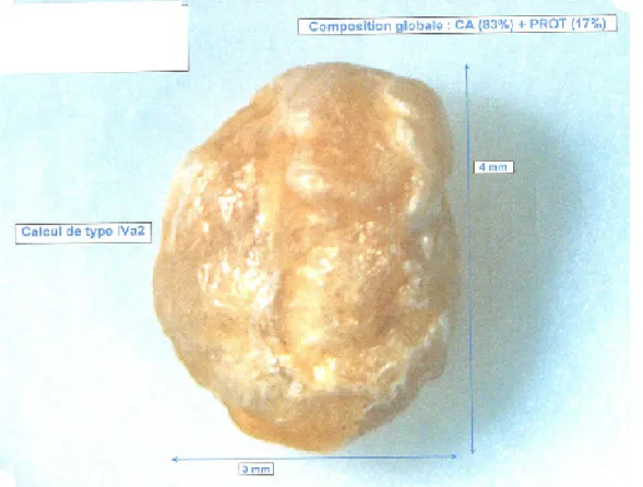
A 68-year-old woman, who has been on CAPD for 1 year, reported to the hospital for «one-way obstruc-tion» type of drainage difficulties. The X-ray demons-trated that the catheter was in place. Thrombolysis was attempted by instillation of Alteplase according to the protocol of the service, followed by 2 weekly injections of heparin. In the aftermath, however, irregular drainage and normal infusion were consistent with continued pe-ritoneal dialysis. One month later, the patient returned to the hospital for an impossible drainage. On examination, we discovered a lithiasis completely obstructing the ca-theter lumen, at the connector level of the extension line. Lithiasis was extracted after section and repair of the ca-theter. The dialysis then resumed without any problem. The lithiasis extracted was oval, irregular, 4 x 3 x 2 mm size with a light yellow-brown color. The analy-sis revealed it was composed of carbonated calcium
hydroxyphosphate (83% carbapatite) with a layer of protein (17%).
The patient had started PD one year earlier for renal fai-lure secondary to chronic tubulointerstitial nephropathy. She had no history of nephritic colic or lithiasis. Six months before the cholelithiasis, she presented a peri-tonitis without identified organism, of good evolution under antibiotherapy. She was treated with CAPD with 3 continuous daily exchanges, including a long noctur-nal exchange of an icodextrin-based solution. The cal-cium concentration of the dialysate was 1.75 mmol / L, the magnesium concentration was 0.50 mmol / L (0.25 mmol / L for icodextrin). On admission, the patient had poorly controlled secondary hyperparathyroidism with 2.15 mmol / L serum calcium and 2.48 mmol / L phos-phoremia. The intact PTH was 215 ng / L.
Secondary hyperparathyroidism has been suggested as a leading cause of peritoneal lithiasis [1, 2]. As in our ob-servation, Antoniou described a significant hyperphos-phoremia due to poor adherence to chelation therapy [1]. However in the case described by Skaro [2]
hyper-journal officiel du Registr e de D ialyse Péritonéale de Langue Française RDPLF www .rdplf.or g
www.bdd.rdplf.org Volume 1, n° 1, Juin 2018
Page 37
parathyroidism was perfectly controlled and the patient had no hyperphosphoremia.
In our case as in that of Antoniou [1], there is a history of peritonitis. However the case reported by Skaro [2] had no peritoneal or emergent infection.
The physicochemical composition of the lithiasis is si-milar to that of the two other cases reported in the li-terature: carbonated calcium hydroxyphosphate with a protein layer. Its shape and color are also similar. In the two cases previously described, the authors evoke the formation of a lithiasis in the peritoneal cavity, and its secondary migration in the PD catheter. However, in our observation, the size of the calculation suggests lithoge-nesis in situ, directly in the catheter lumen. Indeed, the internal diameter of the catheter is about 2.7 mm and our lithiasis was 4 x 3mm. With this size, the lithiasis would not have been able to cross the intraperitoneal distal ori-fice of the catheter from the peritoneal cavity. In addition it was necessary to cut the catheter to extract the lithia-sis. In the case reported by Antoniou [1], the lithiasis measured 3.9 x 3 mm and the catheter had to be depo-sited. The lithiasis described by Skaro [2] was smaller: 2.5 x 1.5 mm, and could be aspirated with the syringe. This observation suggests a local mechanism at the ori-gin of the development of lithiasis in the catheter lumen, despite the multiple rinsing of the catheter by the pe-ritoneal dialysis solution. In the three cases described, the lithiasis was of the same physico-chemical compo-sition; the dialysate contained lactate, and had a calcium concentration of 1.75 mmol / L.
In spite of the rarity of this complication, the presence of lithiasis of the peritoneal dialysis catheter must be evoked in case of drainage problems.
DISCLOSURE
Authors declare no conflict of interest
REFERENCES
1. Antoniou S, Syreggelas D, Papadopoulos C, Dimi-triadis A. Intraluminal lithiasis of a peritoneal catheter. Perit Dial Int 1991; 11: 358-60.
2. Skaro B, Jelicic I, Ljutic D. Intraluminal stone in a PD catheter. A rare complication. Perit Dial Int 2011; 31(3): 371-72.
Received 2018/05/18 accepted after revision 2018/05/31, published 2018/06/13 journal officiel du Registr e de D ialyse Péritonéale de Langue Française RDPLF www .rdplf.or g
www.bdd.rdplf.org Volume 1, n° 1, Juin 2018
Open Access This article is licensed under a Creative Commons Attribution 4.0 International License, which permits use, sharing, adaptation, distribution and reproduction in any medium or
format, as long as you give appropriate credit to the original author(s) and the source, provide a link to the Creative Commons license, and indicate if changes were made. The images or other third party material in this
article are included in the article’s Creative Commons license, unless indicated otherwise in a credit line to the material. If material is not included in the article’s Creative Commons license and your intended use is not permitted by statutory regulation or exceeds the permitted use, you will need to obtain permission directly from the