HAL Id: hal-01204451
https://hal.archives-ouvertes.fr/hal-01204451
Submitted on 28 May 2020
HAL is a multi-disciplinary open access
archive for the deposit and dissemination of
sci-entific research documents, whether they are
pub-lished or not. The documents may come from
teaching and research institutions in France or
abroad, or from public or private research centers.
L’archive ouverte pluridisciplinaire HAL, est
destinée au dépôt et à la diffusion de documents
scientifiques de niveau recherche, publiés ou non,
émanant des établissements d’enseignement et de
recherche français ou étrangers, des laboratoires
publics ou privés.
Copyright
sulfatases from the human gut symbiont bacteroides
thetaiotaomicron reveals the first GAG-specific bacterial
endosulfatase
Jonathan Ulmer, Eric Morssing Vilen, Ramesh Babu Namburi, Alhosna
Benjdia, Julie Beneteau, Annie Malleron, David Bonnaffe, Pierre-Alexandre
Driguez, Karine Descroix, Gilbert Lassalle, et al.
To cite this version:
Jonathan Ulmer, Eric Morssing Vilen, Ramesh Babu Namburi, Alhosna Benjdia, Julie Beneteau, et
al.. Characterization of Gglycosaminoglycan (GAG) sulfatases from the human gut symbiont
bac-teroides thetaiotaomicron reveals the first GAG-specific bacterial endosulfatase. Journal of Biological
Chemistry, American Society for Biochemistry and Molecular Biology, 2014, 289 (35), pp.24289
-24303. �10.1074/jbc.M114.573303�. �hal-01204451�
Characterization of Glycosaminoglycan (GAG) Sulfatases
from the Human Gut Symbiont Bacteroides thetaiotaomicron
Reveals the First GAG-specific Bacterial Endosulfatase
*
Received for publication, April 10, 2014, and in revised form, July 3, 2014 Published, JBC Papers in Press, July 7, 2014, DOI 10.1074/jbc.M114.573303
Jonathan E. Ulmer‡§1, Eric Morssing Vile´n¶1, Ramesh Babu Namburi储1,2, Alhosna Benjdia‡§, Julie Beneteau‡§, Annie Malleron**, David Bonnaffe´**, Pierre-Alexandre Driguez‡‡, Karine Descroix‡‡, Gilbert Lassalle‡‡, Christine Le Narvor**, Corine Sandstro¨m¶, Dorothe Spillmann储1,2,3, and Olivier Berteau‡§1,4
From the‡Institut National de la Recherche Agronomique, ChemSyBio, UMR 1319 Micalis, F-78350 Jouy-en-Josas, France, the
§AgroParisTech, ChemSyBio, UMR 1319 Micalis, F-78350 Jouy-en-Josas, France, the¶Department of Chemistry and Biotechnology,
Swedish University of Agricultural Sciences, P. O. Box 7015, SE-750 07 Uppsala, Sweden, the储Department of Medical Biochemistry and Microbiology, Biomedical Center, Uppsala University, SE-751 23 Uppsala, Sweden, the **ICMMO/G2M/LCOM/UMR
8182(CNRS-UPS), LabEx LERMIT, Universite´ Paris-Sud, 91405 Orsay Cedex, France, and‡‡Sanofi R&D, Early to Candidate Unit, 195 Route d’Espagne, BP13669, 31036 Toulouse Cedex, France
Background:Sulfatases are emerging as key adaptive tools of commensal bacteria to their host.
Results:The first bacterial endo-O-sulfatase and three exo-O-sulfatases from the human commensal Bacteroides
thetaiotao-micron, specific for glycosaminoglycans, have been discovered and characterized.
Conclusion:Commensal bacteria possess a unique array of highly specific sulfatases to metabolize host glycans. Significance:Bacterial sulfatases are much more diverse than anticipated.
Despite the importance of the microbiota in human physiol-ogy, the molecular bases that govern the interactions between these commensal bacteria and their host remain poorly under-stood. We recently reported that sulfatases play a key role in the adaptation of a major human commensal bacterium, Bacte-roides thetaiotaomicron, to its host (Benjdia, A., Martens, E. C., Gordon, J. I., and Berteau, O. (2011) J. Biol. Chem. 286, 25973– 25982). We hypothesized that sulfatases are instrumental for this bacterium, and related Bacteroides species, to metabolize highly sulfated glycans (i.e. mucins and glycosaminoglycans (GAGs)) and to colonize the intestinal mucosal layer. Based on our previous study, we investigated 10 sulfatase genes induced in the presence of host glycans. Biochemical characterization of these potential sulfatases allowed the identification of GAG-specific sulfatases selective for the type of saccharide residue and the attachment position of the sulfate group. Although some GAG-specific bacterial sulfatase activities have been described in the literature, we report here for the first time the identity and the biochemical characterization of four GAG-spe-cific sulfatases. Furthermore, contrary to the current paradigm, we discovered that B. thetaiotaomicron possesses an authentic GAG endosulfatase that is active at the polymer level. This type of sulfatase is the first one to be identified in a bacterium. Our study thus demonstrates that bacteria have evolved more sophisticated
and diverse GAG sulfatases than anticipated and establishes how B. thetaiotaomicron, and other major human commensal bacteria, can metabolize and potentially tailor complex host glycans.
Bacteria are by far the most abundant and diverse microorgan-isms inside the human gastrointestinal tract with Firmicutes and Bacteroidetes being the major phyla (1–3). How these bacteria efficiently compete in this fierce ecosystem is still poorly under-stood despite their major role in human physiology (4). Although the human gut provides a constant source of nutrients and tightly controlled conditions, only a few phyla are abundant, suggesting a selective adaptation of these bacteria to their host (1). Key studies have highlighted that the prominent gut bacterium Bacteroides
thetaiotaomicronhas evolved complex relationships with its host (5, 6). Of particular interest, it has been established that B.
thetaiotaomicronrelies on host glycan foraging to enhance the fitness to its host (7). Recently, we have shown that in this process sulfatases play a pivotal role to allow B. thetaiotaomicron persist-ence in the gastrointestinal tract (8).
Sulfatases catalyze hydrolysis of sulfate groups from a broad range of substrate molecules, including small organic com-pounds to large macromolecules such as mucins or glycosami-noglycans (GAGs).5In humans, sulfatases have been studied in *This work was supported in part by European Community within a
consor-tium PolyModE KBBE-2007-3-3-07.
1These authors contributed equally to this work.
2Supported by grants from the Swedish Cancer Foundation and the
Founda-tion for Proteoglycan Research at Uppsala University.
3To whom correspondence may be addressed: IMBIM, Uppsala University,
SE-751 23 Uppsala, Sweden. Tel.: 46-18-471-43-67; E-mail: dorothe. spillmann@imbim.uu.se.
4To whom correspondence may be addressed: INRA, Institute Micalis (UMR
1319), F-78350 Jouy-en-Josas, France. Tel.: 65-23-08; Fax: 33-1-34-65-24-62; E-mail: olivier.berteau@jouy.inra.fr.
5The abbreviations used are: GAG, glycosaminoglycan; anSME, anaerobic
sulfatase-maturating enzyme; anSMEbt, B. thetaiotaomicron anSME; HS, heparan sulfate; CS, chondroitin sulfate; DS, dermatan sulfate; GlcNAc,
N-acetyl-glucosamine; GalNAc, N-acetyl-galactosamine; GlcN,
glucosa-mine; GlcNS, N-sulfated glucosaglucosa-mine; GlcUA, glucuronate; IdoUA, iduro-nate; GlcNAc6S, N-acetylglucosamine-6-O-sulfate; GlcNS6S, N-sulfated glucosamine-6-O-sulfate; GlcNS3S6S, N-sulfated glucosamine-3,6-O-sul-fate; GlcNAc6S-O-pNP, p-nitrophenyl GlcNAc6S; CE, capillary electrophore-sis; RPIP-HPLC, reversed-phase ion pairing-high performance liquid chro-matography; ⌬HexA, ⌬4,5 unsaturated hexuronate; PDB, Protein Data Bank; Ni-NTA, nickel-nitrilotriacetic acid.
at INRA Institut National de la Recherche Agronomique on June 13, 2018
http://www.jbc.org/
great detail and are shown to be involved in genetic disorders or cancer (9). Most of them are lysosomal, and at least six are exo-enzymes responsible for GAG degradation (10). In addi-tion, one GAG endosulfatase has been identified and shown to be critical for heparan sulfate (HS) remodeling (11). Opposite to the mammalian sulfatases, few bacterial GAG sulfatases have been characterized with three heparin sulfatases identified in the bacterium Flavobacterium heparinum (now called
Pedo-bacter heparinus) (12–14). In addition, several GAG sulfatase activities, acting on chondroitin sulfate (CS), have been reported in B. thetaiotaomicron (15) and Proteus vulgaris (16). Among hydrolases from bacteria to human, sulfatases are unique in requiring a critical amino acid post-translational modification on a key active site residue. This modification leads to the formation of the so-called “formylglycine” (3-oxo-alanine), which is the only naturally occurring amino acid hav-ing an aldehyde function (17). Although the role of formylgly-cine in catalysis is still not completely understood, we have shown this modification to be dependent on three distinct enzymatic systems in bacteria (17–20). In B. thetaiotaomicron only one of these systems, the anaerobic sulfatase-maturating enzyme (anSME), which is part of the large and diverse family of radical S-adenosyl-L-methionine enzymes (21, 22), is involved
in sulfatase activation (20). By inactivating anSME, we have demonstrated that the ability of B. thetaiotaomicron to survive in the gastrointestinal tract is strongly impaired (8). Transcrip-tomic analyses further revealed that, in the absence of a func-tional anSME, the expression profile of genes related to host glycan metabolism, especially encoding putative sulfatase genes, was markedly altered suggesting their involvement in host glycan metabolism.
To decipher the function of these potential sulfatases, we cloned 10 B. thetaiotaomicron genes, nine of which are induced by host glycans and five belong to polysaccharide utilization loci (Table 1) (7, 8). These genes were expressed in Escherichia
coli in the presence of the B. thetaiotaomicron anSME (anSMEbt) that we have previously characterized (20, 23). The activity of the soluble purified proteins was then assayed with synthetic and natural GAG substrates. We especially focused on heparin/HS and chondroitin sulfate/dermatan sulfate (CS/ DS), two major classes of sulfated GAGs, to determine the activity and specificity of these enzymes. Both classes of GAGs are characterized by alternating units of a hexuronate and an amino sugar: N-acetylglucosamine (GlcNAc) or N-acetyl-galactosamine (GalNAc), respectively, with heparan polymer containing GlcUA1–4GlcNAc␣1–4 and CS GlcUA1– 3GalNAc1–4 as basic repeat units.
During biosynthesis, these structures are further modified by extensive sulfation and epimerization. In HS, part of the GlcNAc are N-deacetylated and N-sulfated during biosynthesis resulting in an N-sulfated GlcN unit (GlcNS). The hexuronate, either glucuronate (GlcUA) or its C5-epimer iduronate (IdoUA), may be 2-O-sulfated, whereas the GlcNAc and GlcNS units can further be 6-O- and, rarely, 3-O-sulfated. CS/DS can be sulfated at position 2 on IdoUA and, more rarely on GlcUA, at positions 4 and 6 of the GalNAc unit. These sulfate groups are not only critical for the functional properties of the GAGs
but also confer resistance to the hydrolytic activity of bacterial glycosidases (24).
In B. thetaiotaomicron, we identified two different 6-O-sul-fatases with a strict specificity for either gluco- or galactosami-noglycans. We also report a 2-O-sulfatase that removes sulfates from a hexuronate unit independent of the parent GAG and whose structure is reported. Whereas these three enzymes are exolytic hydrolases, like all bacterial sulfatases reported to date, we also identified, in a totally unanticipated manner, a unique GAG endosulfatase. This novel enzyme very efficiently removes sulfate groups in the 4-O-position from CS/DS disac-charides to large polymeric chains and is thus the first bacterial GAG endosulfatase reported to date active at the polymer level. We not only discovered novel bacterial sulfatases, we also demonstrate the complex interplay between these enzymes for host glycan degradation. Finally, we show that these enzymes are widespread in gut-associated bacteria underlining the importance of such metabolic pathways for a dominant phylum of the human microbiota, the Bacteroides. These novel sulfata-ses constitute attractive targets to manipulate the human microbiota and could potentially influence gut epithelial integ-rity by their unique capacity to selectively modify human GAGs.
EXPERIMENTAL PROCEDURES
Materials—CSA from bovine trachea, CSB from porcine intestinal mucosa, and CSC from shark cartilage were from Sigma. CSD from shark cartilage and CSE from squid cartilage were purchased from Seikagaku. HS from porcine intestine was a gift from G. van Dedem (Diosynth, Oss, The Netherlands), and heparin from bovine lung was purified as described previ-ously (see Table 2 for composition) (16). CS-derived ⌬4,5-un-saturated disaccharides were purchased from Iduron, and HS-derived ⌬4,5-unsaturated disaccharides were obtained from Calbiochem. Monosaccharide substrates N-acetyl glucosamine 6-O-sulfate (GlcNAc6S), N-acetyl glucosamine 3,6-O-sulfate (GlcNAc3S6S), N-sulfate glucosamine (GlcNS), N-sulfate glu-cosamine 3-O-sulfate (GlcNS3S), N-sulfate gluglu-cosamine 6-O-sulfate (GlcNS6S) and N-6-O-sulfate glucosamine 3,6-O-6-O-sulfate (GlcNS3S6S) were purchased from Dextra. All other synthetic or semisynthetic HS derivatives were from Sanofi. Chondroiti-nase ABC was bought from Seikagaku, heparin lyases I–III from IBEX Pharmaceuticals, Inc. (Montreal, Canada), and ⌬4,5-gly-cosidase from Grampian enzymes (Orkney, UK). A Luna 5- C18 reversed phase column (4.6 ⫻ 150 mm) was from Phenomenex.
Cloning of Sulfatase Enzymes—Sulfatases were cloned into the pRSF-Duet1 expression vector (Novagen) at the MCS1 cloning site with an N-terminal His6tag that allowed purifica-tion on the Ni-NTA column. The sulfatase-maturating enzyme from B. thetaiotaomicron (anSMEbt (20)) was cloned into the same vector at the MCS2-cloning site that allowed sulfatase to be activated during expression in E. coli. The expression vectors were transformed into One Shot BL21 StarTM (DE3) E. coli
(Invitrogen). The following primers were used for the corre-sponding sulfatase genes: BT_0756, 5⬘-GGA TCC AAT GGC CTC TGC TGT GCA-3⬘ and 5⬘-CTG CAG TTA TTT TTT TAA TGA TGA TTT CTT CAC TT-3⬘; BT_1596, 5⬘-GGA
at INRA Institut National de la Recherche Agronomique on June 13, 2018
http://www.jbc.org/
TCC AAT GGG ATT AGC CCT TTG TGG-3⬘ and 5⬘-CTG CAG TTA TTT TCT TTT GAG GAT CTC CCG-3⬘; BT_1624, 5⬘-GGA TCC AAT GAG AAA AGA ATT TTA TGG TAT ATT ACC-3⬘ and 5⬘-CTG CAG TTA TAG TGG CAG ACC GTA GCG-3⬘; BT_1628, 5⬘-GGA TCC AAT GCC GGA AGG CCA TC-3⬘ and 5⬘-CTG CAG TTA TTC CTT GTC CCT TTC CG-3⬘; BT_1918, 5⬘-GGA TCC AAT GAT TAA CCT GAA ATG TAC ATT TGC-3⬘ and 5⬘-CTG CAG TTA TCG CTT TTC TTT CGG ATA GTT-3⬘; BT_3095, 5⬘-GCA TGA CTG AAT TCA ATG CAG CGT TTT GTA TTA CGG-3⬘ and 5⬘-GAG TCT ACG TCG ACT TAT TGT TCG GGT TGG AGA TAA TT-3⬘; BT_3101, 5⬘-GGA TCC AAT GAA TAG ACT ATT TTT GAG TGT TTC TGT-3⬘ and 5⬘-CTC CAG TCA CTT TTG TTT GGA AGG CA-3⬘; BT_3333, 5⬘-GGA TCC AAT GAA GAA TGT CTC ACG TTT ACT ACC-3⬘ and 5⬘-CTG CAG TTA TCT TGT CTT TAC CGA TTC AAG C-3⬘; BT_3349, 5⬘-GGA TCC AAT GGG AGG CTT GAC CCT C-3⬘ and 5⬘-CTG CAG TCA GTA AGG TAT ACG GTC CGA A-3⬘; and BT_4656, 5⬘-GGA TCC AAT GCC CGC ACG GAA AAG-3⬘ and 5⬘-CTG CAG TTA TCG ATT TTC CAT CAG TCT TCT G-3⬘.
Expression and Purification of Sulfatase Enzymes—E. coliwas used for cloning and expression of sulfatase genes using previ-ously reported procedures (20). Briefly, plasmid coexpressing sulfatase genes with anSMEbt were selected on LB-agar plates containing 50g/ml kanamycin and grown in LB medium sup-plemented with the same antibiotic at 37 °C under agitation at 200 rpm. When the A600reached 0.7, incubation temperature
was lowered at 21 °C, and sulfatase expression was induced by adding isopropyl-D-1-galactopyranoside (500Mfinal
con-centration). The culture was continued overnight, and bacteria were harvested by centrifugation at 5000⫻ g for 20 min at 4 °C. After resuspension in buffer A (50 mMTris/HCl, pH 7.0, 100
mMKCl, 10 mMMgCl2), the cells were disrupted by sonication
and centrifuged at 100,000⫻ g for 1 h at 4 °C. The cell extract was then loaded onto a Ni-NTA-Sepharose column equili-brated with buffer A. The column was washed extensively with the same buffer. Weakly adsorbed proteins were washed off by applying 3 column volumes of 25 mMimidazole in buffer A,
fol-lowed by 2 column volumes of 100 mMimidazole in the same
buffer. Sulfatases were then eluted by applying 1 column volume of 500 mM imidazole in buffer A. Imidazole was removed using
PD-10 desalting columns (GE Healthcare) equilibrated in buffer A. The sulfatase-containing fractions were immediately concen-trated in Ultrafree cells (Millipore) with a molecular cutoff of 10 kDa. Sulfatase purity was assessed by SDS-PAGE, and their iden-tity was confirmed by mass spectrometry analyses.
Synthesis of the Chromogenic Substrate 4-Nitrophenyl 2-Ac-etamido-2-deoxy-6-O-sodium Sulfonato--D-glucopyranoside (GlcNAc6S-O-pNP)—Triethylamine䡠SO3 (21 mg, 1 eq) was
added to a solution of 4-nitrophenyl 2-acetamido-2-deoxy-
-D-glucopyranoside (40 mg, 0.12 mmol) in dimethylformamide
(5.4 ml) under argon atmosphere at 0 °C. After stirring at 0 °C for 0.5 h and at room temperature for 1 h, a 0.15Maqueous
solution of sodium hydrogen carbonate (2.6 ml) was added at 0 °C and stirred for 16 h. The reaction mixture was then con-centrated under reduced pressure and purified by reverse phase column chromatography (H2O/MeOH) to finally give pure
GlcNAc6S-O-pNP (21 mg, 34%) after lyophilization.1H and 13C NMR data were in agreement with previously reported data
(25).
Synthesis of Methyl (2-O-Sulfonato-␣- L-idopyranosyluro-nate)-(1– 4)-2-acetamido-2-deoxy-6-O-sulfonato-␣- D-glucopy-ranoside, Sodium Salt (IdoUA2S-GlcNAc6S-O-Me)—Sodium hydrogen carbonate (45 mg, 0.54 mmol) and acetic anhydride (25l, 0.27 mmol) were successively added at 0 °C to a satu-rated sodium hydrogen carbonate solution (1.8 ml) of methyl (2-O-sulfonato-␣-L-idopyranosyluronate)-(1–
4)-2-amino-2-deoxy-6-O-sulfonato-␣-D-glucopyranoside (7.0 mg, 13.3mol) (26). After stirring 3 h at 0 °C and then 17 h at room tempera-ture, the reaction mixture was diluted with 0.2Maqueous NaCl
(2 ml) and layered on top of a Sephadex G-25 column (in 0.2M
NaCl). Effluent fractions were pooled, concentrated, and desalted on the same gel filtration column, equilibrated with H2O. Lyophilization gave the sodium salt of methyl (2-O-sulfonato-␣-L-idopyranosyluronate)-(1–
4)-2-acetamido-2-deoxy-6-O-sulfonato-␣-D-gluco-pyranoside (6.2 mg, 82%). [␣]D⫹ 0.063° (c 0.18, H2O); 1H NMR (500 MHz, D 2O)␦ ⫽ 5.18 (d, J1,2⫽ 2.4 Hz, 1 H, H-1 II), 4.79 (d, J 1,2⫽ 3.7 Hz, 1 H, H-1 I), 4.74 (br. s, 1 H, H-5II), 4.39 – 4.29 (m, 3 H, H-6 a I, H-6 b I, H-2II), 4.07– 4.00 (m, 4 H, H-3II, H-5I, H-4II, H-2I), 3.85 (dd, J⫽ 8.8 Hz, J⫽ 10.3 Hz, 1 H, H-3I), 3.77 (dd, J⫽ 8.8 Hz, J ⫽ 9.6 Hz, 1 H, H-4I), 3.43 (s, 3 H, CH 3O), 2.06 (s, 3 H, CH3CO). 13C NMR (125.76 MHz, D2O)␦ ⫽ 101.0 (C-1 II), 99.5 (C-1I), 79.5 (C-4I), 76.8 (C-2II), 71.6 (C-3I), 71.3 (C-4II), 71.2 (2 C, C-5II, C-3II), 70.4 (C-5I), 68.7 (C-6I), 57.0 (CH 3O), 55.4 (C-2 I), 23.7 (CH 3CO).
Screening for Sulfatase Activity—Sulfatase activity was rou-tinely assayed using the chromogenic reagent p-nitrophenyl sulfate (pNP-S) as a substrate. Standard assays were performed with 10Menzyme incubated at 30 °C for 10 min with 50 mM
substrate in 100 mMTris/HCl buffer, pH 7.25, containing 10
mMMgCl2, as described previously (17, 18). The pNP released
was measured spectrophotometrically at 405 nm (⑀ ⫽ 9 000 mol⫺1䡠cm⫺1at pH 7.25).
Sulfatase Assays with Synthetic and Natural Sulfated Gly-cans—Synthetic libraries of sulfated glycans, previously synthe-sized (27), were tested with enzymes as follows.: Onel (10 mM) of
enzyme was added to 68l of 34 mMTris/HCl buffer, pH 7.5,
containing 6l of library solution (1 mg/ml). Incubations were performed overnight at 25 °C before analysis by capillary electro-phoresis. Sulfated mono- and disaccharides (3 mM) were assayed at identical conditions.
To measure sulfatase activity on natural GAGs, sulfatases were incubated at a final concentration of 10Min 20l of 50
mMTris/HCl, pH 7.5, 100 mMKCl, 10 mMMgCl2, containing 1
g of GAG at 30 °C for 8 h. Sulfatase activity was stopped by boiling the sample at 96 °C for 10 min. For specificity analyses, variable enzyme concentrations were used as indicated in the figure legends. Sulfated saturated mono- and unsaturated disaccharides were used at a 20Mfinal concentration and
enzymatically digested as indicated, followed by analysis by reversed-phase ion pairing-high performance liquid chroma-tography (RPIP-HPLC).
Analysis of Sulfatase Products by Capillary Electrophoresis—
Analyses by capillary electrophoresis (CE) were carried out on an Agilent CE apparatus, using a bare fused-silica capillary (64
at INRA Institut National de la Recherche Agronomique on June 13, 2018
http://www.jbc.org/
cm⫻ 50m). New capillaries were conditioned by successive flushes with NaOH (1 and 0.1M) and water for 10, 5, and 10 min,
respectively. Borate buffer (Agilent Technologies) (23 mM, pH 9.0, filtered through 0.2-m filtered before use) was used as CE background electrolyte. The capillary was thermostatically controlled at 25 °C, and CE experiments were performed by applying a positive voltage of 30 kV at the capillary inlet. Prior to each sample injection, the capillary was flushed with the sepa-ration electrolyte for 10 min. Samples were loaded hydrody-namically by applying 50 millibars at the capillary inlet for 4 s. Detection was performed at 192 nm.
Analysis of Sulfatase Products by Reversed-phase Ion Pairing Chromatography—Sulfatase-treated GAGs (1 g) were pre-pared for RPIP-HPLC analysis by exhaustive digestion to disac-charides with either 20 milliunits chondroitinase ABC (for CS/DS) or a mixture of 0.4 milliunit each of heparin lyase I–III (for heparin or HS) at 37 °C overnight as described (28) before heat inactivation of the enzymes. Products were separated by RPIP-HPLC and monitored by post-column fluorescence detection as described earlier (28).
Structure of the BT_1596 Sulfatase—The BT_1596 structure was solved by the Joint Center for Structural Genomics (29) and deposited in the PDB under code 3B5Q. Manual docking of the enzyme substrate was made using the disaccharide ⌬HexA2S-GalNAc4,6S derived from a structurally characterized heparin tetrasaccharide (PDB code 1BFB). The human cerebroside-3-sulfate 3-sulfohydrolyase (ArsA) in interaction with a synthetic substrate (PDB code 1E2S) was superimposed with the BT_ 1596 structure using Coot and the SSM program. The heparin disaccharide was manually docked using ArsA substrate as reference.
NMR Analysis—NMR experiments were performed on a Bruker 600 MHz Avance III spectrometer using a 2.5-mm
1H/13C inverse detection probe, a 5-mm 1H/13C/15N/31P
inverse detection QCI probe, or a 5-mm1H/13C/15N/31P cryo-probe all equipped with z-gradient and controlled by Topspin 2.1 and 3.0 software. The incubated samples were repeatedly exchanged with D2O with intermediate lyophilization. The residual HOD signal was suppressed by saturation of the water peak during the recycle delay or using the WATERGATE pulse sequence. For the one-dimensional NMR experiments with the substrates incubated with the enzymes, the diffusion-edited NMR experiment (ledbpgp2s1d) was used for effective suppres-sion of the residual water and buffer signals while retaining the signals from the substrates. The diffusion delay⌬ and the gra-dient pulse length␦ were 100 and 1.9 ms, respectively. The gradient strength was set to 95%. Assignments of1H and13C
resonances were achieved using COSY, TOCSY, NOESY, HSQC, HMBC, and HSQC-TOCSY experiments from the Bruker pulse sequence library.
RESULTS
Expression and Functional Assay of B. thetaiotaomicron Sulfatases—Based on our and other transcriptomic studies (7, 8), we selected 10 potential sulfatase genes in the B.
thetaiotao-micron genome, induced by host glycans (Table 1). These potential sulfatases were cloned and expressed in E. coli in the presence of the sulfatase-activating enzyme (anSMEbt) from B.
thetaiotaomicron (20). Indeed, we have shown earlier that anSMEbt is critical for the post-translational modification and thus the activation of sulfatases from B. thetaiotaomicron (20). Purified sulfatases were first assayed with the simple chromo-genic substrate pNP-S (Table 1). Half of these enzymes proved to be active on this synthetic substrate identifying them as authentic sulfatases.
We further synthesized a more relevant substrate in which the sulfate group was on a carbohydrate moiety rather than on an aromatic ring. This substrate (GlcNAc6S-O-pNP; see Scheme 1a and “Experimental Procedures”) (25) also contained a chromogenic group allowing the spectroscopic monitoring of the reaction by a coupled enzyme assay (Table 1). All the puri-fied sulfatases were tested on this substrate; however, only two sulfatases, BT_4656 and to a much lesser extent BT_1628, hydrolyzed this reagent. Interestingly, despite its strong activity on this substrate, the BT_4656 sulfatase was inactive on pNP-S (Table 1).
Sulfatase Activity on Synthetic GAG-like Substrates—The B.
thetaiotaomicronsulfatases were then tested against a library of synthetic saturated CS disaccharides with sulfate groups on the 2-O-position of the uronate unit and 4-O- and 6-O-positions of the amino sugar as described previously (27, 30).
Only the sulfatase BT_3349 (Table 1) proved to be active on these disaccharides. This enzyme led to the hydrolysis of all the disaccharides containing a sulfate group in the 4-O-position, whether or not a sulfate group is present in 6-O-position. To confirm BT_3349’s specificity, we incubated this enzyme with the synthetic di-O-sulfated disaccharide GlcUA-GalNAc4,6S (Scheme 1b). Comparison of the NMR spectra of the disaccha-ride in the absence or presence of BT_3349 (Fig. 1A) showed that the signals at⬃4.8 and 4.9 ppm corresponding to the H4 of the GalNAc4,6S (␣, ) unit disappeared in agreement with des-ulfation at the 4-O-position of the GalNAc residue.
These results were consistent with transcriptomic analysis showing that two putative sulfatases genes, BT_3349 and BT_3333, are induced when B. thetaiotaomicron grows with CS as sole carbon source (7). B. thetaiotaomicron is known to pos-sess two distinct CS sulfatase activities, one specific for the
4-O-TABLE 1
B. thetaiotaomicron sulfatases expressed in this study
Genes induced by host glycans (7, 8) are shown in boldface type.
Namea Activity on pNP-Sb Activity on the GlcNAc6S-O-pNPc CS disaccharidesd
BT_0756 None None None
BT_1596 Yes None None
BT_1624 Yes None None
BT_1628 Yes Low None
BT_1918 Yes None None
BT_3095 Yes None None
BT_3101 None None None
BT_3333 None None None
BT_3349 Yes None Yes
BT_4656 None Yes None
aThese are the sulfatase gene numbers.
bSulfatase activity was assayed with p-nitrophenyl sulfate (see “Experimental
Procedures”).
cSulfatase activity assayed with GlcNAc6S-O-pNP. Assay was performed in the
presence of the indicated sulfatase with 10 mMGlcNAc6S-O-pNP and 1 unit of -N-acetylglucosaminidase (Canavalia ensiformis). The released pNP was mea-sured spectrophotometrically at 405 nm.
dSulfatase activity was assayed on synthetic CS libraries as described (see
“Experi-mental Procedures” and Ref. 27) for synthesis and composition).
at INRA Institut National de la Recherche Agronomique on June 13, 2018
http://www.jbc.org/
sulfate group and the other for the 6-O-sulfate group (15). Because we established BT_3349 as a 4-O-sulfatase, BT_3333 was the logical candidate for the 6-O-sulfatase activity reported for that bacterium. Nevertheless, none of the synthetic sub-strates tested (CS disaccharide libraries or GlcNAc6S-O-pNP) proved to be a suitable substrate for the BT_3333 enzyme.
B. thetaiotaomicron GAG-specific 6-O-Sulfatases Require an Unsubstituted, Nonreducing End Hexosamine as Substrate—
Because the synthetic CS disaccharides only partially mimic products formed during bacterial CS degradation, we assayed our library of enzymes on disaccharide mixtures obtained after treatment of HS and CS with bacterial eliminases. The disac-charides produced, in contrast to the synthetic ones, contain a 4,5-unsaturated hexuronate ring at their nonreducing end. Nevertheless, neither CS- nor HS-derived disaccharides (Fig.
1B) proved to be suitable substrates for BT_3333 or the 6-O-sulfatase BT_4656.
As exoenzymes would not be expected to attack the sulfate groups on the sugar in reducing-end position, we treated these disaccharides with a bacterial⌬4,5-glycuronidase that catalyzes the hydrolysis of the unsaturated hexuronate ring (31). The resulting monosaccharide mixtures were assayed with BT_ 3333 and BT_4656 sulfatases. As shown, the monosaccharides GalNAc6S and GlcNAc6S were hydrolyzed by BT_3333 and BT_4656, respectively (Fig. 1B). Using a mixture of differen-tially sulfated CS disaccharides as substrates, we demonstrated that only the 6-O-sulfate group on the GalNAc unit was hydro-lyzed by BT_3333 after removal of the⌬HexA unit (Fig. 1C) (where ⌬ stands for the 4,5-unsaturated ring structure and HexA for the uronate unit).
O HO HO NHAc OpNP OSO3
-GlcNAc6S-
O-pNP
a
O O O NHAc O OSO3 -H S O3 -O HO OH -O O C HOb
GlcA-GalNAc46S
IdoA2S-GlcNAc6S-
O-Me
c
Ac=COCH3 R1=H or SO3 -SO3 or COCH3 R2=-∆HexA2S-GlcNS6S-IdoA-GlcNAc6S-GlcA-GlcNS36S
O O O O N H Ac HO O OSO3 -N H S O3 -HO OSO3 -O OSO3 -HO -OOC O O OH NHSO3 -O3SO O OH -O O C OSO3 -HO O O OH OH -OOCd
O HO -OOCn ~ 3
O O O O NHR2 R1O O OR1 HO OR1 O NHR2 HO OR1 O OR1 Hn
-OOCAVE5026 (Na salt)
f
O O O O OH -OOC OH OSO3 -HO OH NHAc O O OH OSO3 -O O HO OSO3 -NHAc O O OH OOC OSO3 -O OMe HO OSO3 -N H Ac2
-OOC-IdoA2S-GlcNAc-IdoA2S-GlcNAc6S-IdoA2S-GlcNAc6S-IdoA2S-GlcNAc6S-
O-Me
e
3 O O HO Ac N OMe OSO -H O OH -OOC OH OSO3-SCHEME 1. Structure of the synthetic and semisynthetic oligosaccharides used in this study.
at INRA Institut National de la Recherche Agronomique on June 13, 2018
http://www.jbc.org/
4.0 4.2 4.4 4.6 4.8 5.0 ppm H4α H4β H4β H4α
}
* 6 Hβ / α 3 Hα 2 Hα 5 Hα H6α/β Hβ 2H3β Hβ 5 H1β * * * H1β H5α H6α/β Hβ 2 3 Hα Hβ5 O O O NAc OH HO HO -OOC OSO3 -OH OSO3 -+ BT_3349 0 0.5 1.0 1.5 2.0 Ø BT_3333 BT_3349 BT_1596 BT_4656 Ø BT_3333 BT_3349 BT_1596 BT_4656 Ø BT_3333 BT_4656 Ø BT_3333 BT_4656 Sulfate/Saccharide Enzyme HS disaccharides CS disaccharides GalNAc6S GlcNAc6S 0 0.2 0.4 0.6 0.8 1.0 1.2 1.4 1.6 1.8 2.0 GalNAc6S 4S 6S 2S 4S6S 2S4S 2S6S 2S4S6S Recovery (pmol) Saccharide Units CS disaccharide mixture + Gly:ase + BT_3333 + Gly:ase + BT_3333 3.5 3.6 3.7 3.8 3.9 4.0 4.1 4.2 4.3 4.4 ppm H2 H3 H4 H5 H6 H6 H2 H6 H5H3 H4 H6 * * * * * * * O HO HO NHAc O NO2 OSO3 -+ BT_4656 GlcNAc3S6S, GlcNS3S6S GlcNAc3S6S, GlcNS3S6S 0 20 40 60 80 100 120 0 20 40 60 80 100 Substrate remaining (%) [BT_4656] (nM) GlcNAc6S GlcNS6SB
A
D
C
E
FIGURE 1. Identification of BT_3349 as a 4-O-sulfatase and of BT_4656 and BT_3333 as exosulfatases acting on the 6-O-sulfated nonreducing end
hexosamine unit. A, comparison of1H 1D NMR spectra of GlcUA-GalNAc4,6S before (upper trace) and after (lower trace) incubation with BT_3349. The
characteristic signals of H4 (␣/) of GalNAc4,6S at 4.9 and 4.8 ppm are upfield shifted upon incubation with the enzyme. The asterisks denote signals from the glucuronic acid residue. B, mixtures of HS and CS disaccharides were incubated for 1 h at 30 °C with the indicated putative sulfatases. Disaccharides pretreated with glycuronidase to release the nonreducing end⌬HexA unit were incubated with BT_3333 and BT_4656. All samples were analyzed by RPIP-HPLC and quantified by comparison against known amount of standards. The average number of sulfate groups per unit (either disaccharide mixture or monosaccha-ride) is indicated for substrates without (Ø) or with pretreatment by the corresponding enzyme as marked. C, unsaturated CS disaccharide mixture was treated with BT_3333 for 1 h either with or without previous digestion by⌬4.5-glycuronidase that converted ⌬4.5 HexA-GalNAc6S (6S) to GalNAc6S, which was the only structure further digested by BT_3333, whereas all the other unsaturated CS disaccharides present were neither substrates for glycuronidase nor BT_3333. D,
1H NMR spectra of GlcNAc6S-O-pNP before (upper trace) and after (lower trace) incubation with BT_4656. Addition of the enzyme results in an upfield shift of the
H6 signals indicative of desulfation at position 6 of GlcNAc. The asterisks indicate signals from the buffer. E, to assess the influence of substitution for BT_4656 in either position 3 or position 2, GlcNAc6S, GlcNS6S, N-acetylglucosamine 3,6-O-sulfate, and GlcNS3S6S were incubated at a constant concentration with increasing concentrations of BT_4656 for 1 h, before analysis by RPIP-HPLC. Remaining substrate molecules were quantified as a proportion of originally added monosaccharides.
at INRA Institut National de la Recherche Agronomique on June 13, 2018
http://www.jbc.org/
1H NMR analysis of BT_4656 in the presence of
GlcNAc6S-O-pNP showed that signals at⬃4.4 and 4.2 ppm, corresponding to the two H6 protons of the substrate, disap-peared in the NMR spectrum (Fig. 1D), consistent with its iden-tity as an N-acetylglucosamine-6-O-sulfatase. On the contrary, BT_4656 was inactive on a synthetic disaccharide with the GlcNAc6S unit in reducing-end position (i.e. IdoUA2S-GlcNAc6S-O-Me, Scheme 1c and “Experimental Procedures”), in agreement with the experiments performed on the enzymat-ically produced disaccharides.
In HS not only the GlcNAc can be 6-O-sulfated but also the GlcN-sulfated unit. Furthermore, the rare 3-O-sulfation of the GlcN unit may hinder 6-O-sulfatase activity. We therefore tested the impact of additional sulfation on the GlcNAc unit for 6-O-desulfation. Although the substitution with either an
N-acetyl- or an N-sulfate group did not affect sulfatase activity, 3-O-sulfation totally hampered enzyme activity, demonstrating tight substrate specificity (Fig. 1E). Taken together, these data established that the sulfatases BT_3333 and BT_4656 are strict 6-O-sulfatases with an exclusive exolytic mode of action.
Link between the CS and HS Degradation Pathways in B. thetaiotaomicron—Further investigation with B.
thetaiotaomi-cronsulfatases led to the identification of BT_1596 as a sulfatase active on HS-derived disaccharides but also on CS-derived disaccharides (Fig. 1B). Analysis of the different disaccharides present in solution showed that all types of 2-O-sulfated disac-charides, independent of the type of hexosamine unit and the presence of additional sulfate groups, were hydrolyzed by this enzyme. Notably, ⌬HexA2S-GlcNAc, ⌬HexA2S-GlcNAc6S, ⌬HexA2S-GlcNS, and ⌬HexA2S-GlcNS6S were completely de-sulfated (Fig. 2A), as well as GalNAc4S, ⌬HexA2S-GalNAc6S, and ⌬HexA2S-GalNAc4,6S (Fig. 2B). Comparison of BT_1596 on ⌬HexA2S-GalNAc and ⌬HexA2S-GlcNAc indicated similar hydrolytic activities on both CS- and HS-de-rived disaccharides (data not shown). However, BT_1596 had no effect on polymeric CS or HS indicating that this enzyme is an exolytic sulfatase (Fig. 2C).
To confirm its exolytic nature and specificity, BT_1596 was incubated with a heparin-derived hexasaccharide (Scheme 1d) (32), and the reaction was analyzed by NMR (Fig. 2D). Compar-ison of the1H and13C signals in the hexasaccharide before and
after incubation with BT_1596 showed chemical shift differ-ences for the signals H1/C1 to H4/C4 at the nonreducing end. The large upfield shift of H2 by 0.84 ppm from 4.64 to 3.80 ppm together with the upfield shift of C2 by 3 ppm proved desulfa-tion at this posidesulfa-tion. The changes in chemical shifts for the other signals were also characteristic for a change from a ⌬4,5-HexA2S to the nonsulfated ⌬4,5-HexA (33, 34). BT_1596 is thus a 2-O-sulfatase active on unsaturated nonreducing end hexuronate units.
To establish whether the 4,5-unsaturated ring is critical for enzyme recognition, we synthesized a heparin-like octasaccha-ride containing internal 2-O-sulfate groups and an IdoUA2S unit at its nonreducing end, IdoUA2S-GlcNAc-IdoUA2S-GlcNAc6S-IdoUA2S-GlcNAc6S-IdoUA2S-GlcNAc6S-OMe (Scheme 1e) (35). Despite extensive incubation with BT_1596, no modification of the NMR spectrum could be monitored. Notably, no changes were measured regarding both internal
2-O-sulfated units and the saturated 2-O-sulfated nonreducing end unit.
The activity of the BT_1596 sulfatase was further investi-gated by incubating the enzyme with the semisynthetic ultra-low molecular weight heparin AVE5026 (Scheme 1f) (36). AVE5026 consists of a mixture of different oligosaccharides with an average Mr of⬃2400 g/mol (i.e. ⬃8 units) and the
internal IdoUA residues partially sulfated on position 2. NMR analysis showed AVE5026 to be an enzyme substrate (Fig. 2E). It also demonstrated that BT_1596 exclusively hydrolyzes the ester sulfate at the 2-O-position of the⌬4,5HexA2S nonreduc-ing end of all oligosaccharides present in AVE5026. Further-more, no change for the C2 chemical shifts and the H1 signal intensity of the internal IdoUA2S units was observed, confirm-ing that they are not targets of the enzyme (33). Our study thus establishes BT_1596 as a⌬4,5-hexuronate-2-O-sulfatase con-necting HS and CS metabolism in B. thetaiotaomicron, consis-tent with transcriptomic analysis showing its induction in pres-ence of host glycans (7).
Structure of the⌬4,5-Hexuronate-2-O-sulfatase BT_1596—
Investigation of public databases revealed that the structure of the BT_1596 enzyme has been solved by the Joint Center for Structural Genomics (29) (PDB code 3B5Q) and identified as a putative sulfatase. Its structure shows an␣/ topology, similar to other solved arylsulfatase structures (9). It is composed of six -sheets in the larger N-terminal domain and of the canonical four anti-parallel-sheets in the C-terminal domain, both sur-rounded by␣-helices (Fig. 2F). In the highly charged active site, the critical residue (i.e. Ser-64) is located, as expected, at the beginning of an␣-helix (9). Only one structure of a sulfatase in interaction with a substrate analog is available. It was obtained using an alanine mutant of the human cerebroside 3-sulfate 3-sulfohydrolyase (ArsA) and the synthetic substrate p-nitro-catechol sulfate (37). Interestingly, the superposition of ArsA and BT_1596 sulfatase shows that both enzyme structures share high structural similarities (root mean square deviation of 2.1) and the conservation of several amino acids involved in catalysis and substrate binding. Inside the BT_1596-active site, in addition to the catalytic residue Ser-64 (formylglycine 64 in the post-translationally activated enzyme), His-119 and Arg-68 are potentially involved in the elimination of the sulfate ester intermediate, whereas His-180, Lys-117 and Lys-296 may assist the binding of the anionic sulfate of the substrate (Fig. 2G). Moreover, although no cation has been modeled in the BT_1596 crystal structure, a putative conserved metal-binding region is found, composed of Asp-24, 25, Asp-283, and His-284, likely coordinating a metal cation in the active enzyme. Incubation of BT_1596 in the presence of EDTA abolished its activity, further supporting the requirement of a cation, in the enzyme-active site.
Based on this analysis and using the ArsA substrate as a reference, we manually docked a disaccharide containing a ⌬4,5-HexA2S (PDB code 1BFB) inside the BT_1596-active site (Fig. 2G). Interestingly, validations of our model are pro-vided by the fact that the docked disaccharide superimposed with a sulfated piperazine crystallized within the enzyme-active site and that the four sulfate oxygen atoms coincide perfectly with four water molecules located inside the
at INRA Institut National de la Recherche Agronomique on June 13, 2018
http://www.jbc.org/
enzyme-active site. In this model, two charged amino acids (i.e. Lys-296 and Lys-117) are ideally positioned to interact with the sulfate group, although Glu-378 and Asn-95 seem to be responsible for the correct orientation of the unsaturated unit inside the enzyme-active site.
BT_3349 Is a Bacterial Endo-4-O-sulfatase Active from Di- to Polysaccharides—As demonstrated, the 2-O-sulfatase and the two 6-O-sulfatases isolated from B. thetaiotaomicron proved to be strict exolytic hydrolases, active only on the nonreducing end of oligosaccharides, in agreement with what is known for
0 0.5 1.0 1.5 2.0 2.5 3.0 3.5 4.0 NAc NS 6S 2S NS6S NS2S 2S6S NS2S6S Recovery (pmol) HS disaccharide mixture + BT_1596 0 1.0 2.0 3.0 4.0 5.0 6.0 OS 4S 6S 2S 4S6S 2S4S 2S6S 2S4S6S Recovery (pmol) CS disaccharide mixture + BT_1596 Disaccharide Units 0 0.5 1.0 1.5 2.0 2.5 3.0 Heparin HS - + BT_1596 Degree of sulfation (sulfate groups/disaccharide) 3.5 4.0 4.5 5.0 5.5 6.0 ppm H4-∆(2S) ) S 2( ∆-3 H H2-∆(2S) H1-∆(2S) H4-∆ H1-∆ H2-∆ ∆-3 H + BT_1596 3.80 4.00 4.20 4.40 4.60 4.80 ppm 66 68 70 72 74 76 78 80 H3-ΔHexA(2S) H2-ΔHexA(2S) H2-ΔHexA H3-ΔHexA H2-IdoA(2S) H3-IdoA(2S) ppm 5.20 5.30 5.40 5.50 5.60 5.70 5.80 5.90 ppm 100 102 104 106 108 110 112 H4-ΔHexA(2S) -1 H Δ ) S 2( A x e H H1-IdoA(2S) H4-ΔHexA H1-ΔHexA 82 H180 H25 K296 D24 H284 D283 R68 K117 H119 S64
B
A
C
E
D
G
Active site N-terminal domain C-terminal domainF
+at INRA Institut National de la Recherche Agronomique on June 13, 2018
http://www.jbc.org/
bacterial sulfatases. On the contrary, BT_3349 was active on the reducing-end unit of saturated (Fig. 1A) or unsaturated CS disaccharides (Fig. 1B) but not on monosaccharide units (30) suggesting a possible endolytic mode of activity.
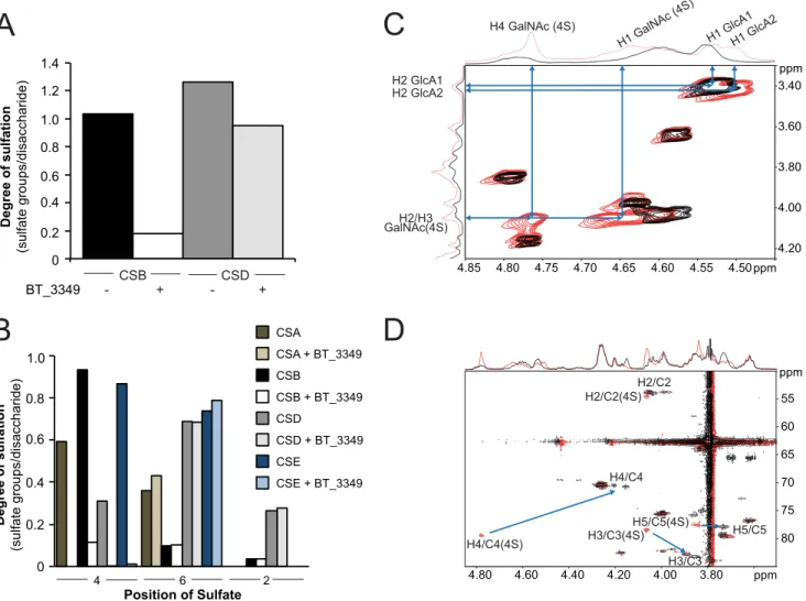
To probe this hypothesis, we assayed BT_3349 on various polymeric CS with different degrees and locations of sulfate modifications (Table 2). With all CS used, we monitored exten-sive polymer desulfation related to the chain content of 4-O-sulfate groups (Fig. 3, A and B). Even with excess of enzyme, only 4-O-sulfate groups were removed from either substrate indicating a high degree of specificity of this enzyme.
To univocally demonstrate 4-O-desulfation, we incubated the BT_3349 sulfatase with CS from shark cartilage (CSD) and followed the reaction by NMR. Analysis of the two-dimensional COSY, TOCSY, and HSQC NMR spectra showed shifts of all GalNAc4S H4/C4 signals by⫺0.61/⫺8.6 ppm, H3/C3 signal by ⫺0.21/⫹5 ppm, and H5/C5 signal by ⫺0.13/⫹0.4 ppm upon enzyme addition (Fig. 3, C and D). These changes in1H and13C
chemical shifts indicate desulfation on the 4-O-position of
GalNAc4S (38 – 40). Taken together these data demonstrated that the vast majority of the 4-O-sulfate groups present on the GalNAc units in the polymer chain were hydrolyzed. Quantitative analysis confirmed that, depending on the CS used, between 90 and 100% of 4-O-sulfate groups were removed (Fig. 3B).
Nevertheless, the substitution of CS units with sulfate groups in other positions and the presence of epimerized hexuronate unit affected the sulfatase activity (Fig. 4A). Mono-4-O-sulfated disaccharides were essentially completely hydrolyzed by⬍0.5 Menzyme at a constant substrate level, whereas the presence
of iduronate or sulfation of the uronate residue (more often on IdoUA than GlcUA) in the chain reduced the efficiency of the enzyme (Fig. 4, cf. CSB versus CSA, -D, or -E). Substitution of the 4-O-sulfated GalNAc with an additional sulfate group in position C6 reduced the efficiency slightly with all 4-O-sulfate groups hydrolyzed by⬃1Menzyme (Fig. 4A, CSE).
As distribution of sulfate groups in different chain types is heterogeneous, analysis at chain level gives only a semi-quanti-tative indication of the impact of modification surrounding the target sulfate group. We therefore employed lyase-generated disaccharides to determine the influence of sulfate groups in position C2 of the hexuronate and C6 of the hexosamine unit on 4-O-sulfatase efficiency. Although 6-O-sulfation increased the enzyme concentration required to hydrolyze a disaccharide sub-strate from⬃0.05M(for mono-4-O-sulfated disaccharides) to
⬃0.5 M (for di-4,6-O-sulfated disaccharides), an additional
10-fold more enzyme (⬃5M) was necessary when a sulfate group
was present in position C2 of the hexuronate (Fig. 4B). Sulfation in all three positions further hindered sulfatase activity.
DISCUSSION
Despite their broad distribution in living organisms, only scarce information is available on sulfatases. Notably, because no sulfatase has been crystallized with its physiological sub-strate, almost no information about their specificity and selec-tivity is available. Furthermore, even if GAG-active sulfatases have been reported in some bacterial species, the identity of many of them remains unknown limiting biochemical characterization and sequence/structure comparisons. Finally, because of the lack of knowledge on these enzymes, their role in bacteria is usually confined to sulfate scavenging, although we have recently demon-strated their critical involvement for bacteria/host relationships in the context of the human microbiota (8).
FIGURE 2. Common 2-O-sulfatase (BT_1596) for HS and CS degradation products. A, incubation of a mixture of lyase-produced HS disaccharides with BT_1596. Recovery of disaccharides incubated with heat-inactivated enzyme (filled black boxes) or active BT_1596 enzyme (open white boxes) after separation by RPIP-HPLC and quantification are indicated. The standard HS disaccharide units are abbreviated as follows: NAc,⌬HexA-GlcNAc; NS, ⌬HexA-GlcNS; 6S, ⌬HexA-GlcNAc6S; 2S, ⌬HexA2S-GlcNAc; NS2S, ⌬HexA2S-GlcNS; NS6S, ⌬HexA-GlcNS6S; NS2S6S, ⌬HexA2S-GlcNS6S. B, analogous experiment with lyase-pro-duced CS disaccharides incubated with BT_1596 and analyzed. C, HS and heparin were incubated without or with BT_1596 for 8 h under conditions described under “Experimental Procedures” and thereafter exhaustively digested by a mixture of heparin lyases (I–III) before analysis by RPIP-HPLC and quantification as described in Fig. 1C. The average sulfation degree was calculated and plotted on the y axis. CS-derived disaccharides are abbreviated as follows: 0S, ⌬HexA-GalNAc; 4S,⌬HexA-GalNAc4S; 6S, ⌬HexA-GalNAc6S; 2S, ⌬HexA2S-GalNAc; 4S6S, ⌬HexA-GalNAc4,6S; 2S4S, ⌬HexA2S-GalNAc4S; 2S4S6S, ⌬HexA2S-GalNAc4,6S.
D,1H-1D NMR spectra of a size-defined, lyase-produced heparin-derived hexasaccharide before (lower trace) and after (upper trace) incubation with BT_1596.
The large upfield shift of H2 and smaller upfield shifts of the other signals from the nonreducing end unsaturated uronate residue (⌬) are indicative of desulfation at the C2 position. E, superimposition of1H-13C HSQC spectra of AVE5026 alone (red, CH and CH
3groups, and blue, CH2groups) and incubated with
BT_1596 (black, CH and CH3groups, and green, CH2groups) show the large shifts experienced by the carbon and proton signals of the nonreducing end residue
upon desulfation at the C2 position, although signals for internal IdoUA2S remain unaltered. The upper spectrum displays the anomeric region and the lower spectrum the C2/H2 to C6/H6 region. F, overall structure of the BT_1596 sulfatase (PDB code 3B5Q) with the disaccharide⌬HexA2S-GalNAc46S (PDB code 1BFB) manually docked into the active site. G, zoom of the BT_1596 sulfatase showing conserved critical residues. Residues involved in cation coordination are shown in salmon, and residues involved in substrate coordination are shown in pink. Serine 64 is likely a C␣-formylglycine in the active enzyme. The green sphere represents a modeled cation.
TABLE 2
Composition of polysaccharide chains used as sulfatase substrates
Disaccharides obtained by exhaustive enzymatic cleavage of chains were quantified, and the relative proportions of different disaccharide units were calculated for each type of chain.
1)CS/DS disaccharides were generated by chondroitinase ABC lyase, and all units
therefore contain a 4.5-unsaturated hexuronate unit (⌬HexA) at the nonreduc-ing end.
2)Disaccharide abbreviations reflect the positions in⌬HexA-GalNAc modified
with a sulfate group (where the uronate unit can be modified at position C2 and the N-acetylgalactosamine at positions 4 and 6).
3)HS/heparin disaccharides were generated by a mixture of heparin lyases I–III. 4)Disaccharide abbreviations reflect sulfate positions in the basic⌬HexA-GlcNR
unit, where R can be either an N-acetyl (NAc) or an N-sulfate (NS) group. O-Sulfation in HS and heparin are indicated for positions C2 of HexA and C6 of GlcN.
5)n.d. means not detected. at INRA Institut National de la Recherche Agronomique on June 13, 2018
http://www.jbc.org/
We thus undertook a comprehensive analysis of potential GAG-specific sulfatases from the major human gut commensal
B. thetaiotaomicron.In our study, we identified four sulfatases (BT_1596, BT_3333, BT_3349, and BT_4656) specific for GAG degradation. Three of these sulfatases are exolytic enzymes, in line with what is known about bacterial sulfatases, although one is an authentic endolytic O-sulfatase.
The BT_4656 sulfatase proved to be an N-acetylglucosamine sulfatase sharing 57.5% identity with the heparin/HS 6-O-sulfatase from P. heparinus (14). Both enzymes exhibit similar properties, notably their specificity toward glucosamine over galactosamine residues. Using a high homology threshold (E-value 0, identity⬎50%), we identified more than 200 related potential sulfatases genes in bacterial genomes. This group contains two other sulfatase genes from B. thetaiotaomicron, BT_3177 and BT_1628 (52.5 and 51.7% identity, respectively). We have shown here that the BT_4656 sulfatase has a different specificity compared with the BT_1628 enzyme, suggesting that these 200 genes are unlikely to be GAG-specific glucosa-mine 6-O-sulfatases, although the BT_1628 exhibited weak
6-O-sulfatase activity. Phylogenetic analysis revealed that the glucosamine 6-O-sulfatase from B. thetaiotaomicron and P.
heparinusdefined a gene cluster containing 61 sulfatase genes. Interestingly, this cluster is composed mostly of genes originat-ing from 38 major gut Bacteroides species, includoriginat-ing
Bacte-roides cellulosilyticus, BacteBacte-roides eggerthii, Bacteroides
fine-goldi, Bacteroides intestinalis, Bacteroides ovatus, Bacteroides
pyogenes, Bacteroides stercoris, B. thetaiotaomicron,
Bacte-roides uniformis, BacteBacte-roides xylanisolvensand several unchar-acterized Bacteroides species.
BT_3333 is the other 6-O-sulfatase we have identified. It rep-resents the first bacterial N-acetylgalactosamine-6-O-sulfatase gene identified so far, although this type of activity has been reported in P. vulgaris (16). This enzyme does not present sig-nificant sequence homologies with the human N-acetylgalac-tosamine-6-sulfatase (UniProt P34059, identity⬍24%) and is thus currently only annotated as an arylsulfatase in bacterial genomes. We were able to identify 64 homologs (E-value 0, identity⬎50%) with a distribution mirroring essentially the one of BT_4656 among gut bacteria.
0 0.2 0.4 0.6 0.8 1.0 1.2 1.4 Degree of sulfation (sulfate groups/disaccharide) CSB CSD - + -BT_3349 H4 GalNAc (4S) GlcA1 1 H H2/H3 H2 GlcA1 ppm 4.50 4.55 4.60 4.65 4.70 4.75 4.80 4.85 ppm 3.40 3.60 3.80 4.00 4.20 ) S 4 ( c A N l a G 1 H 2 A c l G 1 H H2 GlcA2 GalNAc(4S) ppm 3.80 4.00 4.20 4.40 4.60 4.80 ppm 55 60 65 70 75 80 H4/C4(4S) H4/C4 H2/C2(4S) H2/C2 H3/C3 H3/C3(4S) H5/C5(4S) H5/C5
C
A
D
0 0.2 0.4 0.6 0.8 1.0 4 6 2 Position of Sulfate CSA CSA + BT_3349 CSB CSB + BT_3349 CSD CSD + BT_3349 CSE CSE + BT_3349 Degree of sulfation (sulfate groups/disaccharide)B
+FIGURE 3. BT_3349 is an endo-4-O-sulfatase. A, two preparations of CS (B and D) were incubated for 8 h with or without BT_3349 as indicated, before exhaustive degradation of the chains with chondroitinase ABC followed by RPIP-HPLC. The average degree of sulfation was calculated for the recovered disaccharides and plotted. B, four different types of CS chains containing different degree and distribution of sulfate groups (see Table 2 for composition) were treated by BT_3349 before analysis as described in A. Degree of sulfation is plotted for each of the three different positions, C4 and C6 in GalNAc and C2 in ⌬HexA. C,1H-1H COSY of CS before (red) and after (black) addition of BT_3349. D, superimposition of the1H-13C HSQC of CS before (red) and after (black) addition
of BT_3349. The arrows show the change in chemical shifts of H4/C4, H3/C3, and H5/C5 of the GalNAc residues upon desulfation at the C4 position.
at INRA Institut National de la Recherche Agronomique on June 13, 2018
http://www.jbc.org/
Although both of these 6-O-sulfatases are highly specific for either CS or HS units and cannot cross-react, we identified the BT_1596 sulfatase as a⌬4,5-hexuronate-2-O-sulfatase able to efficiently hydrolyze both types of substrates with slight prefer-ence for HS-derived structures. BT_1596 exhibits only 29% sequence identity with the analogous enzyme identified in P.
heparinus(12). The latter enzyme, contrary to BT_1596, has been described as a 2-O-sulfatase kinetically preferentially act-ing on HS-derived unsaturated disaccharides, yet capable to cleave 2-O-sulfated unsaturated CS-derived units at saturating conditions. Whereas both enzymes hydrolyze sulfate groups on 4,5-unsaturated units derived from HS and CS disaccharides, we demonstrate here that the BT_1596 sulfatase is efficient on
longer oligosaccharide and strictly active on the unsaturated units. Indeed, even after an extended period of time, BT_1596 cannot hydrolyze saturated units.
The solved structure of this enzyme shows the typical ␣/-topology of sulfatases and a narrow active site consistent with its strict exolytic activity. Our structural analysis allowed us to identify characteristic residues likely involved in catalysis and cation coordination, although no metal ion was found in the electron density map. A model of P.
hepa-rinus sulfatase was previously built, and several residues were predicted to be essential for substrate interaction (41). None of them were conserved in the structure of the BT_1596 sulfatase.
A
0 10 20 30 40 50 60 70 80 CSA 0 10 20 30 40 50 60 70 80 90 ) %( t n et n o c e di r a h c c a si D CSB HexA-GalNAc HexA-GalNAc4S HexA-GalNAc6S HexA-GalNAc4,6S HexA2S-GalNAc4S 0 10 20 30 40 50 CSD HexA2S-GalNAc6S 0 10 20 30 40 50 60 70 80 90 0 1 2 3 4 5 [BT_3349](uM) CSE 0 1 2 3 4 5 [BT_3349](uM) ) %( t n et n o c e di r a h c c a si D ) %( t n et n o c e di r a h c c a si D ) %( t n et n o c e di r a h c c a si D ∆HexA-GalNAc4S ∆HexA-GalNAc6S ∆HexA2S-GalNAc4S ∆HexA-GalNAc4,6S ∆Hex2S-GalNAc4,6S 0 20 40 60 80 100 120 0 0.01 0.1 1 10 100 ) %( g ni ni a m er et ar t s b u S [BT_3349] (uM)B
FIGURE 4. Endo-4-O-sulfatase (BT_3349) recognizes 4-O-sulfation in different contexts. A, CS chains A, B, D, and E, as indicated (1g), were incubated for 8 h with increasing concentrations of enzyme before exhaustive digestion of chains by chondroitinase. Analysis of resulting disaccharides was performed by RPIP-HPLC. Relative proportions of the different disaccharide units recovered were plotted against enzyme concentrations. B, BT_3349 is affected by surround-ing sulfation. Constant concentrations of CS-derived disaccharides were incubated for 1 h with increassurround-ing concentrations of sulfatase BT_3349 before analysis of products by RPIP-HPLC. Recovered disaccharides were plotted as a proportion of the original starting material.
at INRA Institut National de la Recherche Agronomique on June 13, 2018
http://www.jbc.org/
Structural analysis revealed the presence of two charged amino acids, likely involved in the interaction with the sulfate group. Interestingly, both residues (i.e. Lys-296 and Lys-117) are conserved in the P. heparinus sulfatase. Based on our model, we also identified Glu-378 and Asn-95 as likely candidates for the selectivity of BT_1596 toward⌬4,5HexA2S over IdoUA2S units.
Interestingly, the BT_1596 sulfatase has a more narrow dis-tribution with only 32 homologs (E-value 0, identity⬎85%) found exclusively in a few Bacteroides species including B.
xyla-nisolvens, B. pyogenes, B. ovatus, B. finegoldi, and B. uniformis. Because of its lack of homology with previously known sulfa-tases, all these genes are annotated as putative sulfatase (similar to the yidJ gene from E. coli (18)) or as hypothetical proteins. The P. heparinus 2-O-sulfatase (31) has no homolog at such a threshold indicating the existence of at least two distinct groups of⌬4,5-hexuronate-2-O-sulfatases among bacteria.
Finally, our study allowed us to identify a fourth GAG-spe-cific sulfatase (BT_3349) in B. thetaiotaomicron, which we have demonstrated to be a CS/DS-specific 4-O-sulfatase. We have established that this enzyme efficiently removes sulfate groups from a broad substrate range of saturated disaccharides to high molecular weight polymers. As expected, this enzyme shares no significant homologies with the human N-acetylgalac-tosamine-4-O-sulfatase, ArsB (UniProt ID: P15848), which is a strict exo-enzyme active only on the sulfate group present on the GAG nonreducing end (42).
The BT_3349 enzyme exhibits similar properties to an enzyme from P. vulgaris (16). Although the P. vulgaris sulfata-ses were never further characterized, two sulfatase activities (a CS 4-O- and 6-O-sulfatase) were found in this bacterium like in
B. thetaiotaomicron. Few major differences are apparent between the activities identified in P. vulgaris and the enzymes we characterized from B. thetaiotaomicron. First, we have unequivocally demonstrated that the CS 6-O-sulfatase (N-acetylgalactosamine-6-O-sulfatase) from B. thetaiotaomicron (i.e. BT_3333) is an exo-enzyme active only on the nonreducing end of CS oligosaccharides, whereas the P. vulgaris CS 6-O-sulfatase was reported to be specific for the sulfate groups pres-ent at the reducing end of hexasaccharides (16). Second, the P.
vulgarisCS 4-O-sulfatase was shown to be active at the reduc-ing end of oligosaccharides up to hexasaccharides and, under some conditions, able to act on monosulfated internal units. Here, we demonstrate that the BT_3349 efficiently hydrolyzes sulfate groups from a broad range of substrate size, including disaccharide to high molecular weight CS and DS polymers. We have shown that this enzyme is active from mono- to tri-O-substituted units with a strict specificity for the 4-O-sulfate groups of galactosamine. Furthermore, a noteworthy feature of this enzyme is its ability to extensively and efficiently remove sulfate groups on the intact polymer and, as such, is the first reported bacterial GAG endosulfatase active at polymer level. To date, only one other GAG endosulfatase, the HS 6-O-endo-sulfatase, present in eukaryotes from Drosophila to humans, is able to hydrolyze 6-O-sulfate groups on the chain level without requiring prior polymer degradation. This latter enzyme has attracted considerable interest during the last 10 years because of its major roles in metazoan development and homeostasis
(11). Although no structural data are available, this enzyme has been reported to be able to act as an endosulfatase because of the unique presence of an additional positively charged domain. This domain has been speculated to be critical for the interaction with the highly negatively charged GAG chains. The BT_3349 sulfatase does not possess such an additional domain and has a predicted topology similar to other bacterial sulfatases. This enzyme thus represents a novel group of GAG-specific endosulfatases and is the first identified CS-endosulfa-tase and the first bacterial GAG-specific endosulfaCS-endosulfa-tase ever reported.
Interestingly, whereas genes, encoding for homologs of BT_3349, are also present in several Bacteroides species (e.g. B.
xylanisolvens, B. pyogenes, B. ovatus, B. finegoldi, and B.
unifor-mis), no homologs were found in the P. heparinus genome. Conversely, the sequenced Proteus species are apparently deprived of HS-related sulfatases. The gut Bacteroides are thus uniquely equipped to extensively modify and degrade both types of host GAGs.
Based on our biochemical studies, we can tentatively propose a metabolic pathway for GAG degradation in B.
thetaiotaomi-cron(Fig. 5). The CS and HS degradation pathways might differ in the first step because CS can be extensively desulfated at the 4-O-position in the extracellular environment by the BT_3349 sulfatase. Then CS and HS are cleaved by lyases producing unsaturated di- to oligosaccharides. Because most of these enzymes belong to polysaccharide utilization loci, these enzymes are likely to exert their activity in the bacterial envi-ronment and the resulting oligosaccharides to be imported into the bacterium as described for other types of glycans (43).
The 2-O-sulfate groups present on the unsaturated hexuro-nate units of CS and HS oligosaccharides are cleaved off by the BT_1596 sulfatase. These products are then further hydrolyzed by glycosidases releasing oligosaccharides containing 6-O-sul-fatated galactosamine or glucosamine at their nonreducing end. The BT_4656 and BT_3333 sulfatases are involved in the final steps reducing sulfation of oligo- or monosaccharides. Despite our attempts, we failed to identify in B.
thetaiotaomi-cronan N- and a 3-O-sulfatase whose activities are required for completion of GAG desulfation even though we expressed and assayed BT_3095 and BT_3101, the closest homologs of P.
heparinus N-sulfoglucosamine sulfohydrolase. Although we cannot exclude that these activities would require different assay conditions, it is likely that genes coding for these enzymes are present among the 14 other putative sulfatase genes of this organism. Interestingly, although many bacteria and
Bacte-roidesspecies have either the CS or HS degradation pathway, only a few Bacteroides species possess both complete degrada-tion pathways. These species are microbiota dominant species such as B. thetaiotaomicron, B. ovatus, or B. uniformis.
Although the role of the enzymes we have discovered is likely to metabolize host or food-derived GAGs, the unexpected dis-covery of a bacterial GAG endosulfatase raises the possibility that, as we previously hypothesized (8), Bacteroides use sulfata-ses to shape their glycosidic landscape. Indeed, these enzymes provide B. thetaiotaomicron (and other major human commen-sal Bacteroides) with the unique ability not only to metabolize host glycans but possibly also to modify host macromolecules
at INRA Institut National de la Recherche Agronomique on June 13, 2018
http://www.jbc.org/
FIGURE 5. Proposed functions of B. thetaiotaomicron sulfatases in the bacterial degradation pathways of glycosaminoglycans. Chondroitin and der-matan sulfate can be first desulfated by the unique endo-4-O-sulfatase (BT_3349) before lyases act, whereas heparin/HS are degraded directly into oligosac-charides and disacoligosac-charides. Independently of their origin, GAG oligosacoligosac-charides and disacoligosac-charides are further processed by the⌬4,5-hexuronate-2-O-sulfatase (BT_1596) and hydrolyzed into shorter oligosaccharides or monosaccharides. Oligosaccharides with a nonreducing end hexosamine and monosaccharides then become substrates for the two specific 6-O-sulfatases (BT_3333 and BT_4656). Further action of an N-sulfamidase and 3-O-sulfatase is required to obtain totally desulfated units from HS and heparin.
at INRA Institut National de la Recherche Agronomique on June 13, 2018
http://www.jbc.org/