HAL Id: hal-01227700
https://hal.archives-ouvertes.fr/hal-01227700
Submitted on 11 Nov 2015HAL is a multi-disciplinary open access
archive for the deposit and dissemination of sci-entific research documents, whether they are pub-lished or not. The documents may come from teaching and research institutions in France or abroad, or from public or private research centers.
L’archive ouverte pluridisciplinaire HAL, est destinée au dépôt et à la diffusion de documents scientifiques de niveau recherche, publiés ou non, émanant des établissements d’enseignement et de recherche français ou étrangers, des laboratoires publics ou privés.
Variation of sub-10nm nanoparticle chemical
composition in glass revealed by Atom Probe
Tomography
H Francois-Saint-Cyr, I Martin, P Lecoustumer, C Hombourger, D Neuville,
T.J. Prosa, D.J. Larson, E Gonthier, L Geai, C Guillermier, et al.
To cite this version:
H Francois-Saint-Cyr, I Martin, P Lecoustumer, C Hombourger, D Neuville, et al.. Variation of sub-10nm nanoparticle chemical composition in glass revealed by Atom Probe Tomography. Colloque SFµ 2015, Jun 2015, Nice, France. �hal-01227700�
Variation of sub-10nm nanoparticle chemical composition in glass
revealed by Atom Probe Tomography
H. Francois-Saint-Cyr
1, I. Martin
1, P. LeCoustumer
2, C. Hombourger
1, D. Neuville
3, D. J.
Larson
1, T. J. Prosa
1, E. Gonthier
4, L. Geai
4,
C. Guillermier
5, W. Blanc
6*1CAMECA Instruments Inc., 5500 Nobel Drive, Suite 100, Madison, WI, 53711, USA 2Université Bordeaux 3, Géo-ressources et Environnement, EA4592, 33607 Pessac, France
3Institut de Physique du Globe de Paris, 1 rue Jussieu, 75005 Paris, France 4Bordeaux Imaging Center, Pôle d’Imagerie Electronique, Université de Bordeaux,
Bordeaux, France
5National Resource for Imaging Mass Spectroscopy, Cambridge, MA 02139, USA 6Université Nice Sophia Antipolis, CNRS, LPMC, UMR7336, 06100 Nice, France
*wilfried.blanc@unice.fr; Téléphone : 0492076799, Fax : 0492076754
1. INTRODUCTION
Glasses containing rare-earth (RE)-doped nanoparticles are of particular interest for many applications as they can combine (i) mechanical and financial advantages of the glassy host matrix with (ii) the spectroscopic properties of the nanoparticles provided by the local environment of the RE ions [1,2]. The material’s performance depends on our ability to grow and control the chemical composition of dielectric nanoparticles (DNPs). However, the understanding of the mechanisms involved in the formation of these DNPs is not clear yet due to a lack of information on the characteristics of DNPs at their early stage of formation. Combined imaging techniques at the nanometric scale of nanodomains with chemical analysis of potential chemical gradients are challenging especially when nanoparticules are included into a glass matrix.
In this report, we are taking advantage of a recent technology, Tri-Dimensional (3D) Atom Probe Tomography (APT), to investigate the chemical composition variations in DNPs grown in silicate glass through heat treatments at their early stages of formation. Specifically, we are providing here a comprehensive experimental dataset describing the variation in concentration of elements such as P, Mg, Ge and Er for DNPs which radii spanned in a [1-10 nm] range. This study shows 1- the partition of Er3+ ions within amorphous oxide nanoparticles smaller than 10 nm, 2- a chemical variation with the size of the DNPs, 3- the first direct observation of a plateau at the early stage of nucleation.
2. RESULTS
The sample under investigation is an optical fiber drawn from a preform prepared by conventional Modified Chemical Vapor Deposition (MCVD) technique [3]. The prevalence of large phase immiscibility domains in silicate systems containing divalent metal oxides (Mg for instance) is well known to promote the formation of DNPs through phase separation mechanism. Here, we take advantage of thermal treatments inherent to the MCVD process to obtain DNP which formation is triggered by the introduction of an alkaline earth element
(Mg). Average composition of the fiber core was determined via EPMA analyses. MgO, P2O5, GeO2 mean
concentrations estimated in the SiO2-based glass, are 1.5, 1.5 and 2mol%, respectively. Er content is in the 100 ppms range.
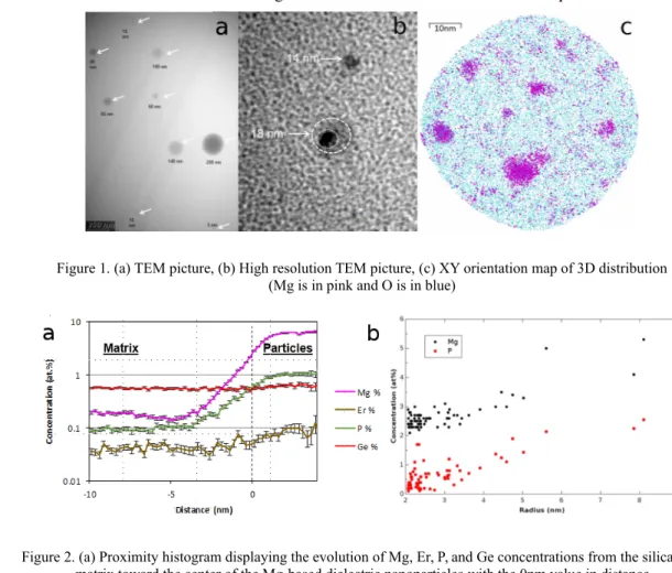
Nanoparticles observed by TEM are shown in Figure 1a. The particles size spans few nm to 200 nm. They are spherical in shape despite the drawing stage involved in the fabrication process. top sliced view of a reconstructed 3D volume obtained by APT reveals a high density of very small DNPs (Figure 1c) not observed in a high resolution TEM image (figure 1b). . As we aim at quantifying best the chemical content of the DNPs, which could not be achieved by any other analytical technique, a mathematical approach called proximity histogram or proxigram [4] was used to interrogate shells of equal thicknesses in the directions normal to an iso-concentration surface. The proxigram displayed in Figure 2a highlights the enrichment in Mg and P in the order of 100-fold and 10-fold respectively, between the matrix and the particle. We have observed a clear uptake trend in Er, while the Ge content remained steady from the matrix toward the center of the DNPs. Thanks to the high sensitivity of APT, we report on the direct measurement of Mg, P and Ge contents in nanoparticles for the entire [1-10nm] range of radii. The variation of the concentration for Mg and P with the size of the nanoparticles is shown in Figure 2b. Concentration for both elements follows the same trend: the concentration is constant up to a radius of 3.5 nm, then increases forparticles of radii larger than 3.5nm. The set of data shown in this study
specifically evidences the existence of a first plateau followed by an increase of the composition of the clusters. These experimental data disprove the capillary approximation of the Classical Nucleation Theory. However, such a plateau is expected in the framework of the Generalized Gibbs Approach formulated by Schmelzer et al. [5]. In order to gather the short-range order (SRO) of the Er atoms, we used the so-called partial radial distribution function (RDF) which consists inprobing the average local environment of each Er atom. s We observed that the close environment of Er is enriched with Mg and P elements when the size of the nanoparticle increases.
Figure 1. (a) TEM picture, (b) High resolution TEM picture, (c) XY orientation map of 3D distribution (Mg is in pink and O is in blue)
Figure 2. (a) Proximity histogram displaying the evolution of Mg, Er, P, and Ge concentrations from the silica matrix toward the center of the Mg-based dielectric nanoparticles with the 0nm value in distance corresponding to the interface between matrix and particles for an iso-concentration surface set at 3at%
for Mg, (b) Concentration of Mg and P vs DNPs size measured by Atom Probe Tomography in silicate glass
3. CONCLUSION
Thanks to the high sensitivity and resolution of APT, we probed the composition of amorphous sub-10-nm nanoparticles formed via heat treatments in silicate glass. We demonstrate the partition of Mg, P and Er ions in the nanoparticles and describe their concentration trends. We also provide the first set of data which describe Mg and P concentrations increase and erbium environment evolves with the size of the DNPs. An increase in the DNPs sizes corresponds to an intake of Er with Mg and P. This result indicates that the design of the DNPs-doped material does not only consider the size in terms of light scattering but also on their potential environment for luminescent ions.
REFERENCES
[1] Goncalves, M.C., et al., Comptes Rendus Chimie, 5, 845-854 (2002)
[2] Blanc, W., et al., “Formation et applications des nanoparticules dans les fibres optiques à base de silice”, in “Du verre au cristal. Nucléation, croissance et démixtion, de la recherche aux applications”, Ed. D. R. Neuville, L. Cormier, D. Caurant, L. Montagne, EDP Sciences (2013) [3] MacChesney, J. B., et al., Proc. IEEE, 62, 1280–1281 (1974)
[4] Hellman, O.C., et al., Microscopy and Microanalysis 6, 437-444 (2000) [5] Schmelzer, W. P., et al. , Journal of Chemical Physics, 112, 3820-3831 (2000)
