HAL Id: tel-01321321
https://tel.archives-ouvertes.fr/tel-01321321
Submitted on 25 May 2016
HAL is a multi-disciplinary open access archive for the deposit and dissemination of sci-entific research documents, whether they are pub-lished or not. The documents may come from teaching and research institutions in France or abroad, or from public or private research centers.
L’archive ouverte pluridisciplinaire HAL, est destinée au dépôt et à la diffusion de documents scientifiques de niveau recherche, publiés ou non, émanant des établissements d’enseignement et de recherche français ou étrangers, des laboratoires publics ou privés.
Cascade profiling of the ubiquitin-proteasome system in
cancer
Anastasiia Rulina
To cite this version:
Anastasiia Rulina. Cascade profiling of the ubiquitin-proteasome system in cancer. Agricultural sciences. Université Grenoble Alpes, 2015. English. �NNT : 2015GREAV028�. �tel-01321321�
THÈSE
Pour obtenir le grade de
DOCTEUR DE L’UNIVERSITÉ GRENOBLE ALPES
Spécialité : Biodiversite du Developpement Oncogenese
Arrêté ministériel : 7 août 2006
Présentée par
Anastasiia Rulina
Thèse dirigée par Maxim Balakirev
préparée au sein du Laboratoire BIOMICS
dans l'École Doctorale Chimie et Sciences du Vivant
Profilage en cascade du
système
ubiquitine-protéasome dans le cancer
Thèse soutenue publiquement le «17/12/2015», devant le jury composé de :
M. Damien ARNOULT Docteur CR1, CNRS, Rapporteur M. Matthias NEES
Professor Adjunct, University of Turku, Rapporteur M. Philippe SOUBEYRAN
Docteur, INSERM, Membre
Mme. Jadwiga CHROBOCZEK Directeur de recherche DR1, CNRS, Membre M. Xavier GIDROL
Directeur de laboratoire, CEA, Président du jury M. Maxim BALAKIREV
3 Я посвящаю эту научную работу моей маме, Рулиной Людмиле Михайловне, с любовью и благодарностью. Мне всегда везло с людьми, которые давали мне вдохновение и любовь для занятий биологией, но именно ты заложила во мне те качества, которые помогли мне пройти весь путь от начала и до конца. Именно твоя поддержка, в любых моих начинаниях давали мне ощущение «твердой земли под ногами».
5
ACKNOWLEDGEMENTS
First and foremost, I would like to thank my supervisor, Maxim Balakirev for guiding me throughout the thesis. Your expertise and endless enthusiasm helped me to work on this complicated subject. During these 3 years you were always there to discuss and share experience, and I hope I have learned the most I could from you.
I would like to express gratitude to all the members of the laboratory, who helped me with the technical part of my thesis. Thank you to Patricia Obeid for introducing me to siRNA screens, as well as for data acquisition and analysis. It was a great pleasure and luck for me to work with such a hard-working, experienced and enthusiastic person. Laurent Guyon, thank you for your job! Your deep and comprehensive data analysis was just priceless for this dissertation. I would also liketo thank Frederic Mittler for cell culture, recent Western Blots and IF; Sophie Gerbaud for performing EdU tests and recent IF images; Frederic Kermarreck for qPCRs; Eric Sulpice for helping with FACS analysis. Also I want to show my appreciation to Amandine Pitaval for explaining immunofluorescence microscopy to me and to Vincent Haguet for performing Wound Healing Assay. I am grateful to the members of my “Comité de suivi de thèse” - Jadwiga Chroboczek and François Berger for their time and useful advice in the course of the project.
I would like to say “merci” to Nicole Assard. It was a pleasure to share working space with you! I appreciate so much your friendliness and positive attitude, as well as accuracy and professionalism. Thanks to the Eric Sulpice and Stephanie Combe’s team for moral support and useful advice about science and personal life. I am also grateful to our wonderful secretary, Patricia Leclyese for helping with the millions of administrative questions I had.
I would like to thank my mother – you have always been a constant source of support. Thanks to all my friends in Grenoble for all the good days we spent together – without you it would be so boring here! Special thanks to Dolega Monika, Brasouskaya Daria, Fefelova Anastasiia, Ilaria Vitulano and Komarynets Olga for the best support during these times. Thank you, Arnaud Lemelle for all the loving, patience and care you give me.
7
MOTIVATION AND CONTEXT
According to the American Cancer Society, the prostate cancer (PCa) is the most frequent malignancy in men. After lung cancer, the PCa is the second leading oncological cause of death. Despite many years of research, the main current treatments are still surgery, radiation and androgen deprivation therapy. The androgen ablation ultimately leads to the development of a more advanced, hormone-refractory (castration-resistant) form of prostate cancer, which responds poorly to standard chemotherapy and is considered to be terminal. It is evident that there is an urgent need for the discovery of alternative targets and the development of new therapeutic approaches for prostate cancer treatment.
The design of new therapeutic agents against prostate cancer depends critically on our knowledge of the molecular mechanisms of cancer origin and progression. In 2007 the term “non-oncogene addiction” was proposed by Elledge and co-authors to explain the increased dependency of cancer cells on the function of normal genes. The phenomenon is based on increased cellular stresses experienced by cancer cells (mitotic, proteotoxic, metabolic, etc.), making them more dependent on stress support systems. Among these, the ubiquitin-proteasome system (UPS), as a major mediator of key cellular functions, represents a perfect model for a loss-of-function screen to search for potential drug targets based on non-oncogene addiction.
This work was carried out in the Biomics laboratory, which is specialized in functional genome-wide screens and biomarker discovery targeting prostate cancer. In the course of the project, we applied a novel systematic approach to the loss-of-function screen of the UPS. Our strategy was to employ the cascade organization of the UPS and its hierarchical mode of function. Compared to standard genome-wide screens, this "cascade profiling" results in a rather compact and more targeted screen, which facilitates hits identification. Using this approach we have identified components of UPS potentially important for prostate cancer cell viability.
9
LIST OF ABBREVIATIONS
2D/3D – two dimensional/three dimensional 7-AAD – 7-Aminoactinomycin D
a.a. – amino acid
ADAMs – A-disintegrin-and-metalloproteinases AIDS – acquired immune deficiency syndrome AJs – adherens junctions
AKT – v-Akt murine thymoma viral oncogene homolog 1 AMACR - alpha-methylacyl-coa racemase
AR – androgen receptor
ATCC - American Type Culture Collection ATG12 – autophagy 12
ATG8 – autophagy 8
ATM – ataxia telangiectasia mutated bp – base pair
BPH – benign prostatic hyperplasia BRCA – breast cancer
BSA – bovine serum albumin
CAND1 – cullin-associated and neddylation-dissociated 1 Caspase – cysteine-aspartic proteases
CHD1 – chromodomain helicase DNA binding protein 1
ChSM – DMEM supplemented with the charcoal/dextran stripped FBS c-Myc – v-Myc avian myelocytomatosis viral oncogene homolog COP1 – constitutive photomorphogenesis protein 1 homolog CR – castration resistant CRL – Cullin-RING-Ligase CUL1-7 – cullins 1-7 DAPI – 4'6-diamidino-2-phenylindole DDR – DNA-damage response DHT - 5α-dihydrotestosterone
DMEM – Dulbecco’s Modified Eagle's Medium DMSO – dimethyl sulfoxide
DNA – deoxyribonucleic acid DSB – double stranded breaks dsRNA – double-stranded RNA DUB – deubiquitylating enzyme E1 – ubiquitin-activating enzymes E2 – ubiquitin-conjugating enzymes E3 – ubiquitin-protein ligases EdU – 5'-ethyl-2'-deoxyuridine
EGFR – epidermal growth factor receptor EMT – epithelial-to-mesenchymal transition ERG – ETS-related gene
ETS – v-Ets avian erythroblastosis virus E26 oncogene homolog 1 ETV1-5 - Ets variants 1-5
FACS – fluorescent activated cell sorting FAK – focal adhesion kinase
FAs – focal adhesions
FAT10 – HLA-F adjacent transcript 10
FAU – Finkel-Biskis-Reilly murine sarcoma virus (FBR-MuSV) ubiquitously expressed FBS – fetal bovine serum
GS – Gleason Score
gSD – global Standard Deviation
HIF1α – hypoxia-inducible factor-1 alpha IRS-1 - insulin receptor substrate 1
ISG15 – interferon-stimulated gene product of 15 kDa IκB – inhibitor of NF-κB
LAP – leukemia associated protein Lys – lysine
8 miRNA – micro RNA
mRNA – messenger RNA
MSMB – microseminoprotein, Beta- MW – molecular weight
NAE – NEDD8 activating enzyme E1 subunit 1
NEDD8 – neural precursor cell expressed, developmentally down-regulated 8 NF-κB – nuclear factor kappa B
NKX3-1 – NK3 Homeobox 1 nt – nucleotide
Opti-MEM – reduced serum modification of eagle's minimum essential media p21 – cyclin-dependent kinase inhibitor 1A
p27 – cyclin-dependent kinase inhibitor P27 p53 – tumor protein p53
PBS – phosphate buffered saline PBS – phosphate-buffered saline PCa – prostate cancer
PCA3 – prostate cancer antigen 3
PDGFRβ – platelet-derived growth factor receptor, beta polypeptide PFA – paraformaldehyde
PHD – plant homeo domain PI3K – phosphoinositide 3-kinase PIN – prostatic intraepithelial neoplasia PSA – prostate specific antigen
PSMA – prostate-specific membrane antigen PTEN – phosphatase and tensin homolog Rb – retinoblastoma 1
RBR – ring between ring
RBX1/2 - RING-box 1/2, E3 Ubiquitin Protein Ligase RdRP – RNA-dependent RNA-polymerase
RING – Really Interesting New Gene RISC – RNA-induced silencing complex RLC – RISC loading complex
RNA – ribonucleic acid RNAi – RNA interference siRNA – small interfering RNA SPOP – speckle-type POZ protein
StdM – DMEM supplemented with standard FBS SUMO – small ubiquitin-like modifier
TGFβ – transforming growth factor beta TICs – tumor-initiating cells
TJs – tight junctions
TMPRSS2 – transmembrane protease, serine 2
TMPRSS2:ERG – genetic fusion of TMPRSS2 and ERG genes Ub – Ubiquitin
UFM1 – ubiquitin-fold modifier 1 ULM – Ubiquitin-like modifiers UPS – Ubiquitin-proteasome system URM1 – for ubiquitin-related modifier 1 UTR – untranslated region
WB – Western Blot
Wnt – wingless-type MMTV integration site family
9
TABLE OF CONTENT
ACKNOWLEDGEMENTS ... 5
MOTIVATION AND CONTEXT ... 7
LIST OF ABBREVIATIONS ... 9
TABLE OF CONTENT ... 9
LITERATURE REVIEW ... 11
CHAPTER 1. PROSTATE CANCER ... 11
1.1 STRUCTURE AND FUNCTION OF PROSTATE ... 11
1.2 EPIDEMIOLOGY OF PROSTATE CANCER ... 13
1.3 SIGNIFICANT PROGNOSTIC FACTORS ... 16
2.3.1 Gleason score ... 16
2.3.2 Prostate specific antigen ... 16
2.3.2 Other potential biomarkers ... 17
1.4 REARRANGMENTS OF ETS (E26 TRANSFORMATION SPECIFIC) TRANSCRIPTION FACTORS ... 18
CHAPTER 2. UBIQUITIN-PROTEASOME SYSTEM (UPS) ... 22
2.1 UBIQUITIN AND UBIQUITIN-LIKE MODIFIERS ... 22
2.2 UPS MACHINERY ... 25
2.3 PROSTATE CANCER & UPS ... 27
2.4 NEDD8-PATHWAY ... 30
3.4.1 NEDD8 as an ubiquitin-like protein ... 30
3.4.2 NEDD8 targets ... 31
3.4.3 Role of CRL/NEDD8 pathway in cancer ... 33
CHAPTER 3. RNA INTERFERENCE ... 35
3.1 DISCOVERY OF RNAi ... 35 3.2 MECHANISM OF RNAi... 36 3.3 RNAi AS A TECHNOLOGY ... 41 1) Stability. ... 42 2) Delivery. ... 42 3) Off-target effects. ... 43
II. MATERIALS AND METHODS ... 45
CELL CULTURE ... 45
CHARCOAL/DEXTRAN STRIPPING OF SERUM ... 45
DOUBLE THYMIDINE BLOCK ... 46
LENS-FREE CELL IMAGING ... 46
SPHEROID FORMATION ASSAY ... 46
SPHEROID SPREADING ASSAY ... 47
TEST FOR SENESCENCE ... 47
RNA EXTRACTION/RT-PCR/PCR/qPCR ... 48
siRNA TRANSFECTION... 48
WESTERN BLOT ... 49
IMMUNOFLUORESCENCE MICROSCOPY OF CELLS ... 50
IMMUNOFLUORESCENCE MICROSCOPY OF SPHEROIDS ... 50
ANALYSIS OF CELL CYCLE ... 51
PROLIFERATION ASSAY BY ATP CONTENT ... 51
PROLIFERATION BY EdU INCORPORATION ... 51
APOPTOSIS BY ACTIVATION OF CASPASES ... 52
SCREEN QUANTIFICATION ... 53
III. RESULTS AND DISCUSSION ... 55
CHAPTER 1. UPS PROFILING IN PROSTATE CANCER ... 55
1.1 INTRODUCTION ... 55
1.2 METHODOLOGY ... 58
1.3 CANCER CELL LINES CHARACTERIZATION ... 60
1.4 PRIMARY SCREEN ... 63
1.5 SECONDARY SCREEN ... 70
1.4.1 Technical notes ... 70
10 UBE2U ... 78 UBE2H ... 82 UBE2A ... 83 CAND1 ... 85 CUL4B ... 88 RBX1 ... 89 CUL2 ... 92 1.6 DISCUSSION ... 92 1.5.1 Screening parameters ... 92
1.5.2 Hits and primary validation ... 95
CHAPTER 2. DISTINCT OUTCOMES OF CRL/NEDD8 PATHWAY INHIBITION IN CANCER CELLS ... 97
2.1 INTRODUCTION ... 97
2.2 DIFFERENT SENSITIVITY OF PROSTATE CANCER CELL LINES TO MLN4924 ... 99
2.3 MLN4924 EFFICIENTLY INHIBITS NEDD8 PATHWAY IN VCaP CELLS ... 101
2.4 DIFFERENTIAL EFFECT OF NEDD8 PATHWAY INHIBITION ON CELL CYCLE PROGRESSION AND VIABILITY ... 103
2.5 KNOCKDOWN OF CRL COMPONENTS CAN HAVE OPPOSITE EFFECT ON CELL PROLIFERATION AND SURVIVAL ... 106
2.6 MLN4924 INDUCES REVERSIBLE GROWTH ARREST IN 3D PROSTATOSPHERE MODEL ... 107
2.7 INHIBITION OF NAE ACTIVATES ANDROGEN RECEPTOR ... 109
2.8 OPPOSITE ROLES OF AR & ERG IN VCaP CELL RESPONSE TO NAE INHIBITION ... 111
2.9 DISCUSSION ... 114
2.8.1 Causes of differential phenotypic outcome. ... 114
2.8.2 Direct effectors of CRL inhibition... 115
2.8.3 General outcomes: transcriptional reprogramming ... 116
2.8.4 Cancer cell plasticity: implication for cancer treatment ... 118
CHAPTER 3. INHIBITION OF CRL/NEDD8 PATHWAY BY MLN4924 CHANGES VCaP MORPHOLOGY ... 120
3.1 INTRODUCTION AND PRELIMINARY OBSERVATIONS ... 120
3.2 MORPHOLOGY CHANGE CORRELATES WITH INCREASED ADHESION OF VCaP CELLS ... 123
3.3 STIMULATION OF SPHEROID SPREADING ... 124
3.4 MLN4924 DOES NOT INDUCE EMT ... 132
3.5 EFFECT ON CELL JUNCTION PROTEINS ... 133
3.5.1 Tight junction ... 134 3.5.2 Adherens junction... 136 3.5.3 Focal adhesion ... 137 3.6 DISCUSSION ... 140 IV. CONCLUSIONS ... 142 V. SUPPLEMENTARY DATA ... 145 VI. REFERENCES ... 171
11
LITERATURE REVIEW
CHAPTER 1. PROSTATE CANCER
While in developing countries respiratory infections and AIDS are the most mortal diseases, in developed countries, medicine and technology allow people to overcome these health problems and live longer, which make heart disease and cancer the leading causes of death (http://www.who.int/). Prostate cancer (PCa) is the most often diagnosed malignancy (1 in 6 men is diagnosed with PCa during their lifetime) and the second leading cause of death from cancers (Jemal et al., 2011; Siegel et al., 2012; Stewart & Wild, 2014). Despite high incidence of PCa, the prognosis is usually positive – only 1 of 100 men will have progression to an aggressive form during the 5 years following diagnosis, and the best treatment option in this case – is watchful waiting. Nevertheless, in some patients PCa progresses to terminal disease with extremely fast metastasis that requires intensive therapy. Apart from that, current treatment options (radio-, chemo- and androgen-deprivation therapy) ultimately lead to the development of more advanced forms of prostate cancer - castration-resistant and metastatic PCa. Metastatic cancer is considered to be incurable and only 1/3 of patients with metastatic PCa survives the 5 years following diagnosis. Better understanding of prostate cancer biology would allow clinicians to distinguish relatively indolent forms of prostate cancer from aggressive ones. In our research we are searching for proteins involved in the viability of prostate cancer cells thus giving an insight in prostate cancer biology. In this chapter I describe: (I) epidemiology of prostate cancer, (II) major biomarkers that clinicians currently use to determine disease stage and prognosis, and (III) the prevalent mutation in prostate cancer, TMPRSS:ERG, and its influence on prostate cancer development.
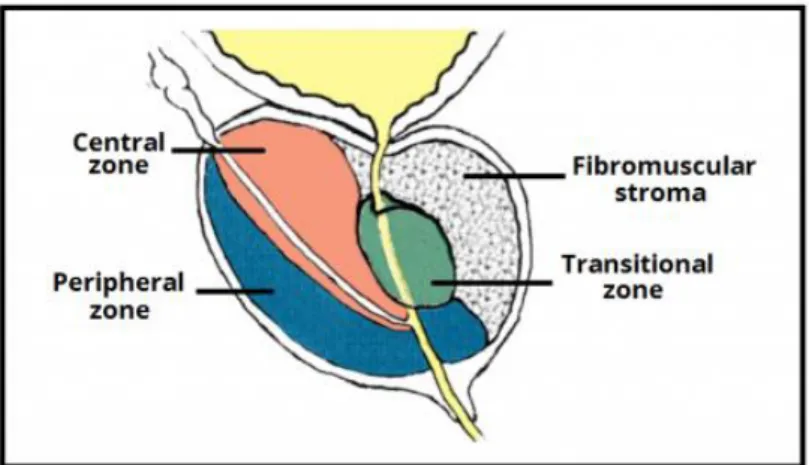
1.1 STRUCTURE AND FUNCTION OF PROSTATE
The prostate is a little (3 cm diameter, about 20 grams) auxiliary exocrine gland of the male reproductive system located in the anteroinferior part of the pelvis where it encloses the urethra and bladder neck. An adult human prostate consists of 3 concentric zones (transition/periurethral, central, peripheral), and an anterior fibro-muscular zone (Figure 1). The majority of cancers are found in the peripheral zone of the prostate, the transition zone in the second place, and almost none in the central zone (De Marzo et al.,
12 2007). In contrast, benign prostatic hyperplasia (BPH), a common nonmalignant condition found in older men, arises mostly from the transition zone. The prostate is both a glandular and muscular body. About a half of the volume of the prostate is taken up by 30-50 small glands (usually thin branching tubules), forming wedge-shaped slices draining into the urethra. The second half of the gland is distributed evenly between fibro-elastic stroma and randomly orientated smooth muscle bundles that help expel semen during ejaculation and form the involuntary urethral sphincter (Shen & Abate-Shen C., 2010).
At the histological level, the glandular epithelium of the prostate is mainly pseudostratified, comprising tall columnar luminal cells, basal and neuroendocrine cells. Tall columnar cells express high levels of androgen receptor (AR), and are responsible for the secretory functions of the prostate. They are controlled by the endocrine system and respond by the production of unique complex of secretions involved in the fertilization of the egg. Basal cells, comprising about 10% of prostatic epithelium, are almost devoid of secretory products. They lie on the basal membrane and are wedged between columnar cells. It is believed that these cells function as stem cells and are ancestors of luminal cells (Merk et al., 1982; Shen & Abate-Shen, 2010). Finally, neuroendocrine cells are singular cells producing growth-support secretions (Davis, 1987; Abrahamsson & Lilja, 1989).
There is a large variety of types of sensory nerve receptors in the prostate: Vater-Pacini corpuscles (lamellar bodies), Krause’s end bulb, etc. An extensive network of nerve ganglia and nodes around the prostate is so large that relatively small pathological changes often lead to severe disorder. Nerve fibers are connected with other nerves of the pelvic organs, primarily with the bladder, seminal vesicles, rectum, vas deferens, and corpus cavernosum. The close interweaving of the pelvic nerves may facilitate the transmission of stimuli (e.g., inflammation) to other pelvic organs. Because of this effect, various disorders of pelvic organs often induce uniform symptoms (Molochkov & Ilyin, 1998).
13
Figure 1. Structure of the prostate. http://teachmeanatomy.info/pelvis/the-male-reproductive-system/prostate-gland/
1.2 EPIDEMIOLOGY OF PROSTATE CANCER
The main risk factors for the development of prostate cancer are: aging, family history, race (for African-Americans this type of cancer is diagnosed more frequently and more often leads to death), hormonal changes, genetic mutations and lifestyle (Reiter & de Kernion, 2002). The prevalent form (95%) of prostate cancer is adenocarcinoma originating from columnal cells in the acini and glandular part of the ducts. Other categories of prostate cancer – such as ductal adenocarcinoma, mucinous carcinoma, and signet ring carcinoma – are extremely rare.
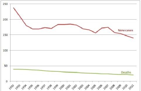
As it was mentioned before, PCa is one of the most often diagnosed malignancies in men. Since the introduction of prostate specific antigen (PSA) screening, the rates of prostate cancer mortality and incidence began to stabilize and then decline since 1990s. PCa incidence decreases 2.4% per year, and the number of PCa-related deaths declines by 3.4% per year (Figure 2). Despite high incidence of this type of cancer, the prognosis is usually positive: PCa is usually confined within the gland and progresses slowly. The percentage of patients surviving 5 years comprises 98.9% (Edwards et al., 2014). Nonetheless, long-term survival is not as good, because the main treatments often lead to the development of highly aggressive metastatic form of PCa (Elledge, 2010).
The clinical course of localized untreated prostate cancer is still unclear. Progression of the disease to the metastatic form within 10-15 years following diagnosis is not frequent (5% of all PCa cases according to American Cancer Society), but the further follow-up has shown that even primarily indolent localized prostate cancer can proceed to a more aggressive form, invading surrounding tissues and giving metastases in the lymph nodes and the bones as the major sites (McNeal JE, 1992). To prevent
14 overtreatment, it is necessary to understand the prolonged natural history of the disease (McNeal, 1969; McNeal, 1992; Johansson et al., 2004; Shen MM & Abate-Shen C., 2010).
Figure 2. Number of new cases and deaths from prostate cancer per 100,000. Figure from Edwards et al., 2014
Therapeutic decisions based on correct diagnosis and accurate staging of prostate cancer is critically important for the fate of the patient. Early diagnosis of prostate cancer includes three main options: (1) measurement of PSA in blood, (2) digital rectal examination and (3) ultrasound-guided transrectal biopsy of the prostate with further immunostaining for specific markers (anti-p63, cytokeratin 5 and 14). The most important prognosis factors are the initial (pre-therapeutic) level of total serum PSA and the Gleason score (G2-G10, discussed below in more details). Patients are also diagnosed by the status of their primary tumors, from organ-confined to fully invasive (T1–T4), with or without lymph node involvement (N0 or N1), and the presence and degree of distant metastases (M0 and M1a–c). Prostate cancer is often preceded by a stage of pre-cancer, and timely identification of this condition significantly helps to determine prognosis and treatment. To date, Prostatic Intraepithelial Neoplasia (PIN) remains as the only well-proven preneoplastic condition with clinical significance (Armah & Parwani1, 2008; Shen & Abate-Shen, 2010).
For early diagnosed organ-confined prostate cancer current treatment options include watchful waiting, surgery (radical prostatectomy) and radiotherapy - external or brachytherapy (implantation of radioactive “seeds”). This type of PCa is curable with very good survival and cure rates, but the disease relapses in approximately 25% of patients. Nevertheless, choice of treatment option is very questionable – localized cancer
15 rarely progresses to a fatal form and some patients receive overtreatment while others die during prostatectomy. For more malignant forms, combined therapy regimens (brachytherapy, hormone therapy and chemotherapy) are applied specifically according to the disease’s stage, patient’s age and personal choice. In the case of advanced cancer, these regimens are usually followed or substituted by androgen deprivation therapy (chemical or surgical castration), which initially will reduce tumor burden, but inevitably leads to the development of a more destructive form of prostate cancer - androgen-independent (or castration-resistant, CR-PCa) prostate cancer (Bardan et al., 2007; Shen & Abate-Shen, 2010). This recurrent disease has a median survival rate of less than 2 years. For CR prostate cancer, the only approved therapy is with docetaxel and provides a modest survival benefit of 2 to 3 months.
To explain the emergence of CR-cancer, Isaacs and Coffey proposed that androgen withdrawal results in the natural selection of the androgen-refractory cells initially present in heterogeneous prostate tumor (Isaacs & Coffey, 1981). Indeed, several observations suggest that prostate tumor-initiating cells (TICs) do not express androgen receptor (AR) (Gu et al., 2007; Kasper, 2009). In addition, prostate neuroendocrine cells, which have been implicated in CR-PCa, are androgen-independent (Cindolo et al., 2007). Despite this evidence, however, it is widely established that in the majority of CR tumors, AR remains the key driver of cancer progression. Notably, castration-resistant tumors express AR as well as AR target genes such as PSA, indicating that pathway activity is intact (Gregory et al., 1998). Androgen ablation has been shown to select for TIC clones with aberrant, androgen-independent AR signaling (Wang & Shen, 2011). This arises through a variety of mechanisms, including mutations that change AR function, inactivation of tumor suppressors, activation of oncogenes, increase in autocrine stimulation, and rearrangement of cell signaling pathways (Knudsen & Penning, 2010; Feldman & Feldman, 2001). As a result, in CR malignant cells, AR executes a transcriptional program distinct from that in androgen-responsive cells resulting in increased cell survival and androgen-independent growth (Wang et al., 2009).
It should be noted that in more than 50% of metastatic prostate tumors AR is not mutated (Heinlein & Chang, 2004; Knudsen & Penning, 2010). This suggests that the aberrant function of AR in androgen-refractory cells results mainly from the changed cellular context, i.e. a variety of cellular factors, which define and complement AR activity in CR prostate cancer. Comparative genomic analysis of prostate cancer, CR tumors and normal prostate cells has revealed numerous genetic alterations and abnormal
16 gene expression profiles in cancer cells (Varambally et al, 2005; Taylor et al, 2010; Berger et al, 2011). However, though very valuable for cancer classification and prognosis, genomic data tell less about molecular mechanisms of prostate cancer progression. A complete understanding of cancer biology requires knowledge of principal actors regulating protein interactions and activities.
1.3 SIGNIFICANT PROGNOSTIC FACTORS 2.3.1 Gleason score
To assess the prognosis of patients, clinicians use Gleason Score based on the architecture of the prostate glands and the relationship between the tumor cells and the surrounding stromal tissue. The Gleason grading system has five levels of tumor progression, grade 1 being the least aggressive, while grade 5 is the most anaplastic. Usually, prostate tumors are not homogenous. Gleason Score is a sum of Gleason grades of the two most typical tumor samples. Thus, it can range from 2 (1 +1) to 10 (5 +5) (Bardan et al., 2007).
In general, the higher the Gleason Score, the more "malignant" the tumor is. However, this rule should be used wisely. Generally, the patients with GS ≥ 7 means a greater risk for the patient and that it should be treated intensively. Nevertheless, some studies using surrogate end points have shown that the prognosis of GS 7 cancers varies considerably (Stark et al., 2009). On the other hand, tumors with GS below 4 are considered to be indolent and almost never progress to the advanced stage, thus the best treatment option in this case – local therapy and watchful waiting. However, after universal introduction of routine PSA diagnostics, it became clear that small fraction of these “indolent” tumors could progress rapidly and require immediate treatment. Consequently, the major clinical challenge is the current inability to readily distinguish between indolent and aggressive tumors in prostate cancer patients with a low Gleason Score (Shen & Abate-Shen, 2010).
2.3.2 Prostate specific antigen
PSA is an organ-specific marker produced by a healthy prostate, and released into the blood only in the case of impairment of normal prostate architecture (Lilja et al., 2008). Thus, while the blood PSA is not a sign of a certain disease or condition, its increased level can indicate the presence of a destructive disease, such as adenoma or
17 tumor. This helps to diagnose and monitor PCa progression. PSA in blood serum exists in three forms: free, associated with either α-1-antichymotrypsin or with α-2-macroglobulin. Two forms of PSA are routinely used in diagnostics: free and α-1-antichymotrypsin-binded, which add up to a "total PSA". In healthy conditions, PSA is present in serum at a very low level, which is age-dependent:
40-49 years - 2.5 ng/ml 50-59 years - 3.5 ng/ml 60-69 years - 4.5 ng/ml Over 70 years - 6.5 ng/ml
Nevertheless, it has been shown that some PCa cases can exist at normal level of PSA (Thompson et al., 2004). Men with increased PSA level (10 ng/ml) are recommended for prostate biopsy to verify the presence of cancer. To make a decision about biopsy in these cases, when serum PSA level is below 10 ng/ml, the ratio of free PSA to total PSA becomes crucial: prostate cancer was shown to be associated with the formation of protein-bound PSA. “PSA velocity” (the rate of PSA increase) and PSA density (the ratio of PSA to prostate volume) are also important prognostic factors (Carter, 2006).
2.3.2 Other potential biomarkers
Routine PSA screening provides a 20% mortality reduction, because it helps in early detection of clinically unapparent, localized tumors with a low GS. However, the clinical course of localized untreated prostate cancer is still unclear. Progression to the metastatic form in 10-15 years following diagnosis is not frequent, but a further follow-up has shown that even primarily indolent localized prostate cancer can proceed to a more aggressive form. To prevent overtreatment, it is necessary to understand the prolonged natural history of the disease. In order to improve specificity and sensitivity of detection, many efforts are being made to identify novel biomarkers. Some of them are already at early stages of development and are being evaluated in clinical trials (Duskova & Vesely, 2014). Examples of such biomarkers are PCA3, PSMA, AMACR, MSMB, etc. One of the promising urine biomarkers is TMPRSS2:ERG fusion transcripts. In contrast to PSA, which often gives false-negative and false-positive results, TMPRSS2:ERG appears only in malignant conditions, which greatly increases the specificity of the test. Some other researchers show the efficiency of a multiplexed approach, where the set of markers is followed simultaneously that make detection more sensitive and specific. These
18 prominent results could be adjusted for clinical use and would increase the efficiency of routing testing for prostate cancer (Rubin, 2012).
1.4 REARRANGMENTS OF ETS (E26 TRANSFORMATION SPECIFIC) TRANSCRIPTION FACTORS
Localized prostate cancer usually contains histologically and genetically distinct areas and thus is regarded as multifocal malignancy. In contrast, despite phenotypical differences of metastases in diverse sites, genomic analysis demonstrates their clonal origin. During the last few decades, there has been an accumulation of data on molecular alterations in PCa, shedding light on the mechanisms of prostate cancer initiation and progression. The frequent characteristic mutations have been identified, which include: (1) ETS-rearrangements found in more than half of prostate cancer cases (discussed in details below); (2) mutations of SPOP component of cullin-RING E3 Ub-ligase complex, which are present in up to 15 % of PCa cases; (3) deletion of CHD1 gene (substrate recognition component of the transcription regulatory histone acetylation complex SAGA) found in up to 15 % of PCa cases. There are also some other less frequent mutations/deletions/rearrangements involving PTEN, AR, NKX3-1, p27, p53, Rb and etc. (Tomlins et al., 2005; Shen & Abate-Shen, 2010; Yoshimoto et al, 2012; Wyatt et al., 2014; Yadav et al., 2015).
The ETS (E26 transformation-specific) family of transcription factors includes 30 proteins unified by the presence of evolutionarily-conserved ETS domain responsible for DNA binding. Also, they contain an N-terminal regulatory domain. The ETS family of proteins participates in the regulation of many key cellular processes including proliferation, differentiation and apoptosis. In prostate cancer, rearrangements that activate ERG, ETV1, ETV4 and ETV5 members of the ETS family have been identified (John et al., 2012; Kumar-Sinha et al., 2008; Tomlins et al., 2005; Tomlins et al., 2007b). The most frequently rearranged gene in prostate cancer is ERG (ETS related gene) (Tomlins et al., 2005). Under normal conditions ERG is constitutively expressed in endothelial cells where it regulates angiogenesis and endothelial apoptosis by affecting expression of many genes, including eNOS, HO-1, ICAM-2, VE-cadherin, von Willebrand's Factor, etc. (Birdsey et al., 2008; Nikolova-Krstevski et al., 2009). The major ETS translocation in PCa is TMPRSS2:ERG. TMPRSS2 is a prostate specific, androgen responsive transmembrane serine protease (Figure 3). TMPRSS2:ERG fusion leads to the ERG expression under control of androgen sensitive promoter elements of
19 TMPRSS2 (St John et al., 2012). There are multiple other 5’ fusion partners of the ETS family in PCa (Figure 3).
Interestingly, these chromosomal rearrangements may be caused by AR function. Studies in androgen-responsive LNCaP cells have shown that AR binding induces chromosomal proximity between the TMPRSS2 and ERG loci that can lead, upon DNA damage, to the formation of TMPRSS2:ERG fusions. In addition, androgen signaling recruits topoisomerase II to AR-binding sites, leading to the induction of double-stranded breaks even in the absence of genotoxic stress (Shen MM & Abate-Shen C, 2010). Thus, ETS fusions could be induced in cells, which, initially, do not harbor these translocations. Combined treatment of prostate cancer LNCaP cells or non-cancerous PNT2 cells with DHT and γ-irradiation leads to the appearance of cells with ETS rearrangements (Lin et
al., 2009; Mani et al., 2009; Bastus et al., 2010; Chiu et al., 2012). Unfortunately, the fate
of these cells has not been followed, and there is no data on whether this rearrangement gives some survival advantages to the cells.
TMPRSS2:ERG is most likely a driver mutation in prostate cancer. The first piece of evidence is the enrichment of this translocation during cancer progression. TMPRSS2:ERG fusion is found only in 20% of prostatic intraepithelial neoplasia (PIN) lesions (Tomlins et al., 2008), and in up to 50% of confined cancers (Taylor et al., 2010), suggesting that this rearrangement is often acquired after cancer initiation, or, instead, it is an early event in cancer development and predisposes cells to progression to malignant state (Tomlins et al., 2005). The second piece of evidence comes from molecular biology: the appearance of this translocation in prostatic cells results in overproduction of various N-truncated ERG isoforms (Figure 4). ERG, in its turn, causes transcriptional activation of oncogenic pathways including Wnt/β-catenin, NF-κB, c-MYC and disruption of AR-dependent signaling (Birdsey et al., 2015; Yu et al., 2010; Sun et al., 2008; Wang et al., 2011). This could lead to the development of PIN but is not sufficient to produce invasive adenocarcinoma (Klezovitch et al., Tomlins et al., 2008; Zong et al., 2009). Additional factors (e.g., AKT activation, enhanced AR signaling and PTEN loss resulting in aberrant phosphoinositide 3-kinase (PI3K) pathway) are required for the development of prostate cancer (Carver et al., 2009; Sowalsky et al., 2013; Krohn et al., 2014).
20 Figure 3. ETS rearrangements. This diagram represents all published ETS-fusions grouped by ETS members.
http://oncofusion.com/pipelines/erg-inhibitors/
Figure 4. ETS fusions in prostate cancer. Fusion places transcriptional factor ERG (or any other ETS family member, i.e. ETV1, ETV4, and ETV5) under the control of AR-responsive promoter that results in overproduction of more or less N-truncated ERG protein. ERG, in its turn, activates its own transcriptional program. Acquisition of additional mutations (e.g., loss of PTEN) leads to the development of prostate cancer.
21 The third piece of evidence comes from follow-up studies. Most studies following the natural history of the disease show strong correlation between the presence of TMPRSS2:ERG fusion and cancer progression to more aggressive forms, metastases and a reduced survival rate (Demichelis et al., 2007; Attard et al., 2008; Hägglöf et al., 2014; Berg et al., 2014). Interestingly, expression of ERG protein has been shown to correlate with markers previously associated with bad prognosis, such as PDGFRβ, hyaluronan, Caveolin-1 and von Willebrand factor (Hägglöf et al., 2014). Unfortunately, none of these studies addressed the clinical outcome of different TMPRSS2:ERG isoforms, while in in
vivo cancer models different isoforms were shown to have antagonistic behavior (Rastogi et al., 2014). However, the T1E4 isoform (fusion of 1st exon of TMPRSS2 with 4th exon of ERG) seems to be the most frequent and clinically relevant. A study, published in 2011 by Markert and colleagues, takes into account several molecular signatures, including TMPRSS2:ERG, and shows that the presence of this translocation is associated with poor prognosis, even though the most aggressive phenotype is defined as stem cell-like with P53−/PTEN− genotype (Markert et al., 2011).
High incidence of these gene fusions in prostate cancer and their cancer-driving behavior makes them attractive drug targets. However, direct inhibition of ERG protein does not give expected results: inhibition of ERG expression in VCaP cells diminishes invasiveness, but has no effect on the viability of the cells (Tomlins et al., 2008). Another study on VCaPs has shown a slight decrease of cell proliferation under ERG-deprived conditions, but only where a high concentration of siRNA was used (starting from 50 nM), despite that the decrease of ERG protein level starts from 2.5 nM concentration (Urbinati et al., 2015). Thus, this effect on viability could be due to some off-target effects. Hence, targeted therapy might benefit from using pathways altered in ERG-expression tumors (Chatterjee et al., 2015; Mancarella et al., 2015) or identification of ERG-specific non-oncogene addiction.
22
CHAPTER 2. UBIQUITIN-PROTEASOME SYSTEM (UPS)
In this thesis we describe siRNA screening of the ubiquitin-proteasome system (UPS) and ubiquitin-like modifiers (ULMs) pathways to identify individual components required for the functioning of PCa cells. We chose to investigate UPS and ULMs pathways because of the previous success of proteasome inhibitors in the treatment of tumors. It should be noted, that despite the documented anti-cancer effect, proteasome inhibition is generally toxic and, thus, may be detrimental for healthy cells. We wanted to benefit from targeting individual components of UPS or ULM, which could make therapy more selective toward tumor cells. Our screens and subsequent validation experiments using a small molecule inhibitor of neddylation revealed that the inhibition of the components of the CRL/NEDD8 pathway can have a complex outcome on the viability of PCa cells. In this chapter I describe the organization and function of the UPS, and of the CRL/NEDD8 pathway as a part of the UPS.
2.1 UBIQUITIN AND UBIQUITIN-LIKE MODIFIERS
It has been estimated that bacteria with an average genome size of few thousands genes produces about 250,000 individual proteins packed so tight that the space between them does not exceed a few molecules of water. Most likely, the eukaryotic cell has more or less the same level of compaction (Petsko & Ringe, 2004). The cell interior is extremely crowded, but also very mobile: the proteins are separated into different compartments and organelles with permanent exchange between them. Complexity is increased by a flow of newly synthesized proteins and their constant degradation. In such a crowded and mobile environment, precise regulation of protein function is essential to avoid chaos. Protein function in vivo can be regulated at transcriptional level via control of gene expression. Other levels of control include regulation of translation, post-translational modification, localization of the protein, covalent or noncovalent binding of effector molecules, and the lifetime of the active protein (Petsko & Ringe, 2004; Cooper, 2000).
Although it was known that, despite the exergonic nature of peptide-bond cleavage protein, degradation is energy-dependent and proteins have different life-time, for many decades protein degradation was believed to be non-specific and thus was never
23 really studied (Ravid & Hochstrasser, 2008). At that time lysosome-mediated protein degradation was the only pathway known, in accordance with this paradigm. The discovery of the ubiquitin-proteasome system made a revolution and was awarded by the Nobel Prize in Chemistry in 2004. Specific labeling of intracellular proteins, by small protein ubiquitin (Ub), which targets them for degradation by a multienzymatic complex called the proteasome, was identified as the main function of this system. Later it was shown that ubiquitination also has regulatory functions and does not necessarily lead to the degradation of proteins (Glickman & Ciechanover, 2002; Herrmann et al., 2007). Ubiquitin, a highly conserved, 76-amino acid protein, is ubiquitously present in eukaryotes, but absent in the Eubacteria and the Archaea. Characterization of ubiquitin was followed by the discovery that it is a member of a group of protein tags, which share similar structure and mechanism of attachment. Ubiquitin and Ubiquitin-Like Modifiers (ULMs) have a similar three-dimensional core structure, the β-grasp fold, but otherwise are distinct (Figure 5). There are 17 known ubiquitin-like proteins (UBLs) belonging to nine phylogenetically distinct classes (Table 1): NEDD8 (RUB for bacteria and plants), SUMO (for small ubiquitin-like modifier), ATG8 (for autophagy 8) and ATG12, Ufm1 (for ubiquitin-fold modifier1), URM1 (for ubiquitin-related modifier 1), ISG15 (interferon-stimulated gene product of 15 kDa), FAT10 (HLA-F adjacent transcript 10), FAU and a diverse assortment of proteins which harbor structurally related folds fused translationally to other domains (Watson et al., 2011; Hochstrasser, 2009; Vierstra, 2012).
Figure 5. Characteristic ubiquitin-fold. a. Ribbon representation of 3D fold of Ub and ULMs. b. Superposition of their 3D-folds. Figure from Sorokin at al., 2009
24
Table 1. Known ULMs (modified figure from Hochstrasser, 2009). E1 and E2 - enzymes, involved in modification of proteins with a given modifier: E1 – Ub-activating enzyme, E2 – Ub-conjugase (discussed in details below in 2.2 UPS MACHINERY)
Modifier Identity with Ub (%) E1 E2 Roles
Ub 100 UBA1 & UBA6 >30 Multiple, including protein homeostasis, cell receptor signaling, endocytic trafficking, transcriptional regulation and cell cycle progression
NEDD8 59 Uba3/NAE1 UBE2M &
UBE2F
Activation of cullin-based E3s
SUMO 18 SAE1/UBA2 Ubc9 Multiple, including protein stability and
localization, transcriptional regulation and cell cycle progression
Atg12 5 Atg7 Atg10 ATG5–ATG12 conjugate forms complex
with ATG16 that functions as an E3 ligase for autophagic vesicle formation
Atg8 8 Atg7 Atg3 Autophagic vesicle formation
Urm1 ~0 UBA4 dimer – Antioxidant pathways; tRNA uracil
thiolation
ISG15 32/37 UBA7 UBE2H Antiviral functions; possibly cell growth
and differentiation
UFM1 14 UBA5 dimer UFC1 Unknown
FAU 38 – – Regulation of immune response
25
2.2 UPS MACHINERY
Ubiquitin and ULM became attached to target molecule in multistep reaction. More time will be given to the description of the machinery and organization of the ubiquitin-ligation pathway, but the principles are the same for other modifiers.
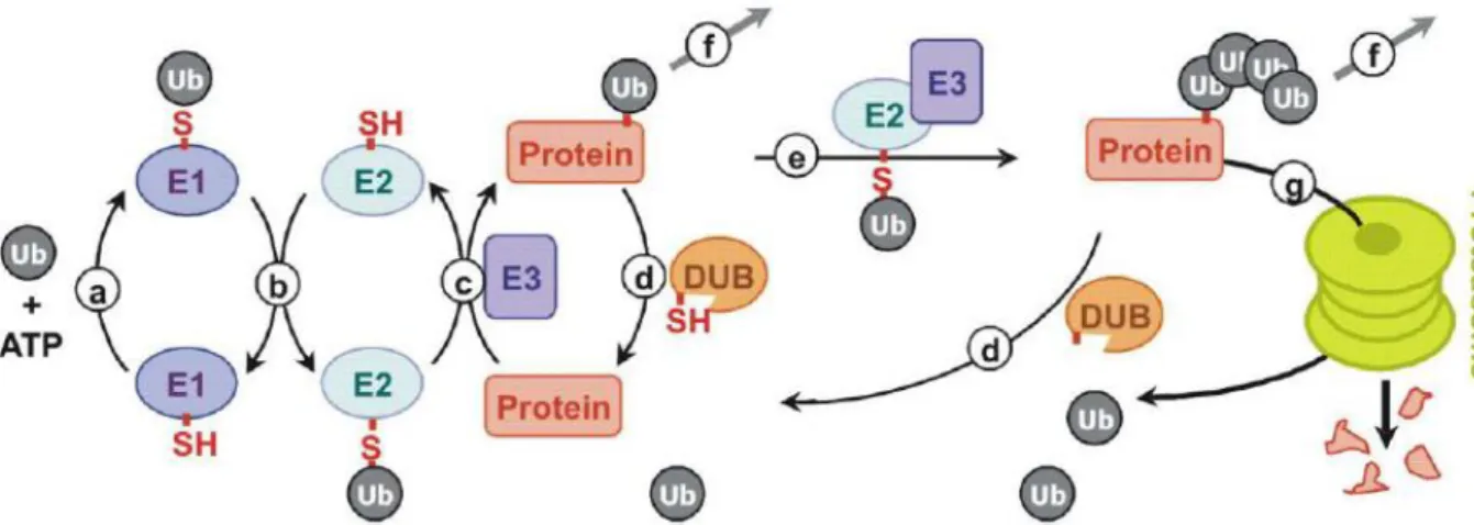
Ubiquitin is conjugated to target proteins by the formation of an iso-peptide bond between the C-terminal carboxyl group of ubiquitin and a lysine side-chain of the target protein. Ub can also be attached by peptide bond to the N-terminus of the protein. This process, termed ubiquitylation, occurs through a cascade reaction and requires three classes of enzymes: ubiquitin-activating enzymes (E1), ubiquitin-conjugating enzymes (E2), and ubiquitin-protein ligases (E3) (Figure 6). E1 activates ubiquitin by forming a high-energy thiol ester bond between an E1 active site-located cysteine and a C-terminal glycine of ubiquitin in a reaction that requires the hydrolysis of ATP. Activated ubiquitin is then transferred to a specific Cys residue of one of ~30 E2s via a thioester linkage. The E3 ubiquitin ligases (E3s) recruit ubiquitin-loaded E2s, recognize specific substrates, and facilitate (or directly catalyze) ubiquitin transfer to either the Lys residues (in most cases) or the N terminus of their molecular targets with the formation of (iso-) peptide bonds. E3s are the key determinants of substrate specificity and are capable of recognizing a few or multiple substrates through specific degradation signals. A single E2 may function with multiple E3s (and vice versa) to provide specificity in a combinatorial way. To date, >500 E3s have been identified. There are several mechanistically distinct classes of E3 enzymes: many of these E3s contain the Homologous to E6-associated protein (E6-AP) Carboxy Terminal (HECT) domain or the Really Interesting New Gene (RING) finger domain. Recently, four RING-like domains, the U-box, the Leukemia Associated Protein (LAP) finger proteins, the Plant Homeo Domain (PHD) and the Ring Between Ring fingers (RBR)-domain family, have also been shown to have E3 activity (Figure 7) (Bernassola et al., 2008; Deshaies & Joazeiro, 2009; Chen et al, 2006; Metzger et al., 2012; Eisenhaber et al., 2007).
26
Figure 6. Enzymatic cascade of UPS. (a) Activation of Ub-moiety by activating enzyme (E1) in energy-dependent reaction. (b) Transthiolesterification reaction between E1 and E2 (conjugating enzyme). (c) E3 ubiquitin ligase transfer Ub from E2~Ub thioester on target protein. (d) Ubiquitin mark can be eliminated by deubiquitylating enzymes (DUBs). Monoubiquitylated substrate can then acquire additional Ub moieties in the form of multiple single attachments (not shown) or an ubiquitin chain (e). After depending on the nature of the chain protein can change its function (common for mono- multi-ubiquitylation and also for poly-Lys63-cahains) (f) or undergo proteasomal degradation (typical for the Lys48 chains (g). Modified figure from Deshaies & Joazeiro, 2009
Figure 7. Classification of E3 ubiquitin ligases by mechanism of action. Two major types of E3 are illustrated. The PHD domain, LAP and U-box E3 have the similar mechanism as RING-ligases. S, substrate of an E3. I. For HECT-ligases, ligation involves an obligate thioester intermediate with the active-site cysteine of the E3. II. Ring-ligases mediate the direct transfer of ubiquitin from E2 to substrate. Modified figure from Chen et al., 2006
27 The fate of ubiquitylated proteins is determined by its nature (mono-, multi- or poly-ubiquitylation) and the type of isopeptide linkage of Ub (chains formed through lysine 6, 11, 27, 29, 33, 48, 63 or mixed are found in vivo and seem to target proteins to different fates). Monoubiquitylation and the formation of multiubiquitin chains by isopeptide bonds other than Lys48, such as Lys6, Lys29/33 and Lys63, perform both proteolytic as well as regulatory function. Poly-ubiquitylation through Lys48 represents a standard signal for proteasome-mediated degradation (Bernassola et al., 2008). Lys63-poly-ubiquitin chains are involved in protein/protein interaction important for kinase signaling activation, receptor endocytosis, protein trafficking, and DNA damage repair (Wertz et al., 2004). Lys6 and Lys11 polyubiquitin linkages have been identified in vivo, and their accumulation correlates with the pathogenesis in neurodegenerative disorders (Bernassola et al., 2008). In addition, recently discovered linear polyubiquitylation through Methionine (Met1) is involved in nuclear factor-κB signaling and cell death, and dysfunctions in linear ubiquitylation underlie chronic inflammation (Iwai et al., 2014).
Like many other dynamic posttranslational protein modification, ubiquitylation is a reversible process. Ub cleavage is performed by deubiquitylating proteins (DUBs). The human genome encodes for nearly 100 Ub-specific DUBs, divided in six families: the UCH, USP, OTU, MJD, MCPIP and JAMM families (Reyes-Turcu et al., 2009).
2.3 PROSTATE CANCER & UPS
The UPS conjugation pathways have multiple essential biological roles. Even if we focus our attention only on cancer-related processes of UPS, the variety of downstream effects is extraordinary (Table 2). Ubiquitin-mediated proteasomal degradation controls cell-cycle progression, protein quality control, signal transduction, and circadian rhythms. The non-proteolytic way of regulation is applied to membrane trafficking, control of genome integrity, and the assembly of signaling complexes (Hochstrasser, 2009). ULM also regulates multiple biological functions. For example, SUMO plays an important role in DNA repair and maintenance of genome stability (Bergink & Jentsch, 2004; Nagai et al., 2011). NEDD8 regulates the Ub-pathway and degradation of some individual proteins including p53. ISG15 modification is part of the cellular response to infection and inflammation (Hochstrasser, 2009). In this light it is not surprising that their function, and often malfunction, are important factors in various human pathogenesis, including numerous cancer types, cardiovascular diseases and neurodegenerative disorders.
28
Table 2. Biological roles of UPS in cancer.
In prostate cancer, E3 ubiquitin ligase adaptor SPOP (speckle-type POZ protein) is probably the most often deregulated component of UPS. It is specifically down-regulated or mutated in 10-15% of all PCa cases, but not in other types of cancer suggesting tissue-specific mechanism of action (García-Flores et al., 2014). Indeed, it was shown that the wt-SPOP protein provides ubiquitylation and degradation of androgen receptor, while mutant isoforms lack this activity (An et al., 2014). Moreover, SPOP mutation is associated with genomic instability (Boysen et al., 2015). Interestingly, mutation of SPOP and the presence of TMPRSS2:ERG translocation are mutually exclusive events (Berger et al., 2011). COP1 (constitutive photomorphogenesis protein 1 homolog) is another tumor suppressor E3-RING gene, which is frequently affected by loss-of-function mutations in cancer (Migliorini et al., 2011). This Ub-ligase controls degradation of such oncoproteins as c-Jun and ETS transcription factor Etv1. In the majority of prostate cancers ETS factors are overproduced because of TMPRSS2:ETS gene fusions (Tomlins et al., 2008). Interestingly, truncated Etv1 encoded by prostate cancer translocation TMPRSS2:ETV1 lacks the critical COP1 binding motifs and is
29 fiftyfold more stable than wild-type Etv1 (Vitari et al., 2011). As a result, almost all clinically-relevant translocations result in COP1-insensitive Etv1, implying that COP1 loss-of-function confers a selective advantage to prostate cancer cells. In contrast to COP1, the gene for E3-HECT Ub-ligase WWP1 is frequently amplified in prostate cancer. WWP1 induces degradation of several components of the TGFβ pathway as well as the Klf5 protein. The latter is one of the key tumor suppressor transcription factors, which are down-regulated in PCa (Chen et al., 2007). Mutations of some UPS genes have been associated with elevated incidence of sporadic cancer as well as with hereditary prostate cancer. For example, the risk of prostate cancer is increased two- to fourfold in men with BRCA1/2 mutations (Agalliu et al., 2009). These tumor suppressor genes code for E3-RING Ub-ligases, which control DNA repair as well as G2/M and DNA replication checkpoints. The most frequent mutations result in truncated BRCA proteins, which have lost E3-ligase activity.
The global alterations of UPS in prostate cancer can also be seen on the protein level. Immunostaining of prostate tissues reveals that upon cancer progression sub-cellular localization of Ub-conjugates is shifted from the nucleus to the cytoplasm (Bataineh & Habbal, 2006). This may result from the accumulation of Ub-rich cytoplasmic protein aggregates caused by defective protein synthesis and degradation. Indeed some UPS components implicated in protein quality control are specifically downregulated in PCa (Tomlins et al., 2007b; Lapointe et al., 2004). Other ULMs also demonstrate global changes in conjugation and intracellular localization. For example, both SUMO and ISG15 pathways are upregulated in PCa cells (Moschos et al., 2010; Kiessling et al., 2009), while the NEDD8 pathway is believed to be significantly inhibited (Meehan et al., 2002). The critical role of the UPS in prostate cancer is thus well recognized and may result from direct control of stability and function of androgen receptor and/or regulation of other cancer-related proteins such as oncoproteins and tumor suppressors. However, though very important for development of new therapeutic approaches, the mechanisms of this regulation, as well as the proteins involved, remain poorly understood.
30
2.4 NEDD8-PATHWAY 3.4.1 NEDD8 as an ubiquitin-like protein
Among ubiquitin-like modifiers NEDD8 is one of the best studied. NEDD8 is the closest to Ub in sequence and structure, but they also have a non-overlapping function. NEDD8 was mainly characterized in the context of its function in the regulation of the biggest class of E3 ubiquitin-ligases – CRLs (cullin-RING-ligases). However, new studies show that NEDD8 probably have other target proteins and functions, the most well-studied being the regulation of p53 protein (Harper et al., 2004; Enchev et al., 2014). Like in the Ub-pathway, neddylation requires a cascade of reactions performed by E1, E2 and E3 enzymes. Neddylation can be reversed by NEDD8-specific proteases. The NEDD8-conjugating cascade starts from the NEDD8-activating enzyme (NAE). NAE is a heterodimer comprising NAE1 and UBA3 subunits. Activated NEDD8 is subsequently transferred to the E2-conjugating enzyme. In metazoans, two NEDD8-specific E2 conjugating enzymes have been described: UBE2M (also known as UBC12) and UBE2F (Huang et al., 2009). Finally, E3 ligases transfer NEDD8 to one of the Lys or N-terminus of the target protein. NEDD8-E3-ligases, with one exception, belong to the RING (really interesting new gene) finger proteins.
The best characterized NEDD8-E3-ligases are RBX1 (RING-box protein 1, neddylates cullins 1-4 and 7) and RBX2 (RING-box protein 2, specific to cullin 5). Neddylation of cullins by RBX1/2 accompanied by DCUN1 proteins (defective in cullin neddylation protein 1-like proteins; there are 5 of them in human). Both RBX1 and RBX2 are part of the multi-subunit complexes Cullin-RING-E3-Ub-Ligases (CRLs), and neddylation of cullins leads to activation of CRLs. Other described NEDD8-E3-ligases from the RING family are MDM2 (which neddylates p53 and p73 and attenuates its transactivation function), c-CBL (which neddylates receptor tyrosine kinases, e.g. EGFR and TGFIIβ, and targets them to cell compartments), RNF111 (which neddylates histone H4 during DNA-damage response), and DIAP1 (which neddylates caspases and thus prevents apoptosis) (Harper, 2004; Watson et al., 2006; Oved et al., 2006; Yang et al., 2007; Broemer et al., 2010; Ma et al., 2013; Zuo et al., 2013). Recently, the HECT-domain family of E3 ligase, SMURF1, was shown to be auto-neddylated at multiple sites, which increases its activity (Enchev et al., 2014). A few deneddylases have been identified, including the CSN5, NEDP1, USP21, Ataxin-3, UCH-L1, and UCH-L3 (Watson et al., 2011). CSN5 and NEDP1 are selective to NEDD8, whereas the other
31 above mentioned NEDD8 ligases also cleave Ub. The principal cullin deneddylase is CSN5, which is active only being a part of 8-subunit COP9 signalosome complex (CSN). NEDP1 complements the activity of CSN and ensures maturation of the NEDD8 precursor (Enchev et al., 2014).
Normally, both Ub and NEDD8-pathway enzymes are highly specific (Souphron
et al., 2008), but in conditions of increased ratio of NEDD8:Ub, ubiquitylating enzyme
E1 UBA1 can activate NEDD8, and redirect it to Ub-conjugation pathway. Unfortunately, the biological role of this effect is unclear. One possibility is that formation of mixed Ub-NEDD8 chains mediates specific stress-response pathways (Leidecker et al., 2012; Hjerpe et al., 2012).
Figure 8. Architecture of Cullin-RING-ligases. http://biology.ucsd.edu/research/faculty/e1bennett 3.4.2 NEDD8 targets
The best-known function of neddylation is regulation of the activity of Cullin-RING ligases (CRLs), the biggest class of E3 ubiquitin ligases. CRLs are multisubunit complexes which consist of the cullin scaffold, a conjugating apparatus represented by E2-conjugase and RING-E3-ligases RBX1/2 and an adaptor module, which determines specificity to substrate protein (Figure 8). There are a few subclasses of CRLs, depending on the type of cullin and adaptor protein involved. In mammals, 7 cullins have been identified (1, 2, 3, 4A, 4B, 5 and 7), forming about 300 distinct CRL complexes. Two other proteins, CUL9 (also known as PARC) and APC2 (anaphase promoting complex
32 subunit 2), also have significant sequence homology to cullins over a ~180 a.a. region and bind RBX1 or a homologous small RING protein, APC11.
The major regulator of CRLs’ function is neddylation. Modification of the cullin by NEDD8 causes change of its conformation, stimulating binding to Ub-loaded E2, bringing together E2 and substrate, and facilitates transfer of Ub from the E2 active site. Upon substrate exhaustion, deneddylation by CSN5 turns off CRL activity and allows changing of substrate specificity (Figure 9, C-E). Another key regulator of CRL is the protein CAND1. Binding of CAND1 is mutually exclusive with neddylation and the adaptor complex of proteins (Figure 9, A-C). CAND1 helps to change the adaptor unit and thus change substrate specificity (Merlet et al., 2009; Duda et al., 2011; Pierce et al., 2013; Abidi & Xirodimas, 2015).
Other ubiquitin E3 ligases reported to be neddylated include VHL, parkin, BRCA1-associated protein 2 (BRAP2) and MDM2. These are all members of the RING domain family. Apart from E3 ligases, other proteins modified by NEDD8 include p53, NF-κB, L11, HIF1α, E2F1 and APP (Hjerpe et al., 2012; Enchev et al., 2014).
Figure 9. CRLs regulation. CAND1 works as an exchange factor for substrate-recognition unit (A-C), while neddylation stabilizes substrate- and E2- conjugated state of the complex (D-E). Ubiquitylated substrate can be degraded by proteasome. Figure demonstrates the mechanism described by Pierce et al., 2013 and Merlet et al., 2009.
33
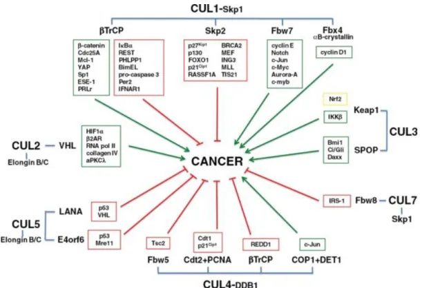
Figure 10. Involvement of CRLs and their substrates in promoting (green boxes/arrows) or inhibiting (red boxes/arrows) growth and survival of cancer cells, thus impacting oncogenesis. Figure from Lee J & Zhou, 2010.
3.4.3 Role of CRL/NEDD8 pathway in cancer
Proteins involved in the regulation of the CRL/NEDD8 pathway are not conventional oncoproteins because their effect depends on their targets. Nevertheless, there are well-established roles in tumor biology for some final effectors (Figure 10). Therefore, the CRL/NEDD8 pathway can function both in promotion and in suppression of cancer development (Lee J & Zhou, 2010). In addition, different sorts of NEDD8-pathway deregulations have been found in cancer. For example, overexpression of NAE and UBC12 and global hyper-neddylation are found in a variety of cancers, including lung adenocarcinomas and squamous-cell carcinomas (Chairatvit & Ngamkitidechaku, 2007; Li et al., 2014), while in prostate cancer the pathway is thought to be down-regulated (Meehan et al., 2002). Another CRL regulatory protein, DCUN1D1, is amplified in cancer, particularly in squamous cell carcinoma. Targeting the expression of DCUN1D1 by short hairpin RNA induces apoptosis, while another member of DCNL family, DCUN1D3, functions as a tumor suppressor by antagonizing the neddylation activity of DCUN1D1 (Sarkaria et al. 2006). NEDD8 was reported to cause an anti-proliferative effect through degradation of Estrogen Receptor-alpha (Fan, 2003). Neddylation of pVHL can also inhibit proliferation because it results in pVHL binding to
34 fibronectin and the assembly of extracellular fibronectin. The fibronectin matrix promotes differentiation and suppresses the proliferative and metastatic potentials of transformed cells in various model systems (Stickle et al., 2004). On the other hand, in its deneddylated form pVHL becomes a part of ECV complex (Elongin B/C-CUL2-VHL) and participates in the destruction of hypoxia-inducible factor which in turn plays an essential role in tumor angiogenesis (Russell & Ohh, 2008).
Collectively, these data demonstrate the important role of NEDD8-pathway in tumorigenesis and suggest that inhibition of neddylation may be a valid therapeutic approach for cancer treatment. The development of a potent small molecule inhibitor of NAE, MLN4924, was reported in 2009 by Millennium Pharmaceuticals. MLN4924 is structurally related to adenosine 5′-monophosphate (AMP), a product of the NAE reaction. MLN4924 forms covalent NEDD8-MLN4924 adduct within the active site of NAE. MLN4924 is a potent inhibitor of NAE (half-maximal inhibitory concentration IC50 = 4 nM), and does not affect related pathways (SUMO, Ub, etc.) or other ATP-dependent enzymes (Soucy et al., 2009). Treatment of cells with MLN4924 resulted in a dose-dependent stabilization of known CRL substrates (Soucy et al., 2009).
35
CHAPTER 3. RNA INTERFERENCE
In this project RNA interference (RNAi) was used to perform loss-of-function screening of the UPS components. The discovery of RNA interference made a revolution in the field of molecular biology and created a powerful tool for the modulation of gene expression. Compared to other methods, RNAi is easy to perform, cost-effective, specific to a selected gene and also low in toxicity. As a result, RNAi currently is the most used method for gene knockdown. After the development of this technology, genome-wide functional screenings became a standard research method, helping to identify the function of a gene, the involvement of a protein in a certain process, and, therefore also potential new drug targets. Most likely, RNAi will also be used in medicine to treat viral infections and cancer, even though there are still some difficulties related to its non-specific effects and its delivery system. RNAi and major aspects of its application for molecular biology research are discussed below.
3.1 DISCOVERY OF RNAi
The term RNAi describes the phenomenon when a double stranded RNA (dsRNA) initiates a cellular response, leading to sequence-dependent recognition of a target mRNA and thus causes modulation of its function (degradation of mRNA, temporary stimulation or inactivation). It has been known by many names, including co-suppression, post-transcriptional gene silencing (PTGS) and quelling. Only after the underlying mechanisms were understood well enough was it given with its current name “RNA-interference”, or RNAi.
The first piece of evidence comes from the 1970s with experiments showing that the introduction of oligonucleotides complementary to the target RNA causes the formation of RNA-RNA duplexes and interferes with the function of the target RNA. First this phenomenon was shown for the E. coli 16s rRNA where antisense RNAs inhibited translation. Later the same effect was shown for many other nucleotide sequences, and sometimes this interaction has regulatory, but not inhibitory effect. One such example is the regulation of the E. coli mobile genetic element Tn10. Hybridization of a small anti-sense transcript of Tn10 to its mRNA contributes to the regulation of Tn10 transposition. Finally, the same effect was shown for mRNAs, where the expression of sequences complementary to the herpes simplex virus I (HSV) thymidine kinase (TK)
36 diminishes activity of the enzyme fivefold (Izant & Weintraub, 1984). However, it has since been shown that not only can complementary (antisense) RNA cause such a silencing, but so can sense oligonucleotides, too.
In 1990 three reports described a similar phenomenon. To make petunia have brighter flowers, petunia genes responsible for flowers coloration (chalcone synthase
CHS and dihydroflavonol-4-reductase DFR) were overexpressed in transgenic plants.
Unexpectedly, ¼ to ½ of resultant petunias had completely white flowers or had uncolored patterns on the naturally-colored violet flowers. None of transgenic plants had flowers darker than the parental genotype. It was found that the level of the introduced mRNA was extremely low. Likewise, the level of mRNA of the corresponding endogenous gene was very low, too (Napoli et al., 1990; van der Krol et al., 1990). Similar results were reported for the tomato polygalacturonase gene. This phenomenon was named “co-suppression” (Smith et al., 1990). In 1994 it was shown that, despite the strong decrease of CHS mRNA, the transcription of this gene was unchanged (de Lange
et al., 1994). This suggested that co-suppression was a posttranscriptional event. The next
important step was made in 1997 by Metzlaff and colleagues, who showed that at least in some cases the introduced genes could cause formation of dsRNA, which induces silencing. For example, the petunia CHS mRNA forms dsRNA region at the 3’-end of transgenic mRNA.
Andrew Fire and Craig C. Mello brought all this evidence together and proved that double stranded RNAs are up to 100 times more efficient comparing to single-stranded antisense RNA (Fire et al., 1998). For this discovery they were awarded with the Nobel Prize in Physiology or Medicine in 2006. This work, performed on nematode C.
Elegans, stimulated research in the field, leading to the discovery of RNAi mechanisms
and key RNAi enzymes. It was shown also, that RNAi is evolutionarily conserved and is used by many eukaryotes, from protozoa to animals, for gene regulation and for protection from viruses. Recently RNAi has become widely used in molecular biology as a tool for analyzing gene function.
3.2 MECHANISM OF RNAi
As mentioned before, RNAi is initiated by the exogenous or endogenous dsRNA. For all vertebrates the efficiency of the interference correlates with the length of dsRNA: the longer dsRNA is, the greater the amount of siRNA produced and the greater number of target sites recognized on the mRNA molecule. The minimal size of dsRNA sufficient