HAL Id: hal-01307684
https://hal.sorbonne-universite.fr/hal-01307684
Submitted on 26 Apr 2016HAL is a multi-disciplinary open access
archive for the deposit and dissemination of sci-entific research documents, whether they are pub-lished or not. The documents may come from teaching and research institutions in France or abroad, or from public or private research centers.
L’archive ouverte pluridisciplinaire HAL, est destinée au dépôt et à la diffusion de documents scientifiques de niveau recherche, publiés ou non, émanant des établissements d’enseignement et de recherche français ou étrangers, des laboratoires publics ou privés.
Marika Urbanski, Marie-Laure Bréchemier, Béatrice Garcin, David
Bendetowicz, Michel Thiebaut de Schotten, Chris Foulon, Charlotte Rosso,
Frédéric Clarençon, Sophie Dupont, Pascale Pradat-Diehl, et al.
To cite this version:
Marika Urbanski, Marie-Laure Bréchemier, Béatrice Garcin, David Bendetowicz, Michel Thiebaut de Schotten, et al.. Reasoning by analogy requires the left frontal pole: lesion-deficit mapping and clinical implications. Brain - A Journal of Neurology , Oxford University Press (OUP), 2016, �10.1093/brain/aww072�. �hal-01307684�
For Peer Review
Reasoning by analogy requires the left frontal pole:
lesion-deficit mapping and clinical implications
Marika Urbanski1,2,3,4,5, Marie-Laure Bréchemier1,2,3,4, Béatrice Garcin1,2,3,4, David Bendetowicz1,2,3,4, Michel Thiebaut de Schotten1,2,3,4,6, Chris Foulon1,2,3,4,6,
Charlotte Rosso7, Frédéric Clarençon8, Sophie Dupont9, Pascale Pradat-Diehl10, Marc-Antoine Labeyrie11, Richard Levy1,2,3,12, Emmanuelle Volle1,2,3,4,6,13
Abstract
Analogical reasoning is at the core of the generalization and abstraction processes that enable concept formation and creativity. The impact of neurological diseases on analogical reasoning is poorly known, despite its importance in everyday life and in society. Neuroimaging studies of healthy subjects and the few studies that have been performed on patients have highlighted the importance of the prefrontal cortex in analogical reasoning. However, the critical cerebral bases for analogical reasoning deficits remain elusive. In the current study, we examined analogical reasoning abilities in 27 patients with focal damage in the frontal lobes and performed voxel-based lesion-behaviour mapping and tractography analyses to investigate the structures critical for analogical reasoning. The findings revealed that damage to the left rostrolateral prefrontal region (or some of its long-range connections) specifically impaired the ability to reason by analogies. A short version of the analogy task predicted the existence of a left rostrolateral prefrontal lesion with good accuracy. Experimental manipulations of the analogy tasks suggested that this region plays a role in relational matching or integration. The current lesion approach demonstrated that the left rostrolateral prefrontal region is a critical node in the analogy network. Our results also
For Peer Review
suggested that analogy tasks should be translated to clinical practice to refine the neuropsychological assessment of patients with frontal lobe lesions.
Author Affiliations :
1. Inserm, U 1127, 75013 Paris, France 2. CNRS, UMR 7225, 75013 Paris, France
3. Sorbonne Universités, UPMC Univ Paris 06, UMR S 1127, 75013 Paris, France 4. ICM, FrontLab, 75013 Paris, France
5. Hôpitaux de Maurice, Medicine and Rehabilitation Department, 94410 Saint-Maurice, France
6. ICM, Brain Connectivity and Behaviour, 75013 Paris, France. 7. AP-HP, Hôpital de la Salpêtrière, Stroke Centre, 75013 Paris, France
8. AP-HP, Hôpital de la Salpêtrière, Neuroradiology Department, 75013 Paris, France 9. AP-HP, Hôpital de la Salpêtrière, Epileptology Department, 75013 Paris, France
10. AP-HP, Hôpital de la Salpêtrière, Physical Medicine and Rehabilitation Department, 75013 Paris, France
11. AP-HP, Hôpital Lariboisière, Neuroradiology Department, 75010 Paris, France
12. AP-HP, Hôpital de la Salpêtrière, Behavioural Neuropsychiatry Unit, 75013 Paris, France. 13. CENIR, ICM, 75013 Paris, France
For Peer Review
Emmanuelle VolleFrontLab - ICM
Hôpital Pitié Salpêtrière, 47, bd de l'hôpital, 75013 Paris emmavolle@gmail.com
Running title: Analogies require the left frontal pole
For Peer Review
Abbreviations:
AF: arcuate fasciculus; ATR: anterior thalamic radiations; AUC: area under the curve; BA Brodmann area; FAB: frontal assessment battery; FMT: fronto-marginal tract; IFOF: inferior fronto-occipital fasciculus; MFG: middle frontal gyrus; MMSE: mini mental state examination; MNI: montreal neurological institute; ns: non-significant; PFC: prefrontal cortex; rlPFC: rostrolateral prefrontal cortex; ROC: receiver operating characteristic; SD: standard deviation; SFG: superior frontal gyrus; UF: uncinate fasciculus; VLSM: voxel-based lesion- symptom mapping
For Peer Review
Introduction
Using analogies, we can learn abstract concepts and create new associations between distant ideas. Analogies are a powerful tool that allows us to infer general representations from similarities between objects/situations and to transfer this general schema to new cases (Gentner, 1983; Gick and Holyoak, 1983; Gentner et al., 1993; Holyoak and Thagard, 1995; 1997; Gentner and Holyoak, 1997). Hence, analogical reasoning is at the core of generalization and abstraction processes (Holyoak and Thagard, 1995; Thibaut et al., 2010a; Hofstadter and Sander, 2013).
Analogical reasoning combines three key mechanisms: relational processing, similarity processing, and schema inference. Reasoning by analogy depends on the ability to consider, integrate, and compare multiple relationships between components of mental representations (Gentner et al., 1993; Robin and Holyoak, 1995; Gentner and Markman, 1997; Holyoak and Thagard, 1997; Halford et al., 2010). The consideration and integration of multiple relationships (relational reasoning) is also thought to be a key factor for fluid intelligence and to rely on prefrontal functions (Robin and Holyoak, 1995; Duncan et al., 1995; Waltz et al., 1999; Geake and Hansen, 2005; Jung and Haier, 2007). In addition, analogical reasoning depends on the ability to detect similarities between these relational representations (Gentner et al., 1993; Gentner and Medina, 1998; Blanchette and Dunbar, 2000). The comparison and mapping of relational representations composing analogous situations result in the inference of an analogy schema, i.e., a general representation of a pattern of relational similarities (Gick and Holyoak, 1983; Bethell-Fox et al., 1984; Gentner et
al., 1993; Gentner and Markman, 1997). When a new analogy schema is inferred, new concepts are formed in a flexible manner. Therefore, analogical reasoning allows the study of the relational integration, similarity matching and schema inference processes that are
For Peer Review
Despite the importance of these high-level functions in human cognition, deficits in analogical reasoning are rarely assessed in clinical practice, leading to poor understanding of their impacts on patients’ daily lives and of their neuroanatomical bases (Ahmed and Miller, 2014). However sparse, previous patient studies have revealed deficits on pictorial and verbal analogy tasks in patient with frontotemporal dementia, suggesting that the prefrontal cortex (PFC) is critical for analogical reasoning (Morrison et al., 2004; Krawczyk et al., 2008). As frontotemporal dementia patients have diffuse prefrontal damage and no voxel-based morphometry analyses have been performed, these studies have not provided evidence of a precise anatomical correlate. In a voxel-based morphometry study of adolescents, traumatic brain injury has been shown to impair performance on a scene analogy task and to alter its correlation with cortical thickness in prefrontal regions (Krawczyk et al., 2010a). The one study that examined focal lesions in adults used a voxel-based lesion symptom mapping (VLSM) approach on (mainly) stroke patients (Schmidt et al., 2012). The results revealed several posterior prefrontal and temporal areas critical for semantic verbal analogies. However, the poor representation of prefrontal damage (n = 17) and of anterior prefrontal lesions in particular in this stroke population limited the conclusions that could be drawn regarding the role of anterior cerebral regions. Among studies that used relational reasoning tasks that are similar to analogy tasks, such as matrix problem tasks (Raven, 1938; Wechsler, 1997), voxel-based lesion studies have also lacked coverage of the rostral PFC region despite larger sample sizes (Gläscher et al., 2009; Baldo et al., 2010), and the conclusions drawn regarding the critical brain regions for these tasks have not always been consistent among studies (Waltz et al., 1999; Tranel et al., 2008; Gläscher et al., 2009; Baldo et al., 2010; Woolgar et al., 2010; Waechter et al., 2013). In related fields that explored abstraction or reasoning, studies in brain-damaged patients have highlighted the critical importance of the left PFC for proverb interpretation (McDonald et al., 2008; Kaiser et al., 2013; Murphy et al.,
For Peer Review
2013), conceptualization (Dubois et al., 2000; Delis et al., 2001; Hoffman et al., 2010; Lagarde et al., 2015), and inductive reasoning (Reverberi et al., 2005).
In healthy volunteers, functional imaging studies on analogy have shown the involvement of various prefrontal regions, including the rostrolateral PFC (rlPFC), in addition to parietal and temporal regions (for a review, see Krawczyk, 2012). A variety of analogy tasks have employed verbal, figurative or abstract material that involved semantic (Bunge et
al., 2005; Wendelken et al., 2008; Green et al. 2010), role-based (Krawcyk et al. 2010a), visuospatial, mathematical, or logical relationships (Christoff et al., 2003; Geake and Hansen 2005; Smith et al., 2007; Wartenburger et al., 2009; Cho et al., 2010; Volle et al., 2010; Preusse et al., 2011; Watson et al., 2012). Although domain-oriented or relation-oriented brain regions have been observed (Krawczyk et al., 2011), the rlPFC has been demonstrated to be a domain-general region involved in both semantic and visuospatial analogies (Wendelken et al., 2012) and in both classical analogy and matrix problem solving tasks (Krawczyk et al., 2011). A recent meta-analysis of functional imaging results has shown that the left rlPFC and dorsolateral PFC are the most consistently activated regions across different analogy studies and tasks (Vartanian, 2012). Other approaches such as voxel-based morphometry on healthy volunteers (Aichelburg et al., 2014) and developmental studies of children (Wright et al., 2007; Crone et al., 2009; Thibaut et al., 2010b; Dumontheil, 2014), have also indicated that the left rlPFC is important for various relational reasoning tasks.
In other words, the literature on healthy subjects indicates that the rlPFC, among other regions, plays an important and domain-general role in analogy but the available evidence cannot demonstrate whether it is critical for this process. Patient studies have provided limited conclusions regarding the roles of rostral frontal areas. To the best of our knowledge, no study has examined whether analogical or relational reasoning depends on the integrity of frontal
For Peer Review
lobe connections. Hence, the precise cerebral bases for analogical reasoning deficits and the effect of rostral PFC damage on analogical reasoning abilities remain to be clarified.
In this study, we employed a lesion-behaviour mapping approach in 27 patients with a focal brain lesion in the PFC, to explore the crucial prefrontal regions for analogy and to test whether the left rlPFC is critical. The patients were administered a visuospatial analogy task that has been previously associated with the left rlPFC in healthy subjects (Volle et al., 2010; Aichelburg et al., 2014). The analogy schemas of this task are comparable to those used in previous studies (Gentner and Medina 1998; Krawczyk et al., 2008; Wartenburger et al., 2009; Watson et al., 2012) or in matrix problems. The two analogy conditions used each required relational reasoning and differed only in whether the analogy schema must be inferred. These conditions were compared to a control task that did not require relational processing. Lesion-deficit relationships were explored using a VLSM technique (Bates et al., 2003). In addition, we used a track-wise lesion-deficit analysis (Thiebaut de Schotten et al., 2014; 2015) to explore the impact of tract disconnection on analogical reasoning. Finally, we examined the sensitivity and specificity of these analogy tasks in patients with damaged frontal lobes and estimated the potential value of the task in clinical practice.
Materials and methods
Participants
Twenty-seven right-handed patients (16 females, mean age of 47.2 years, ranging from 23 to 75 years) who each presented with a single, focal frontal lesion and were seen at the chronic stage (> 2 months) participated in this study. The patients were recruited from the departments of nervous system diseases and neuroradiology at Salpêtrière Hospital, the
For Peer Review
neurological unit at Saint-Antoine Hospital and the neuroradiology department at Lariboisière Hospital in Paris. Patients with a history of psychiatric or neurological disease, drug or psychotropic abuse, MRI contraindication or who were not able to understand the task instructions were excluded. All patients were native French speakers. Descriptive and clinical data are reported in Table 1.
The patient performances were compared to those of a normative group of 54 healthy right-handed, French native speaker controls (Supplementary Table 1), who were matched for age and years of formal education and who had no history of psychiatric or neurological disease, drug or psychotropic abuse, or MRI contraindication and no cognitive impairment (Mini Mental State Examination, MMSE ≥ 27/30; Folstein et al., 1975).
The experiment was approved by the local ethics committee; all participants provided written informed consent according to the Declaration of Helsinki and were paid for their participation.
Neuropsychological testing
A battery of neuropsychological tests was administered to all participants (Supplementary method 1). Cognitive status was measured with the MMSE (Folstein et al., 1975). A short assessment of cognitive and behavioural executive functions was performed using the Frontal Assessment Battery (FAB, Dubois et al., 2000), a semantic and lexical fluency task (Cardebat et al., 1990) and the Stroop test (Stroop, 1935). Semantic knowledge was assessed using short French versions of a naming test and a semantic matching test (as described in Merck et al., 2011).
Experimental design
For Peer Review
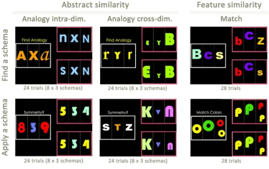
experimental conditions followed the same design and used the same types of stimuli (Fig. 1; Supplementary Method 2). After the instructions were displayed, a first set of stimuli appeared on the left part of the screen (the source set), and two other sets appeared on the right part of the screen (the target sets). The participants were asked to select the target set that matched the source set based on the relationships between the stimuli that composed the sets (Analogy tasks) or based on the similarity of their visual features (Match tasks). The subjects had 11.5 seconds to respond by a button press. The stimuli were letters, numbers or abstract figures, presented in different colours, numbers, sizes or patterns.
Analogy tasks were divided into two conditions: an AnalogyFind and an AnalogyApply condition. In the AnalogyFind condition, the participants had to find the analogy schema by considering the similarities between the structures of each set. The instruction “find analogy” was displayed, and the task required comparing the sets, finding an analogy schema and choosing the target set accordingly (e.g., symmetry of the size of the stimuli). In the AnalogyApply condition, the analogy schema was indicated to the participants by providing them with a verbal term that described it (e.g., “Proportion”). The instruction that contained the verbal description of the schema was displayed on the screen together with the sets; thus, participants still had to consider and compare the multiple relationships between the stimuli, but there was no need to infer or retrieve the schema. Six geometrical or mathematical schemas (proportion, subtraction, addition, mirroring, symmetry and progression) were used and applied to the identity of the stimuli (letters or figures) or to their size, number, brightness, or texture. The features of the stimuli that were not relevant for the analogy schema varied between the source and target, to avoid perceptual matching. For AnalogyApply and AnalogyFind, two types of analogy trials were proposed in the same proportion: intra- and cross-dimensional analogies (Fig. 1).
For Peer Review
In the Match tasks, the source and target sets had to be matched on the basis of six perceptual attributes: colour, quantity, size, texture, figures and letters. As with the Analogy tasks, the Match tasks included two separate conditions, a MatchFind and a MatchApply condition. In the MatchFind condition, the instruction “find match” was displayed, and the participants had to find the perceptual relationship between the source and the correct target set. In the MatchApply condition, the participants were instructed to apply a given matching rule. The instruction that contained a verbal description of the matching rule was displayed on the screen (e.g., “same colours”) together with the sets.
All participants understood the instructions and were able to perform the tasks correctly after training. They performed one session of each of the four experimental conditions in the following order: 28 MatchApply trials, 28 MatchFind trials, 48 AnalogyApply trials and 48 AnalogyFind trials. The trials were randomized within each session.
Behavioural Analysis
The accuracy (percentage of correct responses) was measured for each condition. Analogy and Match mean accuracies were calculated by averaging performance on the AnalogyFind and AnalogyApply conditions and on the MatchApply and MatchFind conditions, respectively. Similarly, the Find and Apply performances were calculated by averaging the Find (AnalogyFind and MatchFind) and Apply (AnalogyApply and MatchApply) conditions. We also examined the performance at cross- and intra-dimensional trials for the AnalogyApply and AnalogyFind tasks separately. To assess the possible specificity of the deficits in the Analogy tasks relative to the control task, we also calculated an index (Analogy index = [Analogy mean accuracy – Match mean accuracy] × 100 / mean accuracy in all Analogy and Match tasks averaged). Similarly, we calculated indices to test
For Peer Review
for possible specificity of deficits in the Find condition relative to the Apply condition (Find index = [mean accuracy in Find conditions – mean accuracy in Apply conditions] × 100 / mean accuracy in the average of all conditions), and in the cross- relative to the intra-dimensional analogies (Cross index = [mean accuracy in cross-dimension Analogy trials – mean accuracy in intra-dimension Analogy trials] × 100 / mean accuracy in the average of all analogy conditions).
Statistical analyses were performed using SPSS software (v22.0; LEAD Technologies, Inc.). Between-group differences were analysed using parametric t-tests (when the assumption of normality was met) or non-parametric tests otherwise (Mann-Whitney test), using exact P values for comparison within our patient group. Correlations between the performances of the patients and age, education, delay or volume of the lesion were analysed using the non-parametric Spearman test (rs).
Image acquisition and preprocessing
Magnetic resonance Acquisition
Patients and controls underwent the same high-resolution T1-weighted structural MRI acquisition on a Siemens 3 Tesla VERIO TIM system that was equipped with a 32-channel head coil. A three-dimensional MPRAGE dataset that covered the whole brain was acquired for each participant across 176 axial slices with a voxel isometric resolution of 1 mm3 (TE = 2.98 msec, TR = 2300 msec, and flip angle = 9°). MRI and behavioural testing took place on the same day for most of the participants or a few days apart at most.
MRI spatial normalization
T1-weighted 3D sequences were preprocessed with SPM8 software (Wellcome Department of Imaging Neuroscience, London, UK), which ran on Matlab (Mathworks Inc.,
For Peer Review
Natick, USA; www.mathworks.com/matlabcentral). The MRIs were spatially normalized to the Montreal Neurological Institute (MNI) template. The ‘unified segmentation’ approach was combined with lesion masking to limit the impact of a brain lesion on the spatial normalization (Crinion et al., 2007; Andersen et al., 2010). This approach has been identified as the best compromise between the normalization accuracy and lesion shrinkage in a recent study (Ripollés et al., 2012). The segmentation parameters were set to the defaults, except for regularization, which was set to medium (Andersen et al., 2010; Ripollés et al., 2012). Spatially normalized images were resliced with a final voxel size of 1.5 × 1.5 × 1.5 mm3. Each normalized MRI was visually checked and compared with the MNI template to evaluate the normalization accuracy (BG, MLB, DB and EV). No patient had to be excluded due to difficulties with normalization.
Lesion-behaviour mapping approach
To investigate lesion-deficit relationships, we ran a VLSM analysis (Bates et al., 2003) using NPM software (http://www.mccauslandcenter.sc.edu/mricro/npm/). The preprocessed and normalized MRIs were used for lesion segmentation. Signal abnormalities due to the lesion were manually segmented using MRIcron (http://www.mccauslandcenter.sc.edu/mricro/mricron/) by trained neurologists (BG, MLB, and DB) supervised by an experienced neurologist (EV), who were blind to the performances of the patients at the time of the lesion segmentation. The resulting segmented lesion volumes in the MNI space were then introduced in the statistical procedure.
Given the non-normal distribution of the performance and the small sample of the patients, we used the non-parametric Brunner-Munzel test and corrected for multiple comparisons for family-wise errors using permutations, with a significance threshold of P < 0.05. Only the voxels that concerned at least three lesions were considered (all of the lesions
For Peer Review
together covered 74% of the frontal lobes; overlaps of at least three lesions represented 30% of the frontal lobes). These analyses provided statistical maps for Analogy and Match mean accuracy scores as well as for the Analogy index.
Track-wise lesion-deficit analysis
To explore the impact of tract disconnection on analogical reasoning, we used two track-wise lesion-deficit approaches.
A priori approach.
First, independent of the VLSM results, we used a diffusion-based atlas of frontal lobe connections (Rojkova et al., 2015) combined with Tractotron software as part of the BCB toolkit (http://www.brainconnectivitybehaviour.eu/), to identify the tracts that could be affected by the lesion of each patient. Tractotron automatically computes the overlap of each segmented lesion with the map of the tracts. We mapped the lesion from each patient onto tractography reconstructions of white matter pathways obtained from a group of healthy controls (Rojkova et al., 2015). We quantified the severity of the disconnection by measuring the probability of the tract to be disconnected (Thiebaut de Schotten et al. 2014). A tract was considered disconnected when a lesion overlapped with a voxel that belonged to this tract with a probability that was above the chance level (probability > 0.5). We a priori selected several projection tracts that have been associated with Analogy performance according to Aichelburg et al. (2014): the long segment of the arcuate fasciculus (AF), the fronto-marginal tract (FMT), the inferior fronto-occipital fasciculus (IFOF), the uncinate fasciculus (UF) and the anterior thalamic radiations (ATRs). Then, we examined the impact of the disconnection of each tract in the left and right hemispheres on analogical reasoning. For each tract of interest, we compared the performance of the patients with and without its disconnection using non-parametric Mann-Whitney tests (with exact P values significant at a P < 0.05).
For Peer Review
VLSM-based approach.
Second, based on the VLSM results, we created a map of the tracts connecting the VLSM Analogy region (“VLSM connectome map”) and calculated the probability that each lesion intersects this map. We built the VLSM connectome map using Disconnectome map software (Aichelburg et al., 2014; Thiebaut de Schotten et al., 2015) as part of the BCB toolkit (http://www.brainconnectivitybehaviour.eu/). The VLSM region was registered to the tractography of a group of healthy controls (Rojkova et al., 2015) using affine and diffeomorphic deformations. The registered VLSM region was used as a seedpoint to track streamlines passing through the region in a normative dataset. The software creates a probability map of the streamlines intersecting the seed such that the value in each voxel of the map varies based on inter-subject variability. Then, we used Tractotron (Thiebaut de Schotten et al., 2014) (http://www.brainconnectivitybehaviour.eu/), to compute the probability that each lesion intersects the VLSM connectome map. Tractotron also identified the tracts connected to the VLSM region (Rojkova et al., 2015). Among these VLSM connected tracts, we calculated the number of tracts that were disconnected by the lesion of each patient (with probability of greater than 0.5) and examined its correlation with analogy performance.
Sensitivity and specificity of analogy tasks and conditions for clinical use
Finally, we aimed to evaluate the clinical value of the analogy tasks for patients with frontal lobe damage. First, our original tasks in their current form might not be suitable for clinical practice because they take time to perform (between 45 and 50 minutes). Therefore, we ran a new analysis on a subsample of the trials, which was composed of the 28 first AnalogyFind trials (intra- and cross-dimensional) and the 28 MatchFind trials that had been
For Peer Review
administered to each participant. As the order was randomized for each participant, the 28 first Analogy trials were not the same among individuals and were randomly selected. We also checked the reliability of all trials statistically and observed good item reliability (Spearman-Brown coefficient = 0.761). The Apply conditions were discarded because the Find conditions may correspond better to real-life analogies, and they were more strongly impacted by brain lesions. The estimated duration of this subsample of trials did not exceed 15 minutes.
Second, to examine the ability of this shorter version to discriminate among patients, we grouped the patients according to their lesion location, independent of the VLSM results. For analysis of accuracy of the short version, the patients were divided into two a priori-defined groups based on integrity of the left rlPFC. The definition of the group was based on previous literature indicating the importance of the left rlPFC in analogy. Because the rostral PFC is difficult to delineate anatomically, a pragmatic definition was used, as described in Tisserand et al. (2002): the rostral prefrontal region corresponds to the most anterior 25 coronal slices (2.5 cm), y > 44 in the MNI coordinates. Within this rostral prefrontal region, we selected its left lateral part (defining the ‘left rlPFC region’) by selecting MNI x coordinates that were lower than − 25. Seven patients had a lesion that affected this anatomically defined rlPFC region and were pooled in the ‘damaged left rlPFC’ group (indicated in Table 1). Their performances were compared to patients who had an intact left rlPFC (‘intact left rlPFC’ group).
We then examined the sensitivity and specificity of this shorter subtask to discriminate brain lesions, by building Receiver Operating Characteristic curves (ROC curves) for each score. These ROC curves show the trade-off between the sensitivity and specificity, and the area under the curve (AUC) estimates the accuracy of the task for predicting the left rostrolateral damage in patients who had frontal lesions. Based on the obtained predictive
For Peer Review
value of the Analogy and index scores and on the normative scores of controls, we grouped the patients according to the presence or absence of a deficit in analogical reasoning (indicated in Table 1) and compared their cognitive profiles and lesion locations.
Results
Behavioural results
The patients exhibited significantly poorer performances compared with the controls on the FAB, fluency tasks, and MMSE, and they showed a greater interference effect on the Stroop test, but their semantic knowledge was preserved (Supplementary Table 1 and Supplementary Fig. 1).
The patients performed significantly more poorly than the healthy participants in terms of both the Analogy conditions separately and the Analogy mean accuracy score. They had lower Match mean accuracy scores, whereas their accuracy in each Match condition separately did not differ from that of the controls. The Analogy index was significantly higher in the patient group, which suggests that their deficit was larger in the Analogy than the Match tasks. The patients scored lower than the healthy subjects in the Find and Apply conditions, but not in the Find index, which suggests that they were equally impaired in the Apply and Find conditions.
Although age, lesion volume and lesion delay, and in some cases education, can be confounding factors in VLSM analysis, there was no significant correlation between Analogy mean accuracy score and age (rs = − 0.276, ns), education (rs = 0.336, ns), lesion volume (rs = − 0.314, ns), or lesion delay (rs = − 0.201, ns), which have not been covaried out.
For Peer Review
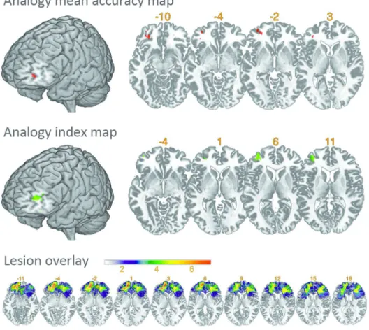
The statistical map of the Analogy mean accuracy score (Fig. 2) showed that a deficit in the Analogy tasks was associated with a left rostral prefrontal area (MNI coordinates centred on -31, 51, -3; z = 3.48; volume = 0.33 cc) that encompassed BA 47/10 and was located at the rostral junction between the superior and middle frontal gyrus (SFG and MFG), extending into pars triangularis. A smaller cluster was located posteriorly, centred on coordinates -34, 41, 3, and another in the orbitofrontal cortex, BA 47 and 11, centred on coordinates -30, 41, -10. These clusters are gathered under the term ‘the VLSM Analogy region’ in the further analyses below.
Table 2 shows that the patients who contributed to the ‘VLSM Analogy region’ (n = 5) did not differ from the other patients in terms of their age, education, lesion volume, delay between lesion and inclusion, and general neuropsychological testing, except for the Stroop test, in which they had a stronger interference effect. The patients contributing to the ‘VLSM Analogy region’ had lesions caused by various mechanisms, including haemorrhage (P02 and P29), tumour excision (P04 and P08) or epilepsy surgery (P22), as indicated in Table 1.
Patients with a lesion in the ‘VLSM Analogy region’ showed significantly greater impairment than the other patients on the Analogy tasks but not on the Match tasks, as shown by a significant between-group difference in the Analogy index (Fig. 3 and Table 2). There was no between-group difference in the Find index or the Cross index. In other words, the ‘VLSM Analogy’ patients were not differentially affected compared with the other patients by the need to infer the analogy schema (no significant difference in the Find index) or to transfer the schema to different dimensions in the source and target (no significant difference in the Cross index). For subsequent analyses, intra- and cross-dimensional trials, as well as Apply and Find analogy trials, were pooled.
For Peer Review
The statistical map for the Analogy Index (Fig. 2) revealed a region that was very close to the ‘VLSM Analogy region’ in the MFG and pars triangularis, which encompassed BA 10, 47, 45 and 46 and was centred on the MNI coordinates -35, 48, and 9 (volume of 1.57 cc). Although the VLSM Analogy and VLSM Index regions did not fully overlap, they both included BA10 and 47 in the left rlPFC, indicating that the left rlPFC was specifically critical to Analogy relative to Match.
Track-wise Lesion-Deficit approach
A priori approach
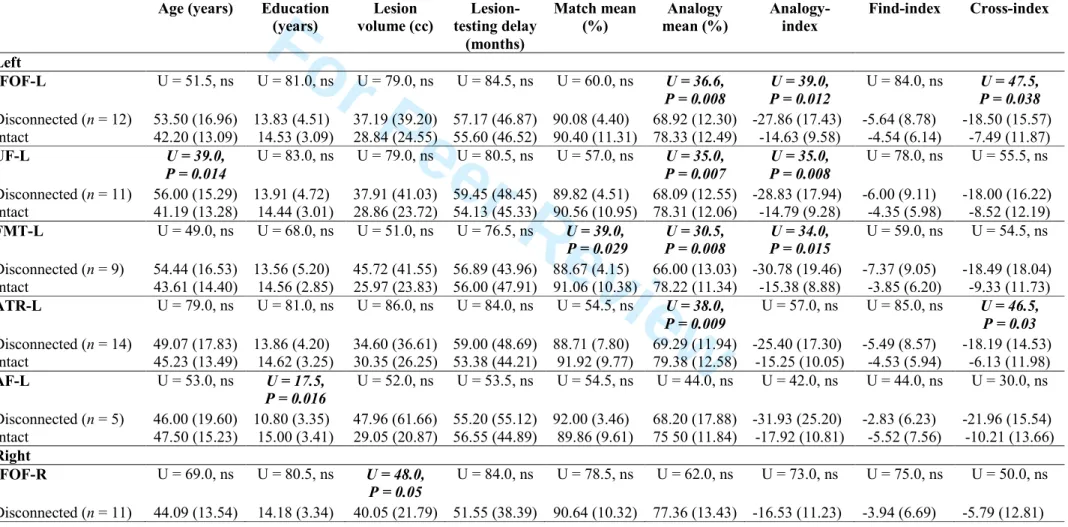
Several tract disconnections had impacts on analogical reasoning abilities. Table 3 shows that disconnections of the left IFOF, UF and FMT were associated with a greater deficit in Analogy tasks (Analogy mean accuracy score and Analogy index). Disconnection of the left ATR was associated with a deficit in the Analogy tasks, but no significant association was observed with the Analogy index. Age, education, lesion delay and lesion volume did not significantly differ between the ‘Disconnected’ and ‘Intact’ groups for these four analogy-related tracts, except that the patients with a UF disconnection had an increased age. Disconnection of the left AF was not associated with a significant deficit, perhaps because it was disconnected in only five patients. None of the selected tracts in the right hemisphere was associated with a deficit in an Analogy or Match tasks when disrupted.
Both the VLSM and disconnection approaches show that left-brain lesions were associated with analogical difficulties. Note that none of the descriptive, clinical or neuropsychological data significantly differed between the right-brain-damaged (n = 9) and left-brain-damaged patients (n = 14) (Supplementary Table 2).
For Peer Review
In a second approach, we built the VLSM connectome map composed of the tracts connected to the VLSM Analogy region. This map is shown in Supplementary Fig. 2. For each patient, the probability of disconnection of the VLSM connectome map and the number of disconnected tracts among the VLSM connected tracts are provided in Supplementary Table 3. The probability of disconnection of the VLSM connectome map was significantly correlated with the Analogy mean accuracy score (rs = - 0.511; P = 0.006) and with the Analogy Index (rs = −0.471; P = 0.013). This result indicates that a lesion that disconnected the ‘VLSM Analogy region’ affected analogical reasoning. The correlations of the probability of disconnection of the VLSM connectome map with age (rs = 0.262), education (rs = − 0.201) and lesion volume (rs = 0.366) were not significant.
Tracts connected to the VLSM Analogy region included the left ATR, FMT, IFOF, UF, orbitopolar tract, superior longitudinal fasciculus (branch 3), fronto-pontine projections, and frontostriatal fasciculus. Among these tracts, we observed significant correlations between the number of disconnected tracts per patient and the Analogy mean accuracy score (rs = - 0.553; P = 0.003) as well as the Analogy Index (rs = - 0.416; P = 0.031). Correlations with age (rs = 0.334), education (rs = - 0.131) and lesion volume (rs = 0.024) were not significant. Among all other tracts (tracts not connected to the VLSM analogy region), the Analogy mean accuracy score and Analogy index were not correlated with the number of disconnected tracts per patient (rs = 0.126 and 0.122, respectively; ns), but lesion volume was correlated (rs = 0.693; P < 0.001). These findings indicate that analogical reasoning depends on connectivity of the VLSM region independent of lesion size.
Value of Analogy tasks in clinical practice
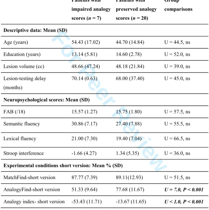
To further explore the value of our analogy task in clinical practice, we analysed a subsample of the original trials. Patients in the ‘damaged left rlPFC’ group (with left rlPFC anatomically defined, n = 7) had poorer performances than those in the ‘intact left rlPFC’
For Peer Review
group (n = 20) in the AnalogyFind-short version condition but not in the MatchFind-short version condition, and their Analogy index-short version was significantly greater (Supplementary Table 4).
We explored the discriminative value of the short version of the analogy tasks with regard to brain damage location (damaged versus intact left rlPFC) using ROC curves (Supplementary Fig. 3). The AUCs showed that the accuracies of the AnalogyFind-short version performance (AUC = 0.925; P = 0.001) and of the Analogy index-short version (AUC = 0.954; P < 0.001) were very good, but that the MatchFind-short version discriminated among the patients poorly (AUC = 0.707; P = 0.109).
Examination of the coordinate points of the ROC curves showed that an AnalogyFind-short version score of below 65.3% (which corresponds to the mean performance of the controls minus 1.5 SD) had a sensitivity of 85.7% and a specificity of between 85 and 90%. An Analogy index-short version that was lower than – 33% (absolute value > 33) had a sensitivity of 85.7% and a specificity of between 90 and 95%. The Analogy index-short version was as sensitive as the AnalogyFind-short version when discriminating the patients, and it had a slightly better specificity. Thus, an Analogy index-short version score that exceeded 1.5 SD from the mean score of the healthy controls (< – 33%) was used as a cut-off to define an analogical reasoning impairment.
To further characterize the value of such a cut-off in brain-damaged patients, we analysed the cognitive profile and visualized the lesion location of the patients as a function of their deficit in analogical reasoning.
Table 4 shows that the two groups did not differ significantly in age, education, lesion volume, mean lesion-testing delay or neuropsychological scores, especially for those tasks that tap into executive functions (see also Table 1). This finding suggests that other cognitive deficits cannot explain the analogy difficulties. Fig. 4 shows that the lesions of the patients
For Peer Review
with impaired analogical reasoning overlapped mainly in the left rlPFC region, whereas the lesions of the patients with preserved analogical reasoning overlapped in the right PFC.
In summary, the short version of the tasks was sufficiently sensitive to confirm the critical role of the left rlPFC in analogical reasoning, with a high accuracy of the Analogy index in distinguishing the patients with a left rlPFC lesion.
Discussion
The current study focused on the impacts of prefrontal lesions on analogical reasoning. The results obtained using three distinct approaches (VLSM, disconnection, and ROC analyses) converge to show the critical role of the left rostral prefrontal region in analogical reasoning. Two new findings emerge from this work. First, analogical reasoning specifically depends on the integrity of the left rlPFC and/or on the integrity of some of its long-range connections. Second, our analogy task very accurately predicts a left rlPFC lesion and could be used as a new tool to assess brain-damaged patients. These findings have important clinical implications because analogical reasoning and the more general functions of the rostral part of the PFC are poorly assessed in clinical practice.
Analogy and the integrity of the prefrontal cortex and/or its connections
Few data are available regarding analogical reasoning abilities in patients with brain damage. Following the two studies that explored neurological patients with diffuse frontal damage (Morrison et al., 2004; Krawczyk et al., 2008), the current VLSM analysis specifies which area in the PFC is critical for analogical reasoning. This critical area is located in the rlPFC, encompasses BA 10 and 47 and is left lateralized (Fig. 2). A lesion of this region is not associated with a deficit in the perceptual matching condition, which suggests that analogical reasoning is relatively specifically impaired when this region is damaged (Table 2 and Fig. 3). As illustrated in Supplementary Fig. 4, this result converges with the conclusions drawn from
For Peer Review
other approaches, such as functional imaging (Volle, et al., 2010; and for reviews Krawczyk, 2012; Vartanian 2012) and morphometry (Aichelburg et al., 2014). The left rlPFC has been observed in functional imaging studies using different analogy tasks that involved distinct types of relationships in the semantic or visuospatial domains, which suggests a domain-general role of the left rlPFC in analogies. However, the precise role of this rlPFC region in analogy, and in cognition in general, is not clearly understood. Previous studies have suggested that this rlPFC region is involved in relational integration (Christoff et al., 2001; Kroger et al., 2002; Ruff et al., 2003; Ramnani and Owen, 2004; Krawczyk et al., 2011), abstraction (Christoff et al., 2009), and the mapping of similarities (Bunge et al., 2005; Garcin et al., 2012). Our task manipulation did not provide evidence of a significant difference that would have suggested a role in inference processes (Find vs. Apply) or in remote mapping that allows for abstract generalization (Cross- vs. Intra-dimensional analogy), although there is evidence from other studies that the left PFC is important for rule induction (Reverberi et al., 2005) and for distant analogies (Green et al., 2010). However, in patients with left rlPFC damage, the deficit was deeper for inferences based on cross-dimensional mapping (Table 2; Fig. 3). Thus, it remains possible that these differences exist but were missed because of a lack of sufficient statistical power or due to insufficient lesion overlap.
A left dominance of PFC for analogical reasoning was previously highlighted in functional imaging studies (Bunge et al., 2009; Vartanian, 2012) and in one repetitive transcranial magnetic stimulation study (Boroojerdi et al., 2001). However, the lateralization of rlPFC functions is not understood. The role of language in analogies could be at play, but it cannot entirely explain a left lateralization because tasks that used non-verbal analogies also recruited the left rlPFC (Wharton et al., 2000; Christoff et al., 2003; Bunge et al., 2009; Hampshire et al., 2011; Watson and Chatterjee, 2012; Wendelken et al., 2012), and analogical reasoning difficulties are not associated with reduced fluencies in our patients.
For Peer Review
VLSM analysis did not identify other critical prefrontal areas for analogies, although previous functional imaging studies have shown that several prefrontal regions are involved in analogical reasoning (Christoff et al., 2001; Bunge et al., 2005; Geake and Hansen, 2005; Green et al., 2006; Cho et al., 2007; Wendelken et al., 2008; Geake and Hansen, 2010; Krawczyk, et al., 2010b; 2011; Volle, et al., 2010; Hampshire et al., 2011; Preusse et al., 2011; Green et al., 2012; Krawczyk, 2012; Wendelken et al., 2012), as well as temporal and parietal regions. Within the PFC, it is possible that the other prefrontal regions that support analogical reasoning are less lateralized, which allows for the contralateral cortex to compensate for this function. However, we cannot exclude the possibility that the analyses missed some other critical prefrontal region because of the lack of statistical power achieved for some of the regions and because only partial coverage of the frontal lobes was obtained. It is likely that the left rlPFC operates via interaction with more posterior regions in the prefrontal, parietal and temporal lobes (Aichelburg et al., 2014; Cocchi et al., 2014; Rojkova
et al., 2015). Because we examined only patients with prefrontal brain lesions, no conclusion could be made on the critical roles of the parietal and temporal lobes. Nevertheless, our track-wise lesion-deficit approach provided some clues about the roles of the interactions of the left rlPFC with other brain regions for analogy performance.
We found that disconnection of the left IFOF, UF, FMT, or ATR was associated with a deficit in Analogy tasks (Table 3). These results are consistent with a previous tractography study in healthy subjects (Aichelburg et al., 2014) and confirm the importance of the left hemisphere for analogical reasoning. Here, anatomical connections between the temporal cortices (via the UF), the occipital cortex (via the IFOF), and subcortical structures via the ATRs appear to play a role in analogical reasoning. The VLSM-based approach further showed that an analogical reasoning impairment was associated with a disconnection of the
For Peer Review
VLSM Analogy region. Owing to these connections, information can converge within the left rlPFC, coming from distinct domains or networks (Sakai et al., 2003; Parkin et al., 2015)
Recent resting-state studies have emphasized the importance of functional networks for high-level cognitive functions and that the disruption of these networks could better explain a deficit in high-level cognition than lesion location per se (Woolgar et al., 2010; Gratton et al., 2012; Warren et al., 2014; Corbetta et al., 2015). In this context, the result that a very circumscribed lesion site is critical for analogical reasoning is puzzling. This lesion site may be critical because damage to this area cannot be compensated for and/or because several tracts that converge at this site must be conjointly damaged to provoke a deficit, as suggested by the correlation between analogy performance and the number of tracts connected to this region that were affected by the lesions. This latter interpretation would match the cortical disconnection mechanism that was previously hypothesized by Norman Geschwind (1965). Overall, these results suggest that the left rlPFC region is a functional “hub” or an essential relay station in the analogy network. This interpretation argues for the role of this region in integrating information of different natures or domains.
Clinical application of the study: a new assessment tool
Although recent cognitive theories based on functional imaging place the rostral PFC at the top of a frontal hierarchical functioning model that subserves reasoning, problem solving, behavioural adaptation and abstraction (Badre and D’Esposito, 2007; Koechlin and Summerfield, 2007; Badre, 2008; Christoff et al., 2009; Krawczyk et al., 2011), functions of the rostral PFC are poorly assessed in clinical practice. Only recently has Burgess and Shallice’s work on multitasking (Shallice and Burgess, 1991) generated specific tasks for physicians (Burgess et al., 2006; 2009). Existing neuropsychological tools offer very few tests of abstract thinking or reasoning, and the critical brain networks for these tests are not well
For Peer Review
reasoning provides cognitive tasks that tap into abstract thinking and relational reasoning abilities, and the cerebral networks associated with these tasks have begun to be clarified. Hence, patients could benefit from the transfer of analogical reasoning tasks to clinical practice. In this study, we simulated a short version of our analogy task that could be transferred to clinical practice and showed that it had very good sensitivity and specificity for predicting left rlPFC injury, which demonstrates that even a small set of analogy trials is valuable for discriminating among different types of patient damage (Supplementary Fig. 3). The Analogy index appeared to be the most specific measure for analogical reasoning deficits.
Impairment in our Analogy tasks was not associated with global executive dysfunction, which suggests that analogical reasoning is a cognitive ability that is not entirely captured by classical executive neuropsychological tests (Table 4). Our results rather support a functional specialization within the PFC, with a distinct role of the rostral PFC compared with the more posterior areas. Previous studies have highlighted the role of inhibition abilities and interference control for analogical reasoning (Morrison et al., 2004; Krawczyk et al., 2008; Bugaiska and Thibaut, 2015). These studies suggest the possibility that analogy deficit in patients with left rlPFC lesion may be due to poor inhibition abilities. Although our VLSM analysis cannot rule out this explanation, other parts of our findings do not favour this hypothesis because patients with impaired analogy scores did not have more interference sensitivity than patients with preserved analogy scores (Table 4). In addition, response inhibition is usually associated with right or medial frontal regions (Stuss et al., 2001; van Veen and Carter, 2005; Volle et al., 2012; Tsuchida and Fellows, 2013; Aron et al., 2014; Hornberger and Bertoux, 2015; Robinson et al., 2015).
Finally, patients with frontal lesions of different aetiologies have been included in this study, which is a limitation because different pathologies affect the brain differently with distinct time courses and mechanisms of plasticity. However, a recent study has demonstrated
For Peer Review
that frontal lesions of different vascular and tumour aetiologies have similar effects on executive testing and fluid reasoning (Cipolotti et al., 2015). This finding supports the idea that the pooling of lesions with various physiopathological mechanisms in the same analysis is a valid methodological approach to exploring the organization of frontal functions. This approach has been used previously (Volle et al., 2008; 2012; Tsuchida and Fellows, 2013; Azuar et al., 2014), including lesions caused by epilepsy surgery (Tranel et al., 2008; Gläscher et al., 2009; Chapados and Petrides, 2013). The pooling of lesions with different physiopathological mechanisms allowed us to more completely cover the possible lesion locations in the PFC, including the rostral part, which is rarely affected by ischaemic strokes. Furthermore, this approach could mitigate statistical and spatial biases due to stroke locations (Nachev et al., 2008; Volle et al., 2013; Mah et al., 2014). In this context, our findings suggest that assessing analogical reasoning in brain-damaged patients has a clinical value that is independent of the lesion aetiology.
Overall, the short version of our analogy task could enrich the classical neuropsychological toolbox for the assessment of high-level cognitive functions that depend on the rostral PFC. The ecological validity of this test in real-world problem solving remains to be demonstrated. Hence, the short version of the Analogy test will be validated in an independent and larger sample of patients with more homogeneous lesions and in a group of controls matched for age and education. We will examine correlations to other relational reasoning or problem solving tasks to further improve the value of the test as a tool for the evaluation of analogical reasoning abilities in clinical practice.
Conclusions
Analogical reasoning plays a significant role in inferring general representations from similarities, which in turn can be applicable to solving new problems. The current lesion study
For Peer Review
with its long-range anatomical connections) is specifically critical for analogical reasoning and that a left rlPFC lesion could impair the relational integration and matching processes that are involved in abstract thinking. Despite a relatively small sample size examined in this study, these results converge clearly with existing neuroimaging findings on analogy. Furthermore, our study provides a sensitive and specific new neuropsychological test that can be transferred to everyday clinical practice, for the assessment of analogical reasoning in patients. These findings could be useful to clinicians by informing them of the expected consequences of rostral prefrontal damage on high-level cognitive functions and proposing a tool for their assessment.
Acknowledgements
The authors thank Prof. Claude Adam, Dr. Carole Azuar, Dr. Dorian Chauvet, Dr. Vincent Degos, Prof. Damien Galanaud, Dr. Florence Laigle, Dr. Anne Leger, and Prof. Vincent Navarro for their help in recruiting the patients.
Funding
This work was supported by the ‘Agence Nationale de la Recherche’ [grants number ANR-09-RPDOC-004-01, EV] and the “Societe Française de Neurologie” (MLB and DB). The
research leading to these results received funding from the program “Investissements d’avenir” ANR-10-IAIHU-06 and from the project PHENOTYPES, no. ANR-13-JSV4- 0001-01.
For Peer Review
Supplementary material is available.For Peer Review
References
Ahmed FS, Miller LS. Adequate proverb interpretation is associated with performance on the Independent Living Scales. Neuropsychol. Dev. Cogn. B Aging Neuropsychol. Cogn. 2014: 1–12.
Aichelburg C, Urbanski M, Thiebaut de Schotten M, Humbert F, Levy R, Volle E. Morphometry of left frontal and temporal poles predicts analogical reasoning abilities. Cereb. Cortex 2014: bhu254.
Andersen SM, Rapcsak SZ, Beeson PM. Cost function masking during normalization of brains with focal lesions: still a necessity? Neuroimage 2010; 53: 78–84.
Aron AR, Robbins TW, Poldrack RA. Inhibition and the right inferior frontal cortex: one decade on. [Review]. Trends Cogn. Sci. 2014; 18: 177–185.
Azuar C, Reyes P, Slachevsky A, Volle E, Kinkingnehun S, Kouneiher F, et al. Testing the model of caudo-rostral organization of cognitive control in the human with frontal lesions. Neuroimage 2014; 84: 1053–1060.
Badre D. Cognitive control, hierarchy, and the rostro-caudal organization of the frontal lobes. [Review]. Trends Cogn. Sci. 2008; 12: 193–200.
Badre D, D’Esposito M. Functional magnetic resonance imaging evidence for a hierarchical organization of the prefrontal cortex. J. Cogn. Neurosci. 2007; 19: 2082–2099.
Baldo JV, Bunge SA, Wilson SM, Dronkers NF. Is relational reasoning dependent on language? A voxel-based lesion symptom mapping study. Brain Lang. 2010; 113: 59–64. Bates E, Wilson SM, Saygin AP, Dick F, Sereno MI, Knight RT, et al. Voxel-based lesion-symptom mapping. Nat. Neurosci. 2003; 6: 448–450.
Bethell-Fox CE, Lohman DF, Snow RE. Adaptive reasoning: Componential and eye movement analysis of geometric analogy performance. Intelligence 1984; 8: 205–238.
For Peer Review
Blanchette I, Dunbar K. How analogies are generated: The roles of structural and superficial similarity. Mem. Cognit. 2000; 28: 108–24.
Boroojerdi B, Phipps M, Kopylev L, Wharton CM, Cohen LG, Grafman J. Enhancing analogic reasoning with rTMS over the left prefrontal cortex. Neurology 2001; 56: 526–8. Bugaiska A, Thibaut J-P. Analogical reasoning and aging: the processing speed and inhibition hypothesis. Aging Neuropsychol. Cogn. 2015; 22: 340–356.
Bunge SA, Helskog EH, Wendelken C. Left, but not right, rostrolateral prefrontal cortex meets a stringent test of the relational integration hypothesis. Neuroimage 2009; 46: 338–342. Bunge SA, Wendelken C, Badre D, Wagner AD. Analogical reasoning and prefrontal cortex: evidence for separable retrieval and integration mechanisms. Cereb. Cortex 2005; 15: 239–49. Burgess P, Alderman N, Forbes C, Costello A, Coates L, Dawson D, et al. The case for the development and use of ‘ecologically valid’ measures of executive function in experimental and clinical neuropsychology. J. Int. Neuropsychol. Soc. 2006; 12: 194–209.
Burgess PW, Alderman N, Volle E, Benoit RG, Gilbert SJ. Mesulam’s frontal lobe mystery re-examined. [Review]. Restor. Neurol. Neurosci. 2009; 27: 493–506.
Cardebat D, Doyon B, Puel M, Goulet P, Joanette Y. [Formal and semantic lexical evocation in normal subjects. Performance and dynamics of production as a function of sex, age and educational level]. Acta Neurol. Belg. 1990; 90: 207–217.
Chapados C, Petrides M. Impairment Only on the Fluency Subtest of the Frontal Assessment Battery after Prefrontal Lesions. Brain 2013; 136: 2966–78.
Cho S, Holyoak KJ, Cannon TD. Analogical reasoning in working memory: resources shared among relational integration, interference resolution, and maintenance. Mem. Cognit. 2007; 35: 1445–1455.
For Peer Review
Cho S, Moody TD, Fernandino L, Mumford JA, Poldrack RA, Cannon TD, et al. Common and dissociable prefrontal loci associated with component mechanisms of analogical reasoning. Cereb. Cortex 2010; 20: 524–533.
Christoff K, Keramatian K, Gordon AM, Smith R, Mädler B. Prefrontal organization of cognitive control according to levels of abstraction. Brain Res. 2009; 1286: 94–105.
Christoff K, Prabhakaran V, Dorfman J, Zhao Z, Kroger JK, Holyoak KJ, et al. Rostrolateral prefrontal cortex involvement in relational integration during reasoning. Neuroimage 2001; 14: 1136–49.
Christoff K, Ream JM, Geddes LP, Gabrieli JD. Evaluating self-generated information: anterior prefrontal contributions to human cognition. Behav. Neurosci. 2003; 117: 1161–8. Cipolotti L, Healy C, Chan E, Bolsover F, Lecce F, White M, et al. The impact of different aetiologies on the cognitive performance of frontal patients. Neuropsychologia 2015; 68: 21– 30.
Cocchi L, Halford GS, Zalesky A, Harding IH, Ramm BJ, Cutmore T, et al. Complexity in relational processing predicts changes in functional brain network dynamics. Cereb. Cortex 2014; 24: 2283-96.
Corbetta M, Ramsey L, Callejas A, Baldassarre A, Hacker CD, Siegel JS, et al. Common behavioral clusters and subcortical anatomy in stroke. Neuron 2015; 85: 927–941.
Crinion J, Ashburner J, Leff A, Brett M, Price C, Friston K. Spatial normalization of lesioned brains: performance evaluation and impact on fMRI analyses. Neuroimage 2007; 37: 866–875. Crone EA, Wendelken C, van Leijenhorst L, Honomichl RD, Christoff K, Bunge SA. Neurocognitive development of relational reasoning. Dev. Sci. 2009; 12: 55–66.
Delis DC, Kramer JH, Kaplan E. The Delis-Kaplan Executive Function System. San Antonio: TX: The Psychological Corporation; 2001.
For Peer Review
Dubois B, Slachevsky A, Litvan I, Pillon B. The FAB: a Frontal Assessment Battery at bedside. Neurology 2000; 55: 1621–1626.
Dumontheil I. Development of abstract thinking during childhood and adolescence: The role of rostrolateral prefrontal cortex. [Review]. Dev. Cogn. Neurosci. 2014; 10: 57–76.
Duncan J, Burgess P, Emslie H. Fluid intelligence after frontal lobe lesions. Neuropsychologia 1995; 33: 261–8.
Folstein MF, Folstein SE, McHugh PR. ‘Mini-Mental State’ - A practical method for grading the cognitive state of patients for the clinician. J. Psychiatric Res. 1975; 12: 189–198.
Garcin B, Volle E, Dubois B, Levy R. Similar or different? The role of the ventrolateral prefrontal cortex in similarity detection. PloS One 2012; 7: e34164.
Geake JG, Hansen PC. Neural correlates of intelligence as revealed by fMRI of fluid analogies. Neuroimage 2005; 26: 555–64.
Geake JG, Hansen PC. Functional neural correlates of fluid and crystallized analogizing. Neuroimage 2010; 49: 3489–3497.
Gentner D. Structure-mapping: A theoretical framework for analogy. Cogn. Sci. 1983; 7: 155–170.
Gentner D, Rattermann MI, Forbus KD. The roles of similarity in transfer: separating retrievability from inferential soundness. Cogn. Psychol. 1993 25:524-75.
Gentner D, Holyoak KJ. Reasoning and learning by analogy. Am. Psychol. 1997; 52: 32–34. Gentner D, Markman AB. Structure mapping in analogy and similarity. Am. Psychol.1997; 52: 45–56.
Gentner D, Medina J. Similarity and the Development of Rules. Cognition 1998; 65: 263–97. Geschwind N. Disconnexion syndromes in animals and man. I. [Review]. Brain 1965; 88:237-94.
For Peer Review
Gick NL, Holyoak KJ. Schema induction and analogical transfer. Cogn. Psychol. 1983; 15: 1– 38.
Gläscher J, Tranel D, Paul LK, Rudrauf D, Rorden C, Hornaday A, et al. Lesion mapping of cognitive abilities linked to intelligence. Neuron 2009; 61: 681–691.
Gratton C, Nomura EM, Pérez F, D’Esposito M. Focal brain lesions to critical locations cause widespread disruption of the modular organization of the brain. J. Cogn. Neurosci. 2012; 24: 1275–1285.
Green AE, Fugelsang JA, Kraemer DJM, Shamosh NA, Dunbar KN. Frontopolar cortex mediates abstract integration in analogy. Brain Res. 2006; 1096: 125–37.
Green AE, Kraemer DJM, Fugelsang JA, Gray JR, Dunbar KN. Connecting long distance: semantic distance in analogical reasoning modulates frontopolar cortex activity. Cereb. Cortex 2010; 20: 70–76.
Green AE, Kraemer DJM, Fugelsang JA, Gray JR, Dunbar KN. Neural correlates of creativity in analogical reasoning. J. Exp. Psychol. Learn. Mem. Cogn. 2012; 38: 264–272.
Halford GS, Wilson WH, Phillips S. Relational knowledge: The foundation of higher cognition. [Review] Trends Cogn. Sci. 2010; 14: 497–505.
Hampshire A, Thompson R, Duncan J, Owen AM. Lateral prefrontal cortex subregions make dissociable contributions during fluid reasoning. Cereb. Cortex 2011; 21: 1–10.
Hoffman P, Jefferies E, Lambon Ralph MA. Ventrolateral prefrontal cortex plays an executive regulation role in comprehension of abstract words: convergent neuropsychological and repetitive TMS evidence. J. Neurosci. Off. J. Soc. Neurosci. 2010; 30: 15450–15456. Hofstadter D, Sander E. Surfaces and essences: Analogy as the fuel and fire of thinking. First Edition. New York: Basic Books; 2013.
For Peer Review
Holyoak KJ, Thagard P. Mental leaps: Analogy in creative thought. Cambridge MA: MIT Press; 1995.
Hornberger M, Bertoux M. Right lateral prefrontal cortex—specificity for inhibition or strategy use? Brain 2015; 138: 833-835.
Jung RE, Haier RJ. The Parieto-Frontal Integration Theory (P-FIT) of intelligence: converging neuroimaging evidence. [Review]. Behav. Brain Sci. 2007; 30: 135–154; discussion 154–187.
Kaiser NC, Lee GJ, Lu PH, Mather MJ, Shapira J, Jimenez E, et al. What dementia reveals about proverb interpretation and its neuroanatomical correlates. Neuropsychologia 2013; 51: 1726–1733.
Koechlin E, Summerfield C. An information theoretical approach to prefrontal executive function. [Review]Trends Cogn. Sci. 2007; 11: 229–35.
Krawczyk DC. The cognition and neuroscience of relational reasoning. [Review]. Brain Res. 2012; 1428: 13–23.
Krawczyk DC, McClelland MM, Donovan CM, Tillman GD, Maguire MJ. An fMRI investigation of cognitive stages in reasoning by analogy. Brain Res. 2010b; 1342: 63–73. Krawczyk DC, Hanten G, Wilde EA, Li X, Schnelle KP, Merkley TL, et al. Deficits in analogical reasoning in adolescents with traumatic brain injury. Front. Hum. Neurosci. 2010a. 4:62.
Krawczyk DC, McClelland MM, Donovan CM. A hierarchy for relational reasoning in the prefrontal cortex. Cortex 2011; 47: 588-97.
Krawczyk DC, Morrison RG, Viskontas I, Holyoak KJ, Chow TW, Mendez MF, et al. Distraction during relational reasoning: The role of prefrontal cortex in interference control. Neuropsychologia 2008; 46: 2020–2032.
For Peer Review
Kroger JK, Sabb FW, Fales CL, Bookheimer SY, Cohen MS, Holyoak KJ. Recruitment of anterior dorsolateral prefrontal cortex in human reasoning: a parametric study of relational complexity. Cereb. Cortex 2002; 12: 477–85.
Lagarde J, Valabrègue R, Corvol J-C, Garcin B, Volle E, Ber IL, et al. Why do patients with neurodegenerative frontal syndrome fail to answer: ‘In what way are an orange and a banana alike?’. Brain 2015; 138: 456–471.
Mah Y-H, Husain M, Rees G, Nachev P. Human brain lesion-deficit inference remapped. Brain 2014: 137: 2522-31.
McDonald CR, Delis DC, Kramer JH, Tecoma ES, Iragui VJ. A componential analysis of proverb interpretation in patients with frontal lobe epilepsy and temporal lobe epilepsy: relationships with disease-related factors. Clin. Neuropsychol. 2008; 22: 480–496.
Merck C, Charnallet A, Auriacombe S, Belliard S, Hahn-Barma V, Kremin H, et al. La batterie d’évaluation des connaissances sémantiques du GRECO (BECS-GRECO) : validation et données normatives. Rev. Neuropsychol. 2011; 3: 235–255.
Morrison RG, Krawczyk DC, Holyoak KJ, Hummel JE, Chow TW, Miller BL, et al. A neurocomputational model of analogical reasoning and its breakdown in frontotemporal lobar degeneration. J. Cogn. Neurosci. 2004; 16: 260–71.
Murphy P, Shallice T, Robinson G, MacPherson SE, Turner M, Woollett K, et al. Impairments in proverb interpretation following focal frontal lobe lesions. Neuropsychologia 2013; 51: 2075–2086.
Nachev P, Coulthard E, Jäger HR, Kennard C, Husain M. Enantiomorphic normalization of focally lesioned brains. Neuroimage 2008; 39: 1215–1226.
Parkin BL, Hellyer PJ, Leech R, Hampshire A. Dynamic network mechanisms of relational integration. J. Neurosci. 2015; 35: 7660–7673.
For Peer Review
Preusse F, van der Meer E, Deshpande G, Krueger F, Wartenburger I. Fluid intelligence allows flexible recruitment of the parieto-frontal network in analogical reasoning. Front. Hum. Neurosci. 2011; 5-22.
Ramnani N, Owen AM. Anterior prefrontal cortex: insights into function from anatomy and neuroimaging. Nat. Rev. Neurosci. 2004; 5: 184–94.
Raven JC. Standardization of progressive matrices. Br. J. Med. Psychol. 1938; 19: 137–150. Reverberi C, Lavaroni A, Gigli GL, Skrap M, Shallice T. Specific impairments of rule induction in different frontal lobe subgroups. Neuropsychologia 2005; 43: 460–472.
Ripollés P, Marco-Pallarés J, de Diego-Balaguer R, Miró J, Falip M, Juncadella M, et al. Analysis of automated methods for spatial normalization of lesioned brains. Neuroimage 2012; 60: 1296–1306.
Robin N, Holyoak KJ. Relational complexity and the functions of prefrontal cortex. In: Cazzaniga MS, editor. The cognitive neurosciences. Cambridge MA: MIT press; 1995. Robinson GA, Cipolotti L, Walker DG, Biggs V, Bozzali M, and Shallice T. Verbal suppression and strategy use: A role for the right lateral prefrontal cortex? Brain 2015; 138: 1084–96.
Rojkova K, Volle E, Urbanski M, Humbert F, Dell’Acqua F, Thiebaut de Schotten M. Atlasing the frontal lobe connections and their variability due to age and education: a spherical deconvolution tractography study. Brain Struct. Funct. 2015; doi:10.1007/s00429-015-1001-3
Ruff CC, Knauff M, Fangmeier T, Spreer J. Reasoning and working memory: common and distinct neuronal processes. Neuropsychologia 2003; 41: 1241–1253.
Sakai K, Passingham RE. Prefrontal interactions reflect future task operations. Nat. Neurosci. 2003; 6: 75–81.