HAL Id: pasteur-01949703
https://hal-pasteur.archives-ouvertes.fr/pasteur-01949703
Submitted on 10 Dec 2018
HAL is a multi-disciplinary open access
archive for the deposit and dissemination of
sci-entific research documents, whether they are
pub-lished or not. The documents may come from
teaching and research institutions in France or
abroad, or from public or private research centers.
L’archive ouverte pluridisciplinaire HAL, est
destinée au dépôt et à la diffusion de documents
scientifiques de niveau recherche, publiés ou non,
émanant des établissements d’enseignement et de
recherche français ou étrangers, des laboratoires
publics ou privés.
Distributed under a Creative Commons Attribution - NonCommercial - ShareAlike| 4.0
International License
Before Hearing Deterioration In Mice
Françoise Lazarini, Lida Katsimpardi, Sarah Levivien, Sébastien Wagner,
Pierre Gressens, Natacha Teissier, Pierre-Marie Lledo
To cite this version:
Françoise Lazarini, Lida Katsimpardi, Sarah Levivien, Sébastien Wagner, Pierre Gressens, et al..
Congenital Cytomegalovirus Infection Alters Olfaction Before Hearing Deterioration In Mice. Journal
of Neuroscience, Society for Neuroscience, 2018, 38 (49), pp.10424-10437.
�10.1523/JNEUROSCI.0740-18.2018�. �pasteur-01949703�
Development/Plasticity/Repair
Congenital Cytomegalovirus Infection Alters Olfaction
Before Hearing Deterioration In Mice
X
Franc¸oise Lazarini,
1,2Lida Katsimpardi,
1,2* Sarah Levivien,
1,2,3* Se´bastien Wagner,
1,2X
Pierre Gressens,
3,4,5Natacha Teissier,
3,4,6†and Pierre-Marie Lledo
1,2†1Institut Pasteur, Perception and Memory Unit, F-75015 Paris, France,2Centre National de la Recherche Scientifique, Unite´ Mixte de Recherche 3571, F-75015 Paris, France,3PROTECT, INSERM, Unite´ 1141, F-75019 Paris, France,4Paris Diderot University, Sorbonne Paris Cite´, F-75018 Paris, France, 5Center for Developing Brain, King’s College, London, WC2R2LS United Kingdom, and6Pediatric Otorhinolaryngology Department, Robert Debre´ Hospital, Assistance Publique–Hoˆpitaux de Paris, F-75019 Paris, France
In developed countries, cytomegalovirus (CMV)-infected newborns are at high risk of developing sensorineural handicaps such as
hearing loss, requiring extensive follow-up. However, early prognostic tools for auditory damage in children are not yet available. In the
fetus, CMV infection leads to early olfactory bulb (OB) damage, suggesting that olfaction might represent a valuable prognosis for
neurological outcome of this viral infection. Here, we demonstrate that
in utero CMV inoculation causes fetal infection and growth
retardation in mice of both sexes. It disrupts OB normal development, leading to disproportionate OB cell layers and rapid major
olfactory deficits. Olfaction is impaired as early as day 6 after birth in both sexes, long before the emergence of auditory deficits.
Olfactometry in males reveals a long-lasting alteration in olfactory perception and discrimination, particularly in binary mixtures of
monomolecular odorants. Although sensory inputs to the OB remain unchanged, hallmarks of autophagy are increased in the OB of
3-postnatal week-old mice, leading to local neuroinflammation and loss of neurons expressing tyrosine hydroxylase and calbindin. At the
cellular level, we found CMV-infected cells and an increased number of apoptotic cells scattered throughout the OB layers, whereas cell
proliferation in the neurogenic subventricular zone was decreased. These cellular observations were long-lasting, persisting up to 16
weeks after birth in both males and females and thus providing a mechanism supporting olfactory loss. Despite obvious differences in
neurogenesis between human and mouse, these findings offer new strategies aimed at early detection of neurological dysfunctions caused
by congenital infections.
Key words: congenital; cytomegalovirus; deafness; hyposmia; interneuron; olfactory bulb
Introduction
The high seroprevalence of herpes virus type-5 or
cytomegalovi-rus (CMV) constitutes a major public health concern due to
pos-sible sequelae in the fetus and newborn and the absence of
vaccines or curative treatments (
Teissier et al., 2011
;
Manicklal et
Received March 21, 2018; revised Sept. 24, 2018; accepted Oct. 10, 2018.
Author contributions: F.L., L.K., N.T., and P.-M.L. wrote the first draft of the paper; F.L., N.T., and P.-M.L. edited the paper; F.L., N.T., and P.-M.L. designed research; F.L., L.K., S.L., and N.T. performed research; S.W. and P.G. contributed unpublished reagents/analytic tools; F.L., L.K., S.L., and N.T. analyzed data; F.L., N.T., and P.-M.L. wrote the paper.
This work was supported by the Institut Pasteur, Paris (GPF 2015 Microbes and Brain “INFECSMELL”), AG2R-La-Mondiale, Inserm Paris VII, and Fondation de l’Avenir (ET4718). We thank Professor Stipan Jonjic´ for providing the
antibody against murine CMV used in this study; Yoann Madec (Emerging Diseases Epidemiology Unit, Institut Pasteur, Paris) for help in conducting the statistical analysis; the Imagopole-Plateforme d’Imagerie Dynamique (PFID) France–BioImaging infrastructure, supported by the French National Research Agency (ANR 10-INSB-04-01, Investments for the Future), for advice and access to the CV1000 system; and Zeina Serhal, Laurent Cotter, and Be´atrice de Cougny for technical help during the course of this study.
The odorant mixtures are the subject of a patent application (EP3245944 published on November 22, 2017) by Institut Pasteur, Centre National de la Recherche Scientifique, and Assistance Publique–Hoˆpitaux de Paris on which F.L., P.-M.L., N.T., and S.L. are named as inventors. The remaining authors declare no competing financial interests.
Significance Statement
In developed countries, congenital cytomegalovirus (CMV)-infected newborns are at high risk of developing sensory handicaps
such as hearing loss, thus requiring prolonged follow-up. In this study, we describe for the first time the functional impact of
congenital CMV infection on the olfactory system and its associated sense of smell. We demonstrate that a mouse model of
congenital CMV infection shows defects in olfactory bulb (OB) normal development and pronounced olfactory deficits affecting
acuity and discrimination of odorants. These major olfactory deficits occur long before the emergence of auditory deficits through
the upregulation of OB autophagy inducing local neuroinflammation and altered neuron content. Our findings provide new
opportunities for designing olfactory means to monitor the possible neurological outcome during congenital CMV infection.
al., 2013
). CMV is transmitted through bodily fluids, likely by the
oral and/or the intranasal route (
Farrell et al., 2016
;
Jackson and
Sparer, 2018
). Infection is usually asymptomatic or can have a
flu-type presentation except when transmitted from mother to
fetus. CMV belongs to the TORCH class of agents affecting the
fetal/neonatal brain that are transmitted in utero or intrapartum.
These agents include toxoplasmosis, rubella, herpes simplex and,
recently, Zika virus (
Coyne and Lazear, 2016
). In developed
countries, CMV infection is the leading cause of congenital
mal-formations. During pregnancy, CMV infects the placenta with
varied outcomes ranging from fetal growth retardation to
irre-versible birth defects, depending on maternal immunity and
ges-tational age (
Pereira and Maidji, 2008
;
Fornara et al., 2017
). Very
few prognostic tools for neurosensory sequelae have been
devel-oped; these include ultrasound examination for macroscopic
brain abnormalities and viral burden at birth (
Forner et al.,
2015
). Approximately 0.5–2% of newborns are CMV infected
and are at high risk of developing learning disabilities and hearing
loss, requiring prolonged follow-up care (
Williamson et al., 1992
;
Townsend et al., 2013
).
CMV exhibits particular tropism for neural stem cells of the
olfactory system of fetuses (
van Den Pol et al., 1999
;
Odeberg et
al., 2006
;
Teissier et al., 2014
), thus lesioning the OB, but no study
has hitherto examined the impact of the virus on olfaction. Other
studies have reported olfactory deficits caused by neurotoxic and
viral lesions (
Lazarini et al., 2012
,
2014
;
Tian et al., 2016
). Because
olfactory cues guide many behaviors, most importantly feeding
and social behaviors especially at early stages in life, it is plausible
that CMV induces behavioral changes resulting from olfactory
defects.
Olfaction is the first sensory modality to develop during fetal
life in mammals, long before vision and audition (
Sarnat and Yu,
2016
). Odor processing begins in olfactory sensory neurons
(OSNs) of the nasal epithelium, and then proceeds into the OB,
followed by primary olfactory cortex (
Whitman and Greer,
2009
). In humans, OSN olfactory receptors appear at gestational
8 weeks (W8;
Bradley and Mistretta, 1975
). OSNs are functional
in the fetus, as shown by evidence for antenatal learning of odors
and olfactory responses of preterm neonates (
Engen and Lipsitt,
1965
;
Schaal et al., 1998
,
2000
). Olfactory reflexes constitute a
useful test in the neurological examination of the term neonates
(
Sarnat, 1978
). Indeed, the neural circuitry underlying the sense
of smell is established between the end of the second trimester of
pregnancy and early childhood, including a critical period
iden-tified in epidemiological studies (
Kennedy et al., 2016
). Axons
from OSNs project to the OB, where they form synapses on the
dendrites of mitral/tufted cells, the main projection neurons of
the OB, in organized structures called glomeruli. The OB
consti-tutes a usual entry portal for many neurotropic viruses, including
CMV, via anterograde transport along OSNs from the nasal
epi-thelium or OB capillaries (
Monath et al., 1983
;
Winkler et al.,
2015
;
Farrell et al., 2016
;
Wheeler et al., 2017
). Fetal OB infection
might cause irreversible neuron damage and thus immediate
olfactory deficits, preceding the delayed hearing loss that is
usually observed. For this reason, early detection of olfactory
impairment might represent a valuable prognostic tool for
these congenital infections.
To address the impact of CMV on olfaction, we investigated
olfactory behavior in an animal model of congenital CMV
infec-tion (
Sakao-Suzuki et al., 2014
). We show that virus-infected
mice display OB development abnormalities and pronounced
olfactory deficits long before the emergence of auditory deficits.
These findings provide the rationale for designing new olfactory
tests to monitor the possible neurological outcomes of these viral
infections.
Materials and Methods
Ethics statement. All animal procedures were performed in accordance
with French legislation and in compliance with the European Commu-nities Council Directives (2010/63/UE, French Law 2013–118, February 6, 2013) according to the regulations of INSERM and Pasteur Institute Animal Care Committees. The Animal Experimentation Ethics Commit-tee (CETEA 89) of the Pasteur Institute approved this study (2015– 0028). Mice were housed in isolators and manipulated in class II safety cabinets in the Pasteur Institute animal facilities accredited by the French Ministry of Agriculture for performing experiments on live rodents.
CMV transplacental inoculation. Pups of timed Oncins France 1 (OF1)
pregnant mice from Charles Rivers Laboratory were intraplacentally in-oculated under anesthesia (isoflurane) with 2l of murine CMV (Smith strain, ATCC VR-1399, at 10(4.75)TCID50/0.2 ml) or PBS at embryonic day 13 (E13) as described previously (Sakao-Suzuki et al., 2014).
Design and groups. Following in utero intraplacental inoculation, mice
(total n⫽ 81) were divided in two groups, control (CTL) and CMV-infected mice (CMV) for two separate experimental sets.
Experimental set 1. Experimental set 1 studied the impact of congenital
CMV infection on growth, olfaction, olfactory epithelium (OE), OB, and subventricular zone (SVZ) of preweaning mice (seeFigs. 1C,2,5). This first set comprises a total of 56 inoculated mice (CTL: n⫽ 21, including 15 males; CMV: n⫽ 35, including 25 males). Olfaction was assessed 6 and 8 d after birth using 18 CTL and 19 CMV mice that were chosen randomly (seeFig. 2). The body weight was assessed at day 8 after birth for all the pups (seeFig. 1C). Immunohistochemistry and immunoblot
were performed at day 21 after birth using 10 mice per group (six animals for immunohistochemistry and four animals for immunoblot) that were chosen randomly (Fig. 5).
Experimental set 2. Experimental set 2 studied the impact of congenital
CMV infection on hearing, olfaction, OB, and SVZ of weaning mice (see
Figs. 1D, E,3,4,6). This second set comprises a total of 25 inoculated mice (CTL: n⫽ 13, including 11 males; CMV: n ⫽ 12, including 8 males). The presence of CMV nucleic acids was assessed by PCR in all the mice at W3 in saliva samples. We chose to investigate hearing and olfactory behavior in only male mice because the sample size for females was too small to reach the needed power for statistical analysis. Hearing was assessed in eight CMV male mice at W4 and W16 after birth and in eight CTL male mice that were chosen randomly (seeFig. 1E). Olfactory tests
were conducted at W4 after birth using all the male mice (CTL: n⫽ 11; CMV: n⫽ 8; seeFigs. 3,4). Following 1 week of training in olfactometers, olfactory tests lasted 7 weeks (seeFig. 1A). Simple and difficult olfactory
discrimination (monomolecular odorants and odorant mixtures) was assessed at W5–7 and W8 –9, respectively. Olfactory perception was as-sessed at W10 –11. Buried food finding was performed at W12 on the same mice. Olfactory memory was also assessed at W16. Immunohisto-chemistry was performed at W16 after birth using 15 mice (CTL: n⫽ 7, including 5 males; CM: n⫽ 8, including 4 males) that were chosen randomly (seeFig. 6).
Data analysis and statistics. Data were analyzed using GraphPad Prism
software. Results are expressed as mean⫾ SEM. The statistical test and sample size (n) for each experiment are specified in the figure legends. Sex ratio was compared between groups using Fisher’s exact test. Corre-lation analysis was performed using nonparametric Spearman’s test. For odorant discrimination tasks in olfactometers, data were analyzed by two-way repeated-measures ANOVA. Other data were analyzed by Gehan–Breslow–Wilcoxon, Mann–Whitney, or Wilcoxon
matched-*L.K. and S.L. contributed equally to this work. †N.T. and P.-M.L. are co-senior authors.
Correspondence should be addressed to either Dr. Franc¸oise Lazarini or Dr. Pierre-Marie Lledo, Perception and Memory Laboratory, Institut Pasteur, 25 rue du Dr. Roux, F-75724 Paris Cedex 15, France, E-mail:[email protected]
https://doi.org/10.1523/JNEUROSCI.0740-18.2018
pairs signed-rank tests as appropriate. p ⬍ 0.05 was considered significant.
PCR analysis. To confirm the CMV infection, saliva samples were
collected at W3 and harvested for viral DNA extraction using a QIamp DNA micro Kit (Qiagen). A set of oligonucleotide primers from the murine CMV gene was selected on the DNA sequence designed by the ATCC (GenBank accession number U68299). The base sequences coding for UL73 for PCR were as follows: forward primer, 5 ⬘-GAA-GGT-TCG-TCG-TCG-TCG-AAG-3⬘; and reverse primer, 5⬘-TAG-CCG-TGT-CTG-AGG-TGA-AGG-3⬘. Template DNA was prepared by a series of phenol– chloroform extractions (DNA mini kit, Qiagen) and 1l of sample DNA was added to the reaction mixture before amplification. The final mixture using a PCR kit Qiagen was as follows: tampon 10⫻ (2.5l), MgCl2(1l), dNTP (1 l), H2O (13.3l), Taq polymerase (0.2
l), forward primer (1 l), reverse primer (1 l), and template DNA (1 l). The DNA reaction mixture was then amplified as follows: step 1, 3 min at 94°C⫻ 1 cycle and step 2, 3 min at 94°C, 30 s at 60°C, 1.5 min at 72°C⫻ 35 cycles. The amplified DNA was electrophoresed on 1.0% agarose gels, stained with ethidium bromide, and photographed.
Odorants. All nonsocial odorants were monomolecular compounds
from Sigma-Aldrich dissolved in water or mineral oil as indicated. Citral has lemon scent; n-butanol has a rancid scent;D-limonene has an orange
scent; citronellal has a citronella scent; anethol has an anise scent; and isoamylacetate has a banana scent.
Auditory brainstem responses (ABRs). Hearing loss was assessed using
ABR (OtoPhylab, RT Conception) as described previously (Nguyen et al., 2009;Scimemi et al., 2014). Needle electrodes were placed subcuta-neously under anesthesia with ketamine (40 mg/kg) and xylazine (4 mg/ kg). The reference electrodes were inserted beneath the pinna of the ears, the ground beneath the skin of the neck, and the active electrode beneath the skin of the back. Headphones with appropriate earplugs were used as acoustic transducers. Clicks were delivered in range of 12–30 Hz. Filter settings were at 150 –3000 Hz. Responses from 1000 sweeps were aver-aged at each intensity level. Initially, the click intensity was reduced by 20 or 10 dB steps sound pressure level (SPL) and then by 5 dB SPL steps near threshold. A contralateral auditory masking was used for high-intensity stimulations (⬎45 dB). The threshold was defined as the lowest intensity at which a clear IV waveform was visible in the evoked trace and was determined by visual inspection of the responses in blind of the mouse group.
Recording and analysis of ultrasonic vocalizations. To assess olfaction in
6- to 8-d-old pups, we used custom-made olfactometers (Lemasson et al., 2005) to measure the number of ultrasonic vocalizations emitted after isolation as an index of odorant detection. The recording of ultrasonic calls began 30 s after placing the pup in the custom-made chamber iso-lator of olfactometers as described previously (Lemasson et al., 2005). Ultrasonic vocalizations were detected using an ultrasonic microphone connected to a bat detector (frequency range 10 –130 kHz, Magenta BAT5 digital bat detector, RSPB) that converts ultrasonic sounds to the audible frequency range. Using the broadband 60 kHz output of the detector, ultrasonic calls were sampled, recorded, and analyzed using Audacity open software (www.audacityteam.org). Ultrasonic emissions were recorded during the 5 min isolation time of the pup into the olfac-tometer during three successive test periods: the first period without any odorant (1 min), the second period with exposure to social or nonsocial odorant (1 min), and the last period with exhaust odorant (1 min and 30 s). The social odorant was a male scent from 10 g of soiled bedding from a group of six unfamiliar male adult OF1 mice. The nonsocial odorant was an odorant scent delivered from 10 ml of liquid 10% mineral oil dilution of citral. After testing, pups were immediately put back with their mothers. The mean rate of ultrasonic emissions (calls/min) was computed for each time block.
Buried food-finding test. To assess olfaction in adult mice, we used the
buried food finding test after 20 h of food deprivation as described pre-viously (Lazarini et al., 2012). Approximately 10 pieces of “Coco Pops” cereal were hidden in the corner of the test cage under the bedding. The mouse was placed in the opposite corner and the latency to find the food within a 15 min period was recorded (defined as the time to locate cereals
and start digging). Thirteen minutes later, mice performed the same test but with visible cereal positioned upon the bedding.
Olfactometry in weaning mice. Mice were partially water deprived by
receiving 1–2 ml/d of water for 1 week and then trained on a “go–no go” discrimination task in computer-controlled olfactometers with custom-made mouse chamber isolator. As described previously (Alonso et al., 2012), mice were trained to respond to the presence of an odorant (pos-itive stimulus, S⫹) by licking the water delivery tube situated out of the odorant sampling port (5 cm distance). They were also trained not to respond on the presentation of another odorant or solvent (negative stimulus, S⫺). A single stimulus (S⫹ or S⫺) was randomly presented at each trial. Each series of 20 trials (block) comprised 10 presentations of the rewarded odorant and 10 presentations of the nonrewarded odorant. Licking response following an S⫹ trial and no licking following an S⫺ trial were scored as correct and called hit and correct rejection, respec-tively. Approximately 10l of water were delivered as a reward in a hit. A licking response following an S⫺ trial and no licking following an S⫹ trial were scored as error and named false alarm and miss, respectively. Accuracy (percentage of correct responses) was scored for each series of 20 trials [(hits⫹ correct rejections)/20 ⫻ 100]. Olfactory performances were assessed using monomolecular odorant compounds and binary odorant mixtures. Mice were given a session of 8 –10 series of 20 trials per day. All odorants were diluted just before the experiments and their concentrations are given as the dilution of the odorant in the saturator bottles.
Odorant detection threshold. Mice were trained in olfactometers to
recognize n-butanol as the rewarded stimulus (S⫹). Mice have to detect successively descending decimal concentrations of n-butanol (S⫹) di-luted in water. In each session, water served as the S⫺. Mice were given one to two sessions per day with one decimal dilution of the odorant per session. The session ended at the criterion performance achievement (ⱖ75% of correct response in the series of 20 trials). If the criterion performance was not achieved in two successive sessions with the same odorant dilution, then the preceding dilution was considered as the de-tection threshold.
Odorant discrimination tasks. Mice were trained to discriminate
be-tween: (1)D-limonene (dilution 10⫺2in mineral oil, S⫹) and citronellal
(dilution 10⫺2 in mineral oil, S) (simple discrimination task); (2)
D-limonene (dilution 10⫺2in mineral oil, S⫹) and anethol (dilution
10⫺2in mineral oil S⫺) (simple discrimination task); and (3) 0.1% isoamylacetate (dilution 10⫺3in water, S⫹) and anethol (S⫺) diluted in mineral oil (simple discrimination task).
Then, olfactory discrimination performance was assessed for two iso-amylacetate–anethol mixture tasks (difficult discrimination task). In the first task, S⫹ was a solution of 0.8% isoamylacetate and 0.2% anethol and S⫺ was a solution of 0.2% isoamylacetate ⫹ and 0.8% anethole. In the second task, S⫹ was a solution of 0.6% isoamylacetate and 0.4% anethole and S⫺ was a solution of 0.4% isoamylacetate and 0.6% anethol.
Long-term memory test. Mice were given four daily training sessions of
eight blocks of 20 trials forD-limonene anethol discrimination task (S⫹
was a solution ofD-limonene at dilution 10 –2 in mineral oil and S⫺ was
a solution of anethol dilution 10 –2 in mineral oil). Mice were then left for 30 d in their home cages and subjected to partial water deprivation for the last 7 d. No water was given on day 29; the following day, each mouse was subjected to a 20-trial memory test for the two odorant tasks. No rein-forcement (reward) was given for correct responses in this session.
Immunohistochemistry, confocal imaging, and quantification. Mice
were anesthetized with sodium pentobarbital (100 mg/kg, i.p., Sanofi) and perfused transcardially with a solution containing 0.9% NaCl and heparin (5⫻ 103U/ml, Sanofi) followed by 4% paraformaldehyde in
phosphate buffer. OE and brain were removed and postfixed by incuba-tion in the same fixative overnight. OE were cryoprotected by incubaincuba-tion in 30% sucrose in PBS overnight and then embedded in Tissue-tek (Sakura). OE 20-m-thick transverse sections were obtained using a cryostat (CM3050S, Leica) and were thaw-mounted onto coated glass slides (Superfrost Plus). Forty-micrometer coronal brain sections were obtained using a vibrating microtome (VT1000S, Leica). Immunostain-ing was performed on free-floatImmunostain-ing sections for brain slices and fixed sections for OE as described previously (Siopi et al., 2016). Sections were
blocked in 0.2% Triton X-100, 4% bovine serum albumin (BSA, Sigma-Aldrich), and 2% goat serum and incubated overnight with primary antibodies at 4°C followed by an incubation step with secondary antibod-ies (biotinylated or Alexa Fluor-conjugated secondary antibodantibod-ies, Jack-son ImmunoResearch Laboratories) at room temperature. The primary antibodies used in this study and their working dilutions are listed in
Table 1. For immunolabeling of either murine CMV (Cekinovic´ et al., 2008), tyrosine hydroxylase (TH), anti-ionized calcium binding adaptor molecule (Iba1), and cluster of differentiation (CD) 68, brain sections were preincubated for 20 min in citrate buffer 0.1M, pH 9.0, at 80°C.
Fluorescent sections were stained with the nuclear dye 4 ⬘,6⬘-diamidino-2-phenylindole (DAPI) and then mounted in Fluoromount solution (eBiosciences). Immunoperoxidase-labeled cells were developed using the ABC system (Vector Laboratories) and 3,3⬘-diaminobenzidine (0.05%, Sigma-Aldrich) as chromogen and then sections were mounted in Depex medium.
For cell counting, six OE consecutive slices from the posterior nasal cavity and four to eight brain slices separated by120m were selected for each animal. Whole OE, OB, or SVZ mosaics were obtained using an Olympus BX51 microscope (20⫻ objective) and Compix imaging soft-ware (Hamamatsu Photonics) for the analysis of immunoperoxidase-labeled cells or the spinning disk confocal microscope Cell Voyager (CV1000, Yokogawa) for the analysis of immunofluorescent-labeled cells. The borders of the OE septum, SVZ, rostral migratory stream (RMS), glomerular layer (GL), and granule cell layer (GCL) were delin-eated blinded to the results. The glomerulus area was assessed by mea-suring the area occupied by olfactory marker protein (OMP). The thickness of the OE was measured on each side of the dorsal septum as the total distance between the basement membrane and the top of the ciliary layer in 0.1% cresyl violet solution (Nordic BioSite)-stained sections. Positive cells were automatically counted using the “spot detector” tool of the Icy open source platform (de Chaumont et al., 2012). For the quantification of OSN-positive cells in the OE, six or seven zones be-tween the basement membrane and the top of the ciliary layer were analyzed per section of the olfactory mucosa situated in the dorsal sep-tum of the posterior nasal cavity zones.
Protein blotting and quantification. OBs were dissected, snap frozen in
liquid nitrogen, and lysed in RIPA lysis buffer (25 mMTris-HCl, pH 7.6,
150 mMNaCl, 1% NP-40, 1% sodium deoxycholate, 0.1% SDS) (Pierce/
Thermo Scientific), Protease Complete (Roche), and phosphatase (phosSTOP, Roche) inhibitors. Protein concentration was measured with Pierce BCA protein Assay Kit. Tissue lysates were mixed with 4⫻ NuPage LDS loading buffer (Invitrogen) and 25g of protein was loaded onto 4 –12% SDS-PAGE gradient gels (Invitrogen) and subsequently transferred by semidry or wet transfer onto a PVDF membrane (Trans-blot Turbo Mini PVDF, Bio-Rad). The (Trans-blots were blocked in 5% BSA in Tris-buffered saline with Tween and incubated overnight with primary antibodies at 4°C. Primary antibodies used in this study and their
work-ing dilutions are listed inTable 1. To detect protein signal, the following horseradish peroxidase (HRP)– conjugated secondary antibodies were used: goat anti-rabbit IgG (H⫹ L)-HRP conjugate (1:6000, 1706515, Bio-Rad) and goat anti-mouse IgG1 heavy chain (HRP) (1:6000, ab97240, abcam). Chemiluminescence detection of proteins was per-formed with Luminata Crescendo Western HRP Substrate (Merck Millipore) with a Chemidoc Imaging System (Bio-Rad). Bands were quantified using Fiji software.
Results
Impact of congenital CMV infection on growth and hearing
In utero intraplacental inoculation of murine CMV was
per-formed in timed pregnant mice (E13) under deep anesthesia (
Fig.
1
A, B). Earlier studies demonstrate that this model recapitulates
many features of congenital human CMV infection (
Sakao-Suzuki et al., 2014
). First, we confirm that transplacental CMV
inoculation causes fetal infection using saliva PCR assay for CMV
genome detected in virus-inoculated mice, but not in CTL mice
(saliva specimen collected at W3: n
⫽ 12 CMV, n ⫽ 13 CTL).
Second, we found a strong male bias at birth for both CTL and
CMV groups (male/female ratio: CTL: 3.25; CMV: 2.36, Fisher’s
exact test, p
⫽ 0.62, n ⫽ 34 CTL, n ⫽ 47 CMV). These findings
suggest a differential survival rate of embryos due to both the
transplacental inoculation and the virus injection. In humans,
the sex ratio seems more favorable to girls in case of congenital
CMV infection (
Watt et al., 2016
). Third, we confirmed that our
model induces developmental retardation reminiscent of
con-genital CMV infection in humans (
Fig. 1
C, mean
⫾ SEM body
weight at 8 d: CTL: 7.065
⫾ 0.1 g; CMV: 6.44 ⫾ 0.11 g, Mann–
Whitney U (52)
⫽ 151, p ⫽ 0.0005, n ⫽ 21 CTL, n ⫽ 35 CMV).
Severe growth retardation was observed in 25% of 8-postnatal
day-old offsprings exposed to infection in utero (nine pups of 34),
consistent with a previous report (
Li and Tsutsui, 2000
). Third,
we confirmed that this model develops a progressive hearing loss
phenotype reminiscent of what has been reported in infants with
congenital CMV infection (
Fig. 1
D, E, CMV vs CTL). Recordings
of ABRs show an age-dependent sensorineural hearing loss in
CTL and CMV-infected mice (
Fig. 1
E, W4 vs W16: hearing
threshold: CTL: Wilcoxon matched-pairs signed-rank test, p
⫽
0.0156; CMV: Wilcoxon matched-pairs signed-rank test, p
⫽
0.0078, n
⫽ 8 CTL, n ⫽ 8 CMV). ABRs showed no difference in
hearing thresholds between control and infected mice at W4 after
birth, but increased by
⬃20 dB in W16 infected mice (
Fig. 1
E,
CMV vs CTL: hearing threshold: W4: Mann–Whitney U
(14)⫽ 24,
p
⫽ 0.44; W16: Mann–Whitney U
(16)⫽ 2, p ⫽ 0.0005, n ⫽ 8 CTL,
n
⫽ 8 CMV), consistent with previous reports (
Juanjuan et al.,
2011
;
Schachtele et al., 2011
;
Bradford et al., 2015
). Altogether,
these findings indicate that transplacental CMV inoculation
causes fetal infection, growth retardation, and progressive
hear-ing deterioration in mice.
Impact of congenital CMV infection on neonate olfaction
To address olfactory abilities in very young pups following
con-genital CMV infection, we recorded their ultrasonic vocalizations
emitted when exposed to odorants in an olfactometer (
Fig. 2
).
We found that pups with placental CMV infection exhibited
im-paired olfactory perception as early as 6 d after birth (
Fig. 2
). After
separation from their mothers and isolation in an olfactometer
chamber, preweaning pups aged 6 – 8 d produced ultrasonic calls
that promoted mother– offspring interaction (
Fig. 2
A–F ),
con-sistent with previous reports (
Branchi et al., 1998
;
Lemasson et
al., 2005
). CMV-infected pups also emitted such
ultravocaliza-tions following isolation as a distress signal (
Fig. 2
E, H,I ). No
Table 1. Detailed information on the primary antibodies used in this study
Antibody (anti-) Manufacturer, catalog # Marker of Working dilution
Actin Sigma-Aldrich, A5441 Housekeeping protein 1:6000 Atg5 Cell Signaling Technology, 12994 Autophagy initiation 1:1000 Beclin1 Cell Signaling Technology, 3495 Autophagosome induction 1:1000 CB Swant, 300 Calbindin interneurons 1:2000 CMV S. Jonjic gift IE1 protein of the murine virus 1:1000 CR Swant, 7699/3H Caretinin interneurons 1:2000
Active casp3 Cell Signaling Technology, asp175 Apoptotic cells 1:300 CD68 Serotec, MCA1957GA Activated microglia 0.5g/ml Iba1 Wako Chemicals, 016-20001 Microglia 1:500 Ki67 Abcam, 15580 Proliferating cells 1:1000 Lamp1 Abcam, 24170 Lysosomes (autophagy) 1:1000 LC3 Sigma-Aldrich, L7543 Autophagic flux 1:2000 OMP Wako, 544-10001 Olfactory sensory neurons 1:1000 P62 Abnova, H00008878-M01 Autophagic flux 1:1000 pAMPK1 Cell Signaling Technology, 2531 Metabolic signalling 1:1000 pmTOR Cell Signaling Technology, 2971 Immune signalling 1:1000 TH ImmunoStar, 22941 Dopaminergic neurons 1:4000
Figure 1. Impact of CMV congenital infection on hearing. A, Timetable of the experiments. Mice were infected with CMV or received PBS at E13. They were analyzed using olfactometers and with buried food-finding tests. Their ABRs were recorded twice, at W4 and W16. B, Animal model of CMV infection in pregnancy. Murine CMV or PBS was intraplacentally inoculated in each embryo of pregnant mice under deep anesthesia. C, Body weight of the 8-postnatal day-old mice after infection with murine CMV at day 13 of gestation (n⫽21CTL,n⫽35CMV).CTLmicewereinjectedwith saline only. Outliers were identified by ROUT. Variances between the CTL and CMV groups without outliers are different (F(33,19)⫽2.433,*p⬍0.05),underlyingthegrowthretardationof9/34CMV
pups. D, Click-evoked ABR waveforms at different sound intensities. E, ABR hearing thresholds for a click. n⫽ 8 male mice per group. p-values were calculated by Mann–Whitney test (C, D: comparison between CTL and CMV groups) or Wilcoxon matched-pairs signed–rank test (D: comparison between W4 and W16 for each mouse group). *p⬍ 0.05; **p ⬍ 0.01, ***p ⬍ 0.001; mean⫾ SEM in bar graphs.
Figure 2. Impact of CMV congenital infection on neonate olfaction. A, Emission and quantitation of ultrasonic vocalizations. The recording of ultrasonic calls began 30 s after placing the pups in the test chamber of the olfactometer. Ultrasonic vocalizations were detected using an ultrasonic microphone connected to a bat detector that converts ultrasonic sounds to the audible frequency range. B, Timetable of the experiments. Mice were infected in utero with CMV or received PBS at E13. They were analyzed using olfactometers at 6 and 8 d after birth. C, Typical wave traces of spontaneous call series from preweaning 6-day-old pups after congenital CMV infection. CTL mice were inoculated with PBS only. D, Experimental paradigm. Ultrasonic emission responses were recorded during the first period without odorant (1 min), followed by a period of odorant exposure (1 min) and finally the last period of exhaust odorant (1 min and 30 s). E–G, Emission of ultrasonic calls for citral odorant on day 6 after birth (n⫽ 18 CTL, n ⫽ 19 CMV). H, I, Emission of ultrasonic calls for male scent odorant on day 8 after birth (n ⫽ 8 CTL, n ⫽ 11 CMV). p-values were calculated by Mann–Whitney test (E) or Wilcoxon matched-pairs signed–rank test (H, I ). **p⬍ 0.01, ***p ⬍ 0.001, ****p ⬍ 0.0001; mean ⫾ SEM in E.
difference in the number of emitted ultravocalizations was found
between CTL and CMV-infected pups (
Fig. 2
E, CTL vs CMV:
number of calls/min in the first minute of isolation with no
ol-factometer emission of odorant: Mann–Whitney U
(35)⫽ 169.5,
p
⫽ 0.97, n ⫽ 18 CTL, n ⫽ 19 CMV), as well as between male and
female pups of the two groups (data not shown). As expected and
consistent with previous studies (
Branchi et al., 1998
;
Lemasson
et al., 2005
), CTL pups decreased their emission of calls in
re-sponse to exposure to nonsocial or social odorant molecules such
as citral or male scent, respectively (
Fig. 2
F, H, no odorant vs
citral,
Fig. 2
F: number of calls/min: Wilcoxon matched-pairs
signed–rank test, p
⬍ 0.0001, n ⫽ 18 CTL; no odorant vs male
scent,
Fig. 2
H: number of calls/min: Wilcoxon matched-pairs
signed–rank test, p
⬍ 0.0078, n ⫽ 8 CTL). In contrast, congenital
CMV infection impaired the ultrasonic call responses triggered
by the two scents, indicating an alteration of olfactory perception
induced by the virus (
Fig. 2
E, G,I ).
Figure 2
, E and G, reveals
olfactory dysfunction as seen by the lack of inhibition of
ultravo-calizations when pups were exposed to citral as early as the sixth
day after birth (
Fig. 2
E, CTL vs CMV: number of calls/min when
citral exposition: Mann–Whitney U
(35)⫽ 61.5, p ⫽ 0.0005, n ⫽
18 CTL, n
⫽ 19 CMV;
Fig. 2
G, CMV:
number of calls /min when citral
exposi-tion, Wilcoxon matched-pairs signed–
rank test, p
⫽ 0,67, n ⫽ 19 CMV).
Similarly,
Figure 2
I shows impairment in
detecting male scent in CMV-infected
mice (no odorant vs male scent: number
of calls/min: Wilcoxon matched-pairs
signed–rank test, p
⬎ 0.99, n ⫽ 11 CMV).
Impact of congenital CMV infection on
odorant detection in weaning mice
Given the early detrimental effects of
congenital CMV infection in neonate
ol-faction, we assessed olfactory-driven
be-havior of infected mice at W16 after in
utero viral inoculation to investigate
po-tential long-lasting effects. Consistent
with defective olfaction, CMV-infected
mice exhibited impaired hidden food
search (
Fig. 3
A, CMV vs CTL: latency:
Ge-han–Breslow–Wilcoxon test,
2⫽ 4.032,
p
⫽ 0.045, n ⫽ 11 male CTL, n ⫽ 8 male
CMV), but not when food was made
visi-ble (
Fig. 3
B, CMV vs CTL: latency:
Gehan–Breslow–Wilcoxon test,
2⫽
0.007, p
⫽ 0.935, n ⫽ 11 male CTL, n ⫽ 8
male CMV). This latter control paradigm
indicates that the deficit in this behavioral
test is genuinely olfactory and not related
to another modality. Three olfactory
di-mensions could be compromised by CMV
infection: odorant detection,
discrimina-tion, and/or learning. To specify whether
they were equally impaired, congenitally
infected mice were tested 1 month after
birth using automated olfactometers (
Fig.
3
C).
We investigated olfactory sensitivity by
determining the detection threshold for
n-butanol odorant using the descending
method of limits in a two-odorant rewarded
discrimination task. Mice were subjected to two sessions per day
with a one decimal dilution of the odorant per session. As shown in
Figure 3
D, CMV-infected mice needed more trials to learn to
distin-guish between n-butanol and its water solvent (CMV vs CTL: block
number to criterion: Gehan–Breslow–Wilcoxon test,
2⫽ 8.945,
p
⫽ 0.028, n ⫽ 11 male CTL, n ⫽ 8 male CMV). CMV infection
resulted in an increase of approximately three orders of magnitude
in the detection threshold of the odorant (
Fig. 3
E, left, CMV vs CTL:
threshold: Mann–Whitney U
(15)⫽ 9, p ⫽ 0.0104, n ⫽ 11 male CTL,
n
⫽ 6 male CMV). Although all control mice were able to detect
diluted n-butanol, two CMV-infected mice of eight were unable to
achieve the performance criterion even for pure n-butanol (
Fig. 3
E,
right). We therefore conclude that congenital CMV infection
dra-matically impairs odorant detection in neonate as well in adult mice,
thus affecting several critical olfactory-driven behaviors.
Impact of congenital CMV infection on odorant
discrimination and olfactory memory
Given the strong effects of CMV on olfactory perception, we
investigated the possible impact on olfactory discrimination. The
same paradigm was used in weaning male mice for simple
olfac-Figure 3. Impact of CMV congenital infection on olfactory perception in adult male mice. A, B. Latency to find the buried (A) or visible (B) food reward in the buried food-finding test. Results are percentage of mice that did not find the food over a 15 min period. C, Go–no go procedure. The olfactometer isolator comprises an odorant sampling port and a water delivery tube to reward mice. Animals are trained to distinguish between 2 odorants: a positive stimulus (S⫹) and a negative stimulus (S⫺). A licking response following an S⫹trialandnolickingfollowinganS⫺trialwerescoredascorrect.D,percentageofmicethatdidnotreach the criterion performance (75% correct responses in the block) on a discrimination task using n-butanol (10⫺3dilution) and its solvent. A block is a series of 20 trials with random 10 S⫹and10S⫺.E,EffectsofcongenitalCMVinfectiononolfactorydetection. Results are expressed as detection thresholds (⫺log10of odorant dilution; mean⫾ SEM) for n-butanol (left) or the percentage of
mice for the last dilution performance criterion (right). Results in A, B, and F are expressed as the mean⫾SEMofcorrectresponses.
n⫽ 8 CMV, n ⫽ 11 CTL. p-values were calculated by Gehan–Breslow–Wilcoxon test (A, B, D) and Mann–Whitney test (E). *p ⬍
tory discrimination between two odorants and for difficult
olfac-tory discrimination between binary mixtures of odorants (
Fig. 4
)
using strong concentrations. We found that CMV-infected mice
have altered odorant discrimination, in particular when binary
mixtures of monomolecular odorants were used (i.e., difficult
task). CMV infection alters the ability of mice to learn to
discrim-inate between isoamylacetate and anethol (
Fig. 4
A,
repeated-measures two-way ANOVA, F
virus(1,17)⫽ 5.837, p ⫽ 0.027). In the
difficult discrimination task of isoamylacetate–anethol mixtures
(
Fig. 4
B), the 6/4 versus 4/6 mixtures could not be correctly
dis-criminated by CTL and CMV-infected mice (
Fig. 4
B, right).
When using more contrasted mixture (e.g., 8/2 vs 2/8 mixtures)
only CTL mice discriminated the odorants, whereas
CMV-infected animals performed the task at chance level (
Fig. 4
B, left,
Figure 4. Impact of CMV congenital infection on olfactory discrimination in adult male mice. A, B, Graph depicting the percentage of correct responses in each block of the easy (A) discrimination task between isoamylacetate (S⫹) and anethol (S⫺) or the difficult (B) discrimination tasks between their binary mixtures. The mixture ratio of isoamylacetate and anethol is indicated on the graph. C–E, Percentage of mice that did not reach the performance criterion for the discrimination task between isoamylacetate and anethol (C),D-limonene and citronellal (D), andD-limonene and
anethol (E). F, Long-term memory test. To assess olfactory memory, mice were trained during 5 consecutive days to recall distinguishingD-limonene and anethol. Mice were then retested at W16
following the end of the training session (W12). In A, B, and F, a score of 50% corresponds to the success rate expected on the basis of chance alone (dashed line). Results in A, B, and F are expressed as the mean⫾ SEM of correct response. n ⫽ 8 CMV, n ⫽ 11 CTL. p-values were calculated by ANOVA with repeated-measures (A, B), Gehan–Breslow–Wilcoxon test (C–E), or Mann–Whitney test (F ). *p⬍ 0.05, **p ⬍ 0.01.
repeated-measures two-way ANOVA, F
virus(1,17)⫽ 14.63, p ⫽
0.0014). Even when infected mice successfully completed
two-odorant discrimination tasks in easy problems (simple
discrimi-nation between two odorants), they needed more trials to
complete these tasks. CMV-infected mice required more trials
than CTL to learn to discriminate between isoamylacetate and
anethol (
Fig. 4
C, CMV vs CTL: block number to criterion:
Ge-han–Breslow–Wilcoxon test,
2⫽ 4.56, p ⫽ 0.033, n ⫽ 11 male
CTL, n
⫽ 8 male CMV),
D-Limonene and Citronellal (
Fig. 4
D,
CMV vs CTL: block number to criterion:
Gehan–Breslow–Wilc-oxon test,
2⫽ 7.425, p ⫽ 0.006, n ⫽ 11 male CTL, n ⫽ 8 male
CMV) or
D-limonene and anethol (
Fig. 4
E, CMV vs CTL: block
number to criterion: Gehan–Breslow–Wilcoxon test,
2⫽ 4.735,
p
⫽ 0.03, n ⫽ 11 male CTL, n ⫽ 8 male CMV).
Analysis of long-term olfactory memory suggests no
differ-ences between CMV-infected and CTL mice despite a trend for
lower score in infected animals (
Fig. 4
F, CMV vs CTL: percentage
correct responses at W16: Mann–Whitney test, U
(17)⫽ 27.5, p ⫽
0.1788, n
⫽ 11 CTL, n ⫽ 8 CMV). Altogether, these data indicate
that CMV-inoculated mice exhibit poorer odorant detection and
olfactory discrimination, whereas their olfactory memory seems
intact.
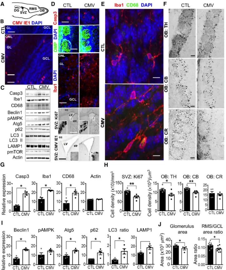
Congenital CMV infection leads to OB neuroinvasion,
apoptosis, and inflammation
Bridging the nose to higher brain structures, the OB circuit
con-stitutes the first central relay station of the olfactory system. The
olfactory deficits we reported here may result from damage to the
OB circuit by CMV-induced lesions in the placenta and/or
im-pairment of the olfactory system (
Fig. 5
A).
Immunohistochemi-cal analysis of brains at W3 after birth reveal viral expression in all
the OB layers of CMV-infected male mice (
Fig. 5
B), as well all in
the SVZ (
Fig. 5
D, down), consistent with previous studies (
Han et
al., 2007
;
Cekinovic´ et al., 2008
). These data indicate that, in our
animal model, transplacental CMV inoculation causes embryo
infection, neuroinvasion, and long-term viremia in the OB
last-ing at least 3 weeks after birth. Moreover, immunohistochemistry
and immunoblot experiments revealed that viral expression in
the OB is accompanied by increased apoptosis (
Fig. 5
B, C). In
particular, we found an increased number of cleaved Caspase 3
(Casp3)-positive apoptotic cells (
Fig. 5
B), as well as substantially
increased levels of Casp3 in CMV-infected mice at W3 compared
with CTL mice by immunoblot analysis of OB proteins (
Fig. 5
G,
CMV vs CTL: Casp3: Mann–Whitney test, U
(6)⫽ 0, p ⫽ 0.0286,
n
⫽ 4 mice per group).
The OB hosts a large population of resident microglial cells,
the innate immune cells of the brain. OB microglial cells can be
activated in response to pathological tissue changes, thus
migrat-ing to the site of injury, where they proliferate and acquire new
functions including phagocytic activity and cytokine secretion
(
Lazarini et al., 2012
,
2014
;
Hasegawa-Ishii et al., 2017
). Recent
studies have indicated a critical role of OB microglia in olfactory
processing (
Seo et al., 2016
;
Denizet et al., 2017
;
Reshef et al.,
2017
). We used Iba1, a marker of both unactivated and activated
microglia, to visualize these immune cells in the OB of
CMV-infected mice at W3 (
Fig. 5
D, middle, E). We found that
congen-ital CMV infection activates OB microglia, as indicated by a
change in morphology (
Fig. 5
E), an increased number of
Iba1-positive cells in all OB layers (
Fig. 5
E), and an increased
expres-sion of Iba1 and CD68, markers of activated microglia (
Fig. 5
C–
E). Consistent with immunochemistry results, we found
substantially increased levels of Iba1 and CD68 in CMV-infected
mice at W3 compared with CTL mice by immunoblot analysis of
OB proteins (
Fig. 5
C,G, CMV vs CTL: Mann–Whitney test, Iba1:
U
(6)⫽ 0, p ⫽ 0.0286; CD68: U
(6)⫽ 0, p ⫽ 0.0286, n ⫽ 4 mice per
group). Together, these findings reveal that congenital CMV
in-fection causes fetal inin-fection and leads to viral expression in the
OB at W3 that is accompanied by increased apoptosis and
inflammation.
Congenital CMV infection alters OB neuron content
The level of neuroinflammation and apoptosis in the OB of
CMV-infected mice at W3 prompted us to investigate whether
infection and inflammation disrupted normal OB development
and if a specific neuronal population was targeted. We observed
neither global change in the bulbar architecture nor histologic
changes in the lamination of the OB (
Fig. 5
D, middle). In
partic-ular, the mitral cell layer was similar in shape and size to those of
CTL, suggesting no damage of these projection cells. Despite the
presence of viral particles and microglial activation, OSN
inner-vation to the OB of CMV-infected mice was also similar to CTLs
(
Fig. 5
D). Consistent with those observations made in the OB, the
OE was similar in shape and size to those of CTL (thickness of the
OE, mean
⫾ SEM: CTL: 70.02 ⫾ 2.91
m; CMV: 72.08 ⫾ 2.86
m, CMV vs CTL: Mann–Whitney test, U
(11)⫽ 18, p ⫽ 0.701,
n
⫽ 6 CTL, n ⫽ 7 CMV). The number of OSNs also remained
unchanged, suggesting no damage of sensory neurons (mean
⫾
SEM cell density of OMP-positive cells (
⫻10
4)/mm
3: CTL:
40.44
⫾ 4.20; CMV: 37.61 ⫾ 3.67, CMV vs CTL: Mann–Whitney
test, U
(10)⫽ 14, p ⫽ 0.631, n ⫽ 5 CTL, n ⫽ 7 CMV).
We subsequently assessed whether the increase in cell
apopto-sis induced by CMV infection could lead to substantial changes in
neuronal subpopulations by visualizing CB-, calretinin (CR)-,
and TH-positive interneurons (
Fig. 5
F ). CB- and CR-positive
cells belong to the periglomerular neuron population that
sur-rounds the glomeruli. TH, the rate-limiting enzyme required for
the synthesis of dopamine, is localized in periglomerular neurons
and short axon cells. CB-, CR,- and TH-positive cells also contain
GABA and glutamic acid decarboxylase, the rate-limiting enzyme
for GABA biosynthesis. We found that CMV-infected mice
showed a reduced population of TH-positive cells in the OB
glomeruli at W3 after birth (
Fig. 5
F, H, CMV vs CTL: Mann–
Whitney test, U
(10)⫽ 4, p ⫽ 0.026; n ⫽ 6 mice per group). We
also observed a decreased population of CB-positive cells in the
glomeruli of these infected mice at W3 (
Fig. 5
F, H, CMV vs CTL:
Mann–Whitney test, U
(9)⫽ 0, p ⫽ 0.0043; n ⫽ 5 CTL, n ⫽ 6
CMV). In contrast, the cell density of CR-positive cells was
sim-ilar to CTL mice, indicating a higher vulnerability for bulbar
dopaminergic and CB- expressing neurons to CMV infection
(
Fig. 5
F, H, CMV vs CTL: Mann–Whitney test, U
(6)⫽ 6, p ⫽
0.6571; n
⫽ 4 mice per group). The depletion of TH- and
CB-positive cells observed in infected animals might reflect a direct
viral tropism for this neuronal category or result from
neuro-inflammation-induced cell death and/or reduced neurogenesis.
It is important to note that the neurogenic SVZ provides the
postnatal OB with interneurons throughout its lifespan in
ro-dents. This area gives rise to thousands of neuroblasts per day that
migrate via the RMS toward the OB, where they functionally
integrate with preexisting neuronal circuits and differentiate
mostly into granule cells and periglomerular interneurons (
Fig.
5
A;
Lledo et al., 2006
). Interestingly, we found a decrease in cell
proliferation in the SVZ at W3 (
Fig. 5
D, H, CMV vs CTL male
mice: Mann–Whitney test, U
(9)⫽ 0, p ⫽ 0.0043; n ⫽ 6 CTL, n ⫽
5 CMV), supporting the hypothesis that CMV might affect SVZ
neural progenitor cell proliferation and thus postnatal
neurogen-Figure 5. Impact of CMV congenital infection on the postnatal olfactory system at W3. A, Sagittal section of a murine brain showing the neurogenic dentate gyrus (DG) of the hippocampus, the neurogenic SVZ, the RMS, and the OB. Neuroblasts born in the neonatal and postnatal SVZ migrate via the RMS until the OB, where they differentiate into GCL or GL interneurons. B, D–F, Representative staining of coronal SVZ (D, bottom) and OB slices with DAPI (B, D, E) murine CMV IE1 (B, D, bottom), cleaved caspase 3 (Casp3, D, top), OMP expressed by OSN (D, middle), Iba (D, middle; E), CD68 expressed by activated microglia and macrophages (E), Ki67 (D), TH (F ), CB (F ), and CR (F ), antibodies showing CMV⫹, apoptotic Casp3⫹, OSN, macrophages, microglia, Ki67⫹ neural progenitor cells, TH⫹, CB⫹ and CR⫹ cells in CTL and congenital CMV-infected mice at W3 after birth. C, G–I, Screening of the OB proteins from congenital CMV-infected mice at W3 for autophagy induction (C, I ), cell apoptosis (C, G), and microglial reaction (C, G). Lysates were extracted from the OB of CTL and congenital CMV-infected mice at W3 and analyzed by immunoblot (C) using antibodies to detect Casp3, Iba1, CD68, Beclin1, phospho-AMPK, Atg5, p62, LC3 I/II, LAMP, phospho-mTOR, and actin (three mice each). The levels of Casp3, Iba, CD68, Beclin1, pAMPK, Atg5, p62, LC3 II/ LC3 I, LAMP, pmTOR, and actin were quantified (G,I ) by band intensity with Fiji software. H, J, Ki67⫹, TH⫹, CB⫹, and CR⫹ cell densities in the SVZ, GL, glomerulus (glom), and GCL at W3 following congenital CMV inoculation. All mice are W3-old males injected at E13 with PBS (CTL) or CMV. For immunoblot analysis (G, I ), n⫽4micepergroup.Forcelldensityanalysis(H)and area ratio (J ), n⫽ 4–6 mice per group. For glom size (J), n ⫽ 409 glom from 4 CTL, n ⫽ 352 glom from 4 CMV. Results are shown as mean ⫾ SEM. p-values were calculated by Mann–Whitney test. *p⬍ 0.05, **p ⬍ 0.01. Scale bars: 100m in D, bottom; 50 m in B, D, middle, and F; 25 m in D, bottom; and 5 m in D, top, and E.
esis in mice (
Mutnal et al., 2011
). However, further experiments
are needed to characterize the precise populations that are lost in
the SVZ, the fate of the proliferating survivor cells, and the impact
on neuronal progenitor migration and survival.
Finally, we assessed whether the decrease in the amount of
interneurons could change the OB size. Our anatomical
investi-gations demonstrated that CMV OB neuroinvasion was
accom-panied by differential changes in the relative size of the different
layers of the OB at W3, including the RMS, GL, and GCL (
Fig.
5
J ), which is composed of GABAergic interneurons that regulate
the neuronal activity of output OB neurons. To address the viral
impact on the OB innervation from the periphery, we measured
the glomerulus area using OMP staining. Congenital CMV
infection decreased the mean size of glomeruli in infected
male animals at W3 (
Figs. 5
J,
6
D; glomerulus area: CMV vs CTL;
Mann–Whitney test, U
(6)⫽ 0, p ⫽ 0.0286, n ⫽ 4 CTL (409
glomeruli), n
⫽ 4 CMV (304 glomeruli). Additionally, the RMS/
GCL area ratio is decreased in infected animals (
Fig. 5
J, CMV vs
CTL; Mann–Whitney test, U
(16)⫽ 14, p ⫽ 0.025, n ⫽ 10 CTL,
n
⫽ 8 CMV). We found a correlation between RMS and GCL
sizes in CTL OBs (Spearman r
⫽ 0.782, p ⫽ 0.011, n ⫽ 11 CTL),
but not in CMV-infected animals (Spearman’s r
⫽ 0.333, p ⫽
0.428, n
⫽ 8 CMV), suggesting that congenital CMV infection
differentially alters the development of these OB cell layers, thus
leading to disproportionate OB structure. A slight difference
be-tween CTL and CMV was found for either body weight (mean
⫾
SEM in CTL: 18.53
⫾ 0.50 g; in CMV: 16.94 ⫾ 0.54 g; Mann–
Whitney test, U
(46)⫽ 160.5, p ⫽ 0.0257, n ⫽ 17 CTL, n ⫽ 31
CMV or brain weight (mean
⫾SEM in CTL: 500.8 ⫾ 14.42 mg; in
CMV: 464.4
⫾ 7.934 mg, Mann–Whitney test, U
(46)⫽ 172, p ⫽
0.0490, n
⫽ 17 CTL, n ⫽ 31 CMV). However, no correlation was
found between body and brain weight for each group (data not
shown). Finally, no correlation was found between RMS and GL,
GCL and GL, GL and the mouse body weight, or brain weight and
body weight at W3 in the both groups (data not shown).
There-fore, CMV-infected preweaning male mice exhibit OB
propor-tionate and disproporpropor-tionate developmental abnormalities,
neuroinflammation, and fewer bulbar interneuron cells that
to-gether might contribute to an olfactory deficit.
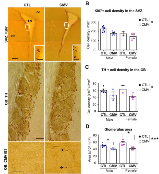
Figure 6. Long-lasting impact of CMV congenital infection on the adult OB. A, Representative staining of coronal SVZ (top) and OB (middle, bottom) slices with Ki67 (top), TH (middle) and murine CMV IE1 (bottom). B–D, Ki67⫹celldensitiesintheSVZ(B),TH⫹celldensitiesintheGL(C),andglomerulussize(D)atW16afterbirthfollowingcongenitalCMVinoculation.Forcelldensityanalysis,
n⫽ 5 male CTL, n ⫽ 2 female CTL, n ⫽ 4 male CMV, n ⫽ 4 female CMV. For glomerulus size, n ⫽ 212 glomeruli from 2 CTL females, n ⫽ 409 glomeruli from 4 CMV females, n ⫽ 499 glomeruli
from 4 CTL males, n⫽ 262 glomeruli from 4 CMV males. Results in D are shown as mean ⫾ SEM. p-values were calculated by Mann–Whitney test. *p ⬍ 0.05, ***p ⬍ 0.001. Cc, Corpus callosum; LV, lateral ventricle; str, striatum. Scale bars: 100m in A, top; 50 m in A, middle and bottom).