The Atger3 promoter confers circadian clock-regulated transcription with
peak expression at the beginning of the night
Dorothee Staiger
∗, Klaus Apel and Gian Trepp
1Institute for Plant Sciences, Swiss Federal Institute of Technology, ETH Center, 8092 Zurich, Switzerland (∗author for correspondence);1present address: University of Fribourg, 1705 Fribourg, Switzerland
Received 25 January 1999; accepted in revised form 17 May 1999
Key words: Arabidopsis thaliana, circadian rhythm, germin-like protein, promoter
Abstract
In Arabidopsis thaliana, steady-state abundance of the Atger3 transcript encoding a germin-like cell wall protein follows a circadian rhythm, reaching its highest level at the beginning of the night. As a first step towards dissecting the molecular mechanisms underlying these transcript oscillations, the Atger3 genomic locus was characterised. Transcriptional fusions of 1.8 kb and 967 bp Atger3 promoter fragments to the β-glucuronidase (GUS) reporter gene mediate high-amplitude circadian oscillations of the GUS transcript in transgenic Arabidopsis. 50deletion to −490 greatly reduces overall transcript abundance while retaining a basal oscillation. Further deletion to −299 abolishes preferential GUS expression in the evening. Taken together, these data indicate that clock-response elements contributing to high-amplitude Atger3 oscillations largely reside between−299 and −967. Histochemical staining for GUS activity indicates that the Atger3 promoter is active in cotyledons, young leaves, petioles, the inflorescence axis, pedicels, sepals, ovary, style and siliques but not in roots, petals and anthers.
Introduction
In most organisms, many cellular functions follow an endogenous daily rhythm. This adaption to the al-ternation of day and night allows anticipation rather than mere reaction to reiterative changes in ambi-ent light and temperature. These approximately 24 h, ‘circadian’, rhythms continue under constant external conditions. Under the natural cycle of light and dark-ness they are synchronised to follow these external changes to ensure cellular functions to occur at an ap-propriate time of the day (Bünning, 1977; Sweeney, 1987). In this way, interfering reactions such as pho-tosynthesis and nitrogen fixation in cyanobacteria can be directed to different phases of the light/dark cycle (Mitsui et al., 1986). Rhythmic processes in higher plants include leaf movement (Engelman et al., 1992), stem growth (Lecharny and Wagner, 1984), stomatal opening (Gorton et al., 1989), photosynthetic
activ-The nucleotide sequence data reported will appear in the EMBL, GenBank and DDBJ Nucleotide Sequence Databases under the accession number AJ132237.
ity (Hennessey and Field, 1991), photoperiodic flower induction (Bünning, 1936) as well as rhythmic expres-sion of specific genes (for a review, see Beator and Kloppstech, 1996).
Circadian regulation of mRNAs has first been demonstrated for those encoding the light-harvesting chlorophyll a/b-binding proteins (Lhc) (Kloppstech, 1985; Piechulla and Gruissem, 1987). Lhc transcript levels increase before dawn, peak in the morning and are almost undetectable throughout most of the night. These oscillations persist in the absence of external timing cues, when plants are transferred to continu-ous light, indicating an endogencontinu-ous control. Run-on transcription experiments in isolated nuclei demon-strated that the Lhc transcription rate is under circadian control in tomato and maize (Giuliano et al., 1988; Taylor, 1989). Reporter gene fusions have led to the identification of cis-acting promoter elements mediat-ing circadian regulation of Lhc expression in several plant species (Fejes et al., 1990; Millar and Kay, 1991; Carré and Kay, 1995; Ono et al., 1996; Piechulla et al., 1998).
For another morning-specific gene in Arabidopsis encoding ribulose-bisphosphate carboxylase activase, a promoter fragment extending from −313 to +13 relative to the transcription start site has been shown to mediate a low-amplitude circadian oscillation (Liu et al., 1996).
Although recently a few clock-controlled genes have been identified that reach their highest expression in the evening (Redinbaugh et al., 1990; Carpenter et al., 1994; Heintzen et al., 1994a,b; Zhong and Mc-Clung, 1996), the molecular basis of their regulation has not been investigated. Characterisation of such genes, with mRNA cycling roughly antiphase to the Lhc phase, would permit the establishment of addi-tional rhythmic molecular markers in Arabidopsis and thus would help to address questions of how temporal information from the endogenous clock is conveyed on differently phased output rhythms (for reviews, see McClung, 1998; Staiger and Heintzen, 1999).
In a systematic search for clock-regulated genes in Sinapis alba we previously identified a transcript with peak abundance about 12 h after onset of illumination (Heintzen et al., 1994a). This transcript codes for a cell-wall protein that shows homology to germin. Ger-min has first been found to be expressed during germi-nation in wheat (Lane et al., 1992). It turned out to be almost identical to barley oxalate oxidase and to have the same enzymatic activity. Therefore it has been sug-gested to contribute to cell wall remodelling, by virtue of the enzymatic breakdown of oxalate with concomi-tant release of hydrogen peroxide (Lane et al., 1993). Subsequently, several germin-type proteins have been described that are expressed at various developmental stages, upon diverse stress treatments and in response to fungal infection in cereals (Hurkman et al., 1994; Dumas et al., 1995; Hurkman and Tanaka, 1996; Zhou et al., 1998; Berna and Bernier, 1999). As H2O2serves as second messenger in the control of defence gene expression and contributes to oxidative cross-linking of cell wall proteins, germin-type proteins have been implicated as part of plant defence mechanisms.
Germin-like proteins (GLPs) have also been identi-fied in dicotyledonous plants (Michalowski and Bohn-ert, 1992; Heintzen et al., 1994a). In Arabidopsis, expressed sequence tags for at least twelve GLP genes have been found (Membre et al., 1997; Carter et al., 1998). So far no oxalate oxidase activity has been demonstrated for these proteins and their exact func-tion has remained elusive.
Circadian oscillations have been found for the transcript encoding the germin-like cell wall protein
SaGLP in Sinapis alba (Heintzen et al., 1994a) and its Arabidopsis counterpart AtGER3 (Membre et al., 1997; D. Staiger, unpublished) as well as for a tran-script in Pharbitis nil (Ono et al., 1996). Diurnal regulation of mRNA abundance was also shown for a germin-like protein in barley (Vallelian-Bindschedler et al., 1998).
In order to begin to understand the clock regula-tion of the Atger3 gene, we have isolated the genomic sequence from Arabidopsis and tested the 50-upstream region for rhythmicity and organ-specific expression using β-glucuronidase reporter gene fusions in trans-genic Arabidopsis.
Materials and methods
Isolation of a genomic Atger3 clone
The Atger3 clone was isolated from a λ EMBL4 li-brary (kindly provided by Dr Christiane Nawrath and Dr Csaba Koncz) by standard plaque screening proce-dures. Hybridisation with an Atger3 cDNA fragment, labelled with the Prime it II kit (Stratagene), was per-formed according to Yang et al. (1993). Final washes were done with 0.2× SSC at 62◦C.
An EcoRI fragment comprising the Atger3 locus was isolated from plaque-purified phage and sub-cloned into pBSK− (Stratagene). The sequence of both strands was determined by dideoxy sequencing (Sanger et al., 1977).
Primer extension
The transcription start site was determined by primer extension. An antisense oligonucleotide spanning positions 70 to 99 was end-labelled with γ -32P ATP and polynucleotide kinase and hybridised to 2 µg polyadenylated Arabidopsis RNA from plants harvested at zt12 (zeitgeber time 12; 12 h after lights on). Reverse transcription and analysis on 6% polyacrylamide urea gels was performed essentially according to Ausubel et al. (1987).
Chimeric gene constructs
Constructs are schematically depicted in Figure 3. To generate the translational Atger3 fusion with β-glucuronidase, an NcoI site was engineered at the ATG start codon by PCR. For construct TL, the 1.9 kb 50 -upstream region was inserted in front of the GUS gene in a vector based on pRT104 (Töpfer et al., 1987).
For the transcriptional fusions, a BglII site was en-gineered at the transcription start site by PCR. The tobacco mosaic virus omega element (Gallie et al., 1987) was used as a heterologous 50-untranslated leader. The 1.8 kb promoter region (construct TR) and the truncated fragments 11 and 12 (cf. Figure 3) were cloned between the BglII and SmaI site in pRT104 containing a SmaI-BglII linker upstream of the omega element/GUS gene fusion.
Cauliflower mosaic virus (CaMV) 35S RNA en-hancer fusions were constructed by inserting the At-ger3 promoter subfragments spanning−299 to +3 and −134 to +3 via blunt-ended restriction sites at their 50 end and the newly introduced BglII site at the tran-scription start site between the EcoRV site (−90) of the CaMV enhancer and the omega element in front of GUS in a vector based on pRT104 to yield the constructs CaMV-13 and CaMV-14, respectively.
All constructs were verified by sequencing. The promoter-GUS fusions were subcloned into pBin19 (Bevan, 1984) and transformed into Agrobacterium tumefaciens.
Transformation of Arabidopsis plants
The translational fusion was introduced into A. thaliana ecotype C24 by Agrobacterium-mediated root transformation (Valvekens et al., 1988). The tran-scriptional fusions were transformed into A. thaliana ecotype Columbia by vacuum infiltration (Bechtold et al., 1993). Kanamycin-resistant seedlings were se-lected and grown to flowering in soil. Southern blot analysis verified that the transgenic plants contained intact Atger3-GUS fusion constructs.
Northern blot kinetics
T2 seeds were germinated on 0.5× MS plates (Murashige and Skoog, 1962) containing 1% sucrose and 50 µg/ml kanamycin. Resistant plantlets were transferred to 0.5× MS plates. Three to four weeks after sowing, plants were harvested at 4 h intervals throughout a light/dark cycle (LD), as indicated in the figure legends, and on the second day after transfer to continuous light (LL).
RNA isolation and northern blotting were per-formed as described (Heintzen et al., 1994a, b, 1997). After determination of OD260 the individual RNA samples were adjusted to the same concentra-tion (1 µg/µl) to increase accuracy of the photometer reading and double-checked by a second OD measure-ment. 15 µg of total RNA were separated on 1.2%
agarose formaldehyde gels. Blots were consecutively hybridized with the β-glucuronidase-coding region, the Atger3 cDNA and a 26S rDNA probe to confirm equal loading.
Histochemical staining
Samples collected at the time of the circadian max-imum (zt12) were infiltrated with 0.1% 5-bromo-4-chloro-3-indolyl glucuronide in 50 mM sodium phos-phate buffer pH 7 containing 0.1% Triton X-100, 5 mM K3Fe(CN)6, 5 mM K4Fe(CN)6, 10 mM EDTA and stained at 37◦C overnight (Jefferson et al., 1996). Subsequently, samples were washed with 50 mM sodium phosphate and cleared of chlorophyll with 70% ethanol.
To generate tissue sections, specimens were fixed with 0.25% glutaraldehyde/3% paraformaldehyde in 0.1 M sodium phosphate buffer and stained with Ruthenium Red. Samples were dehydrated in a graded ethanol series and embedded in Technovit 7100 methacrylate resin according to the supplier’s instruc-tions (Kulzer Heraeus, Friedrichsdorf, Germany). Sec-tions were inspected with an Axiophot microscope.
Results
Isolation of the A. thaliana germin-like protein 3 gene 200 000 pfu of an A. thaliana genomic library in EMBL4 were screened with an Atger3 cDNA frag-ment which we previously isolated from an evening-specific λgt10 library (Membre et al., 1997; D. Staiger, unpublished). Nine positive clones were de-tected, which fell into two classes. Restriction map-ping and Southern hybridisation indicated that these clones largely overlapped, with one class extending about 1 kb further at the 50end.
A 6.6 kb EcoRI fragment of the larger clone was subcloned into pBSK− (Stratagene) and mapped in detail. A 2.9 kb EcoRI-KpnI fragment was identi-fied containing the entire coding region as well as 1.9 kb upstream of the ATG start codon. Collinearity of the clone with the A. thaliana genomic sequence was proven by Southern blotting: digests of A. thaliana DNA with EcoRI/XhoI and XmnI/SnaBI were probed with a radioactively labelled fragment covering the 50 -untranslated and putative promoter region. Identical bands were recognised by this probe in the genomic DNA as well as in the clone (not shown).
Figure 1. Scheme showing the relative location of the Atger3 genomic region (accession number AJ132237) and the ubiquitin4 genomic region (accession number U33014; Burke et al., 1988; Callis et al., 1995). Numbering is relative to the Atger3 transcription start site (+1), as determined by primer extension (cf. Figure 2). The overlap between the two genomic regions is delineated by the dotted line.
Sequence analysis
The sequence of both strands was determined after generating suitable subclones. A scheme of the Atger3 genomic fragment comprising 1.9 kb of upstream re-gion, the entire coding region and 0.3 kb downstream of the stop codon is presented in Figure 1. Comparison with the Atger3 cDNA (accession number Y12673; Membre et al., 1997) revealed the absence of introns. Two presumptive ATG translation initiation codons are found in frame at positions 47 and 53 relative to the start site of transcription. The deduced amino acid sequence contains an extracellular targeting peptide of 18 to 20 amino acids.
The Atger3 deduced amino acid sequence shares 67% sequence identity with the coding region of an auxin-binding protein, ABP20, from Prunus persica (Ohmiya et al., 1988), indicating that among the Ara-bidopsis germin-like proteins AtGER3 is related to ABP20.
Database searches indicated that the closest neigh-bour located downstream of Atger3 is the ubiquitin4 gene (accession number U33014; Burke et al., 1988; Callis et al., 1995; Carter et al., 1998; F. Bernier, per-sonal communication). The Atger3 promoter starting from position−1156 as well as the coding sequence and the 30-untranslated region are entirely contained within the described polyubiquitin genomic region (cf. Figure 1). The 30end of our genomic clone is located 555 bp upstream of the ubiquitin4 ATG start codon. Determination of the transcription start site
Poly(A) containing RNA from plants harvested at the circadian maximum (zt12) was used to delineate the transcription start site by primer extension. The RNA was hybridised to an end labelled antisense oligonu-cleotide spanning positions 70 to 99 within the coding region and was reverse-transcribed. Two strong and one weaker extension products clustered around bases
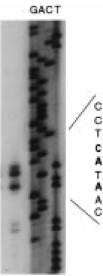
Figure 2. Determination of the Atger3 transcription start site. The gel shows primer extension products using an antisense oligonu-cleotide spanning positions 70 to 99 adjacent to dideoxy sequenc-ing ladders (lanes G, A, C, T). Part of the sequence (inverse of the sequencing reaction) is indicated with the C and A residues corresponding to the extension products in bold.
1757–1760 were detected (Figure 2), suggesting het-erogeneity in the mRNA 50ends or premature stops of the reverse transcriptase. The C corresponding to the longest extension product was taken as the transcrip-tion start site and numbered+1. The identified mRNA 50end conforms well to a consensus transcription start site CTCATCA deduced from 79 plant genes (Joshi, 1987).
Clock-response elements are located in the 50-flanking region
To determine whether clock-responsiveness of the At-ger3 gene resides within the 50-flanking region, the 1.9 kb fragment upstream of the ATG start codon was fused to the β-glucuronidase open reading frame (Fig-ure 3). Transgenic T2 plants harbouring this transla-tional fusion construct TL were grown in 8 h light/16 h
Figure 3. Scheme of the chimeric promoter-GUS constructs. Num-bering is relative to the Atger3 transcription start (+1), as determined by primer extension (cf. Figure 2). All constructs contain the CaMV 35S RNA terminator. TL, translational fusion; TC, transcriptional fusion; 1, deletion.
Figure 4. The Atger3 upstream region mediates oscillations of a linked β-glucuronidase reporter transcript. Plants harbouring the translational fusion TL of Atger3 (−1811 to +47) linked to the GUS coding region were grown in short days (8 h light) and subsequently transferred to continuous light (LL). Plants were harvested at 4 h intervals in light/dark cycles and on the second day in LL. The northern blot was hybridised with the GUS-coding region (upper row) and the endogenous Atger3 cDNA (lower row). Light and dark periods are represented by open and solid bars, respectively. The inserted bar in LL indicates the time interval corresponding to dark-ness during light/dark cycles (subjective night). Zt, zeitgeber time; h, hours after transfer to LL.
dark cycles and subsequently transferred to continuous light. Steady-state abundances of the β-glucuronidase reporter transcript as well as of the endogenous tran-script were determined by northern analysis. Fig-ure 4 shows that the β-glucuronidase transcript under control of the Atger3 upstream region oscillates in light/dark cycles, reaching its highest level around zt8 to zt12 (upper row), similar to the behaviour of the endogenous Atger3 transcript (lower row). These oscillations persist in constant light, indicating that the sequence upstream of the ATG start codon con-tains elements involved in circadian regulation of the Atger3 gene. Almost identical GUS mRNA kinetics were observed for four independent transgenic lines (not shown).
Transcriptional control of Atger3 oscillations
To determine whether the transcription of the Atger3 gene is regulated by the clock, 1.8 kb of the promoter were fused to the omega element as a heterologous 50
-Figure 5. Atger3 transcript oscillations are generated at the tran-scriptional level. Plants harbouring 1.8 kb of the Atger3 promoter fused to the tobacco mosaic virus omega element as a heterologous 50-untranslated region in front of GUS (construct TC) were grown in long days (16 h light) and subsequently transferred to continuous light (LL). Plants were harvested at 4 h intervals in light/dark cycles and on the second day in LL. The northern blot was hybridised with the GUS-coding region (upper row) and the endogenous At-ger3 cDNA (lower row). Light and dark periods are represented by open and solid bars, respectively. The inserted bar in LL indicates the time interval corresponding to darkness during light/dark cycles (subjective night). Zt, zeitgeber time; h, hours after transfer to LL.
untranslated region (construct TC; Figure 3). T2 plants of ten independent kanamycin-resistant transformants were tested for GUS mRNA levels. High GUS tran-script levels are found in the evening (zt12) whereas almost no GUS transcript is detectable in the morning (zt0–zt4). A detailed RNA kinetic for a representative line harvested at four-hour intervals in 16 h light/8 h dark cycles and on the second day after transfer to continuous light is shown in Figure 5. The temporal pattern of GUS mRNA levels under control of the Atger3 promoter (upper row) corresponds to the os-cillations of the endogenous Atger3 transcript (lower row), demonstrating that the transcription of the At-ger3 gene is under control of the endogenous clock. Maximal Atger3 expression occurs around zt12 to zt16 in long-day conditions (16 h light) as compared to zt8 to zt12 in short-day conditions (8 h light) (cf. Figure 4; Heintzen et al., 1997).
The peak-to-trough difference in expression was retained when the promoter was shortened to−967: GUS expression of independent transgenic lines har-bouring construct 11 is significantly higher in plants harvested at the circadian maximum (zt12) than in plants harvested at the circadian minimum (zt4), shown for five representative lines in Figure 6A. Fur-ther truncation to −490 resulted in a large drop in expression level. GUS mRNA levels still are slightly higher in the evening (zt12) than in the morning (zt4), indicating that a basal circadian regulation is retained in construct 12 (Figure 6 B).
To be able to discern whether shorter promoter deletion would drive circadian oscillations, the 35S CaMV enhancer was positioned in front of these short fragments to increase overall transcript abundance
Figure 6. GUS expression driven by truncated Atger3 promoter fragments. Independent lines of transgenic plants harbouring dele-tion constructs 11 (A), 12 (B), CaMV-13 (C) and CaMV-14 (D) (cf. Figure 3) were grown under long-day conditions (16 h light/8 h dark). RNA was isolated from plants harvested at the circadian maximum (zt12) and the circadian minimum (zt4). The northern blot was hybridised with the GUS-coding region. Note that the blot with construct 12 shown in B was exposed about five times as long as the blot with 11 shown in A. The ethidium bromide-stained RNA gels are shown below each autoradiagram as loading control.
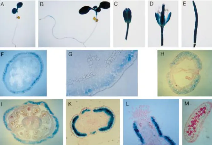
(Figure 3). This approach was also applied to deter-mine nodule-specific cis elements within a soybean leghaemoglobin promoter (Stougaard et al., 1987). Transgenic plants harbouring the constructs CaMV-13 with a deletion to position −299 and CaMV-14 ending at−134 were analysed for GUS mRNA lev-els at the circadian maximum (zt12) and the circadian minimum (zt4). GUS transcript levels were not signifi-cantly different in plants harvested at zt12 as compared to plants harvested at zt4, as shown for representative lines of each construct in Figures 6C and 6D, although the individual lines displayed some variation in overall expression levels. Taken together, these data indicate the presence of at least two cis-acting elements con-tributing to high-amplitude Atger3 oscillations within the promoter, residing between positions −299 and −490 and between −491 and −967, respectively. Histochemical localisation of GUS expression Histochemical staining for GUS activity in transgenic Arabidopsis harbouring the transcriptional fusion TC revealed GUS expression in cotyledons (Figure 7A, B), young leaves, petioles (B), flowers and pedicels (C, D) as well as siliques (E). No GUS expression was
found in roots (A, B). In the inflorescence axis, highest expression for the translational fusion TL is found in the subepidermal cortical cells (F, G). In flowers, GUS expression is found in the sepals except the vascular tissue (I). Expression is also found in the subepidermal cell layers of the ovary and style (K, L) but is absent from petals and anthers (I, M). In the siliques, staining is found in the parenchymal cells (H).
Discussion
Previous work has identified a transcript encoding the germin-like cell wall protein SaGLP in Sinapis alba that oscillates with peak abundance in the evening (Heintzen et al., 1994a). Here we characterise the 50-upstream region of its Arabidopsis counterpart At-ger3. In particular, we wanted to determine whether the promoter mediates circadian clock regulation at the transcriptional level.
We could show that 1.9 kb upstream of the transla-tion start codon (construct TL) mediate oscillatransla-tions of a linked GUS reporter transcript with peak abundance at the end of the daily light period. The oscillations persist under continuous light. No attempts were made to monitor GUS expression in continuous darkness in the transgenic plants: Atger3 expression rapidly damps to levels below the detection limit in constant dark, similar to the Saglp transcript (Heintzen et al., 1994a; D. Staiger, unpublished observation).
Clock regulation occurs predominantly at the level of transcription, as the Atger3 promoter fragment from −1811 to +3 relative to the transcription start site linked to a heterologous 50-untranslated leader (con-struct TC) directs rhythmic GUS mRNA expression. Clock regulation of gene expression in higher plants appears to occur mostly at the level of transcription. Rhythmic transcription has also been found for cata-lase3 in maize (Redinbaugh et al., 1990) and the Lhc genes in wheat, maize, tomato and Arabidopsis (Giuliano et al., 1988; Nagy et al., 1988; Taylor, 1989; Millar and Kay, 1991). In the case of the Arabidopsis Lhc1∗3 (cab1) gene a post-transcriptional control mechanism seems to operate in addition to rhythmic transcription, leading to a relatively con-stant steady-state Lhc1∗3 transcript level (Millar and Kay, 1991). For nitrate reductase2 in Arabidopsis, nuclear run-on experiments revealed predominantly post-transcriptional control of the mRNA oscillations (Pilgrim et al., 1993).
Figure 7. Histochemical localisation of GUS activity in transgenic Arabidopsis plants harbouring the transcriptional Atger3-GUS fusion TC (A–E) or the translational Atger3-GUS fusion TL (F–M), respectively, (cf. Figure 3). A, 6-day old seedling; B, 11-day old plant; C, flower bud; D, flower; E, silique; F, cross-section through inflorescence axis; G, detail of F; H, cross-section through silique; I, cross-section through flower; K, cross section through ovary; L, longitudinal section through style at anthesis; M, longitudinal section through anther.
Circadian-regulated expression with a high peak-to-trough difference is also detected when the Atger3 promoter is shortened to−967 (construct 11). Con-struct 12 retaining 490 bp upstream of the transcrip-tion start site is still expressed at a slightly higher level at the circadian maximum as compared to the circa-dian minimum although overall transcript abundance is greatly reduced. Constructs 13 and CaMV-14 with 50-end points at−299 and −134, respectively, show about equal expression at the circadian max-imum and the circadian minmax-imum. Taken together, these data indicate that sequence elements contribut-ing to preferential expression at the beginncontribut-ing of the night largely reside between−967 and −299. A clock response element mediating a basal oscillation seems to be present between−299 and −490. A second ele-ment contributing to high-amplitude cycling is located between −491 and −967. The weak expression of construct 12 precluded a definite decision whether motifs located within the −491/−967 region would
act as general activating sequences or selectively boost expression at the time of the circadian maximum. This distinction may be possible with the use of more sen-sitive assay techniques such as in vivo measurements of promoter-luciferase fusions (Carré and Kay, 1995). Formally we cannot entirely exclude that an additional element mediating a low-level amplitude oscillation may have gone unnoticed in the CaMV constructs be-cause such a clock response element might not have acted in concert with the CaMV enhancer.
Recently, the MYB-type protein CCA1 origi-nally identified as a trans-acting factor binding to a phytochrome-responsive promoter element of an Ara-bidopsis Lhc gene has been shown to be part of a negative feedback loop involved in rhythm genesis (Wang et al., 1997; Wang and Tobin, 1998). CCA1 overexpression negatively affects the oscillations of transcripts with different phases such as Lhc and the endogenous cca1 transcript that peak in the morning and the Atgrp7/ccr2 transcript peaking later in the day.
Whether CCA1 or related factors also regulate the At-ger3 transcript with maximal expression at zt12 is not known. Interestingly, one motif identical to the CCA1 binding site AAA/CAATCT, as determined previously (Wang et al., 1997; Wang and Tobin, 1998), is located at −197 and four additional sequence elements dis-playing a 7 out of 8 bp identity are present at−315, −478, −780 and −1011 of the Atger3 promoter.
An almost perfect direct repeat of an 18 bp se-quence is found between −1091 and −1046. Its significance for Atger3 expression in not known at present.
The Atger3 promoter also mediates organ-specific expression of a linked GUS reporter gene. Expression was found in cotyledons and young leaves, but not in roots. In a northern blot analysis Atger3 mRNA pre-viously was detected in leaves, flower buds and open flowers, but not in siliques (Membre et al., 1997). This difference might be due to a different developmental state. In the inflorescence axis, expression is found in the subepidermal cortical cell layers. The expression pattern closely resembles the spatial expression of the mustard counterpart SaGLP, as determined by in situ hybridisation (Heintzen et al., 1994a). In flowers, ex-pression was found in sepals, ovary and style but not in petals and anthers. This organ-specific expression pattern could be due either to transcriptional activa-tion or negative regulaactiva-tion in distinct organs. Further dissection of the promoter is required to distinguish between these possibilities.
AtGER3 previously has been mapped to chro-mosome 5 between the nga106 and g4560 markers (Membre et al., 1997). Database searches with the genomic clone now revealed a tight head-to-tail link-age of the Atger3 gene to the polyubiquitin genomic region (Burke et al., 1988; Callis et al., 1995; Carter et al., 1998). The 30end of the Atger3 clone is located 555 bp upstream of the ubiquitin4 ATG start codon, thus leaving ca. 850 bp intergenic region between the Atger3 stop codon and the ubiquitin4 start codon. No-tably, the Atger3 coding region is contained within a 2 kb DNA fragment that previously has been used as a 50-specific hybridisation probe for the ubiquitin4 gene (Burke et al., 1988). This 50probe detected a 1.35 kb transcript whereas the ubiquitin coding regions ad-ditionally detected mRNAs of 1.9, 1.7 and 0.85 kb. Differences in organ-specific expression levels were revealed for the 1.35 kb transcript depending on the choice of the hybridisation probe. Notably, in heat-stressed Arabidopsis plants a small decrease of the 1.35 kb band was detected with the coding region as
probe, whereas a large decrease relative to untreated plants was observed upon hybridisation with the 50 probe. From this finding the authors concluded that an additional transcript must be present in the 1.35 kb size class whose regulation is distinct from that of ubiquitin4. It may be interesting to determine whether part of this differential behaviour might be due to the oscillating Atger3 transcript.
In conclusion, we have shown that the Atger3 promoter mediates cycling of a linked reporter gene with preferential expression at the beginning of the night. Sequence motifs contributing to high-amplitude cycling reside largely between positions −299 and −967. The Atger3 promoter should prove useful to generate markers for clock output differentially phased to the well known oscillations of Lhc promoter activity with peaks in the morning.
Acknowledgements
Our thanks to Drs Christiane Nawrath and Csaba Koncz for the EMBL4 genomic library. We are in-debted to Daniel Dreesmann for critical reading of the manuscript and to Dr Dieter Rubli for his help in preparing the figures. This work has been supported by grants from the Swiss National Science Foundation and the ETH to D.S.
References
Ausubel, F.M., Brent, R., Kingston, R.E., Moore, D.D., Seid-man, J.G., Smith, J.A. and Struhl, K. 1987. Current Protocols in Molecular Biology, John Wiley, New York.
Beator, J. and Kloppstech, K. 1996. Significance of circadian gene expression in higher plants. Chronobiol. Int. 13: 319–339. Bechtold, N., Ellis, J. and Pelletier, G. 1993. In planta
Agrobacterium-mediated gene transfer by infiltration of adult Arabidopsis thaliana plants. C.R. Acad. Sci. Paris, Sci. Vie/Life Sci. 316: 1194–1199.
Berna, A. and Bernier, F. 1999. Regulation by biotic and abiotic stress of a wheat germin gene encoding oxalate oxidase, a H2O2
-producing enzyme. Plant. Mol. Biol. 39: 539–549.
Bevan, M. 1984. Binary Agrobacterium vectors for plant transfor-mation. Nucl. Acids Res. 12: 8711–8721.
Bünning, E. 1936. Die endogene Tagesrhythmik als Grundlage der photoperiodischen Reaktion. Ber. Dt. Bot. Ges. 54: 590–607. Bünning, E. 1977. Die physiologische Uhr. Springer-Verlag,
Berlin/Heidelberg/New York.
Burke, T.J., Callis, J. and Vierstra, R.D. 1988. Characterization of a polyubiquitin gene from Arabidopsis thaliana. Mol. Gen. Genet. 213: 435–443.
Callis, J., Carpenter, T., Sun, C.W. and Vierstra, R.D. 1995. Structure and evolution of genes encoding polyubiquitin and ubiquitin-like protein in Arabidopsis ecotype Columbia. Genet-ics 139: 921–939.
Carpenter, C.D., Kreps, J.A. and Simon, A.E. 1994 Genes encoding glycine-rich Arabidopsis thaliana proteins with RNA-binding motifs are influenced by cold treatment and an endogenous circadian rhythm. Plant Physiol. 104: 1015–1025.
Carré, I.A. and Kay, S.A. 1995. Multiple DNA-protein complexes at a circadian-regulated promoter element. Plant Cell 7: 2039– 2051.
Carter, C., Graham, R.A. and Thornburg, R. 1998. Arabidopsis thaliana contains a large family of germin-like proteins: charac-terization of cDNA and genomic sequences encoding 12 unique family members. Plant Mol. Biol. 38: 929–943.
Dumas, B., Freyssinet, G. and Pallet, K.E. 1995. Tissue-specific expression of germin-like oxalate oxidase during development and fungal infection of barley seedlings. Plant Physiol. 107: 1091–1096.
Engelmann, W., Simon, K. and Phen, C.J. 1992. Leaf movement rhythms in Arabidopsis thaliana. Z. Naturforsch. 47c: 925–928. Fejes, E., Pay, A., Kanevsky, I., Szell, M., Adam, E., Kay, S.
and Nagy, F. 1990. A 268 bp upstream sequence mediates the circadian clock-regulated transcription of the wheat Cab-1 gene in transgenic plants. Plant Mol. Biol. 15: 921–932.
Gallie, D.R., Sleat, D.E., Watts, J.W., Turner, P.C. and Wilson, T.M.A. 1987. The 50-leader sequence of tobacco mosaic virus enhances the expression of foreign gene transcripts in vitro and in in vivo. Nucl. Acids Res. 15: 3257–3273.
Giuliano, G., Hoffman, N.E., Ko, K., Scolnik, P.A. and Cash-more, A.R. 1998. A light-entrained circadian clock controls transcription of several plant genes. EMBO J. 7: 3635–3642. Gorton, H.L., Williams, W.E., Binns, M.E., Gemmel, C.N., Leheny,
E.A. and Shepard, A.C. 1989. Circadian stomatal rhythms in epidermal peels from Vicia faba. Plant Physiol. 90: 1329–1334. Heintzen, C., Fischer, R., Melzer, S., Kappeler, K., Apel, K. and
Staiger, D. 1994a. Circadian oscillations of a transcript encoding a germin-like protein that is associated with cell walls in young leaves of the long-day plant Sinapis alba L. Plant Physiol. 106: 905–915.
Heintzen, C., Melzer, S., Fischer, R., Kappeler, S., Apel, K. and Staiger, D. 1994b. A light- and temperature-entrained circadian clock controls expression of transcripts encoding nuclear pro-teins with homology to RNA-binding propro-teins in meristematic tissue. Plant J. 5: 799–813.
Heintzen, C., Nater, M., Apel, K. and Staiger, D. 1997. AtGRP7, a nuclear RNA-binding protein as a component of a circadian-regulated negative feedback loop in Arabidopsis thaliana. Proc. Natl. Acad. Sci. USA 94: 8515–8520.
Hennessey, T.L. and Field, C.B. 1991. Circadian rhythms in photo-synthesis. Plant Physiol. 90: 1329–1334.
Hurkman, W.J., Lane, B.G. and Tanaka, C.K. 1994. Nucleotide sequence of a transcript encoding a germin-like protein that is present in salt-stressed barley (Hordeum vulgare L.) roots. Plant Physiol. 104: 803–804.
Hurkman, W.J. and Tanaka, C.K. 1996. Germin gene expression is induced in wheat leaves by powdery mildew infection. Plant Physiol. 111: 735–739.
Jefferson, R.A., Burgess, S.M. and Hirsh, D. 1996. β-glucuronidase from Escherichia coli as a gene-fusion marker. Proc. Natl. Acad. Sci. USA 83: 8447–8451.
Joshi, C.P. 1987. An inspection of the domain between putative TATA box and translation start site in 79 plant genes. Nucl. Acids Res. 15: 6643–6653.
Kloppstech, K. 1985. Diurnal and circadian rhythmicity in the ex-pression of light-induced plant nuclear messenger RNAs. Planta 165: 502–506.
Lane, B.G., Cuming, A.C., Fregeau, J., Carpita, N.C., Hurk-man, W.J., Bernier, F., Dratewka-Kos, E. and Kenney, T.D. 1992. Germin isoforms are discrete temporal markers of wheat development. Eur. J. Biochem. 209: 961–969.
Lane, B.G., Dunwell, J.M., Ray, J.A., Schmitt, M.R. and Cuming, A.C. 1993. Germin, a protein marker of early plant development, is an oxalate oxidase. J. Biol. Chem. 268: 12239–12242. Lecharny, A. and Wagner, E. 1984. Stem extension rate in
light-grown plants. Evidence for an endogenous circadian rhythm in Chenopodium rubrum. Plant Physiol. 60: 437–443.
Liu, Z., Taub, C.C. and McClung, C.R. 1996. Identification of an Arabidopsis thaliana ribulose-1,5-bisphosphate carboxy-lase/oxygenase activase (RCA) minimal promoter regulated by light and the circadian clock. Plant Physiol. 112: 43–51. McClung, C.R. 1998. It’s about time: putative components of an
Arabidopsis circadian clock. Trends Plant Sci. 3: 454–456. Membre, N., Berna, A., Neutelings, G., David, A., David, D.,
Staiger, D., Vasquez, J.S., Raynal, M., Delseney, M. and Bernier, F. 1997. cDNA sequence, genomic organisation and differen-tial expression of three Arabidopsis genes for germin/oxalate oxidase-like proteins. Plant Mol. Biol. 35: 459–469.
Michalowski, C.B. and Bohnert, H.J. 1992. Nucleotide sequence of a root-specific transcript encoding a germin-like protein from the halophyte Mesembryanthemum crystallinum. Plant Physiol. 100: 537–538.
Millar, A.J. and Kay, S.A. 1991. Circadian control of cab gene tran-scription and mRNA accumulation in Arabidopsis. Plant Cell 3: 541–550.
Mitsui, A., Kumazawa, S., Takahashis, A., Ikemoto, H. and Arai, T. 1986. Strategies by which nitrogen-fixing unicellular cyanobacteria grow photoautotrophically. Nature 323: 720–722. Murashige, T. and Skoog, F. 1962. A revised method for rapid growth and bio assays with tobacco tissue cultures. Physiol. Plant. 15: 473–497.
Nagy, F., Kay, S.A. and Chua, N.-.H 1988. A circadian clock regulates transcription of the wheat Cab-1 gene. Genes Dev. 2: 376–382.
Ohmiya, A., Tanaka, Y., Kadowaki, H.-i. and Hayashi, T. 1988. Cloning of genes encoding the auxin-binding proteins (ABP19/20) from peach: significant peptide sequence similarity with germin-like proteins. Plant Cell Physiol. 39: 492–499. Ono, M., Sage-One, K., Inoue, M., Kamada, H. and Harada, H.
1996. Transient increase in the level of mRNA for a germin-like protein in leaves of the short-day plant Pharbitis nil during the photoperiodic induction of flowering. Plant Cell Physiol. 37: 855–861.
Piechulla, B. and Gruissem, W. 1987. Diurnal mRNA fluctuations of nuclear and plastid genes in developing tomato fruits. EMBO J. 6: 3593–3599.
Piechulla, B., Merforth, N. and Rudolph, B. 1998. Identification of tomato Lhc promoter regions necessary for circadian expression. Plant Mol. Biol. 38: 655–662.
Pilgrim, M.L., Caspar, T., Quail, P.H. and McClung, C.R. 1993. Circadian and light-regulated expression of nitrate reductase in Arabidopsis. Plant Mol. Biol. 23: 349–364.
Redinbaugh, M.G., Sabre, M. and Scandalios, J.G. 1990. Expres-sion of the maize Cat3 catalase gene is under influence of a circadian rhythm. Proc. Natl. Acad. Sci. USA 87: 6853–6857. Sanger, F., Nicklen, S. and Coulson, A.R. 1977. DNA sequencing
with chain-terminating inhibitors. Proc. Natl. Acad. Sci. USA 74: 5463–5467.
Staiger, D. and Heintzen, C. 1999. The circadian system of Arabidopsis thaliana: forward and reverse genetic approaches. Chronobiol. Int. 16: 1–16.
Stougaard, J., Sandal, N.N., Gron, A., Kühle, A. and Marker, K.A. 1987. 50analysis of the soybean leghaemoglobin lbc3gene:
regulatory elements required for promoter activity and organ specificity. EMBO J. 6: 3565–3569.
Sweeney, B.M. 1987. Rhythmic Phenomena in Plants. Academic Press, San Diego, CA.
Taylor, W. 1989. Transcriptional regulation by a circadian rhythm. Plant Cell 1: 259–264.
Töpfer, R., Matzeit, V., Gronenborn, B., Schell, J. and Steinbiss, H.-H. 1987. A set of plant expression vectors for transcriptional and translational fusions. Nucl. Acids Res. 15: 5890.
Vallelian-Bindschedler, L., Mösinger, E., Metraux, J.-P. and Schweizer, P. 1998. Structure, expression and localization of a germin-like protein in barley (Hordeum vulgare L.) that is insolubilized in stressed leaves. Plant Mol. Biol. 37: 297–308. Valvekens, D., Van Montagu, M. and Van Lijsebettens, M. 1988.
Agrobacterium tumefaciens-mediated transformation of Ara-bidopsis thaliana root explants by using kanamycin selection. Proc. Natl. Acad. Sci. USA 85: 5536–5540.
Wang, Z.-Y., Kenigsbuch, D., Sun, L., Harel, E., Ong, M.S. and To-bin, E.M. 1997. A Myb-related transcription factor is involved in the phytochrome regulation of an Arabidopsis Lhcb gene. Plant Cell 9: 491–507.
Wang, Z.-Y. and Tobin, E.M. 1998. Constitutive expression of the CIRCADIAN CLOCK ASSOCIATED 1 (CCA1) gene disrupts circadian rhythms and suppresses its own expression. Cell 93: 1207–1217.
Yang, H., McLesse, J., Weisbart, M., Dionne, J.-L., Lemaire, I. and Aubin, R.A. 1993. Simplified high throughput protocol for northern hybridization. Nucl. Acids Res. 21: 3337–3338. Zhong, H.H. and McClung, C.R. 1996. The circadian clock gates
expression of two Arabidopsis catalase genes to distinct and opposite circadian phases. Mol. Gen. Genet. 251: 196–203. Zhou, F., Zhang, Z., Gregersen, P.L., Mikkelsen, J.D., de Neergaard,
E., Collinge, D.B. and Thordal-Christensen, H. 1998. Molecular characterization of the oxalate oxidase involved in the response of barley to the powdery mildew fungus. Plant Physiol. 117: 33– 41.