Kan Min Frederik Hahn Kai Ziebarth
Short anterior correction
of the thoracolumbar/lumbar curve
in King 1 idiopathic scoliosis: the behaviour
of the instrumented and non-instrumented
curves and the trunk balance
Received: 29 July 2005 Revised: 28 November 2005 Accepted: 21 January 2006 Published online: 17 March 2006 Ó Springer-Verlag 2006
Abstract This is a retrospective clinical, radiological and patient outcome assessment of 21 consecu-tive patients with King 1 idiopathic adolescent scoliosis treated by short anterior selective fusion of the major thoracolumbar/lumbar (TL/L) curve. Three-dimensional changes of both curves, changes in trunk bal-ance and rib hump were evaluated. The minimal follow-up was
24 months (max. 83). The Cobb angle of the TL/L curve was 52° (45–67°) with a flexibility of 72% (40–100%). The average length of the main curve was 5 (3–8) segments. An average of 3 (2–4) segments was fused using rigid single rod implants with side-loading screws. The Cobb angle of the thoracic curve was 33° (18–50°) with a flexibility of 69% (29–100%). The thoracic curve in bending was less than 20° in 17 patients, and 20–25° in 4 patients. In the TL/L curve there was an improvement of the Cobb angle of 67%, of the apex vertebral rotation of 51% and of the apex vertebral translation of 74%. The Cobb angle of the thoracic curve improved 29% spontaneously. Shoulder balance improved significantly from an average preoperative imbalance of 14.5–3.1 mm at the last follow-up.
Seventy-five percent of the patients with preoperative positive shoulder imbalance (higher on the side of the thoracic curve) had levelled shoul-ders at the last follow-up. C7 offset improved from a preoperative 19.8 (0–40) to 4.8 (0–18) mm at the last follow-up. There were no significant changes in rotation, translation of the thoracic curve and the clinical rib hump. There were no significant changes in thoracic kyphosis or lumbar lordosis. The average score of the SRS-24 questionnaire at the last follow-up was 91 points (max. 120). We conclude that short ante-rior selective fusion of the TL/L curve in King 1 scoliosis with a thoracic curve bending to 25° or less (Type 5 according to Lenke classifi-cation) results in a satisfactory cor-rection and a balanced spine. Short fusions leave enough mobile lumbar segments for the establishment of global spinal balance. A positive shoulder imbalance is not a contra-indication for this procedure. Struc-tural interbody grafts are not necessary to maintain lumbar lordosis.
Keywords Idiopathic scoliosis Æ Anterior correction Æ Trunk balance Æ Shoulder balance Æ Rib
K. Min (&) Æ F. Hahn Æ K. Ziebarth Orthopedics, Universtity of Zurich, Zurich, Switzerland
E-mail: kan.min@balgrist.ch Tel.: +41-1-3861111 Fax: +41-1-3861269
Introduction
Type I idiopathic scoliosis described by King et al. [8] is a s-shaped curve in which both thoracic and lumbar curves cross the midline, the lumbar curve being larger and/or less flexible than the thoracic curve. Where surgical correction was indicated they recommended the posterior fusion of both curves to the fourth lum-bar vertebra. The anterior correction of thoracic as well as thoracolumbar/lumbar (TL/L) scoliosis was first described by Dwyer [5]. Zielke [21] modified the tech-nique by replacing the wires with threaded rods to connect the vertebral screws. Hall put forward the technique of short fusion in TL/L scoliosis using the Zielke implant and described the selection of fusion levels [7]. Since the introduction of rigid rod implants in the early 1990s there have been some reports on the anterior correction of TL/L scoliosis, most of the au-thors describing the fusion of the whole Cobb curve from the upper to the lower end vertebrae [1, 11, 17, 18]. There are very few reports about the behaviour of the non-instrumented thoracic curve after short ante-rior fusion of the TL/L curve in King 1 type idiopathic scoliosis [2, 3]. The possibility of spinal imbalance is a concern in fusing the lower curve selectively in King 1 type scoliosis [4]. To our knowledge there are no re-ports about the changes in shoulder balance, trunk balance and clinical rib hump after short anterior selective fusion of the lower curve in King I type sco-liosis. In this report we describe the three-dimensional changes of the upper and lower curve, the changes in body balance, shoulder symmetry and thoracic rib hump after selective short fusion of the TL/L curves in King I idiopathic adolescent scoliosis.
Materials and methods
Twenty-one consecutive patients with King 1 adolescent idiopathic scoliosis operated in our institution between March 1997 and July 2002 with an average follow-up of 45 (24–83) months were retrospectively analysed. All patients had major TL/L curves and minor thoracic curves. Only the major curves were instrumented (Fig.1). Patients with a single TL/L curve without compensatory thoracic curve were excluded from this study. There were 19 female and 2 male patients. The average age at the time of operation was 16.3 (10.5–28.3) years. Risser stage of every patient at the time of surgery is listed in Table1. The average size of the major curve was 52° (45–67°) and the flexibility in side bending was 72% (40–100%). Five were right sided and 16 were left sided curves. The average size of the thoracic curve was 33° (18–50°) with a flexibility of 69% (29–100%). In 17 patients the thoracic curve bended to
less than 20°. In the remaining four patients the thoracic curve was between 20° and 25° in bending. The thoracic curve crossed the midline in all patients.
Short segment fusion of the TL/L curve was carried out according to Hall [2,7]. The standing ap whole spine radiograph was used for choosing fusion levels. If the apex of the curve was a vertebral body then the apex vertebra, one vertebra above and one vertebra below were included. In one patient in whom the Cobb angle was more than 65°, two vertebrae above and two ver-tebrae below were included in addition to the apex vertebra. If the apex was an intervertebral disc then two vertebrae above and two vertebrae below were included for fusion. A lateral retroperitoneal or transpleural– retroperitoneal approach was used with rib resection performed two levels above the uppermost instrumented vertebra. Complete 360°-discectomies were carried out exposing the posterior longitudinal ligament without opening the spinal canal. Instrumentation was per-formed with a 6 mm single rod stainless steel implant (USS, SynthesÒ, Oberdorf, Switzerland). The contoured rod was first fixed to the uppermost and the lowest instrumented vertebrae. The side-loading vertebral screws were helpful to derotate the apical vertebrae separately. Partial rod derotation was carried out addi-tionally if necessary. The resected ribs were used for interbody bone grafting. No interbody structural sup-port was used. Segmental compression on the convexity was carried out for the final coronal plane correction. An overcorrection was avoided in curves less than 50° and a slight overcorrection was performed in curves more than 50°. An average of 2.9 (2–4) segments was fused. A single thorax drain was inserted before closing the thoracotomy. Intraoperative spinal cord monitoring with sensory and motor evoked potentials was routinely used. All operations were performed by the same sur-geon (KM). Sitting began on the first day, and walking on the second day after the surgery. The thorax drain was removed on the second to third day.
Data collection and analysis were carried out by two orthopaedic surgeons not involved in the treatment of the patients (FH, KZ). Standing coronal and sagittal radiographs of the whole spine taken before the surgery, 3 months after the surgery and at the last follow-up as well as preoperative supine bending radiographs were reviewed. The size of the scoliosis in Cobb angle, the apical vertebra rotation (AVR) and the apical vertebra translation (AVT) for each of the major and minor curves were measured. AVR was measured according to Perdriolle [15]. AVT was measured as the lateral dis-tance of the middle of the apex vertebra to the centre sacral vertical line for both the curves. The thoracic kyphosis was measured from T4 to T12 and the lumbar lordosis from L1 to S1. C7 offset was measured as the lateral distance of the C7 plumb-line to the centre sacral line. The obliquity of T1 (T1O), the tilt of the last
instrumented vertebra (TLIV) and the obliquity of the end vertebra of the instrumented curve (EVO) were measured. The shoulder balance and the size of the rib hump were clinically assessed. Clinical shoulder balance was measured as the difference in height of the tip of the acromions. A difference in shoulder level of 10 mm or more was regarded as shoulder imbalance. When the shoulder on the side of the thoracic curve was higher it
was defined as positive shoulder imbalance and the opposite was defined as negative shoulder imbalance. The rib hump was measured using a scoliometer with the patient in a standing forward inclined position. All radiological and clinical measurements were carried out before the surgery (preop), 3 months after the surgery (postop) and at the last follow-up (f-up). Patient satis-faction was assessed at the last follow-up with the self
Fig. 1 (patient number 7): a Preoperative radiographs (lum-bar curve 56°, thoracic curve 42°), b supine bending radio-graphs (lumbar curve 23°, tho-racic curve 20°), c Radiographs 5 years postoperatively (lumbar curve 25°, thoracic curve 34°)
administrated score (SRS-24) questionnaire [6, 20]. A repeated-measures analysis of variance (ANOVA) was used to compare changes within the parameters.
Results
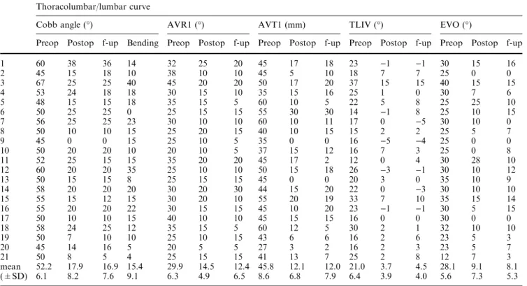
The instrumented curve improved an average of 67% from 52° (45–67°) to 18° (0–38°) (P<0.001) postopera-tively. There were no significant changes till the last follow-up. The apex vertebra rotation of the major TL/L curve (AVR1) improved 51% from 30° (20–45°) to 15°
(5–25°) (P<0.001) postoperatively. AVR1 was 12°
(5–30°) at the last follow-up but this change was not significant. The translation of the apex vertebra of the major curve (AVT1) improved 74%, from 46
(27–60) mm preoperatively to 12 (0–30) mm postoper-atively and at the last follow-up. The TLIV was cor-rected 82% from 21° preoperatively (12–37°) to 4° ()5° to +15°) (P<0.001) postoperatively and at the last follow-up. There was a 67% improvement of the end vertebra obliquity (EVO), from 28° (12–40°) to 9° (0–28°) (P<0.001) postoperatively and 8° (0–16°) at the last follow-up. There were no significant changes during the follow-up period (Table2).
The unfused thoracic curve improved an average of 29% from 33° (18–50°) to 24° (5–40) (P<0.001) post-operatively. There were no significant changes to the last follow-up. The apex vertebra rotation of the thoracic curve (AVR2) improved 17% from 7.9° (0–15°) to 6.9°
(0–20°) postoperatively and 7.1° (0–20°) at the last fol-low-up. The AVT of the thoracic curve (AVT2) did not
change significantly from preoperative 14.9 (6–30) mm to 14.6 (0–32) mm at the last follow-up (Table3).
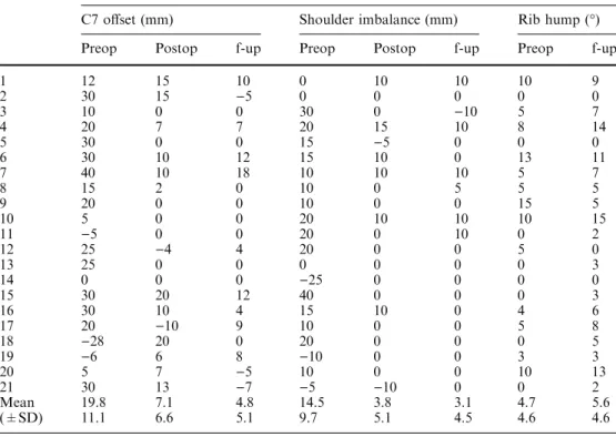
The C7 offset improved from an average of 19.8 (0–40) mm before the surgery to 7.1 (0–20) mm post-operatively (P<0.001) and to 4.8 (0–18) mm at the last follow-up (P<0.05). Three patients before the surgery and three patients after the surgery had a C7 offset shift to the side of the thoracic curve, which are shown in Table3 with negative values. Positive values in other patients indicate the C7 offset shift to the side of the TL/ L curve. T1O did not change from an average of 5°
(0–15°) preoperatively to the last follow-up.
Preoperatively 15 patients had a positive shoulder imbalance (10–40 mm), two patients had a negative imbalance (10, 25 mm) and four patients had balanced shoulders. Ten of the patients with positive imbalance and all patients with negative imbalance had balanced shoul-ders at the last follow-up. No patients with a preoperative positive shoulder imbalance had worsening of shoulder balance at the last follow-up (Table4). One patient with preoperative balanced shoulders had a slight positive imbalance of 10 mm at the last follow-up. The average differences in clinical shoulder level in the whole group were 14.5 (0–40) mm preoperatively, 3.8 (0–15) mm postoperatively and 3.1 (0–10) mm at the last follow-up.
The clinical rib hump was on average 4.7°(0–15°) before the operation and 5.6° (0–15°) at the last follow-up (Table4). The average thoracic kyphosis was 25.8° (6–40°) preoperatively, 22.8° (5–46°) postoperatively and 28.3° (5–46°) at the last follow-up. The average lumbar lordosis was 57.0° (35–73°) preoperatively, 51.5° (33–78°) postoperatively and 58.7° (37–83°) at the last
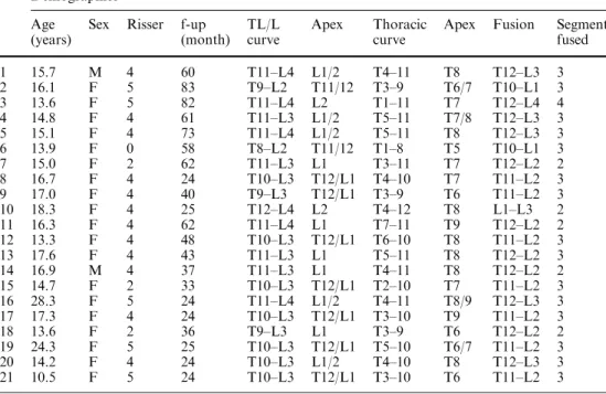
Table 1 Demographics and the number of segments fused
Demographics Age
(years)
Sex Risser f-up (month)
TL/L curve
Apex Thoracic curve
Apex Fusion Segments fused 1 15.7 M 4 60 T11–L4 L1/2 T4–11 T8 T12–L3 3 2 16.1 F 5 83 T9–L2 T11/12 T3–9 T6/7 T10–L1 3 3 13.6 F 5 82 T11–L4 L2 T1–11 T7 T12–L4 4 4 14.8 F 4 61 T11–L3 L1/2 T5–11 T7/8 T12–L3 3 5 15.1 F 4 73 T11–L4 L1/2 T5–11 T8 T12–L3 3 6 13.9 F 0 58 T8–L2 T11/12 T1–8 T5 T10–L1 3 7 15.0 F 2 62 T11–L3 L1 T3–11 T7 T12–L2 2 8 16.7 F 4 24 T10–L3 T12/L1 T4–10 T7 T11–L2 3 9 17.0 F 4 40 T9–L3 T12/L1 T3–9 T6 T11–L2 3 10 18.3 F 4 25 T12–L4 L2 T4–12 T8 L1–L3 2 11 16.3 F 4 62 T11–L4 L1 T7–11 T9 T12–L2 2 12 13.3 F 4 48 T10–L3 T12/L1 T6–10 T8 T11–L2 3 13 17.6 F 4 43 T11–L3 L1 T5–11 T8 T12–L2 3 14 16.9 M 4 37 T11–L3 L1 T4–11 T8 T12–L2 2 15 14.7 F 2 33 T10–L3 T12/L1 T2–10 T7 T11–L2 3 16 28.3 F 5 24 T11–L4 L1/2 T4–11 T8/9 T12–L3 3 17 17.3 F 4 24 T10–L3 T12/L1 T3–10 T9 T11–L2 3 18 13.6 F 2 36 T9–L3 L1 T3–9 T6 T12–L2 2 19 24.3 F 5 25 T10–L3 T12/L1 T5–10 T6/7 T11–L2 3 20 14.2 F 4 24 T10–L3 L1/2 T4–10 T8 T12–L3 3 21 10.5 F 5 24 T10–L3 T12/L1 T3–10 T6 T11–L2 3
follow-up. The changes of the rib hump, the kyphosis and the lordosis were statistically not significant.
Patients’ satisfaction was assessed with the SRS-24 of the Scoliosis Research Society at the last follow-up. An average of 90.1 (68–111) points was achieved for the total score, representing 75% of the maximal 120 points. The subscales are listed in Fig.2.
There were no neurological or pulmonary complica-tions. Average operation time was 248 (180–300) min, estimated intraoperative blood loss was 587 (190–1,200) ml on an average. There were no early or late infections. Solid bony fusion was achieved in all patients without any implant failures.
Discussion
This study shows that short segment instrumentation of the major TL/L curve was effective in achieving a balanced spine in King1 idiopathic scoliosis. We expected that in carrying out short fusions more mobile lumbar segments would be left for the spinal balancing. For this reason we used the Hall’s criteria for selection of fusion levels [2, 7] instead of an end-to-end fusion. Some authors recommend the selective TL/L correction in King 1 curves only when the thoracic curve bends to less than 20° [4, 14, 16]. In three of our patients the
thoracic curve bended to 20° and in one patient to 25°. The postoperative results of these patients were com-parable to the other patients in our series whose tho-racic curves bended to less than 20°. According to our data the thoracic curve should bend to 25° or less for a selective TL/L fusion. This corresponds to the type 5 scoliosis in the classification described by Lenke et al. [10]. Our data support Lenke et al. [9] in that a thoracic curve in a type 5 scoliosis with bending up to 25° can be regarded as non-structural and that a selective fusion of the major TL/L curve is recommendable. We fused an average of 2 segments shorter than the length of the whole curve, the average length of our fusions being 2.9 segments. An achieved coronal correction of 67% of the main curve was nearly of the same extent as in the preoperative side bending (70%), the residual deformity of the major curve being 17° on an average. The improvement of the AVR of the main curve (AVR1) was 51%. These results are comparable to
other studies [3, 4, 11, 16, 17].
The spontaneous improvement of the unfused tho-racic curve in our series with short selective fusion was 29% which is comparable to the results with end-to-end fusions of the TL/L curve of others [3, 4,11,16,17]. If we can accept a residual deformity of less than 40° in the thoracic curve, then a King 1 type curve with a thoracic curve of up to 50° that bends to 25° or less, in other
Table 2 Results of the instrumented thoracolumbar/lumbar curve Thoracolumbar/lumbar curve
Cobb angle (°) AVR1 (°) AVT1 (mm) TLIV (°) EVO (°)
Preop Postop f-up Bending Preop Postop f-up Preop Postop f-up Preop Postop f-up Preop Postop f-up 1 60 38 36 14 32 25 20 45 17 18 23 )1 )1 30 15 16 2 45 15 18 10 38 10 10 45 5 10 18 7 7 25 0 0 3 67 25 25 40 45 20 20 50 17 20 37 15 15 40 15 15 4 53 24 18 18 30 15 10 35 15 16 25 1 0 30 7 6 5 48 15 15 18 35 15 5 60 10 5 22 5 8 25 25 10 6 50 25 25 0 25 15 15 55 30 30 14 )1 8 25 10 15 7 56 25 25 23 30 10 10 60 10 11 17 0 )5 30 10 0 8 50 10 10 15 25 20 15 40 10 15 15 2 2 25 5 7 9 45 0 0 15 25 10 5 35 0 0 16 )5 )4 25 0 0 10 50 20 20 10 20 10 5 37 15 12 16 7 3 25 0 8 11 52 25 15 15 35 20 20 45 17 2 12 0 4 30 28 10 12 60 20 20 35 25 10 10 50 15 18 26 )3 )1 30 10 12 13 50 15 15 8 25 15 15 45 0 0 20 3 0 35 10 9 14 58 20 20 20 30 20 30 44 15 20 22 0 )3 30 10 10 15 55 15 12 15 30 20 10 55 20 19 33 7 10 35 15 14 16 55 20 20 22 30 15 15 45 10 20 23 )1 )1 30 5 15 17 50 10 10 15 40 10 10 45 15 15 16 0 0 30 0 0 18 58 24 25 12 35 15 5 60 12 5 30 2 1 32 10 10 19 50 7 10 10 25 10 15 43 6 6 16 2 6 23 5 3 20 45 14 16 5 20 5 5 27 3 2 16 2 3 23 5 7 21 50 8 5 4 25 15 15 41 13 7 25 2 8 12 7 3 mean 52.2 17.9 16.9 15.4 29.9 14.5 12.4 45.8 12.1 12.0 21.0 3.7 4.5 28.1 9.1 8.1 (±SD) 6.1 8.2 7.6 9.1 6.3 4.9 6.5 8.6 6.8 7.9 6.4 3.9 4.0 5.6 7.3 5.3 AVR1apex vertebra rotation, AVT1apex vertebra translation, TLIV tilt of last instrumented vertebra, negative values indicate the tilt in
words a Lenke type 5 curve with a thoracic curve of up to 50°, is eligible for selective fusion according to our present data. A thoracic curve that bends to more than
25° is normally so large and rigid that it needs a cor-rective fusion. According to Sanders et al. [16] it is possible to predict whether a non-structural thoracic
Table 3 Results of the non-instrumented thoracic curve
AVR2apex vertebra rotation,
AVT2apex vertebra translation
Thoracic curve
Cobb angle (°) AVR2 (°) AVT2 (mm)
Preop Postop f-up Bending Preop Postop f-up Preop Postop f-up
1 50 40 36 15 15 10 15 17 25 30 2 30 20 20 15 5 0 0 21 3 14 3 40 20 20 15 10 15 15 12 15 12 4 44 34 36 20 15 20 20 10 23 25 5 30 15 15 10 5 5 5 22 7 6 6 30 20 25 10 5 0 0 17 22 26 7 42 34 34 20 5 5 5 10 13 9 8 26 22 20 10 10 5 5 7 9 9 9 35 20 3 0 5 0 0 14 13 15 10 40 32 40 20 10 15 15 30 28 32 11 18 18 16 0 5 0 0 12 0 0 12 30 26 26 10 5 5 5 16 8 4 13 30 15 17 0 10 10 5 6 3 5 14 32 32 23 15 10 10 10 6 3 3 15 30 5 15 0 0 5 5 29 16 4 16 40 30 35 15 15 15 15 6 10 23 17 35 30 30 25 5 5 0 13 5 10 18 32 30 21 15 10 5 10 15 9 20 19 31 25 28 10 10 5 5 20 9 18 20 28 20 18 5 5 10 10 15 20 26 21 26 12 28 0 5 0 5 13 0 15 Mean 33.3 23.8 24.1 11.0 7.9 6.9 7.1 14.9 11.5 14.6 (±SD) 7.1 8.4 8.9 7.5 4.0 5.7 5.9 6.6 8.1 9.4
Table 4 Trunk balance and rib hump
Positive values indicate offset on the side of the major curve, negative values indicate offset on the side of the thoracic cur-ve. Shoulder imbalance is posi-tive if the shoulder on the side of the thoracic curve is higher, and is negative when the oppo-site is true. Clinical rib hump measured with scoliometerC7 offsetvertical offset of the C7 vertebra from the centre sacral line
Trunk balance and rib hump
C7 offset (mm) Shoulder imbalance (mm) Rib hump (°) Preop Postop f-up Preop Postop f-up Preop f-up
1 12 15 10 0 10 10 10 9 2 30 15 )5 0 0 0 0 0 3 10 0 0 30 0 )10 5 7 4 20 7 7 20 15 10 8 14 5 30 0 0 15 )5 0 0 0 6 30 10 12 15 10 0 13 11 7 40 10 18 10 10 10 5 7 8 15 2 0 10 0 5 5 5 9 20 0 0 10 0 0 15 5 10 5 0 0 20 10 10 10 15 11 )5 0 0 20 0 10 0 2 12 25 )4 4 20 0 0 5 0 13 25 0 0 0 0 0 0 3 14 0 0 0 )25 0 0 0 0 15 30 20 12 40 0 0 0 3 16 30 10 4 15 10 0 4 6 17 20 )10 9 10 0 0 5 8 18 )28 20 0 20 0 0 0 5 19 )6 6 8 )10 0 0 3 3 20 5 7 )5 10 0 0 10 13 21 30 13 )7 )5 )10 0 0 2 Mean 19.8 7.1 4.8 14.5 3.8 3.1 4.7 5.6 (±SD) 11.1 6.6 5.1 9.7 5.1 4.5 4.6 4.6
curve will remain more than 40° postoperatively when flexibility and skeletal maturity data are taken into account. However, they did not mention the expectable changes in the clinical shoulder balance or of the rib hump deformity. In our series the residual deformity of the thoracic curve did not negatively influence trunk balance as the C7 offset was reduced to an average of 4.8 mm and shoulder imbalance improved to an average of 3.1 mm at the last follow-up. There was an overall improvement of 15 mm in C7 offset and 11.4 mm in shoulder balance which represented a good correction of trunk balance compared to the literature [2, 11, 17]. Positive shoulder imbalance proved not to be a contra-indication for the selective fusion of the TL/L curve. In our study all except one patient with a positive shoulder imbalance improved and the majority of them (70%) had balanced shoulders at the last follow-up. None of the positively imbalanced shoulders worsened. Our data do not support the statement that a balanced shoulder or a negative imbalance is a prerequisite for selective TL/L fusion as stated by others [11]. Despite the 74% improvement of the coronal translation of the apex of the TL/L curve (AVT1), the translation of the thoracic
curve (AVT2) remained largely unchanged. This was in
contrast to the concern that a worsening of the thoracic curve translation may occur because of the selective correction of the lower curve. Worsening of the Cobb angle of the unfused thoracic curve was not observed in any of our patients. It seems that the short fusion of the TL/L curve leaves sufficient mobility in the remaining lumbar spine to accommodate both curves and to establish the global trunk balance.
The changes in the rotation of the thoracic curve and the rib hump in the whole group were statistically not
significant even though a slight increase of rib hump was observed in a few patients. Patients should be informed before the operation that no improvement of the tho-racic rib hump can be expected. The rib hump might appear more prominent as the lumbar hump in relation becomes smaller.
Single rod implants without structural interbody support did not cause lumbar kyphosis in our patients, a concern stated by some authors [2, 13, 18]. There were no significant changes of the thoracic kyphosis and lumbar lordosis. Some authors described the use of in-terbody supports like titanium cages or femoral ring allografts in the anterior fusion of the whole curve [17] whereas others showed that interbody structural sup-ports were not necessary in maintaining the sagittal profile in end-to-end fusions [12]. According to our results such structural supports are not necessary in anterior short segmental fusions to maintain lumbar lordosis.
Most of the changes in trunk balance, in the fused and unfused curves happened during the first 3 months after the surgery. No significant changes took place after that time in the group of 21 patients. A significant loss of correction during the average follow-up of 43 months was not observed.
An average of 83% of the possible maximal points in the corresponding subscale in the SRS-24 questionnaire indicates that patients were satisfied with the operation. Eighty percent of the patients would repeat the treat-ment under the same conditions. Especially the subscale function from the back condition seems to be little affected with a higher-than-average of 84%. To our knowledge only two studies exist which present data of the SRS-24 in healthy controls [6, 19]. Compared to SRS-24 Outcome measurement 90.1 27.3 9.4 10.4 6.1 12.7 11.7 12.5 satisfaction general self-image pain total 0 20 40 60 80 100 120 140
function from back condition
postoperative self-image general level of activity
postoperative function
Fig. 2 Total score and sub-scales of the SRS-24. Dark grey columnmaximal score per scale
their data the results of our patients are close to the values of normal controls.
Conclusions
Short selective fusion of the TL/L curve in King 1 or Lenke 5 type idiopathic scoliosis results in a balanced
spine at the 2 year follow-up. The trunk balance improves significantly. An average of two mobile segments is saved compared to an end-to-end fusion. A higher shoulder on the side of the thoracic curve is not a contraindication for this procedure. The thoracic rib hump remains unchanged. Worsening of the unfused thoracic curve is not observed in any patient. According to the SRS-24 questionnaire the patients were satisfied with the results.
Reference
1. Benli IT, Akalin S, Kis M, Citak M, Ku-rtulus B, Duman E (2000) The results of anterior fusion and Cotrel-Dubousset-Hopf instrumentation in idiopathic scolio-sis. Eur Spine J 9:505–515
2. Bernstein RM, Hall JE (1998) Solid rod short segment anterior fusion in thoracolumbar scoliosis. J Pediatr Orthop B 7:124–131 3. Bitan FD, Neuwirth MG, Kuflik PL,
Casden A, Bloom N, Siddiqui S (2002) The use of short and rigid anterior instru-mentation in the treatment of idiopathic thoracolumbar scoliosis: a retrospective review of 24 cases. Spine 27:1553–1557 4. Burton DC, Asher MA, Lai SM (2002)
Patient-based outcomes analysis of pa-tients with single torsion thoracolum-bar-lumbar scoliosis treated with anterior or posterior instrumentation: an average 5- to 9-year follow-up study. Spine 27:2363–2367
5. Dwyer AF, Newton NC, Sherwood AA (1969) An anterior approach to scolio-sis. A preliminary report. Clin Orthop Relat Res 62:192–202
6. Haher TR, Gorup JM, Shin TM, Ho-mel P, Merola AA, Grogan DP, Pugh L, Lowe TG, Murray M (1999) Results of the Scoliosis Research Society instrument for evaluation of surgical outcome in adolescent idiopathic scoli-osis. A multicenter study of 244 pa-tients. Spine 24:1435–1440
7. Hall JH, Millis MB, Snyder BD (eds) (1997) Short segment anterior
instru-mentation for thoracolumbar scoliosis, 2nd edn. Lippincott-Raven Publisher, Philadelphia
8. King HA, Moe JH, Bradford DS, Winter RB (1983) The selection of fu-sion levels in thoracic idiopathic scoli-osis. J Bone Joint Surg Am 65:1302– 1313
9. Lenke LG (2005) Lenke classification system of adolescent idiopathic scolio-sis: treatment recommendations. AAOS Instr Course Lect 54:537–542
10. Lenke LG, Betz RR, Harms J, Bridwell KH, Clements DH, Lowe TG, Blanke K (2001) Adolescent idiopathic scolio-sis: a new classification to determine extent of spinal arthrodesis. J Bone Joint Surg Am 83-A:1169–1181 11. Lenke LG, Edwards CC II, Bridwell
KH (2003) The Lenke classification of adolescent idiopathic scoliosis: how it organizes curve patterns as a template to perform selective fusions of the spine. Spine 28:S199–S207
12. Lowe TG, Alongi PR, Smith DA, O’Brien MF, Mitchell SL, Pinteric RJ (2003) Anterior single rod instrumenta-tion for thoracolumbar adolescent idi-opathic scoliosis with and without the use of structural interbody support. Spine 28:2232–2241
13. Lowe TG, Betz R, Lenke L, Clements D, Harms J, Newton P, Haher T, Me-rola A, Wenger D (2003) Anterior sin-gle-rod instrumentation of the thoracic and lumbar spine: saving levels. Spine 28:S208–S216
14. Millis H, Hall J, Emans J (1990) Short (3 segment) anterior instrumentation and fusion for idiopathic thoracolum-bar scoliosis. Orthop Trans 14:742–774
15. Perdriolle R, Vidal J (1987) Morphol-ogy of scoliosis: three-dimensional evo-lution. Orthopedics 10:909–915 16. Sanders AE, Baumann R, Brown H,
Johnston CE 2nd, Lenke LG, Sink E (2003) Selective anterior fusion of tho-racolumbar/lumbar curves in adoles-cents: when can the associated thoracic curve be left unfused? Spine 28:706–713 17. Sweet FA, Lenke LG, Bridwell KH,
Blanke KM (1999) Maintaining lumbar lordosis with anterior single solid-rod instrumentation in thoracolumbar and lumbar adolescent idiopathic scoliosis. Spine 24:1655–1662
18. Turi M, Johnston CE II, Richards BS (1993) Anterior correction of idiopathic scoliosis using TSRH instrumentation. Spine 18:417–422
19. Watanabe K, Hasegawa K, Hirano T, Uchiyama S, Endo N (2005) Use of the scoliosis research society outcomes instrument to evaluate patient outcome in untreated idiopathic scoliosis patients in Japan: part II: relation between spinal deformity and patient outcomes. Spine 30:1202–1205
20. Wilson PL, Newton PO, Wenger DR, Haher T, Merola A, Lenke L, Lowe T, Clements D, Betz R (2002) A multi-center study analyzing the relationship of a standardized radiographic scoring system of adolescent idiopathic scoliosis and the Scoliosis Research Society out-comes instrument. Spine 27:2036–2040 21. Zielke K, Stunkat R, Beaujean F (1976)
Ventrale Derotations-Spondylodese. Arch Orthop Unfallchir 85:257–277