ORIGINAL ARTICLE
Influence of caries infiltrant contamination on shear bond
strength of different adhesives to dentin
Liuhe Jia&Bogna Stawarczyk&Patrick R. Schmidlin&
Thomas Attin&Annette Wiegand
Received: 13 February 2012 / Accepted: 9 April 2012 / Published online: 25 April 2012 # Springer-Verlag 2012
Abstract
Objectives To analyze whether the contamination with a caries infiltrant system impairs the adhesive performance of etch-and-rinse and self-etching adhesives on dentin. Materials and methods Dentin contamination with the car-ies infiltrant system (Icon, DMG) was simulated by apply-ing either hydrochloric acid (15 % HCl, Icon Etch, 15 s), the resin infiltrant (Icon infiltrant, 4 min), or both prior to the application of the respective adhesives (each group n010). In the control groups, the etch-and-rinse adhesive (Optibond FL, Kerr) and the self-etching adhesive (iBOND Self Etch, Hereaus) were applied without former contamination with the infiltrant system. Additionally, the adhesive performance of the resin infiltrant alone was tested. Shear bond strength of a nano-hybrid composite was analyzed after thermocy-cling (5,000×, 5–55°C) of the specimens and analyzed by
ANOVA/Scheffé post hoc tests (p<0.05) and Weibull sta-tistics. Failure mode was inspected under a stereomicro-scope at×25 magnification.
Results Contamination with the resin infiltrant alone did not impair shear bond strength, while contamination with hydrochloric acid or with hydrochloric acid and the resin infiltrant reduced shear bond strength (MPa) of the adhe-sives (Optibond FL: 20.5±3.6, iBOND Self Etch: 17.9±2.6) significantly. Hydrochloric acid contamination increased the number of adhesive failures. The adhesive performance of the caries infiltrant system alone was insufficient.
Conclusion The contamination with the caries infiltrant sys-tem impaired the shear bond strength of conventional dental adhesives.
Clinical relevance Contamination of the caries infiltrant system on dentin should be avoided due to the detrimental effect of hydrochloric acid etching.
Keywords Caries infiltrant . Dentin . Adhesive . Shear bond strength . Hydrochloric acid . Phosphoric acid
Introduction
Originally, the caries infiltration technique has been intro-duced to arrest non-cavitated caries lesions by sealing the diffusion pathways of demineralized enamel with a low-viscosity resin. Compared to dental adhesives or fissure sealants, triethylene glycol dimethacrylate (TEGDMA)-based infiltrants were optimized for rapid capillary penetra-tion and are able to penetrate enamel caries lesions almost completely after removal of the intact surface layer by etching with hydrochloric acid.
However, in recent studies, it was questioned whether the range of application of the caries infiltrant system could be L. Jia
:
P. R. Schmidlin:
T. Attin:
A. Wiegand (*)Department of Preventive Dentistry, Periodontology and Cariology, Center of Dental Medicine, University of Zurich, Plattenstrasse 11,
8032 Zürich, Switzerland
e-mail: [email protected] L. Jia
Department of Dentistry, Beijing Jishuitan Hospital, Xinjiekou East Street 31,
100035 Beijing, China B. Stawarczyk
Department of Fixed and Removable Prosthodontics and Dental Material Science, Center of Dental Medicine, University of Zurich, Plattenstrasse 11,
8032 Zürich, Switzerland B. Stawarczyk
Department of Prosthodontics, Ludwig-Maximilians University, Goethestrasse 70,
also extended to cavitated lesions [1–3]. Paris et al. [3] showed that the caries infiltrant is able to penetrate most parts of demineralized enamel in lesions with cavitation but not capable to fill up the cavitation itself. However, resin infiltration might be successfully com-bined with adhesive conditioning to allow for restora-tion of cavitated and infiltrarestora-tion of demineralized areas. Studies by Wiegand et al. [1] and Jia et al. [2] showed that the caries infiltrant system achieved the same bond strength on sound and demineralized enamel as conven-tional adhesives and did not impair but even enhanced the bond strength of the adhesives when applied in combination. These results indicate that cavitated initial enamel lesions could be successfully restored with com-posite, while in the same step demineralized enamel at the margin of the cavitation can be preserved by resin infiltration. However, as larger cavitated carious lesions usually also involve dentin, it is of clinical relevance to which extent the adhesion to dentin is affected by contamination with the caries infiltrant system, especial-ly emphasizing that dentin adhesion is much more com-plex as compared to bonding to enamel due to its high organic content and high hydrophilicity.
The commercially available caries infiltration system comprises 15 % hydrochloric acid for the removal of the outer enamel surface layer, ethanol for drying, and a TEGDMA-based resin for infiltration of the lesion. The aim of the present study was to analyze to which extent the bond strength of an etch-and-rinse and a self-etch adhe-sive on dentin is affected by contamination with the whole caries infiltrant system or single components (hydrochloric acid and TEGDMA-based resin). Moreover, the adhesive properties of the caries infiltrant system itself should be assessed.
The null hypothesis was that contamination by the infil-trant material or its components does not impair bonding to dentin and allows for comparable shear bond strengths as compared to the control treatments.
Material and methods Specimen preparation
Cylindrical dentin specimens (6.6 mm in diameter, n0110) were prepared from the roots of freshly extracted, non-damaged bovine incisors. The specimens were then embed-ded in chemically cured acrylic resin (ScandiQuick, Scan-Dia, Hagen, Germany) and ground flat with P400SiC paper (Buehler, Lake Bluff, USA). The cementum layer was com-pletely removed and checked by stereomicroscope analysis (M3B, Wild, Heerbrugg, Switzerland).
Specimen allocation and bonding procedure
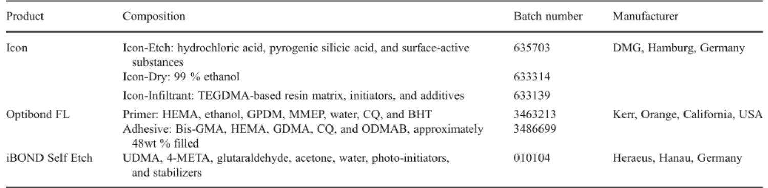
Specimens were randomly divided into 11 groups of ten specimens each. The compositions of the adhesives and the infiltrant resin system are listed in Table1.
The dentin surface was intentionally contaminated with the caries infiltrant system (Icon, DMG, Hamburg, Ger-many) before the respective etch-and-rinse (Optibond FL, Kerr, California, USA) or self-etching adhesive (iBOND Self Etch, HeraeusKulzer, Hanau, Germany) was applied. To analyze the potentially adverse effect of the different components of the infiltrant system separately and in com-bination, the contamination was simulated by applying ei-ther hydrochloric acid (15 % HCl, Icon Etch), the resin infiltrant (Icon infiltrant), or both. Contamination with hydrochloric acid was restricted to 15 s to avoid severe over-etching of dentin [4].
In the control groups, the etch-and-rinse and self-etching adhesives were applied and light-cured (20 s, 800 W/cm2, bluephase, IvoclarVivadent, Schaan, Liechtenstein) accord-ing to the manufacturers’ recommendation without contam-ination with the caries infiltrant system. The simulated contamination and bonding procedures in the different groups are listed in Table2.
Additionally, the adhesive properties of the infiltrant alone (without adhesive application) after etching with 15 % hydrochloric (120 s or 15 s, respectively) or 37 % phosphoric acid (15 s) were analyzed.
Composite application and shear bond measurements A nano-hybrid composite (TetricEvoCeram, IvoclarViva-dent, Schaan, Liechtenstein) was then applied to the dentin surface using a transparent plastic hollow cylinder with an inner diameter of 3 mm [1,2,5]. The composite was packed against the surface in a 2-mm thick increment, which was then light-cured for 60 s. Light intensity was confirmed by a radiometer (Optilux Model 100, SDS Kerr Danbury, USA) after each ten specimens. Bonding procedures were carried out by one operator (LJ) throughout all experiments.
Prior to shear bond strength testing, specimens were submitted to thermocycling (Willytec, Gräfelfing, Germany, 5,000 cycles, 5–55°C, dwell time: 20 s, transfer time: 10 s) [6, 7]. Shear bond strength was tested with a universal testing machine (ZwickZ010, Zwick, Ulm, Germany). A shear force was applied to the adhesive interface through a chisel-shaped loading device at a crosshead speed of 1 mm/ min parallel to the dentin surface. Load at fracture was recorded, and shear bond strength (σ) was calculated by a software (TestXpert 11.02, Zwick, Ulm, Germany) using the load at failure F (N) and the adhesive area A (mm2):σ0F/A. The debonded area was examined for failure mode anal-ysis with a stereomicroscope at×25 magnification (M3B,
Wild, Heerbrugg, Switzerland). Failure mode was consid-ered as adhesive if it occurred at the interface and as cohe-sive if at least parts of either dentin or composite were affected.
Statistical analysis
Mean shear bond strength (± standard deviation) for each group was computed. Statistical analysis of the contamina-tion and the control groups was done by two-way ANOVA, factors being the adhesives and the kind of contamination.
Within each adhesive, one-way ANOVA followed by Scheffé’s post hoc tests were performed to analyze differ-ences between contamination groups (p≤0.05).
Furthermore, for the calculation of the Weibull statistics, the least square estimates of the modulus and characteristic bond strength were computed according to the mean rank plotting (SPSS Version 20, SPSS INC, Chicago, USA) [8]. This statistical program allows only the calculation of the absolute estimates but not of the 95 % CI. Also, post hoc tests for Weibull parameters could not be obtained, so that a statis-tical comparison between the tested groups was not possible. Table 1 Composition of the caries infiltrant and the adhesive systems
Product Composition Batch number Manufacturer Icon Icon-Etch: hydrochloric acid, pyrogenic silicic acid, and surface-active
substances
635703 DMG, Hamburg, Germany Icon-Dry: 99 % ethanol 633314
Icon-Infiltrant: TEGDMA-based resin matrix, initiators, and additives 633139
Optibond FL Primer: HEMA, ethanol, GPDM, MMEP, water, CQ, and BHT 3463213 Kerr, Orange, California, USA Adhesive: Bis-GMA, HEMA, GDMA, CQ, and ODMAB, approximately
48wt % filled
3486699 iBOND Self Etch UDMA, 4-META, glutaraldehyde, acetone, water, photo-initiators,
and stabilizers
010104 Heraeus, Hanau, Germany
Bis-GMA bisphenol A diglycidyl methacrylate, TEGDMA triethylene glycol dimethacrylate, HEMA 2-hydroxyl methacrylate, GPDM glycerol phosphate dimethacrylate, MMEP mono-2-methacryloyloxyethyl phthalate, CQ camphorquinone, BHT butylhydroxytoluene, GDMA glycerol dimethacrylate, ODMAB 2-(ethylhexyl)-4-(dimethylamino)benzoate, UDMA urethane dimethacrylate,4-META 4-mathacryloyloxyethyl trimellitate anhydride
Table 2 Bonding procedures and simulated contamination in the different groups
Group Contamination Etching Infiltrant Adhesive Optibond FL None (control) 37 % H3PO4(15 s)
a
– Optibond FL primer (15 s), Optibond FL adhesive (15 s), light curing (20 s) HCl (Icon Etch) 15 % HCl (15 s)a – Optibond FL primer (15 s) Optibond
FL adhesive (15 s), light curing (20 s) HCl (Icon Etch) and resin
infiltrant (Icon infiltrant)
15 % HCl (15 s) Ethanol (Icon Dry, 30 s),aResin infiltrant (3 min), Light curing (40 s), Resin infiltrant (1 min), Light curing (40 s)
Optibond FL primer (15 s), Optibond FL adhesive (15 s), light curing (20 s)
Resin infiltrant (Icon infiltrant) 37 % H3PO4(15 s)a Resin infiltrant (3 min), Light
curing (40 s), Resin infiltrant (1 min), Light curing (40 s)
Optibond FL primer (15 s), Optibond FL adhesive (15 s), light curing (20 s) iBOND Self
Etch
None (control) – – iBOND Self Etch (20 s ), light curing (20 s) HCl (Icon Etch) 15 % HCl (15 s)a iBOND Self Etch (20 s ), light curing (20 s) HCl (Icon Etch) and resin
infiltrant (Icon infiltrant)
15 % HCl (15 s) Ethanol (Icon Dry, 30 s),aResin infiltrant (3 min), Light curing (40 s), Resin infiltrant (1 min), Light curing (40 s)
iBOND Self Etch (20 s ), light curing (20 s)
Resin infiltrant (Icon infiltrant) – Resin infiltrant (3 min), Light curing (40 s), Resin infiltrant (1 min), Light curing (40 s)
iBOND Self Etch (20 s ), light curing (20 s)
The components of the caries infiltrant system were applied with the smooth surface-tip provided by the manufacturer. The adhesives were applied with a microbrush. The resin infiltrant and the adhesives were applied with light brushing motion. Excess infiltrant was removed by 5 s air blowing prior to light curing
a
Relative frequencies of cohesive failures in each group were calculated at 95 % CI. Shear bond strength data of the caries infiltrant system after etching with hydrochloric or phosphoric acid but without application of an adhesive were close to zero and were therefore excluded from the statistical analysis.
Results
The shear bond strengths of the control groups amounted to 20.5±3.6 MPa (Optibond FL) and 17.9±2.6 MPa (iBond Self Etch).
Two-way ANOVA showed that contamination mode, adhesive material, and the interaction between both factors were significant with respect to shear bond strength. Within each adhesive, one-way ANOVA revealed significant differ-ences between the contamination groups.
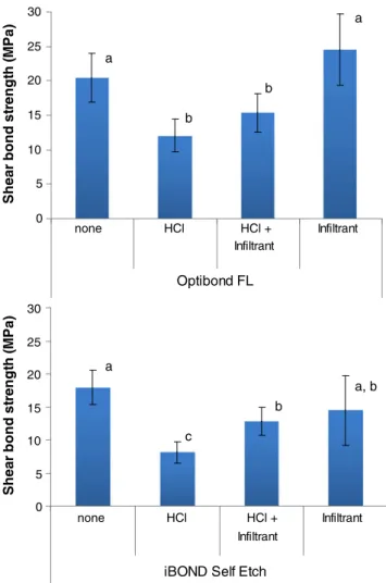
For both adhesives, contamination with hydrochloric acid significantly reduced the shear bond strength. Application of the resin infiltrant after etching with hydrochloric acid and prior to adhesive application slightly increased shear bond strength as compared to the hydrochloric acid etching alone, but values were still significantly lower than in the control groups. Contamination with the resin infiltrant alone did not hamper bonding strength of both adhesives (Fig.1).
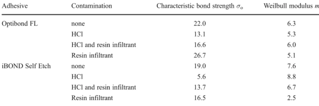
The Weibull parameters are presented in Table 3. The characteristic strength values were highest for the control groups and Optibond FL applied on dentin contaminated with the resin infiltrant. Weibull modulus m ranged from 5.1 to 6.3 for the Optibond FL groups and from 2.5 to 8.8 for the iBOND Self Etch groups.
With regard to the failure mode, the frequency of cohe-sive failures was decreased by the contamination with hydrochloric acid and by the whole caries infiltrant system but not by the resin infiltrant alone (Table4).
The adhesive performance of the caries infiltrant alone (after etching with hydrochloric or phosphoric acid) was insufficient. Shear bond strength values amounted to 0.3± 0.7 MPa (15 s hydrochloric acid), 0.1 ±0.3 MPa (120 s hydrochloric acid), and 0.5 ± 1.2 MPa (15 s phosphoric acid). All failures of these groups were adhesive.
Discussion
This in vitro study showed that the adhesive performance of an etch-and-rinse and a self-etching adhesive was signifi-cantly reduced by contamination with the caries infiltrant system (15 % HCl etching followed or not by resin infiltrant application) but not by the TEGDMA-containing resin infil-trant alone. Our results highlighted that the hydrochloric acid etching rather than the contamination with the resin
infiltrant is detrimental for the adhesive properties of the conventional adhesives. Therefore, the null hypothesis that contamination by the infiltrant material or its com-ponents does not impair bonding to dentin and allows for comparable shear bonds strengths as compared to the control treatments is partly rejected. Moreover, the caries infiltrant alone did not exhibit any adhesive prop-erties on dentin, while it generated similar shear bond strength compared to conventional adhesives on sound and demineralized enamel [1, 2].
Shear bond strength was tested on bovine dentin, which has been proposed as suitable alternative for human dentin although shear bond strength is slightly higher on bovine root dentin compared to human coronal dentin [9]. More-over, it should be considered that shear bond strength values in the present study might be higher than in the presence of intrapulpal pressure simulation [10,11]. However, as rela-tive differences rather than absolute values are of interest, the use of bovine dentin in adhesion testing—even without
0 5 10 15 20 25 30 HCl + Infiltrant Infiltrant Optibond FL She a r bond st re ngt h ( M Pa) 0 5 10 15 20 25 30 HCl + Infiltrant Infiltrant
iBOND Self Etch
She a r bond st re ngt h ( M Pa ) a a b b a a, b b c none HCl HCl none
Fig. 1 Shear bond strength (MPa, mean± standard deviation) of the different contamination groups in dentin treated with Optibond FL or iBOND Self Etch. Values which are not significantly different are marked with the same letter
simulation of intrapulpal pressure—is widely accepted [12–14].
While hydrochloric acid etching is essential for the re-moval of the surface layer of non-cavitated enamel lesions to allow for the penetration of the resin infiltrant into the body of the lesion [15], hydrochloric acid contamination of sound dentin was shown to induce a more severe deminer-alization even in concentrations below 1 % [4] as compared to 37 % phosphoric acid. As a consequence of over-etching, the thick layer of demineralized collagen is not capable of being completely impregnated by adhesives containing monomers with relatively high viscosity, such as Bis-GMA [16,17].
In the present study, the application time of hydrochloric acid was reduced from 2 min (as recommended by the manufacturer for removal of the enamel surface layer) to 15 s to simulate only a contamination and avoid significant over-etching, which can be considered as an adaptation of the etch-and-rinse protocol using phosphoric acid. Although the surface was not extensively dried in these groups to avoid collapse of the exposed dentin network, shear bond strength was significantly reduced for both adhesives, indi-cating an incomplete penetration of demineralized dentin.
This assumption is also confirmed by the fact that solely adhesive failures were observed in these groups.
Contamination with the whole caries infiltrant system (hydrochloric acid etching, ethanol, and resin infiltrant ap-plication) resulted in slightly improved bond strength of the adhesives compared to the hydrochloric acid contamination alone, probably due to the improved penetration depth of the resin infiltrant. Due to the low viscosity, high penetration capability [18], and relatively high application time (total 4 min), we assume that the TEGDMA-containing infiltrant penetrates the severely demineralized dentin to a higher extent compared to the conventional adhesives. Contamina-tion with the whole caries infiltrant system also included ethanol application on the dentin surfaces. Moisture control by ethanol pretreatment might increase the shear bond strength of hydrophilic monomers, like TEGDMA, to dentin [19]. However, shear bond strength after contamination with the caries infiltrant system was still significantly reduced compared to the control groups.
Contamination with the resin infiltrant alone did not impair bonding strength of Optibond FL and iBOND Self Etch significantly. In case of Optibond FL, the resin infil-trant contamination was performed on phosphoric acid etched dentin, thus TEGDMA might infiltrate the collagen network to a higher extent than Optibond FL. Therefore, the amount of cohesive failures was considerably increased compared to the dentin contaminated with hydrochloric acid. The pretreatment with the TEGDMA-containing infil-trant probably results in an oxygen-inhibited layer [20], which allows a chemical connection between the infiltrant and the adhesive. As a consequence, shear bond strength was slightly (although not significantly) increased as com-pared to the control.
In contrast to etch-and-rinse adhesives, self-etching adhe-sives do not require a separate etching step, thus the resin infiltrant could not infiltrate demineralized dentin but only cover the surface of untreated dentin. However, although the shear bond strength data suggest that the application and polymerization of the resin infiltrant on the dentin surfaces did not impair the demineralizing and adhesive efficacy of the self-etching adhesive, the Weibull modulus m is remarkably Table 3 Weibull parameters
(Characteristic strengthσo(MPa)
and Weibull modulus m in the different groups)
Adhesive Contamination Characteristic bond strengthσo Weilbull modulus m
Optibond FL none 22.0 6.3
HCl 13.1 5.3
HCl and resin infiltrant 16.6 6.0 Resin infiltrant 26.7 5.1
iBOND Self Etch none 19.0 7.6
HCl 5.6 8.8
HCl and resin infiltrant 13.7 6.7 Resin infiltrant 16.5 2.5
Table 4 Adhesive and cohesive failures and relative frequency of cohesive failures (95 % CI) in the different groups
Adhesive Contamination Number of failure Relative frequency (%) of cohesive failures (95 % CI) adhesive cohesive Optibond FL none 4 6 60 (26.2; 87.8) HCl 10 0 0 (0.0; 30.8) HCl and resin infiltrant 8 2 20 (2.5; 55.6) Resin infiltrant 0 10 100 (69.2; 100.0) iBOND Self Etch none 6 4 40 (12.2; 73.8) HCl 10 0 0 (0.0; 30.8) HCl and resin infiltrant 10 0 0 (0.0; 30.8) Resin infiltrant 8 2 20 (2.5; 55.6)
low in this group, indicating low reliability [21]. Also, the amount of cohesive failures is reduced compared to the control. Except for this group, the shear bond strength data were sup-ported by the Weibull parameters, showing highest character-istic strength values for the control groups and Optibond FL applied on dentin contaminated with the TEGDMA-based resin infiltrant and similar Weibull moduli m between 5.1 and 8.8.
Generally, it has to be borne in mind that the whole surface of the dentin specimens and not only parts were contaminated, which is not necessarily occurring clinically. Due to the high wettability of TEGDMA resins, it is likely that the exposed dentin area of a cavity becomes completely contaminated when the infiltrant is applied, while the con-tamination with hydrochloric acid can be easier controlled due to the higher viscosity and green color of the gel. Thus, clinically, the contamination with hydrochloric acid gel can be probably restricted to smaller areas, so that the dentin adhesion might be less affected and the overall effect on the adhesion of the restoration is limited.
In conclusion, contamination of dentin during condition-ing of enamel margins of cavitated lesions should be avoided. The crucial factor affecting shear bond strength is hydrochloric acid etching of dentin but not resin infiltrant application.
Acknowledgments The first author was supported by a Swiss Fed-eral Scholarship. The authors want to thank DMG, Germany, for providing the caries infiltrant system.
Conflict of interest The authors declare that they have no conflict of interest.
References
1. Wiegand A, Stawarczyk B, Kolakovic M, Hammerle CH, Attin T, Schmidlin PR (2011) Adhesive performance of a caries infiltrant on sound and demineralised enamel. J Dent 39:117–121 2. Jia LH, Stawarczyk B, Schmidlin PR, Attin T, Wiegand A (2012)
Effect of caries infiltrant application on shear bond strength of different adhesive systems to sound and demineralized enamel. J Adhes Dent. In press
3. Paris S, Bitter K, Naumann M, Dorfer CE, Meyer-Lueckel H (2011) Resin infiltration of proximal caries lesions differing in ICDAS codes. Eur J Oral Sci 119:182–186
4. Ruse ND, Smith DC (1991) Adhesion to bovine dentin—surface characterization. J Dent Res 70:1002–1008
5. Schmidlin PR, Stawarczyk B, Wieland M, Attin T, Hammerle CH, Fischer J (2010) Effect of different surface pre-treatments and
luting materials on shear bond strength to PEEK. Dent Mater 26:553–559
6. Ozcan M, Cura C, Brendeke J (2010) Effect of aging conditions on the repair bond strength of a microhybrid and a nanohybrid resin composite. J Adhes Dent 12:451–459
7. Ozcan M, Barbosa SH, Melo RM, Galhano GA, Bottino MA (2007) Effect of surface conditioning methods on the microtensile bond strength of resin composite to composite after aging condi-tions. Dent Mater 23:1276–1282
8. Stawarczyk B, Ozcan M, Trottmann A, Hämmerle CH, Roos M (2012) Evaluation of flexural strength of hipped and presintered zirconia using different estimation methods of Weibull statistic. J Mech Behav Biomed Mater. In press
9. Schilke R, Bauss O, Lisson JA, Schuckar M, Geurtsen W (1999) Bovine dentin as a substitute for human dentin in shear bond strength measurements. Am J Dent 12:92–96
10. Sengun A, Ozturk B, Ozer F (2003) The effect of simulated intra-pulpal pressure on bond strength to enamel and dentine. J Oral Rehabil 30:550–555
11. Pioch T, Staehle HJ, Schneider H, Duschner H, Dorfer CE (2001) Effect of intrapulpal pressure simulation in vitro on shear bond strengths and hybrid layer formation. Am J Dent 14:319–323 12. Lopes MB, Sinhoreti MA, Correr Sobrinho L, Consani S (2003)
Comparative study of the dental substrate used in shear bond strength tests. Pesqui Odontol Bras 17:171–175
13. Krifka S, Borzsonyi A, Koch A, Hiller KA, Schmalz G, Friedl KH (2008) Bond strength of adhesive systems to dentin and enamel—human vs. bovine primary teeth in vitro. Dent Mater 24:888–894
14. Burke FJ, Hussain A, Nolan L, Fleming GJ (2008) Methods used in dentine bonding tests: an analysis of 102 investigations on bond strength. Eur J Prosthodont Restor Dent 16:158–165
15. Meyer-Lueckel H, Paris S, Kielbassa AM (2007) Surface layer erosion of natural caries lesions with phosphoric and hydrochloric acid gels in preparation for resin infiltration. Caries Res 41:223– 230
16. Hashimoto M, Ohno H, Endo K, Kaga M, Sano H, Oguchi H (2000) The effect of hybrid layer thickness on bond strength: demineralized dentin zone of the hybrid layer. Dent Mater 16:406–411
17. Hashimoto M, Ohno H, Kaga M, Sano H, Tay FR, Oguchi H, Araki Y, Kubota M (2002) Over-etching effects on micro-tensile bond strength and failure patterns for two dentin bonding systems. J Dent 30:99–105
18. Meyer-Lueckel H, Paris S (2010) Infiltration of natural caries lesions with experimental resins differing in penetration coeffi-cients and ethanol addition. Caries Res 44:408–414
19. Hashimoto M, Fujita S, Endo K (2011) Bonding of self-etching adhesives on dehydrated dentin. J Adhes Dent 13:49–54 20. Shawkat ES, Shortall AC, Addison O, Palin WM (2009) Oxygen
inhibition and incremental layer bond strengths of resin compo-sites. Dent Mater 25:1338–1346
21. Stawarczyk B, Ozcan M, Hämmerle CH, Roos M (2012) The fracture load and failure types of veneered anterior zirconia crowns: an analysis of normal and Weibull distribution of complete and censored data. Dent Mater 28:478–487