HAL Id: hal-00682736
https://hal.archives-ouvertes.fr/hal-00682736
Submitted on 28 Oct 2013
HAL is a multi-disciplinary open access
archive for the deposit and dissemination of
sci-entific research documents, whether they are
pub-lished or not. The documents may come from
teaching and research institutions in France or
abroad, or from public or private research centers.
L’archive ouverte pluridisciplinaire HAL, est
destinée au dépôt et à la diffusion de documents
scientifiques de niveau recherche, publiés ou non,
émanant des établissements d’enseignement et de
recherche français ou étrangers, des laboratoires
publics ou privés.
Many putative endocrine disruptors inhibit
prostaglandin synthesis.
David M. Kristensen, Maria L. Skalkam, Karine Audouze, Laurianne Lesné,
Christele Desdoits-Lethimonier, Hanne Frederiksen, Søren Brunak, Niels E.
Skakkebæk, Bernard Jégou, Jacob B. Hansen, et al.
To cite this version:
David M. Kristensen, Maria L. Skalkam, Karine Audouze, Laurianne Lesné, Christele
Desdoits-Lethimonier, et al.. Many putative endocrine disruptors inhibit prostaglandin synthesis..
Environmen-tal Health Perspectives, National Institute of EnvironmenEnvironmen-tal Health Sciences, 2011, 119 (4), pp.534-41.
�10.1289/ehp.1002635�. �hal-00682736�
Prostaglandins (PGs) belong to the group of short-lived lipid signaling compounds that are formed from arachidonic acid (AA), after its mobilization from membrane phospho lipids by any of a broad array of stimuli (Smith et al. 2000). he molecules act locally in a paracrine or autocrine fashion and are involved in such processes as early male sexual development and masculinization, development of sexual behavior, induction of labor, inflammatory responses, pain, calcium movement, vaso-dilation, and hormone regula tion (Adams and McLaren 2002; Amateau and McCarthy 2004; Gupta 1989; Gupta and Goldman 1986; Smith et al. 2000). Deregulation of the PG pathway has also been implicated in the patho-physiology of several diseases, such as cancer and cardiovascular and inlammatory diseases (FitzGerald 2003). In addition, prenatal inhi-bition of the pathway with acetaminophen/ paracetamol (Ace), which belongs to the group of mild analgesics that are pharma ceuti cal cyclo oxygenase (COX) enzyme inhibitors, has recently been associated with atopic diseases in childhood (Perzanowski et al. 2010; Rebordosa et al. 2008; Shaheen et al. 2002, 2005).
We recently noticed that phthalates, known endocrine-disrupting compounds
(EDCs), share a high degree of structural simi-larity with salicylates such as aspirin [acetyl sali-cylate (ASA)] that inhibit the COX-mediated conversion of AA to PGs, and previous studies have indicated that certain phthalates could interfere with the pathway (Tavares and Vine 1985). In the present study, we investigated whether known EDCs could interfere with the PG pathway in a juvenile mouse Sertoli cell line, gestational day (GD) 14.5 fetal rat testes, and in primary human mast cells.
Materials and Methods
Cell culture and PG screen assay. SC5 mouse
juvenile Sertoli cells (Hofmann et al. 1992) were cultured in Dulbecco’s modiied Eagle’s medium with 10% fetal calf serum (FCS), 100 U/mL penicillin, 100 µg/mL strep-tomycin, and 1 mM l-glutamine (all from Invitrogen, San Diego, CA, USA) at 37°C
with 5% CO2. Only phthalate-free
poly-styrene flasks and 12-well plates (NUNC, Roskilde, Denmark) were used. he day before
each experiment, 105 SC5 cells were seeded in
each well in a 12-well plate. The next day, media were removed and 1 mL fresh medium containing either a test compound or vehicle was added. Peroxisome proliferator–activated
receptor-α (PPARα) and retinoid X recep-tor (RXR) agonists (613333 and LGD1069, respectively) were kindly provided by P. Sauerberg (Novo Nordisk, Bagsværd, Denmark); all other receptor ligands and EDCs (listed in Table 1) were purchased from Sigma-Aldrich (St. Louis, MO, USA) or Cayman Chemicals (Ann Arbor, MI, USA). he compounds were tested in at least three concentrations with the far left row in the plate containing the highest concentration and with decreasing concentrations toward the right side of the plate. Negative controls were always in the far right next to the lowest concentration of test compound. Twenty-four hours after exposure, media were removed and snap frozen on dry ice for later PG measure-ments, and cells were either harvested for RNA or used for cytotoxicity tests. To mini-mize the number of cells lost during medium changes, all pipetting was carried out with media added in droplets, and care was taken to minimize contact of the pipette tip with the bottom of the well.
Culture and differentiation of primary human mast cells. Primary human mast cells
were generated from CD133+ cells from bufy
Address correspondence to D.M. Kristensen, University Department of Growth and Reproduction, Section GR5064, Rigshospitalet, Blegdamsvej 9, 2 1 0 0 C o p e n h a g e n , D e n m a r k . T e l e p h o n e : 45-35-45-51-27. Fax: 45-35-45-60-54. E-mail: david@ moebjerg.com
Supplemental Material is available online (doi:10. 1289/ehp.1002635 via http://dx.doi.org/).
We are grafetul to P.O. Schioctz and E.M. Raaby (Aarhus University Hospital) for blood cells and cell culture reagents, and S. Mandrup (University of Southern Denmark) for lentiviral constructs.
This work was supported by the European Commission (EU-F7 grants 212844 and 212502), the Villum Kann Rasmussen Foundation, the Novo Nordisk Foundation, A.P. Møller Foundation for Advancement of Medical Science, INSERM, and the Ministère de l’Enseignement Supérieur et de la Recherche.
he sponsors had no part in study design, data col-lection and analysis, decision to publish, or prepara-tion of the manuscript.
H.L. and D.M.K. have received some support from the Novo Nordisk Foundation; however, the founda-tion had no part in the study or preparafounda-tion of the manuscript. he other authors declare that they have no actual or potential competing inancial interests.
Received 28 June 2010; accepted 16 November 2010.
Many Putative Endocrine Disruptors Inhibit Prostaglandin Synthesis
David M. Kristensen,1 Maria L. Skalkam,1 Karine Audouze,2 Laurianne Lesné,3 Christele Desdoits-Lethimonier,3
Hanne Frederiksen,1 Søren Brunak,2 Niels E. Skakkebæk,1 Bernard Jégou,3 Jacob B. Hansen,4 Steffen Junker,5
and Henrik Leffers1
1Department of Growth and Reproduction, Rigshospitalet, University of Copenhagen, Copenhagen, Denmark; 2Center for Biological
Sequence Analysis, Department of Systems Biology, Technical University of Denmark, Lyngby, Denmark; 3Institut National de la Santé
et de la Recherche Médicale (INSERM), Université de Rennes I, Campus de Beaulieu, Rennes, France; 4Department of Biomedical
Sciences, Panum Institute, University of Copenhagen, Copenhagen, Denmark; 5Department of Human Genetics, University of Aarhus,
Aarhus, Denmark
BACKGROUND: Prostaglandins (PGs) play key roles in development and maintenance of homeostasis of the adult body. Despite these important roles, it remains unclear whether the PG pathway is a target for endocrine disruption. However, several known endocrine-disrupting compounds (EDCs) share a high degree of structural similarity with mild analgesics.
OBJECTIVESAND METHODS: Using cell-based transfection and transduction experiments, mass
spectrometry, and organotypic assays together with molecular modeling, we investigated whether inhibition of the PG pathway by known EDCs could be a novel point of endocrine disruption. RESULTS: We found that many known EDCs inhibit the PG pathway in a mouse Sertoli cell line and in human primary mast cells. he EDCs also reduced PG synthesis in ex vivo rat testis, and this reduction was correlated with a reduced testosterone production. he inhibition of PG synthesis occurred without involvement of canonical PG receptors or the peroxisome proliferator–activated receptors (PPARs), which have previously been described as targets of EDCs. Instead, our results suggest that the compounds may bind directly into the active site of the cyclooxygenase (COX) enzymes, thereby obstructing the conversion of arachidonic acid to PG precursors without inter-fering with the expression of the COX enzymes. A common feature of the PG inhibitory EDCs is the presence of aromatic groups that may stabilize binding in the hydrophobic active site of the COX enzymes.
CONCLUSION: Our indings suggest a hitherto unknown mode of action by EDCs through inhibi-tion of the PG pathway and suggest new avenues to investigate efects of EDCs on reproductive and immunological disorders that have become increasingly common in recent decades.
KEYWORDS: antiandrogens, benzophenones, cyclooxygenase, endocrine disruptors, parabens, phthalates, PPARs, prostaglandins. Environ Health Perspect 119:534–541 (2011). doi:10.1289/ ehp.1002635 [Online 16 November 2010]
Endocrine disruptors inhibit the prostaglandin pathway
coat preparations as described previously
(Holm et al. 2008). In brief, CD133+ cells
were separated using the AC133 cell isolation
kit and a magnetic LS+ separation column
according to the manufacturer’s instruc-tions (Miltenyi Biotech, Bergisch Gladbach,
Germany). The purified CD133+ cells were
suspended at 5 × 10⁵ cells/mL in StemSpan
medium (Stem Cell Technologies, Vancouver, British Columbia, Canada) supplemented with 100 ng/mL human recombinant stem cell factor (rhSCF; R&D Systems, Abingdon, UK), 50 ng/mL human recombinant inter-leukin-6 (rhIL-6; R&D Systems), 1 ng/mL human recombinant interleukin-3 (rhIL-3; PeproTech, London, UK), and 100 µg/mL penicillin/streptomycin (GIBCO BRL, Grand Island, NY, USA) and grown for 3 weeks, after which rhIL-3 was omitted from the cul-ture media. From week 6, 10% FCS (GIBCO BRL) was added, and mast cells were analyzed after 1 week. Cells were cultured in suspen-sion for the entire period, and medium was renewed weekly.
Mature mast cells were sensitized by incuba tion with 2 µg/mL human myeloma IgE, kindly provided by L.K. Poulsen (National University Hospital, Copenhagen, Denmark) for 24 hr at 37°C in StemSpan medium (Stem Cell Technologies) supplied with 100 ng/mL rhSCF and 50 ng/mL rhIL-6. The sensitized mast cells were washed, and
104 cells were resuspended in 100 µL PIPES
[piperazine-N,N´-bis(2-ethanesulfonic acid)] buffer with rhSCF and rhIL-6. Cells were activated by incubation with 100 µL anti-IgE (5 µg/mL; Dako, Glostrup, Denmark) for 30 min at 37°C.
Animals and culture of GD14.5 testes.
Animal experiments were approved by the local ethics committee, and the animals were treated humanely with regard for alle-viation of sufering. Pregnant female Sprague-Dawley rats bred in-house (Groupe d’Etude de la Reproduction Chez l’Homme et les Mammiferes–INSERM U625; Rennes, France) were anesthetized by intra peritoneal injection with 40 mg/kg sodium pento-barbital (Sanofi-Synthélabo, Libourne, France) on GD14.5. The testes were asep-tically removed from male fetuses under a binocular microscope and then immediately explanted in vitro.
Testes were cultured on Millipore fil-ters (0.45 µm pore size; Millipore Corp., Bedford, MA, USA), as previously described (Chauvigne et al. 2009; Habert et al. 1991; Lassurguere et al. 2003). Briefly, each GD14.5 fetal testis was removed with the adjacent mesonephros, placed on a filter loating in a culture dish on 0.5 mL M199 medium (Invitrogen) supplemented with 50 µg/mL gentamicin (Life Technologies, Cergy-Pontoise, France) and 2.5 µg/mL
fungi zone (Life Technologies), and
incu-bated in a humidiied atmosphere (5% CO2
at 37°C) for 24, 48, or 72 hr. Two testes were cultured in 500 µL medium containing either vehicle [dimethyl sulfoxide (DMSO)] or a test compound. Half of the volume of the culture medium was refreshed every 24 hr.
Electroporation. SC5 cells (8 × 10⁵) were
electroporated using Amaxa Nucleofector (Lonza, Basel, Switzerland) in 100 µL electro poration buffer [20 mM HEPES, pH 7.0; 137 mM NaCl; 5 mM KCl;
0.7 mM Na2HPO4; 6 mM glucose; 0.1 mM
β-mercaptoethanol) containing 10 µM or 100 µM mono-n-butyl phthalate (MBP), 10 µM di-n-butyl phthalate (DBP), or etha-nol vehicle. Immediately after electro poration, cells were transferred to media without phtha-late and cultured in 12-well pphtha-lates. Cells were
cultured 6 hr before media were harvested for prostaglandin D2 (PGD2) measure ment.
Cytotoxicity assay. After phthalate
expo-sure, cells were counted and the cell number was compared with controls. We tested cyto-toxicity of the phthalates after 24 hr exposure using a TOX-8 In Vitro Toxicology Assay Kit (Sigma Aldrich, St. Louis, MO, USA).
Testosterone and PG measure ment. Half
the medium of each testis culture was recov-ered every 24 hr and stored at –80°C until analysis by testosterone radioimmunoassay using a Coat-A-Count Total Testosterone Kit (Siemens, Los Angeles, CA, USA) without prior extraction. PGD2 and prostaglandin E2 (PGE2) were determined by Prostaglandin D2-MOX enzyme immuno assay (EIA) and Prostaglandin E2 EIA Kit–Monoclonal (Cayman Chemicals), respectively. he plates
Table 1. IC50 values for inhibition of PG secretion from mouse SC5 cells.
Test compound Abbreviation IC50
Pharmaceutical inhibitors
Acetyl salicylate (aspirin) ASA 1.64 × 10⁻⁶
Acetaminophen/paracetamol Ace 3.82 × 10⁻⁷
Ibuprofen Ibu 1.12 × 10⁻⁸
Indomethacin Indo 4.24 × 10⁻⁷
Phthalates
Dimethyl phthalate DMP No effect
Diethyl phthalate DEP 1.9 × 10⁻⁵
Di-n-propyl phthalate DPP 2.1 × 10⁻⁶
Di-n-butyl phthalate DBP 2.11 × 10⁻⁶
Diisobutyl phthalate DiBP 1.01 × 10⁻⁶
Butylbenzyl phthalate BBzP 2.45 × 10⁻⁵
Di-n-pentyl phthalate DPeP 1.49 × 10⁻⁴
Di-n-benzyl phthalate DBzP 4.17 × 10⁻⁴
Di-n-nonyl phthalate DNP No effect
Diisononyl phthalate DiNP No effect
Di-2-ethylhexyl phthalate DEHP No effect
Parabens Ethylparaben EPa 7.59 × 10⁻⁶ n-Propylparaben PPa 2.85 × 10⁻⁶ n-Butylparaben BPa 2.43 × 10⁻⁶ Isobutylparaben iBPa 1.09 × 10⁻⁶ n-Pentylparaben PePa 4.83 × 10⁻⁷ Benzylparaben BzPa 1.30 × 10⁻⁶ n-Nonylparaben NPa 2.91 × 10⁻⁶ Benzophenones Benzophenone 3 BP3 1.97 × 10⁻⁷ Benzophenone 7 1.20 × 10⁻⁶ Benzophenone 4 4.56 × 10⁻⁵ Benzophenone 12 6.94 × 10⁻⁵
Estrogen and estrogenic compounds
17β-Estradiol 11.28 × 10⁻² Diethylstilbestrol DES 2.72 × 10⁻⁵ Zearalenol 3.18 × 10⁻⁶ Genistein 4.28 × 10⁻⁴ Bisphenol A BPA 2.72 × 10⁻⁶ Coumestrol 1.25 × 10⁻⁵ Nonylphenol No effect Antiestrogenic compounds Tamoxifen Increase at 10 µM
4-OH tamoxifen Increase at 10 µM
ICI 182780 ICI No effect
Androgens and antiandrogen
Testosterone Increase at 10 µM
Dihydrotestosterone Increase at 10 µM
Flutamide 1.87 × 10⁻⁶
were read at 405 nM with a reference wave-length of 620 nM.
Real-time polymerase chain reaction (PCR) analysis. We isolated RNA using the
NucleoSpin RNA II purification kit with DNase I treatment as described by the manu-facturer (Macherey-Nagel, Düren, Germany). One microgram of DNase I–treated RNA was reverse transcribed with avian myelo-blastosis virus reverse transcriptase (USB Corp., Cleveland, OH, USA) using dT20 primers and random hexamers, and was ulti-mately resuspended in 100 µL Tris-EDTA bufer. Quantitative reverse transcriptase PCR (RT-PCR) analy sis was performed in triplicate
in a Stratagene Mx3000P system (Stratagene, La Jolla, CA, USA) with Brilliant SYBR Green QPCR Master Mix (Stratagene), using 35 cycles for amplification. PCR products were run on 2% agarose gels and visualized by ethidium bromide staining. Representative bands from each primer combination were excised and sequenced for veriication (Euroins MWG Operon, Ebersberg, Germany). Primers [see Supplemental Material, Table 1 (doi:10.1289/ehp.1002635)] were obtained from DNA Technology (Aarhus, Denmark).
PPAR reporter and PPAR trans activation experiments and viral transduction. We
per-formed PPAR response element reporter
(TK-PPRE-luc) and PPAR trans activation (PPARδ-LBD/Gal4, PPARγ-LBD/Gal4, pM, and UAS-luc) experiments as described previ-ously (Christensen et al. 2009; Hansen et al.
2001). SC5 cells (104) were transfected using
FuGENE HD (Roche, Basel, Switzerland) in 96-well plates with the plasmids and cytomegalo virus–Renilla. he experiments were initiated 24 hr after transfection, and cells were harvested 48 hr after transfection. Harvested cells were assayed for luciferase activity using Promega Dual-Luciferase Reporter Assay (Promega, Madison, WI, USA). Viral experi-ments were performed as described by Hansen et al. (2001) and Nielsen et al. (2008).
Figure 1. COX-2 enzyme expression and PGD2 secretion in the SC5 juvenile mouse Sertoli cell line. (A) Membrane-bound COX-2 enzyme located in the
endoplas-mic reticulum and nuclear envelope (weak 4´,6-diamidino-2-phenylindole (DAPI) nuclear counterstaining); bar = 50 µm. Inhibition of PGD2 secretion from mouse SC5 cells by ASA (B), Ace (C), Ibu (D), and Indo (E), normalized (norm) to control values. Data are mean ± SE for three experiments performed in triplicate.
125 100 75 50 25 0 125 100 75 50 25 0 125 100 75 50 25 0 125 100 75 50 25 0 –10 –9 –8 –7 –6 –5 –4 –3 –2 –10 –9 –8 –7 –6 –5 –4 –3 –2 –10 –11 –9 –8 –7 –6 –5 –4 –3 –2 –10 –9 –8 –7 –6 –5 –4 –3 –2 ASA (mol/L) Ace (mol/L)
Ibu (mol/L) Indo (mol/L)
PGD2 (norm to control) PGD2 (norm to control)
PGD2 (norm to control) PGD2 (norm to control)
Figure 2. Endocrine disruptors inhibit PG synthesis in the SC5 juvenile mouse Sertoli cell line. DBP (A), BPa (B), BP3 (C), and BPA (D) dose-dependently inhibit
secretion of PGD2 after incubation for 24 hr [normalized (norm) to control values]. (E) Incubation for 24 hr with MBP and MEHP, monoesters of DBP and DEHP, showed that they had no inhibitory effect on PGD2 secretion from SC5 cells. (F) LC-MS/MS analysis for MBP and MEHP after exposure to parental compounds DBP and DEHP and the monoesters revealed that MBP and MEHP were not taken up by SC5 cells. (G) Electroporation with MBP showed that the compound has inhibitory effect on PGD2 secretion from SC5 cells. (H) PGE2 is dose-dependently inhibited by DBP, BPa, BP3, and BPA in SC5 cells after 24 hr incubation. Data are mean ± SE for three experiments performed in triplicate.
*p < 0.05, **p < 0.01, and #p < 0.001, compared with controls by two-tailed Student’s t-test.
125 100 75 50 25 0 125 100 75 50 25 0 1.25 1.00 0.75 0.50 0.25 0 1.25 1.00 0.75 0.50
PGD2 (norm to control) PGD2 (norm to control)
PGD2 (norm to control) PGE2 (norm to control)
–8 –7 –6 –5 –4 –8 –7 –6 –5 –4 10 µM DBP 0.1 µM Buffer 10 µM 1 µM 10 µM MBP 100 µM MBP DBP BPa BP3 BPA * * ** ** **
DBP (mol/L) BPa (mol/L)
125 100 75 50 25 0 125 100 75 50 25 0
PGD2 (norm to control) PGD2 (norm to control)
–8 –8
–9
–10 –7 –6 –5 –4 –7 –6 –5 –4
BP3 (mol/L) BPA (mol/L) 1.25 1.00 0.75 0.50 0.25 0 900 800 700 600 500 400 300 200 100 0
PGD2 (norm to control) Monoester (µg/L)
10 nM 10 µM 100 nM 1 µM 1 mM 100 µM 10 µM DBP 10 µM MBP 10 µM DEHP 10 µM MEHP # # # # # # # MBP MEHP
Endocrine disruptors inhibit the prostaglandin pathway
Liquid chromatography–tandem mass spectrometry (LC-MS/MS). All experiments
were performed three times in triplicate in
12-well plates with 105 cells in each well. After
24 hr, cells were exposed to 10 µM DBP, 10 µM MBP, 10 µM di-2-ethylhexyl phthalate (DEHP), 10 µM mono-2-ethylhexyl phtha-late (MEHP), or vehicle. In addition, media containing phthalates were also incubated in 12-well plates without cells to test for possible contamination. he next day, cells were har-vested by trypsinization, washed three times with phosphate-buffered saline, centrifuged to a pellet in an Eppendorf tube, and imme-diately frozen at –80°C together with samples of the media. Methanol (60 µL) was added to each tube; pellets were then sonicated at 36°C for 15 min and centrifuged. he supernatants were immediately transferred to phthalate-free glass tubes.
Monoester phthalates (MBP and MEHP) were measured as described by Frederiksen et al. (2008). he limits of detection (LODs) were 0.94 ng/mL and 0.18 ng/mL for MBP and MEHP, respectively. Secondary DEHP metabo lites [mono-(2-ethyl-5-hydroxyhexyl) phthalate (5-OH-MEHP; LOD < 0.60 ng/mL), mono-(2-ethyl-5-oxohexyl) phthalate (5-OXO-MEHP; LOD < 0.14 ng/mL), and mono-(2-ethyl-5-carboxypentyl) phthalate (5-CX-MEPP; LOD < 0.43 ng/mL)] were also measured in SC5 cells, but levels were consistently < LODs.
Molecular modeling of chemical binding to the COX active site of COX-2. We assessed
molecular modeling on COX-2 protein by standard computer modeling studies using MOE 2007.09 (Chemical Computing Group Inc., Köln, Germany). We obtained the crystal structure of murine COX-2, which is very simi-lar to human COX-2 (Kurumbail et al. 1996), from the Protein Data Bank (accession no. 1PXX; Research Collaboratory for Structural Bioinformatics 2010; Rowlinson et al. 2003). Each compound was docked using alpha tri-angle for the placement phase and London dG scoring for the scoring function.
Statistical analysis. All results are presented
as mean ± SE of all experimental replicates, except for quantitative RT-PCR, where results are presented as mean ± SD. We assessed sta-tistical signiicance using a two-sided unpaired Student’s t-test; p < 0.05 indicates statistical signiicance.
Results
EDCs dose-dependently inhibit PG synthe-sis. Overnight incubation of 105 SC5 cells
(Figure 1A) in a 12-well plate with 1 mL medium resulted in approximately 300 pg/mL PGD2 and approximately 15 ng/mL PGE2. his secretion was dose-dependently inhibited by Ace, ASA, ibuprofen (Ibu), and indometha-cin (Indo) after 24 hr incubation (Figure 1B–E).
Similar dose-dependent inhibition of PGD2 secretion from Sertoli cells was evident after incubation with many EDCs, including bis-phenol A (BPA), genistein, diethyl stil bestrol (DES), and flutamide (Figure 2A–D; for an extended list, see Table 1). We found no signs of cytotoxicity. he most potent inhibition of PGs occurred with benzo phenone 3 (BP3), diisobutyl phthalate (DiBP), and isobutyl-paraben (iBPa), which were more potent than ASA and Ace. We saw no reduction in secre-tion of PGs after 24 hr incubasecre-tion with natural estrogen and testosterones. Instead, testoster-one, dihydro testostertestoster-one, and tamoxifen, all at 10 µM, actually increased PG production.
Effect of phthalate monoesters. MBP, the
monoester of DBP, had no inhibitory efect on PGD2, nor did MEHP (Figure 2E) or DEHP (data not shown). his was puzzling because MBP, DEHP, and MEHP are all known to have endocrine-disrupting efects on male development (Scott et al. 2009). LC-MS/MS revealed that DBP and DEHP entered the cells, where they were converted
to monoesters, whereas the monoesters MBP and MEHP were excluded probably because of their negative charge (Figure 2F). Because MBP has been argued to be the active metabo lite of DBP, we electroporated 10 µM and 100 µM MBP into the SC5 cells, which at 100 µM resulted in a significant inhibi-tion of PGD2 after 6 hr (Figure 2G). hus, these results suggest that DBP passes into the cells, where it is metabolized to MBP, which (possibly together with DBP) subsequently inhibits PG synthesis. LC-MS/MS showed no other metabolites of DEHP in the cells except MEHP, indicating that the cells had a very limited capacity to create secondary metabo-lites such as 5-OH-MEHP, 5-OXO-MEHP, and 5-CX-MEPP (Koch et al. 2005).
To investigate whether MEHP and MBP could modulate PGD2 production in fetal testes, we incubated GD14.5 rat testes with 10 µM of either compound. PGD2 secretion was reduced after 24 hr for both compounds; however, the change was not statistically significant for MEHP (Figure 3A,B). After
Figure 3. Inhibition of PG synthesis in fetal testes from GD14.5 rats (A–E) and SC5 juvenile mouse Sertoli
cells (F). (A) MBP (10 µM) inhibits secretion of PGD2 after 24 hr culture. (B) MEHP (10 µM) weakly inhibits PGD2 secretion after 24 hr. (C) The inhibitory action of MEHP is evident after stimulation of PGD2 syn-thesis with 100 µM AA for all time points, also implying that the inhibition is downstream from AA. The CYP17 inhibitor ketoconazole reduced testosterone production (D) but did not affect PGD2 synthesis (E). (F) Stimulation of PGD2 secretion from SC5 cells with 1 and 100 µM AA is inhibited by DBP, BPa, BP3, and BPA, normalized (norm) to control values. Similar action is seen with pharmaceutical inhibitors ASA, Ace, and Ibu, indicating that DBP, BPa, BP3, and BPA are inhibiting the COX enzymes. Data are mean ± SE for three experiments performed in triplicate.
*p < 0.05, **p < 0.01, and #p < 0.001, compared with controls by two-tailed Student’s t-test.
7.5 5.0 2.5 0 30 20 10 0 6 5 4 3 2 1 0 70 60 50 40 30 20 10 0 8 6 4 2 0 11 10 9 8 7 6 5 4 3 2 1 0 PGD2 (pg/mL) Testosterone (nM) P G D 2 (p g/ m L) P G D 2 (n or m to c on tr ol ) PGD2 (pg/mL) PGD2 (pg/mL) 24 * 48 72 24 48 72 24 48 72 24 p = 0.08 48 72 24 ** ** * 48 72 Control 10 µM MBP Control 10 µM MEHP Control 10 µM ketoconazole Control 10 µM ketoconazole 100 µM AA 100 AA + 10 µM MEHP 100 µ M A A an d 10 µ M B P3 100 µ M A A an d 10 µ M D BP 100 µ M A A an d 10 µ M A SA 100 µ M A A an d 10 µ M A ce 100 µ M A A an d 10 µ M Ib u 100 µ M A A an d 10 µ M B PA 100 µ M A A an d 10 µ M B Pa 1 µM AA an d 10 µ M D BP 1 µM AA and 1 0 µM BPA 1 µM AA an d 10 µ M A SA 1 µM AA and 1 0 µM Ace 1 µM AA and 1 0 µM Ibu 1 µM AA and 1 0 µM BP3 1 µM AA and 1 0 µM BPa 100 µ M A A 1 µM AA # # # # # # 100 µM AA 1 µM AA # # # ** ** ** ** ** ** Culture time (hr) Culture time (hr)
Culture time (hr) Culture time (hr)
PGD2 production was increased by stimula-tion of the GD14.5 testes with 100 µM AA, 10 µM MEHP exposure resulted in signii-cant inhibition of PGD2 secretion through-out 72 hr of culture (Figure 3C). he CYP17 inhibitor ketoconazole (Scott et al. 2009) reduced testosterone production without afecting PGD2 synthesis (Figure 3D,E).
COX enzymes are the likely point of inhibi-tion. We used the SC5 cell assay to investigate
whether PGE2 synthesis also was inhibited by DBP, n-butylparaben (BPa), BP3, and BPA (Figure 2H). For all, we found dose responses similar to those for PGD2, implying that the point of inhibition is upstream from PGD2 and PGE2 synthases in the PG pathway. The previous experi ments with GD14.5 rat testes showed that AA did not prevent MEHP-mediated inhibition of PGD2. To verify these results, we incubated SC5 cells for 24 hr with either Ace, ASA, Ibu, DBP, BPa, BP3, or BPA and then stimulated with 1 µM or 100 µM AA in medium containing the same compounds for 1 hr. The results showed that all com-pounds had an inhibitory efect (Figure 3F), signifying that the point of inhibition most likely is COX-1 and COX-2.
The inhibitory effect of the EDCs is not mediated through the canonical PGD2 and PGE2 receptors. With data suggesting that
the point of inhibition by the EDCs is the COX enzymes, we focused on the mode of action. Because DBP has some structural resemblance to PGs (Tavares et al. 1984), a mechanism through the PG receptors seemed plausible. However, exposure to BW245c, an agonist for PGD2 receptor (DP1) did not afect the synthesis of PGD2, and AH6809, an inhibitor of both DP1 and PGE2 recep-tor (EP1), had no effect on the inhibirecep-tory efect of DBP on PGD2 secretion from SC5 cells [see Supplemental Material, Figure 1a,b (doi:10.1289/ehp.1002635)], suggesting that the PGD2 and PGE2 receptors were not involved in the inhibitory action of the compounds.
The inhibitory effect of the EDCs is not mediated through PPARs. Because phthalates,
BPA, and other known EDCs are activators of PPARs (Diamanti-Kandarakis et al. 2009), we investigated whether the inhibitory efect was mediated through PPARs. Two differ-ent PPARδ (also known as PPARβ) agonists (GW0742 and GW501516) and a PPARδ antagonist (GSK0660) dose-dependently inhibited PGD2 secretion from SC5 cells [see Supplemental Material, Figure 1c–e (doi:10.1289/ehp.1002635)]. Moreover, the PPARγ and RXR agonists rosiglitazone and LGD1069 (see Supplemental Material, Figure 1f and Figure 1i, respectively) had sim-ilar but weaker efects, whereas two diferent PPARα agonists (613333 and GW590735) had no effect (see Supplemental Material,
Figure 1g,h). Surprisingly, retroviral over-expression of PPARδ, PPARγ, and PPARα in SC5 cells (see Supplemental Material, Figure 2a) resulted in no net change in PGD2 inhibition after incubation with DBP, BPa, BP3, or BPA (see Supplemental Material, Figure 2b). Lentivirus-mediated short hair-pin RNA (shRNA) knockdown of PPARδ or
PPARγ, which efectively reduced the
respec-tive mRNA levels (see Supplemental Material, Figure 2c), further indicated no association between the inhibitory efect of DBP, BPa, BP3, or BPA on PG synthesis and the PPAR genes (see Supplemental Material, Figure 2d).
As an independent confirmation of these data, we transfected SC5 cells with a PPAR-responsive luciferase reporter plasmid (TK-PPRE-luc), and the next day we exposed the cells to DBP, BP3, BPa, or n-propyl paraben (PPa) and PPAR agonists or antagonists for 24 hr. he results showed no PPAR-activated transcription after exposure to DBP and BP3, whereas BPa and PPa slightly increased PPAR activity [see Supplemental Material, Figure 2e (doi:10.1289/ehp.1002635)]. Focusing on the mouse ligand-binding domain (LBD) of PPARδ and PPARγ, we transfected cells with PPARδ-LBD/Gal4 and PPARγ-LBD/ Gal4 expression vectors together with a Gal4-responsive luciferase reporter plasmid; the next day cells were exposed to DBP, DiBP, BP3, PPa, or BPa. Again, we observed no increase in transcriptional activation (see Supplemental Material, Figure 2f,g), confirming that the inhibitory activity of the EDCs on PG synthe-sis is unlikely to be mediated by PPARs.
The inhibitory effect of EDCs is not mediated by consistent changes in COX gene expression. Many of the compounds identiied
as PG synthesis inhibitors (Table 1) are also known to have estrogenic efects (Diamanti-Kandarakis et al. 2009). However, the lack of consistency between the strength of PG inhi-bition and the known estrogenic potency and lack of inhibition by 17β-estradiol and anti-estrogens (i.e., ICI 182780, tamoxifen, and 4-hydroxy-tamoxifen) imply that the effect is not mediated through estrogen receptors. However, to further investigate the possible role of expression levels of Cox1 and Cox2 (Pghs1 and Pghs2) genes in SC5 cells, we per-formed real-time PCR on RNA (complemen-tary DNA) from cells exposed to some of the compounds that changed PG secretion. We observed no significant changes in expres-sion levels for the two Cox genes, except for an increase in expression level after exposure to BP3 [see Supplemental Material, Table 2 (doi:10.1289/ehp.1002635)]. hus, the inhi-bition of PG synthesis was not associated with decreased expression of the COX genes.
EDCs interfere with PG secretion in human immune mast cells. To test whether
human PG synthesis also was inhibited, we
focused on the immune system, where PGD2 secretion from mast cells plays a key role in immediate-type hypersensitivity reactions such as anaphylactic reaction, acute asthma, and allergic rhinitis (Ishizaka et al. 1983). Primary
in vitro diferentiated human mast cells (105)
were sensitized with human myeloma IgE and exposed to test compounds for 24 hr, followed by activation of the surface recep-tor FcεRI with anti-IgE. After 30 min the cells had secreted approximately 25 ng/mL PGD2 on average. This PGD2 pulse was dose-dependently inhibited by Ace, ASA, DBP, BPa, BP3, and BPA (Figure 4A).
Modeling suggests that the EDCs directly inhibit COX enzymes. Phthalates and parabens
are structurally similar compounds, and there seemed to be a correlation between the length of the alkyl side chain of DiBP and iBPa and the potency of inhibition (Figure 4B,C and Table 1). For example, for both phthalates and parabens, compounds with isobutyl side chains had the most pronounced inhibitory effects, which suggests a similar mode of action. Because of the high level of structural similarity between phthalates and some com-mercial COX inhibitors, as exemplified by MBP and valeryl salicylate (Figure 4D), we conducted simulation of the compounds into the binding pocket of COX-2. The binding site of COX enzymes is a hydrophobic channel with possible hydrogen bonding at the mouth with Tyr355 and Arg120 and at the bottom of the channel with Tyr385. he ASA acetylation site, Ser530, is positioned below Tyr385 and is another possible target for hydrogen bond-ing (Kurumbail et al. 1996; Luong et al. 1996; Picot et al. 1994). Modeling showed that mean predicted dissociation constant
(pre-dicted pKi) scores of the binding of DBP, BPa,
BP3, and BPA simulated into the COX-2 active site were higher than the Andrews mean
pKi [see Supplemental Material, Table 3a
(doi:10.1289/ehp.1002635)] that estimates docking in a random binding site, implying that these compounds could be accommo-dated in the ligand binding pocket of COX-2.
he simulations also provided an explana-tion for the observed diferences in potencies seen for the phthalates and parabens. Placing molecules of each paraben from methyl to butyl side chains (n = 1–4) within the active site of COX-2, in a position allowing the best match with hydrogen bonding to Ser530 and Tyr385 by the carbonyl groups in the ester bonds, showed that differences in the
pre-dicted pKi scores [see Supplemental Material,
Table 3a (doi:10.1289/ehp. 002635)] were similar to the observed differences in
half-maximal inhibitory concentration (IC50)
(Table 1). We obtained similar results with predictions for both phthalate diesters and monoesters. hus, the binding ainity of the phthalates and parabens can be explained by
Endocrine disruptors inhibit the prostaglandin pathway
hydrophobic and van der Waals inter actions in the channel lined with hydrophobic resi-dues. he strength of the inter actions increases with increasing length of the alkyl group and with branching, as demon strated by the high hydrophobic inter action potential of com-pounds with an isobutyl side chain (DiBP and iBPa). he importance of the hydropho-bic binding for ligand–COX-2 inter action is well documented (Soliva et al. 2003). he simulation also suggests that with increasing side chain length (n > 5), compounds begin to get too large for the binding site, which reduces the affinity, possibly explaining the decrease in potency of longer chained phtha-lates such as DEHP, di-n-nonyl phthalate, and diisononyl phthalate. Consistent with this, the modeling also suggests that for the larger phthalates, metabolites are more likely candidates for inhibitory action than are the parent compounds. Hence, the second-ary metabo lites of DEHP (5-OH-MEHP, 5-OXO-MEHP, and 5-CX-MEPP) all had higher ainity in the model than did DEHP and MEHP, because they may form hydro-gen bonds both with Arg120 and Tyr355 at
the mouth of the channel and with Tyr385 at the bottom [see Supplemental Material, Table 3b), supporting the notion that the active metabo lite of DEHP is not MEHP but one of the secondary compounds that were not detected in SC5 cells.
Discussion
In this study we found that many putative EDCs inhibit the PG pathway. Using various experiments, including viral transduction and transfection assays, we observed that PG inhi-bition is independent of PGD and PGE recep-tors (DP and EP) and PPAR receprecep-tors and that it occurs without changes in the expres-sion of the Cox genes. Instead, our data sug-gest that the compounds directly interfere with the activity of the COX enzymes in a manner similar to mild analgesics such as ASA, Ace, and Ibu.
Sertoli cells have been hypothesized to be a central point of endocrine disruption during prenatal development of the testes (Skakkebaek et al. 2001), and signaling from fetal Sertoli cells is sensitive to PGs. PGD2 has been shown to be involved in early sexual
diferentiation (Adams and McLaren 2002), and other studies have shown that the PG pathway in general is important for the mas-culinization of the male reproductive tract (Gupta 1989; Gupta and Bentlejewski 1992; Gupta and Goldman 1986). We used the SC5 juvenile mouse Sertoli cell line to screen for inhibition of PG synthesis because it pro-duces high amounts of PGs without prior stimulation.
The monoesters MBP and MEHP did not enter the SC5 cells as readily as did the diesters (DBP and DEHP), possibly because the charged molecules cannot pass the plasma membrane. Accordingly, we found no efect on PGD2 secretion from the cells. However, electroporation of MBP into the cells showed that MBP does inhibit PGD2 secretion. We cannot explain why MBP inhibited the secre-tion of PGD2 in the fetal rat testes, but we speculate that MBP uptake may difer from that of SC5 cells.
Neither DEHP nor its primary metabo-lite, MEHP, inhibited the secretion PGD2 from SC5 cells. LC-MS/MS showed that the secondary metabolites were not detectable in the cells, and modeling suggested that DEHP and MEHP did not it well into the active site of the COX-2 enzymes. However, exposing GD14.5 fetal rat testes to MEHP reduced PGD2 secretion, which became significant after stimulation of PG synthesis with AA. We therefore speculate that the responsible metab-olite is not MEHP, but 5-OH-MEHP, the irst metabolite of MEHP, which has also been found to have an anti androgenic efect in fetal testis (Chauvigne et al. 2009) but is not detect-able in SC5 cells.
All the investigated compounds that had an inhibitory action on PG synthesis have one or more apolar benzene rings, a struc-tural feature known to play a central role in COX inhibition by pharmaceutical inhibitors (Soliva et al. 2003). his can be attributed to inter actions between the apolar rings and the hydrophobic amino acids lining the channel of COX enzymes, an inter action predicted to stabilize the binding with van der Waals inter-actions. Supporting this concept, EDCs with-out aromatic rings, such as perluoro octanoic acid, perluorosulfonic acid, and citral, showed no PG inhibitory effect in SC5 cells (data not shown). Furthermore, phenol alone and substituted phenols in general inhibit COX enzyme activity through binding in the active site, thereby obstructing enzyme kinetics (Hsuanyu and Dunford 1992), which signi-ies the inhibitory efect of the benzene group on the PG pathway. he best known of these substituted phenols is Ace, but others are cate-chol, cate cholamines (e.g., adrenalin), hydro-quinone, eugenol (the principal component of natural analgesic clove oil), and resveratrol (an active component of red wine) (Graham et al.
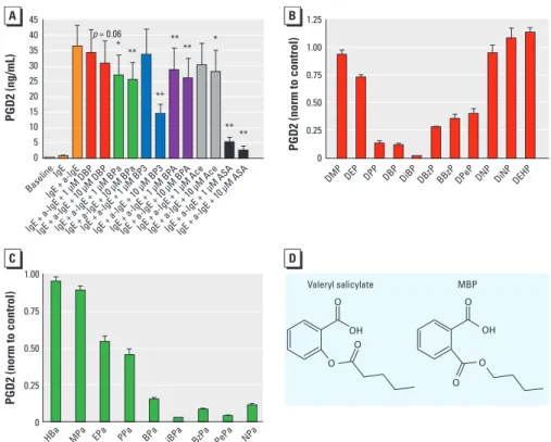
Figure 4. Endocrine disruptors share structural features with mild analgesics and inhibit PGD2 secretion
from primary human mast cells. (A) Secretion of PGD2 from primary human mast cells after stimulation with IgE and anti-IgE is dose-dependently inhibited by exposure to DBP, BPa, BP3, BPA, ASA, and Ace (n = 8). (B and C) Secretion of PGD2 after exposure to different compounds (at 10 µM), indicating that compounds with an isobutyl side chain (DiBP and iBPa) cause the most potent inhibition (n = 3). Data are normalized (norm) to control values. (D) Phthalates share structural similarities with salicylates, here exemplified with valeryl salicylate and MBP. Data are mean ± SE. Abbreviations: BzPa, benzyl paraben; DBzP, dibenzyl phthalate; DEHP, diethylhexyl phthalate; DEP, diethyl phthalate; DiBP, diisobutyl phthalate; DiNP, di-isononyl phthalate; DMP, dimethyl phthalate; DNP, di-n-nonyl phthalate; DPeP, di-n-pentyl phtha-late; DPP, di-n-propyl phthaphtha-late; EPa, ethylparaben; HBa, 4-hydroxy benzoic acid; iBPa, isobutylparaben, MPa, methylparaben, NPa, n-nonylparaben; PePa, n-pentylparaben; PPa, n-propylparaben.
*p < 0.05, **p < 0.01, compared with controls by two-tailed Student’s t-test. 45 40 35 30 25 20 15 10 5 0 1.25 1.00 0.75 0.50 0.25 0 1.00 0.75 0.50 0.25 0 PGD2 (ng/mL) PGD2 (norm to control) PGD2 (norm to control) DMP
HBa MPa EPa PPa BPa iBPa BzPa PePa NPa
DEP DPP DBP DiBP DBzP BBzP DPeP DNP DiNPDEHP
Baseline IgE IgE + a-IgE
IgE + a-IgE + 1 µM DBP IgE + a-IgE + 1 µM BP A
IgE + a-IgE + 1 µM AceIgE + a-IgE + 10 µM AceIgE + a-IgE + 1 µM ASAIgE + a-IgE + 10 µM ASA IgE + a-IgE + 10 µM BP
A
IgE + a-IgE + 1 µM BP3IgE + a-IgE + 10 µM BP3 IgE + a-IgE + 1 µM BPaIgE + a-IgE + 10 µM BPa IgE + a-IgE + 10 µM DBP p = 0.06 ** ** ** ** ** ** * * Valeryl salicylate MBP O O OH OH O O O O
2001; Zykova et al. 2008). Our data suggest that parabens and more complex molecules with multiple phenol groups, such as BP3, BPA, DES, and genistein, should be added to the already characterized substituted phenols with inhibitory efects on PG synthesis.
Tavares and Vine (1985) reported that certain phthalates could interfere with forma-tion of products from the COX and lipoxyge-nase enzymes in rat peritoneal leucocytes. In the present study we focused exclusively on the PG pathway and therefore did not deter-mine whether lipoxygenases are also inhibited. Fujimoto et al. (2005) meas ured PG inhibition in rabbit kidney medulla microsomes and con-cluded that nonyl phenol directly inhibits COX activity. However, they also found that BPA and DBP did not have an inhibitory effect on the PG cascade, which is in conlict with our results and results from a study of perito-neal leukocytes (Tavares and Vine 1985). We cannot explain this discrepancy, but it may be related to the use of diferent experimental designs; also, the proposed binding of EDCs to the COX enzymes should be conirmed using alternative experimental methods.
Genistein and other isoflavones have been reported to decrease PG synthesis in a neck cancer cell line (Ye et al. 2004), in pros-tate cancer cells (Swami et al. 2007, 2009), and in prostate cancer patients (Swami et al. 2009), where the compounds report-edly blocked the development and progres-sion of prostate cancer. In a recent study Swami et al. (2009) reported that genistein decreased expression of COX enzymes with-out affecting COX promoter activity. The authors argued that genistein most likely inhibits PG synthesis through repression of transcriptional activation by growth fac-tors (Swami et al. 2009). However, Ye et al. (2004) reported that genistein inhibited the PG pathway without affecting COX gene expression, consistent with our data suggest-ing that inhibition results from direct efects on COX enzyme activity. Interestingly, genistein has previously been reported to have dual effects in rats (Eustache et al. 2009). Low doses of genistein (1 mg/kg/day) and vinclozolin (1 mg/kg/day) were more anti androgenic when added simultane-ously than when added one at the time, but at higher doses (10 mg/kg/day genistein and 30 mg/kg/day vinclozolin) the anti androgenic effect of vinclozolin was attenuated by genistein (Eustache et al. 2009). Although speculative, it is possible that the low-dose efect could be due to an anti androgenic efect by inhibition of PG synthesis.
Finally, if PG inhibition is involved in the mode of action of some EDCs, it raises the worrying possibility that pharmaceutical PG inhibitors such as ASA, Ace, and Ibu may act as endocrine disruptors. In our investigation
of this we found that Ace indeed reduced the anogenital distance in rat pups after prenatal exposure and that prenatal exposure to ASA reduced testosterone production in fetal testis (Kristensen et al. 2010). Accordingly, in a prospective birth cohort study we found that use of ASA, Ace, and Ibu was associated with cryptorchidism in newborn boys (Kristensen et al. 2010), the best-known risk factor for reduced fertility and testicular germ cell cancers in adulthood (Boisen et al. 2004).
If inhibition of PG synthesis is the mecha-nism of anti androgenicity of compounds such as phthalates, chronic inhibition of the PG pathway by a large number of EDCs com-bined with several short-term high-dose expo-sures to mild analgesics could have an impact on male reproductive health. Furthermore, a growing number of studies has shown that prenatal and early childhood exposure to Ace is associated with atopic diseases (Beasley et al. 2008; Perzanowski et al. 2010; Rebordosa et al. 2008; Shaheen et al. 2002, 2005). Data in the present study show that some of the EDCs are more potent inhibitors of human primary mast cell responses after activation than is Ace; thus, this may suggest a link between exposure to environmental pollutants and disturbances of the immune system.
To conclude, the present study shows an unrecognized point of endocrine disruption through inhibtion of PG synthesis. herefore, more research is needed to investigate whether EDCs could play a role in the increase of immunological and reproductive diseases through inhibition of the PG pathway.
REFERENCES
Adams IR, McLaren A. 2002. Sexually dimorphic development of mouse primordial germ cells: switching from oogenesis to spermatogenesis. Development 129:1155–1164. Amateau SK, McCarthy MM. 2004. Induction of PGE2 by estradiol
mediates developmental masculinization of sex behavior. Nat Neurosci 7:643–650.
Beasley R, Clayton T, Crane J, von Mutius E, Lai CK, Montefort S, et al. 2008. Association between paracetamol use in infancy and childhood, and risk of asthma, rhinoconjunctivitis, and eczema in children aged 6–7 years: analysis from phase three of the ISAAC programme. Lancet 372:1039–1048. Boisen KA, Kaleva M, Main KM, Virtanen HE, Haavisto AM,
Schmidt IM, et al. 2004. Difference in prevalence of con-genital cryptorchidism in infants between two Nordic countries. Lancet 363:1264–1269.
Chauvigne F, Menuet A, Lesne L, Chagnon MC, Chevrier C, Regnier JF, et al. 2009. Time- and dose-related effects of di-(2-ethylhexyl) phthalate and its main metabolites on the function of the rat fetal testis in vitro. Environ Health Perspect 117:515–521.
Christensen KB, Minet A, Svenstrup H, Grevsen K, Zhang H, Schrader E, et al. 2009. Identification of plant extracts with potential antidiabetic properties: effect on human peroxisome proliferator-activated receptor (PPAR), adipo-cyte differentiation and insulin-stimulated glucose uptake. Phytother Res 23:1316–1325.
Diamanti-Kandarakis E, Bourguignon JP, Giudice LC, Hauser R, Prins GS, Soto AM, et al. 2009. Endocrine-disrupting chemi-cals: an Endocrine Society scientific statement. Endocr Rev 30:293–342.
Eustache F, Mondon F, Canivenc-Lavier MC, Lesaffre C, Fulla Y, Berges R, et al. 2009. Chronic dietary exposure to a low-dose mixture of genistein and vinclozolin modifies the
reproductive axis, testis transcriptome, and fertility. Environ Health Perspect 117:1272–1279.
FitzGerald GA. 2003. COX-2 and beyond: approaches to prosta-glandin inhibition in human disease. Nat Rev Drug Discov 2:879–890.
Frederiksen H, Taxvig C, Hass U, Vinggaard AM, Nellemann C. 2008. Higher levels of ethyl paraben and butyl paraben in rat amniotic fluid than in maternal plasma after subcutaneous administration. Toxicol Sci 106:376–383.
Fujimoto Y, Usa K, Sakuma S. 2005. Effects of endocrine disrup-tors on the formation of prostaglandins and arachidonyl- CoA formed from arachidonic acid in rabbit medulla microsomes. Prostaglandins Leukot Essent Fatty Acids 73:447–452.
Graham GG, Robins SA, Bryant KJ, Scott KF. 2001. Inhibition of prostaglandin synthesis in intact cells by paracetamol (acetaminophen). Inflammopharmacology 9:131–142. Gupta C. 1989. The role of prostaglandins in masculine
differ-entiation: modulation of prostaglandin levels in the dif-ferentiating genital tract of the fetal mouse. Endocrinology 124:129–133.
Gupta C, Bentlejewski CA. 1992. Role of prostaglandins in the testosterone-dependent wolffian duct differentiation of the fetal mouse. Biol Reprod 47:1151–1160.
Gupta C, Goldman AS. 1986. The arachidonic acid cascade is involved in the masculinizing action of testosterone on embryonic external genitalia in mice. Proc Natl Acad Sci USA 83:4346–4349.
Habert R, Devif I, Gangnerau MN, Lecerf L. 1991. Ontogenesis of the in vitro response of rat testis to gonadotropin-re-leasing hormone. Mol Cell Endocrinol 82:199–206. Hansen JB, Zhang H, Rasmussen TH, Petersen RK, Flindt EN,
Kristiansen K. 2001. Peroxisome proliferator-activated receptor δ (PPARδ)-mediated regulation of preadipocyte proliferation and gene expression is dependent on cAMP signaling. J Biol Chem 276:3175–3182.
Hofmann MC, Narisawa S, Hess RA, Millan JL. 1992. Immortalization of germ cells and somatic testicular cells using the SV40 large T antigen. Exp Cell Res 201:417–435. Holm M, Andersen HB, Hetland TE, Dahl C, Hoffmann HJ,
Junker S, et al. 2008. Seven week culture of functional human mast cells from buffy coat preparations. J Immunol Methods 336:213–221.
Hsuanyu Y, Dunford HB. 1992. Prostaglandin H synthase kinetics. The effect of substituted phenols on cyclooxygenase activity and the substituent effect on phenolic peroxidatic activity. J Biol Chem 267:17649–17657.
Ishizaka T, Conrad DH, Schulman ES, Sterk AR, Ishizaka K. 1983. Biochemical analysis of initial triggering events of IgE-mediated histamine release from human lung mast cells. J Immunol 130:2357–2362.
Koch HM, Bolt HM, Preuss R, Angerer J. 2005. New metabo-lites of di(2-ethylhexyl)phthalate (DEHP) in human urine and serum after single oral doses of deuterium-labelled DEHP. Arch Toxicol 79:367–376.
Kristensen DM, Hass U, Lesné L, Lottrup G, Jacobsen PR, Desdoits-Lethimonier C, et al. 2010. Intrauterine exposure to mild analgesics is a risk factor for development of male reproductive disorders in human and rat. Hum Reprod 26(1):235–244.
Kurumbail RG, Stevens AM, Gierse JK, McDonald JJ, Stegeman RA, Pak JY, et al. 1996. Structural basis for selec-tive inhibition of cyclooxygenase-2 by anti-inflammatory agents. Nature 384:644–648.
Lassurguere J, Livera G, Habert R, Jegou B. 2003. Time- and dose-related effects of estradiol and diethylstilbestrol on the morphology and function of the fetal rat testis in culture. Toxicol Sci 73:160–169.
Luong C, Miller A, Barnett J, Chow J, Ramesha C, Browner MF. 1996. Flexibility of the NSAID binding site in the structure of human cyclooxygenase-2. Nat Struct Biol 3:927–933. Nielsen R, Pedersen TA, Hagenbeek D, Moulos P, Siersbaek R,
Megens E, et al. 2008. Genome-wide profiling of PPARγ:RXR and RNA polymerase II occupancy reveals temporal activa tion of distinct metabolic pathways and changes in RXR dimer composition during adipogenesis. Genes Dev 22:2953–2967.
Perzanowski MS, Miller RL, Tang D, Ali D, Garfinkel RS, Chew GL, et al. 2010. Prenatal acetaminophen exposure and risk of wheeze at age 5 years in an urban low-income cohort. Thorax 65:118–123.
Picot D, Loll PJ, Garavito RM. 1994. The X-ray crystal structure of the membrane protein prostaglandin H2 synthase-1. Nature 367:243–249.
Endocrine disruptors inhibit the prostaglandin pathway
Rebordosa C, Kogevinas M, Sorensen HT, Olsen J. 2008. Pre-natal exposure to paracetamol and risk of wheezing and asthma in children: a birth cohort study. Int J Epidemiol 37:583–590. Research Collaboratory for Structural Bioinformatics. 2010.
RCSB Protein Data Bank. Available: http://home.rcsb.org/ [accessed 17 February 2011].
Rowlinson SW, Kiefer JR, Prusakiewicz JJ, Pawlitz JL, Kozak KR, Kalgutkar AS, et al. 2003. A novel mechanism of cyclooxygenase-2 inhibition involving interactions with Ser-530 and Tyr-385. J Biol Chem 278:45763–45769. Scott HM, Mason JI, Sharpe RM. 2009. Steroidogenesis in the
fetal testis and its susceptibility to disruption by exogenous compounds. Endocr Rev 30(7):883–925.
Shaheen SO, Newson RB, Henderson AJ, Headley JE, Stratton FD, Jones RW, et al. 2005. Prenatal paracetamol exposure and risk of asthma and elevated immuno globulin E in childhood. Clin Exp Allergy 35:18–25.
Shaheen SO, Newson RB, Sherriff A, Henderson AJ, Heron JE,
Burney PG, et al. 2002. Paracetamol use in pregnancy and wheezing in early childhood. Thorax 57:958–963. Skakkebaek NE, Rajpert-de Meyts E, Main KM. 2001. Testicular
dysgenesis syndrome: an increasingly common develop-mental disorder with environdevelop-mental aspects. Hum Reprod 16:972–978.
Smith WL, DeWitt DL, Garavito RM. 2000. Cyclooxygenases: structural, cellular, and molecular biology. Annu Rev Biochem 69:145–182.
Soliva R, Almansa C, Kalko SG, Luque FJ, Orozco M. 2003. Theoretical studies on the inhibition mechanism of cyclo-oxygenase-2. Is there a unique recognition site? J Med Chem 46:1372–1382.
Swami S, Krishnan AV, Moreno J, Bhattacharyya RS, Gardner C, Brooks JD, et al. 2009. Inhibition of prostaglandin synthesis and actions by genistein in human prostate cancer cells and by soy isoflavones in prostate cancer patients. Int J Cancer 124:2050–2059.
Swami S, Krishnan AV, Moreno J, Bhattacharyya RB, Peehl DM, Feldman D. 2007. Calcitriol and genistein actions to inhibit the prostaglandin pathway: potential combination therapy to treat prostate cancer. J Nutr 137(1 suppl):205S–210S. Tavares IA, Bennett A, Gaffen JD, Morris HR, Taylor GW. 1984.
The biological activities of phthalate esters on rat gastric muscle. Eur J Pharmacol 106:449–452.
Tavares IA, Vine ND. 1985. Phthalic acid esters inhibit arachi-donate metabolism by rat peritoneal leucocytes. J Pharm Pharmacol 37:67–68.
Ye F, Wu J, Dunn T, Yi J, Tong X, Zhang D. 2004. Inhibition of cyclooxygenase-2 activity in head and neck cancer cells by genistein. Cancer Lett 211:39–46.
Zykova TA, Zhu F, Zhai X, Ma WY, Ermakova SP, Lee KW, et al. 2008. Resveratrol directly targets COX-2 to inhibit carcino-genesis. Mol Carcinog 47:797–805.